Original Article
Does 23-gauge vitrectomy decrease complications
and operation time in treating retinal
diseases? A meta-analysis
Fenghua Li1, Jianling Chen2, Ling Sun2
1Department of Ophthalmology, Linyi People’s Hospital, Linyi 276000, China; 2Department of Emergency, Linyi People’s Hospital, Linyi 276000, China
Received December 22, 2015; Accepted March 21, 2016; Epub June 15, 2016; Published June 30, 2016
Abstract: Objective: To compare the therapeutic effects and operation time of 23-G vitrectomy and conventional 20-G vitrectomy on complications (retinal breaks, retinal detachment). Methods: Related studies published prior to October 2014 were retrieved via exhaustive searching of English and Chinese scientific literature databases. High quality clinical cohort studies, focused on 23-G and 20-G vitrectomy in retinal diseases, were carefully selected according to our study inclusion criteria. Statistical analyses were also conducted by comprehensive Meta-analysis 2.0 (CMA 2.0) software. Results:Fifty-five published articles (47 in English and 8 in Chinese) were initially identified as relevant to our keyword search, of which 10 studies were included in this meta-analysis, including 1464 patients treated for retinal diseases (718 treated with 23-G vitrectomy, 746 treated with 20-G vitrectomy). Our meta-analysis showed that the incidence of retinal breaks in 23-G vitrectomy were lower than those in 20-G vitrectomy (P = 0.017). However, no such difference was found in the incidence of retinal detachment between these two surgical procedures (P = 0.467). As for operation time, our results showed that 23-G vitrectomy procedure was performed in less time compared with patients receiving 20-G vitrectomy (P < 0.001). Conclusions: The 23-G transconjunctival sutureless vitrectomy may be superior to conventional 20-G vitrectomy in reducing the incidence of retinal breaks and shortening operation time.
Keywords: 23-G vitrectomy, 20-G vitrectomy, retina, minimally invasive, complication, meta-analysis
Introduction
Poor final visual acuity often results from the treatment of retinal diseases, although new surgical and pharmacological modalities have been rapidly developed [1]. Pars plana vitrec-tomy (PPV), a surgical procedure initially re- ported in 1971 by Machemer et al., has re- volutionized retinal surgery since its advent, and is performed with 20-G instruments [2]. The first small caliber instrument was des-cribed in 2002, which used 25-G inserted through a ‘port’ and did not require suturing. Currently, 23-gauge (23-G), 25-gauge (25-G), and 27-gauge (27-G) techniques are available and transconjunctival sutureless pars plana vit-rectomy (TSV) is considered safe for treating patients suffered from vitreoretinal patholo-gies, particularly macular disorders including preretinal membranes and macular holes [3-6].
disad-10425 Int J Clin Exp Med 2016;9(6):10424-10433 vantages of 20-gauge (20-G) and 25-G
vitrec-tomy, while maximizing the benefit to patients [3, 10]. In comparison to the conventional 20-G vitrectomy, 23-G vitrectomy can reduce trauma to sclera and conjunctiva, and save time at the beginning and at the end of the operation [11]. In addition, it has been manifested that the 23-G vitrectomy has a better safety profile and visual outcomes for a number of different pos-terior segment conditions [12]. In recent years, several studies have examined the difference between 20-G and 23-G vitrectomy in relation to the incidence of complications and the total operation time, some of which reported that 23-G TSV was superior to conventional 20-G vitrectomy [9, 13-16], while some other studies found no statistically significant difference between these two techniques [11, 17]. In view of the conflicting data from previous studies, we performed this meta-analysis to compare the incidence of complications and total opera-tion times between 23-G vitrectomy and con-ventional 20-G vitrectomy.
Materials and methods
Search strategy
English and Chinese databases including Pub- Med (since 1966), Ovid (since 1948), Embase (since 1966), China National Knowledge In- frastructure (CNKI, since 1994), and Wanfang Data (since 1986) were electronically searched for related articles published prior to October 2014. Manual search was performed to identi-fy additional studies from cross-references. The search terms which combined free text words with key words were: 23-G vitrectomy, 20-G vitrectomy, retinal detachment, retinal pigment epithelial detachment, and retinal breaks.
Selection criteria
Published studies were included if they met the following inclusion criteria: (1) Study de- sign: Clinical cohort study; (2) Subject of study: Comparisons of complications and opera- tion time between 23- and 20-G vitrectomy in treatment of retinal diseases; (3) Population: Patients histopathologically diagnosed with re- tinal diseases; (4) Outcomes: Reported com- plications in the operations or the operation time. Exclusion criteria were: (1) incomplete data; (2) repeat publications; (3) non-English or non-Chinese studies.
Data extraction and quality assessment
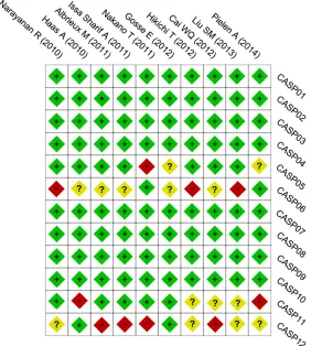
A standardized data-collection form was em- ployed by two investigators to extract data in- dependently from the eligible studies, mainly including the following aspects: first author’s name, year of publication, country, ethnicity, language, disease, age, number of cases and controls, operation time, occurrence of retinal detachment and retinal breaks. Disagreement on any extracted item, if any, was resolved through the discussion with several other inves-tigators. Two investigators performed the meth-odological quality assessment according to the Critical Appraisal Skills Programme (CASP) Checklists (http://www.casp-uk.net/), and a third investigator was involved when there was a disagreement. The CASP criteria as 12 items on the checklist as: 1) if a clearly fo- cused issue was addressed (CASP01); 2) if the cohort were recruited in an acceptable way (CASP02); 3) if the exposure was accu- rately measured to minimize bias (CASP03); 4) if the outcome was accurately measured to minimize bias (CASP04); 5) if the confounding factors had been taken account in the design and/or analysis (CASP05); 6) if the follow up of subjects was complete and long enough (CASP06); 7) if the results of the study were complete (CASP07); 8) how precise are the results (CASP08); 9) if the results credible (CASP09); 10) if the results can be applied to the local population (CASP10); 11) if the re- sults of this study fit with other available evi -dence (CASP11); 12) what are the implications of this study for practice (CASP12).
Statistical analysis
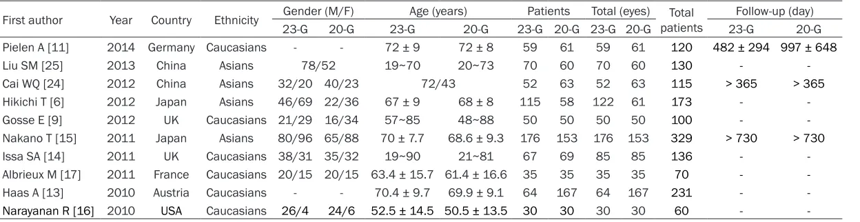
10426 Int J Clin Exp Med 2016;9(6):10424-10433 Table 1. Characteristics of included studies focused on 23-G vitrectomy and 20-G vitrectomy
First author Year Country Ethnicity Gender (M/F) Age (years) Patients Total (eyes) patientsTotal Follow-up (day)
23-G 20-G 23-G 20-G 23-G 20-G 23-G 20-G 23-G 20-G
Pielen A [11] 2014 Germany Caucasians - - 72 ± 9 72 ± 8 59 61 59 61 120 482 ± 294 997 ± 648
Liu SM [25] 2013 China Asians 78/52 19~70 20~73 70 60 70 60 130 -
-Cai WQ [24] 2012 China Asians 32/20 40/23 72/43 52 63 52 63 115 > 365 > 365
Hikichi T [6] 2012 Japan Asians 46/69 22/36 67 ± 9 68 ± 8 115 58 122 61 173 -
-Gosse E [9] 2012 UK Caucasians 21/29 16/34 57~85 48~88 50 50 50 50 100 -
-Nakano T [15] 2011 Japan Asians 80/96 65/88 70 ± 7.7 68.6 ± 9.3 176 153 176 153 329 > 730 > 730
Issa SA [14] 2011 UK Caucasians 38/31 35/32 19~90 21~81 67 69 85 85 136 -
-Albrieux M [17] 2011 France Caucasians 20/15 20/15 63.4 ± 15.7 61.4 ± 16.6 35 35 35 35 70 -
-Haas A [13] 2010 Austria Caucasians - - 70.4 ± 9.7 69.9 ± 9.1 64 167 64 167 231 -
-Narayanan R [16] 2010 USA Caucasians 26/4 24/6 52.5 ± 14.5 50.5 ± 13.5 30 30 30 30 60 -
10427 Int J Clin Exp Med 2016;9(6):10424-10433 with Ph < 0.05. For quantifying the degree of
heterogeneity, I-squared (I2) statistic [19] was
calculated with higher values (ranging from 0%~100%) indicating higher degree. A Ph < 0.05 or I2 > 50% suggested that the studies
were heterogeneous, thus a random effects model was applied, otherwise a fixed-effects model was used [20]. With the sensitivity analy-sis of variables, the impact on the overall re- sults by removing one single study was evalu-ated. Funnel plots, classic fail-safe N [21, 22] as well as Egger regression analyses were per-formed for the assessment of publication bias to evaluate the reliability of the results [23]. All these bilateral tests were conducted with P < 0.05 implying a statistical significance. Results
Database search results
Fifty-five articles were initially retrieved, of which 6 duplicates, 8 letters or reviews, 2 non-human studies and 11 articles not re- lated to the research topics were screened
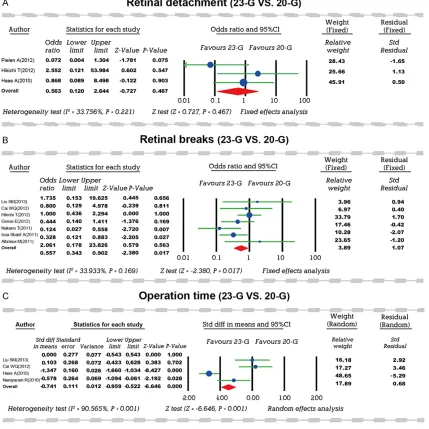
incidence of intraoperative retinal breaks between 23-G TSV and 20-G vitrectomy. No heterogeneity was found according to the results of Cochran’s Q-statistic test (P > 0.05), therefore a fixed-effects model was carried out. Results of our meta-analysis suggested that the incidence of retinal breaks during 23-G vit-rectomy was lower than during 20-G vitvit-rectomy (OR = 0.557, 95% CI = 0.343~0.902, P = 0.017) (Figure 2B). Subgroup analyses by ethnicity demonstrated that the incidence of retinal breaks between these two operations was statistically significant in Caucasians (OR = 0.432, 95% CI = 0.210~0.887, P = 0.022), while no such significance was observed in Asians (OR = 0.684, 95% CI = 0.357~1.312, P = 0.254) (Figure 3). As for retinal detach-ment, three of the included studies compared the incidence of retinal detachment during 23-G and 20-G vitrectomy. No heterogeneity was found (P > 0.05), therefore a fixed-effects model was carried out. Meta-analysis revealed no statistically significant difference in the inci -dence of retinal detachment between these
Figure 1. Critical Appraisal Skills Programme assessments for each included study.
[image:4.612.95.377.76.390.2]out. Additionally, 14 studies were excluded through over- all assessment of the 28 remaining studies, and anoth-er 4 articles wanoth-ere furthanoth-er removed for partially related data. Thus, 10 cohort stu- dies published between 2010 and 2014 [6, 9, 11, 13-17, 24, 25] met our inclusion cri-teria and were enrolled in this meta-analysis. These 10 literatures included 1464 pa- tients with retinal diseases, with a sample size range of 60~329. In relation to the subjects investigated, 6 stud-ies were performed in Cau- casian population and 4 stud-ies in Asian population. The baseline characteristics of studies included in the meta-analysis are presented in Table 1. Results of CASP as- sessment for each included study are shown in Figure 1. Meta-analysis results
10428 Int J Clin Exp Med 2016;9(6):10424-10433
Figure 2. Forest plots: the difference of complications and operation time between 23-G and 20-G vitrectomy.
[image:5.612.92.522.559.706.2]10429 Int J Clin Exp Med 2016;9(6):10424-10433 two operations (OR = 0.563, 95% CI = 0.120~
2.644, P = 0.467) (Figure 2A). A total of 4 studies documented the difference in opera-tion times between 23- and 20-G vitrectomy. Heterogeneity (P < 0.05) was observed among the three studies and a random effects model was utilized for analysis. Meta-analysis results showed that the 23-G TSV was performed in
markedly less time than the conventional 20-G vitrectomy (SMD = -0.741, 95% CI = -0.959~-0.522, P < 0.001) (Figure 2C).
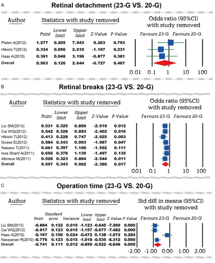
Sensitivity analysis and publication bias
[image:6.612.94.522.71.595.2]Results of sensitivity analysis indicated that except for studies reported by Nakano et al.
10430 Int J Clin Exp Med 2016;9(6):10424-10433
10431 Int J Clin Exp Med 2016;9(6):10424-10433 (2011) and Issa et al. (2011), showing
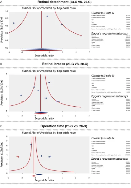
differ-ence in the retinal breaks between 23-G and 20-G vitrectomy and the study by Haas et al. (2010) presenting the operation time differ-ence (a reverse outcome existed after their removal), the other 7 publications had no sig -nificant influence on the OR and SMD in rela -tion to the incidence of complica-tions and oper-ation time between these two operoper-ations (Figure 4). Symmetrical funnel plots showed that there was no publication bias in the differ-ence of complications between 23- and 20-G vitrectomy, which was further verified by the results of classic fail-safe N and Egger regres-sion analyses (all P > 0.05) (Figure 5). However, funnel plots demonstrating the difference in operation time was asymmetric, which indicat-ed the existence of publication bias; further identification of publication bias was obtained from the results of classic fail-safe N and Egger regression analyses (all P < 0.05) (Figure 5). Discussion
TSV, including 23-G vitrectomy, is widely uti-lized by retinal surgeons for its advantages such as patient comfort, reduced operation time and earlier postoperative recovery than conventional 20-G vitrectomy [26]. However, 23-G vitrectomy may also cause complications such as retinal breaks, postoperative retinal detachment, decompression retinopathy and intraoperative instrument breakage [8]. We performed this meta-analysis to investigate the difference in the incidence of complications and total operation time between 23-G vitrec-tomy and conventional 20-G vitrecvitrec-tomy. Specifically, we studied two complications, reti -nal breaks and reti-nal detachment, in relation to these two operative methods. The results of our meta-analysis clearly revealed that the inci-dence of retinal breaks during 23-G vitrectomy were significantly lower than during 20-G vitrec -tomy. According to previous publications, the incidence of retinal breaks triggered by stan-dard 20-G vitrectomy was between 4% and 17% [27-30]. For small-incision vitrectomy, the incidence of retinal breaks ranged from 0% to 15.8% [31-34]. Nakano et al. argued that these broad-ranged results could be caused by the methods used for identification of retinal breaks or due to variations in the surgical indi-cations for vitrectomy [15]. Moreover, a
cannu-lated entry system, which might decrease vitre-ous base traction upon instrument insertion, is used in TSV. Angled scissors, which is often used for delamination during 20-G surgery, cannot be used during cannulated entry sys-tems, and the insertion of curved or straight instruments is likely to reduce traction in TSV surgery [14]. In addition, the cutter mouth in 23-G vitrectomy is closer to the end of the cut-ter compared to 20-G vitrectomy, which causes less risk of breaks by allowing more controlled segmentation of fibrovascular tissue [35].
With respect to retinal detachment, the results of our meta-analysis revealed no statistically significant difference in the incidence of retinal detachment between these two operations. The 23-G vitrectomy is a relatively new proce-dure, with its own unique surgical challenges and complications. According to Albrieux et al., although the scleral indentation of 23-G vitrec-tomy was more delicate, its self-sealing nature of the incisions may pose theoretical concerns of increased risk of vitreous incarceration, hypotony and postoperative endophthalmitis [17].
To better compare the advantages of 20-G and 23-G vitrectomy, we also conducted meta-anal-ysis on the operation times of these two surger-ies. Our results demonstrated that 23-G vitrec-tomy was performed in markedly lesser time than the conventional 20-G vitrectomy, indicat-ing that 23-G vitrectomy was superior to 20-G vitrectomy in reducing total operation time. The potential reason could be that the air/fluid exchange and the dye in 20-G vitrectomy requires 2 minutes, while in 23-G vitrectomy, the dye can be injected onto the macula direct-ly without the need of air/fluid exchange [36].
10432 Int J Clin Exp Med 2016;9(6):10424-10433 Acknowledgements
We appreciate the reviewers and our instruc-tors for their valuable comments and advices on this article.
Disclosure of conflict of interest
None.
Address correspondence to: Dr. Ling Sun, Depart- ment of Emergency, Linyi People’s Hospital, No. 27, Jiefang Road, Linyi 276000, China. Tel: +86-0539-8078716; Fax: +86-+86-0539-8078716; E-mail: [email protected]
References
[1] Vasilaki A, Thermos K. Somatostatin ana-logues as therapeutics in retinal disease. Pharmacol Ther 2009; 122: 324-333.
[2] Machemer R, Buettner H, Norton EW, Parel JM. Vitrectomy: a pars plana approach. Trans Am Acad Ophthalmol Otolaryngol 1971; 75: 813-820.
[3] Eckardt C. Transconjunctival sutureless 23-gauge vitrectomy. Retina 2005; 25: 208-211.
[4] Fujii GY, De Juan E Jr, Humayun MS, Chang TS, Pieramici DJ, Barnes A, Kent D. Initial experi-ence using the transconjunctival sutureless vitrectomy system for vitreoretinal surgery. Ophthalmology 2002; 109: 1814-1820. [5] Oshima Y, Wakabayashi T, Sato T, Ohji M, Tano
Y. A 27-gauge instrument system for transcon-junctival sutureless microincision vitrectomy surgery. Ophthalmology 2010; 117: 93-102, e2.
[6] Hikichi T, Kosaka S, Takami K, Ariga H, Ohtsuka H, Higuchi M, Matsushita T, Matsushita R. Incidence of retinal breaks in eyes undergoing 23-gauge or 20-gauge vitrectomy with induc-tion of posterior vitreous detachment. Retina 2012; 32: 1100-1105.
[7] Narayanan R, Tibra N, Mathai A, Chhablani J, Kuppermann BD. Sutureless 23-gauge versus 20-gauge vitrectomy with silicone oil injection in rhegmatogenous retinal detachment. Retina 2012; 32: 1013-1016.
[8] Warrier SK, Jain R, Gilhotra JS, Newland HS. Sutureless vitrectomy. Indian J Ophthalmol 2008; 56: 453-458.
[9] Gosse E, Newsom R, Lochhead J. The inci-dence and distribution of iatrogenic retinal tears in 20-gauge and 23-gauge vitrectomy. Eye (Lond) 2012; 26: 140-143.
[10] Nagpal M, Wartikar S, Nagpal K. Comparison of clinical outcomes and wound dynamics of
sclerotomy ports of 20, 25, and 23 gauge vit-rectomy. Retina 2009; 29: 225-231.
[11] Pielen A, Guerra NI, Bohringer D, Junker B, Buhler AD, Stahl A, Agostini HT, Ehlken C. Intra- and postoperative risks and complications of small-gauge (23-G) versus conventional (20-G) vitrectomy for macular surgery. Eur J Ophthalmol 2014; 24: 778-785.
[12] Krishnan R, Tossounis C, Fung Yang Y. 20-gauge and 23-gauge phacovitrectomy for idiopathic macular holes: comparison of com-plications and long-term outcomes. Eye (Lond) 2013; 27: 72-77.
[13] Haas A, Seidel G, Steinbrugger I, Maier R, Gasser-Steiner V, Wedrich A, Weger M. Twenty-three-gauge and 20-gauge vitrectomy in epiret-inal membrane surgery. Retina 2010; 30: 112-116.
[14] Issa SA, Connor A, Habib M, Steel DH. Comparison of retinal breaks observed during 23 gauge transconjunctival vitrectomy versus conventional 20 gauge surgery for proliferative diabetic retinopathy. Clin Ophthalmol 2011; 5: 109-14.
[15] Nakano T, Uemura A, Sakamoto T. Incidence of iatrogenic peripheral retinal breaks in 23-gauge vitrectomy for macular diseases. Retina 2011; 31: 1997-2001.
[16] Narayanan R, Sinha A, Reddy RK, Krishnaiah S, Kuppermann BD. Faster visual recovery af-ter 23-gauge vitrectomy compared with 20-gauge vitrectomy. Retina 2010; 30: 1511-1514.
[17] Albrieux M, Rouberol F, Bernheim D, Romanet JP, Chiquet C. Comparative study of 23-gauge vitrectomy versus 20-gauge vitrectomy for the treatment of rhegmatogenous retinal detach-ment. Graefes Arch Clin Exp Ophthalmol 2011; 249: 1459-1468.
[18] Chen H, Manning AK, Dupuis J. A method of moments estimator for random effect multi-variate meta-analysis. Biometrics 2012; 68: 1278-1284.
[19] Peters JL, Sutton AJ, Jones DR, Abrams KR, Rushton L. Comparison of two methods to de-tect publication bias in meta-analysis. JAMA 2006; 295: 676-680.
[20] Zintzaras E, Ioannidis JP. Heterogeneity testing in meta-analysis of genome searches. Genet Epidemiol 2005; 28: 123-137.
[21] Sterne JA, Egger M. Funnel plots for detecting bias in meta-analysis: guidelines on choice of axis. J Clin Epidemiol 2001; 54: 1046-1055. [22] Wikstrom EA, Naik S, Lodha N, Cauraugh JH.
Balance capabilities after lateral ankle trauma and intervention: a meta-analysis. Med Sci Sports Exerc 2009; 41: 1287-1295.
10433 Int J Clin Exp Med 2016;9(6):10424-10433 [24] Cai WQ, Zhen Z, Li T, Chen FE, Xu X. Outcome
analysis of 23-G vitrectomy versus 20-G vitrec-tomy for treatment of retinal detachment. Journal of Shanghai Jiaotong University (Medical Science) 2012; 32: 151-154.
[25] Liu SM, Zhong J, Fan YC. A comparative analy-sis of 23-gauge versus 20-gauge vitrectomy for treatment of retinal detachment. Practical Journal of Clinical Medicine 2013; 10: 113-115.
[26] Ahn SJ, Woo SJ, Ahn J, Park KH. Comparison of postoperative intraocular pressure changes between 23-gauge transconjunctival suture-less vitrectomy and conventional 20-gauge vit-rectomy. Eye (Lond) 2012; 26: 796-802. [27] Carter JB, Michels RG, Glaser BM, De Bustros
S. Iatrogenic retinal breaks complicating pars plana vitrectomy. Ophthalmology 1990; 97: 848-853; discussion 854.
[28] Park SS, Marcus DM, Duker JS, Pesavento RD, Topping TM, Frederick AR Jr, D’Amico DJ. Posterior segment complications after vitrec-tomy for macular hole. Ophthalmology 1995; 102: 775-781.
[29] Sjaarda RN, Glaser BM, Thompson JT, Murphy RP, Hanham A. Distribution of iatrogenic reti-nal breaks in macular hole surgery. Ophthalmology 1995; 102: 1387-1392.
[30] Moore JK, Kitchens JW, Smiddy WE, Mavrofrides EC, Gregorio G. Retinal breaks ob-served during pars plana vitrectomy. Am J Ophthalmol 2007; 144: 32-36.
[31] Rizzo S, Genovesi-Ebert F, Murri S, Belting C, Vento A, Cresti F, Manca ML. 25-gauge, suture-less vitrectomy and standard 20-gauge pars plana vitrectomy in idiopathic epiretinal mem-brane surgery: a comparative pilot study. Graefes Arch Clin Exp Ophthalmol 2006; 244: 472-479.
[32] Kusuhara S, Ooto S, Kimura D, Itoi K, Mukuno H, Miyamoto N, Akimoto M, Kuriyama S, Takagi H. Outcomes of 23- and 25-gauge transcon-junctival sutureless vitrectomies for idiopathic macular holes. Br J Ophthalmol 2008; 92: 1261-1264.
[33] Tan HS, Mura M, de Smet MD. Iatrogenic reti-nal breaks in 25-gauge macular surgery. Am J Ophthalmol 2009; 148: 427-430.
[34] Hikichi T, Matsumoto N, Ohtsuka H, Higuchi M, Matsushita T, Ariga H, Kosaka S, Matsushita R. Comparison of one-year outcomes between 23- and 20-gauge vitrectomy for preretinal membrane. Am J Ophthalmol 2009; 147: 639-643 e631.
[35] Spirn MJ. Comparison of 25, 23 and 20-gauge vitrectomy. Curr Opin Ophthalmol 2009; 20: 195-199.