Original Article
Apoptosis of human hepatocellular carcinoma cells
SMMC-7721 induced by C-3 methylidene
thiazolidinedione acetic acid
Hong-Xia Liang1, Yi-Hua Yu1, Xiao-Hua Li1, Nai-Fu Tang1, Guo-Qiang Hu2, Bin Liu1
1Institute of Nursing and Health, Henan University, Kaifeng, Henan, China; 2College of Pharmacy, Henan Univer-sity, Kaifeng, Henan, China
Received July 9, 2018; Accepted September 12, 2018; Epub January 15, 2019; Published January 30, 2019
Abstract: Objective: The objective of this study was to investigate the effect of rufloxacin derivative C-3 methylidene
thiazolidinedione acetic acid on apoptosis of human hepatocellular carcinoma cells SMMC-7721. Methods: Hu-man hepatocellular carcinoma cells SMMC-7721, esophageal cancer cells EC-9706, colon cancer CaCO-2 cells, and hepatocytes L-02 were cultured in vitro with various concentrations of 6-fluoro -7-(4-methylpiperazin-1-yl)-8,
1-(sulfolatosyl)-1,4-dihydro-4-oxo-3-(3-carboxymethyl-2, 4-thiazolidinedione- 5-methylidene) quinoline (R16). MTT
assay was used to detect the inhibitory effect of R16 on proliferation in all cell lines. DAPI fluorescent staining and TUNEL assay were used to detect the changes of apoptosis. PI staining and flow cytometry were used to detect
cell cycle changes. p53 and caspase-3 protein expression levels were detected by Western blot. Results: R16
sig-nificantly inhibited cell proliferation of SMMC-7721, EC-9706 and CaCO-2 cells under the concentration of 2-20
µmol·L-1. The 24 hour IC
50 values were 3.912, 4.215 and 3.380 mol·L-1, respectively. The 24 hour IC50 value of L-02
cells was 35.224 µmol·L-1. The 24 hour IC
50 value of rufloxacin was 226.924 µmol·L-1 for SMMC-7721. The 24 hour
IC50 value of sunitinib was 7.846 µmol·L-1 for SMMC-7721. Following R16 treatment, cell cycle of SMMC-7721 was
arrested at G1-S phase and the apoptosis rate was significantly higher compared to the control group (P<0.05). R16 significantly increased p53 and caspase-3 expression in SMMC-7721 cells. In addition, the active fragment of caspase-3 was significantly increased. Conclusions: Rufloxacin-rhodanine derivatives exhibited a selective inhibitory effect on cancer cells and significantly induced apoptosis of human hepatocellular carcinoma cells SMMC-7721.
Keywords: Rufloxacin-Rhodanine derivatives, hepatocellular carcinoma cells, cell proliferation, apoptosis, cell
cycle
Introduction
Quinolones are widely used as anti-infective agents in clinical practice [1]. This type of agents are inhibitors of DNA gyrase, a type II DNA topoisomerase [2]. The acting mechanism of this enzyme is to perform DNA strands cleav-age, change DNA topological structures and reconnect the structures. The enzyme plays an important role in DNA replication, transcrip-tion and repair [3]. In eukaryotic cells, some sequence fragments of topoisomerase II have a high degree of homology with DNA gyrase [4]. Some of the fluoroquinolones carboxylic acids commonly used in clinical application, such as ciprofloxacin, rufloxacin, and ofloxacin, have a mild inhibitory effect on eukaryotic topoisomer-ase II activity [5]. Therefore, quinolone
Human hepatocellular carcinoma SMMC-7721 cells
Figure 1. Structure of
6-fluoro-7-(4-methylpiperazin- 1-yl)-8,1-(thioethylene)-1,4-dihydro-4-oxo-3-[5-(3-car-boxymethyl-2,4-thiazolidinedione) ylidene]-quinoline.
activity were synthesized. However, these com-pounds failed prior to clinical evaluation due to their in vivo toxicity, low bioavailability and inability to be metabolized [9]. The carboxyl group on C-3 of fluoroquinolone carboxylic acid is required for anti-bacterial activity but has no effect on anti-cancer activity [10]. The five-membered azole heterocyclic ring has been used as a backbone with dominant pharmaco-phores in the design of drug molecules, among which thiazolidinediones have gained a lot of attention due to their pharmacological activi-ties. Based on the research of fluoroquinolone C-3 rhodanine unsaturated ketones, according to isosteres and splicing principles, thiazolidin-edione was used, instead of thio-thiazolidinedi-one, as the isostere of fluoroquinolones C-3 carboxyl group. A series of fluoroquinolone derivatives were designed and synthesized with good solubility, low molecular weight, un- saturated ketone structures and similar struc-tures to of chalcones and tyrosine kinase inhib-itor sunitinib. The anti-tumor activity of this type of compounds was screened. The results showed that this type of derivatives all had good anti-tumor activity. Among these com-pounds, 6-fluoro-7-(4-methylpiperazin-1-yl)-8, 1- (sulfolatosyl)-1,4-dihydro-4-oxo-3-(3-carboxy-methyl-2,4-thiazolidinedione-5-methylidene) quinoline (R16) exhibited the strongest activity, with an IC50 up to 3.912 µmol/L and further research and development value (Figure 1).
Materials and methods
Main reagents and instruments
Inducer: Rufloxacin derivative C-3 methylidene thiazolidinedione acetic acid was designed and synthesized by the Institute of Chemical Biology of Henan University. The agent was dissolved in
dimethyl sulfoxide (DMSO, Solarbio Co.) at a beginning concentration of 1 × 10⁻² mol·L-1.
Cell lines and major reagents
Human hepatocellular carcinoma cells SMMC-7721 and human esophageal cancer cells EC-9706 were cultured in DMEM media (Gibco) containing fetal bovine serum (Hangzhou Four Seasons Bioengineering Material Inc.) at a con-centration of 0.1% (v/v). Colon cancer CaCO-2 cells and hepatocytes L-02 were cultured in RPMI -1640 media (Gibco) containing fetal bovine serum at a concentration of 0.1% (v/v). Cells were placed in an incubator with 0.05% (v/v) CO2 at 37°C. Tetramethyl azolium salt (mTT) was purchased from Solarbio Co. DAPI was purchased from Sigma. DeadEndTM Fluo-
rometric TUNEL System was purchased from Promega. Mouse monoclonal anti-casepase-3 antibody was purchased from Novus. Rabbit polyclonal anti-p53 antibody was purchased from Baiqi Co. Mouse anti-β-actin polyclonal antibody was purchased from Zhongshan Jin- qiao Co. HRP-labeled goat anti-rabbit and goat anti-mouse antibodies were purchased from Santa Cruz Co. The remaining reagents were domestic analytical reagents.
Major equipment
The CO2 incubator (Forma 3121) and plate reader (Multiskan Ascent) were purchased from Thermo. The inverted microscope was pur-chased from Leica Microsystems. BX51 fluores-cent microscope was provided by Olympus. High speed centrifuge was purchased from Eppendorf. Gel imaging system was purchas- ed from UVP, LLC. Flow cytometer was provided by AceaBio Co. Electrophoresis and semi-dry blotting membrane transfer system were pur-chased from Six-One Electronic Equipment Manufacturing.
Methods
Effects of drugs on cell proliferation by MTT assay: Cells were inoculated on a 96-well plate at a concentration of 16 × 107·L-1. Various
Figure 2. Proliferation inhibition effect of R16, Rufloxacin, and Sunitinib on human cancer cells and L-02 cells.
SMMC-7721 cells (A), EC-9706 cells (B), CaCO-2 cells (C) and L-02 (D) cells were treated with various concentration
of R16 for 24-72 hours. SMMC-7721 cells were treated with various concentration of Rufloxacin (E) and Sunitinib
(F) for 24-72 hours.
a plate reader. The absorbance of wells con-taining no cells but the same volume of culture media and DMSO was used as control.
Cell Growth Inhibition Rate = [1-(OD value of treated group - OD value of control group)/(OD value of untreated group - OD value of control group)] × 100%.
Observation of cell morphological changes by DAPI staining
Cells were inoculated at a concentration of 4 × 10⁷·L-1 in the 6-well plate where cover slides
were placed at the bottom of wells. After cell adhesion, various concentrations of R16 was added and cultured for 24 hours. Cells were washed twice by PBS and fixed in paraformal-dehyde for 20 minutes. 20 µl DAPI working solution was added on the cover slides. After 10 minute staining at room temperature, slides were washed three times by PBS, blocked by 78% neutral glycerol, and observed under the fluorescent microscope.
The apoptosis rate determined by TUNEL assay
Cells were inoculated at a concentration of 4 × 10⁷·L-1 in the 6-well plate where cover slides
were placed at the bottom of wells. Various concentrations of R16 was added and cultured for 24 hours. TUNEL assay was performed
fol-lowing the kit instruction from Promega and the apoptosis rate was calculated.
Observation of cell cycle flow cytometry with PI
staining
Cells were inoculated in a cell flask at a concen-tration of 1 × 10⁹·L-1 and cultured for 12 hours.
Various concentrations of R16 was added and cultured for 24 hours. Cells were collected into an EP tube, washed once by PBS, fixed by 70% ethanol, washed by PBS once and stained by PI for 30 minutes in the dark at room tempera-ture. Cells were detected by flow cytometry.
Detection of protein expression by western blot
Cells were treated by various concentrations of R16 for 24 hours. 200 μL RIPA lysis buffer was added to thoroughly lyse cells. Cells were cen-trifuged at 4°C (12000 r·min-1) for 5 minutes for
Human hepatocellular carcinoma SMMC-7721 cells
The effect of e16 on SMMC-7721 cell cycle pattern
SMMC-7721 cells were treated by R16 at a concentration of 2.543 µmol·L-1, 3.912 µmol·L-1,
and 5.427 µmol·L-1, respectively, for 24 hours.
Compared with the control group, R16 treated group showed significantly increased number of cells at G0/G1 phase (P=0.011, 0.001, and <0.001), no significant changes of cell number at S phase (P=0.054, 0.906, and 0.683), sig-nificantly decreased number of cells at G2/M phase (P=0.024, <0.001, and <0.001), and sig-nificantly increased number of cells at Sub- G1 phase (P=0.031, 0.001 and <0.001). These results showed that R16 arrested SMMC-7721 cell cycle at c-S phase, and inhibited cell divi-sion and proliferation, as shown in Figure 5.
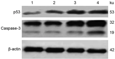
The effect of R16 on cellular p53 and cas-pase-3 expression
SMMC-7721 were treated by R16 at a concen-tration of 2.543 µmol·L-1, 3.912 µmol·L-1, and
5.427 µmol·L-1, respectively, for 24 hours. p53
and caspase-3 protein expression levels were detected by Western blot. Compared with the control group, p53 expression was significantly increased following R16 treatment, in a con-centration dependent manner. Caspase-3 ex-
Statistical analysis
Data were analyzed by EXCEK and SPSS 17.0 software. Quantitative data are expressed using _x ± s. Comparisons between groups were analyzed by t-test.
Results
Inhibition of proliferation of various types of cells by R16
SMMC-7721, EC-9706, and CaCO-2 cells were treated by various concentrations of R16 for 24, 48, and 72 hours, respectively. R16 exhib-ited a substantial inhibitory effect on prolifera-tion of cancer cells in a time and concentraprolifera-tion dependent manner. The IC50 values for 24 hour treatment were 3.912 µmol·L-1 (r2=0.8293),
4.215 µmol·L-1 (r2=0.8926) and 3.380 µmol·L-1
(r2=0.9568), respectively. L-02 cells were
treat-ed by R16 for 24, 48, and 72 hours, and failtreat-ed to show significant inhibitory effect on cell pro-liferation. The IC50 value for 24 hour treatment was 35.224 µmol·L-1 (r2=0.9287). The synthetic
raw material of R16, rufloxacin had no signifi-cant inhibitory effect on SMMC-7721 prolifera-tion, with a 24 hour IC50 of 226.924 µmol·L-1
(r2=0.7981). Sunitinib significantly inhibited
[image:4.612.89.377.71.301.2]SMMC-7731 proliferation, with a IC50 value of
Figure 3. SMMC-7721 cells apoptosis under fluorescent microscope stained by DAPI (× 200). A: Control; B: R16 (2.543 μmol·L-1); C: R16 (3.912 μmol·L-1);
D: R16 (5.427 μmol·L-1).
24 hour treatment of 7.46 µmol·L-1 (r2=0.9650),
signifi-cantly higher than that of R16 on SMMC-7721 cells (Figure 2).
Induction of SMMC-7721 apoptosis by R16
The results of DAPI staining showed that after 24 hour treatment of R16, SMMC-77- 21 cells exhibited apoptotic morphological changes, incl- uding nuclear fragmentation and dissolution, and chroma-tin condensation, shrinkage, and marginalization (Figure 3). TUNEL assay results sh- owed that with an increasing concentration of R16, apop-totic cells were significantly increased in a concentration dependent manner (Figure 4
Figure 4. Induction of apoptosis of SMMC-7721 cells treated with R16 for 24 hours evaluated by TUNEL assay. Rep-resentative images were taken, nuclear stain (DAPI, Left) and apoptotic stain (TUNEL, Right) overlaid. A: Control; B:
R16 (2.543 μmol·L-1); C: R16 (3.912 μmol·L-1); D: R16 (5.427 μmol·L-1) Magnifcation, 100 ×.
Table 1. Apoptotic effects of R16 on
SMMC-7721 cells (_x ± s, n=3)
Group Dose/μmol·L-1 Apoptotic ratio/%
Control 4.15±4.57 R16 2.543 33.85±4.19**
3.912 47.62±7.81** 5.427 66.54±6.92**
**p<0.01 vs control.
pression was also increased, as well as active fragments (Figure 6).
Discussion
In this study, the anti-cancer unsaturated ke- tone skeleton of rhodanine was assembled with fluoroquinolone skeleton at the C-3 posi-tion to construct a series of target compounds of amide fluoroquinolones (rhodanine unsatu-rated ketone). MTT assay was used to screen the target compounds. Twelve fluoroquinolone-rhodanine derivatives had an IC50 below 50 μmol·L-1 after 24 hour treatment on human
hepatocellular carcinoma cells SMMC-7721. Among these derivatives, the IC50 of R16 was 3.893 μmol·L-1, which was significantly lower
than that of the parent compound rufloxacin (226.924 μmol·L-1), and also lower than that of
the clinical anti-cancer agent sunitinib (7.846 μmol·L-1). This results indicate that it was wor-
thy further research and development to
inves-tigate rufloxacin-rhodanine derivatives, which were constructed by assembling rufloxacin and rhodanine unsaturated ketone skeleton, as anti-cancer agents.
In order to understand the difference of R16 on various cancer cells, three types of cancer cells lines (human hepatocellular carcinoma cells SMMC-7721, esophageal cancer cells EC- 9706, and colon cancer CaCO-2 cells) were investigated in this study to detect the inhibito-ry effect of R16 on cell growth. The results show that R16 exhibited a significantly inhibi-tory effect on proliferation of various types of cancer cells. This effect was shown in a time and concentration dependent manner. In this study hepatocyte cell line L-02 was also cul-tured in vitro to detect inhibitory effect of R16 on cell growth. The IC50 on L-02 was 35.244 μmol·L-1, significantly higher than that of R16
on SMMC-7721 cells. This suggested the selec-tivity of R16 on cancer cells and possibly less toxic effects in its future use in clinical practice.
[image:5.612.90.288.354.424.2]Human hepatocellular carcinoma SMMC-7721 cells
Figure 5. Effect of R16 on the cell cycle distribution in SMMC-7721 cells. Cell cycle distribution was demonstrated
by Flow cytometry analysis in SMMC-7721 cells after treatment with 0 (A), 2.543 (B), 3.912 (C) and 5.427 μmol·L-1
(D) R16 respectively for 24 hours.
Figure 6. Effects of R16 on p53 and caspase-3 pro-tein expression in SMMC-7721 cells. 1: Control; 2:
R16 (2.543 μmol·L-1); 3: R16 (3.912 μmol·L-1); 4: R16
(5.427 μmol·L-1).
following R16 treatment by PI staining and flow cytometry. The results show that, compared with the control group, the cell number at G0/G1 phase was significantly increased, accompa-nied by decreased cell number at G2/M phase and no change in S phase, indicating that cell cycle was arrested at G1-S phase. This change was associated with DNA replication. DNA to- poisomerase II is able to catalyze the mutual transformation of DNA topological isomers, ca- talyze the disconnection and binding of DNA double strands, and promote DNA helix unwind-ing to complete the replication process [13]. DNA topoisomerase II toxin led to irreversible broken DNA and damage. The action may acti-vate p53 protein and induce apoptosis [14, 15]. In our experiments, changes of p53 ex- pression were detected by Western-blot. The results suggest that R16 killed SMMC-7721 by DNA damage, and enhanced p53 expression and induction of the apoptotic pathway. The changes in nuclear morphology in SMMC-7721 cells were observed by DAPI staining. Apoptotic SMMC-7721 cells were increased following 24 hour treatment of R16, shown by changes in
nuclear morphology including nucleus shrink-age and fragmentation, chromatin condensa-tion, marginalizacondensa-tion, and appearance of apo- ptotic bodies [16]. TUNEL assay showed signifi-cantly enhanced caspase-3 expression and cleavage fragments with the increased R16 concentration. The results indicate that R16 induced apoptosis by activating capase-3 pa- thway.
In summary, modified rufloxacin-rhodanine derivatives arrested cell cycle at G1-S phase, and significantly inhibited division and prolifera-tion of cancer cells, such as hepatocellular car-cinoma cells, in a selective manner. The anti-cancer effect of R16 was mainly accomplished by induction of apoptosis [17].
Acknowledgements
This work was supported by the National Natural Science Foundation of China (2107- 2045) and Henan Natural Science Foundation (112102310307).
Disclosure of conflict of interest
None.
Address correspondence to: Bin Liu, Institute of Nursing and Health, Henan University, Jin Ming Avenue, College of Nursing and Health, Kaifeng 475004, China. Tel: +86- 371-23880399; Fax: +86- 371-23880399; E-mail: byx0552@163.com
References
[image:6.612.90.288.256.358.2][2] Patel MN, Bhatt BS and Dosi PA. Topoisomer-ase inhibition, nucleolytic and electrolytic con-tribution on DNA binding activity exerted by biological active analogue of coordination compounds. Appl Biochem Biotechnol 2012; 166: 1949-1968.
[3] Schmidt BH, Osheroff N and Berger JM. Struc-ture of a topoisomerase II-DNA-nucleotide complex reveals a new control mechanism for ATPase activity. Nat Struct Mol Biol 2012; 19: 1147-1154.
[4] Berger JM, Gamblin SJ, Harrison SC and Wang JC. Structure and mechanism of DNA topoi-somerase II. Nature 1996; 379: 225-232. [5] Williams GM, Brunnemann KD, Smart DJ,
Mo-lina D, Jeffrey AM, Duan JD, Krebsfaenger N, Kampkoetter A and Schmuck G. Relationship
of cellular topoisomerase IIα inhibition to cyto
-toxicity and published geno-toxicity of fluoroqui -nolone antibiotics in V79 cells. Chem Biol In-teract 2013; 203: 386-390.
[6] Jiang M, Huang O, Xie Z, Wu S, Zhang X, Shen A, Liu H, Chen X, Wu J, Lou Y, Mao Y, Sun K, Hu S, Geng M and Shen K. A novel long non-cod-ing RNA-ARA: adriamycin resistance-associat-ed. Biochem Pharmacol 2014; 87: 254-283. [7] Wang Z, Liang S, Lian X, Liu L, Zhao S, Xuan Q,
Guo L, Liu H, Yang Y, Dong T, Liu Y, Liu Z and
Zhang Q. Identification of proteins responsible
for adriamycin resistance in breast cancer cells using proteomics analysis. Sci Rep 2015; 5: 9301.
[8] Ahmed A and Daneshtalab M. Nonclassical bi-ological activities of quinolone derivatives. J Pharm Pharm Sci 2012; 15: 52-72.
[9] Liang JH and Han X. Structure-activity relation-ships and mechanism of action of macrolides derived from erythromycin as antibacterial agents. Curr Top Med Chem 2013; 13: 3131-3164.
[10] Bringmann G, Dreyer M, Faber JH, Dalsgaard PW, Staerk D, Jaroszewski JW, Ndangalasi H, Mbago F, Brun R and Christensen SB. Ancistro-tanzanine C and related 5,1’- and 7,3’-coupled naphthylisoquinoline alkaloids from Ancistro-cladus tanzaniensis. J Nat Prod 2004; 67: 743-748.
[11] Sun JP, Shi ZY, Liu SM, Kang YH, Hu GQ, Huang-fu CS, Deng JB and Liu B.
Trimethoxy-benzalde-hyde levofloxacin hydrazone inducing the
gr-owth arrest and apoptosis of human hepato-carcinoma cells. Cancer Cell Int 2013; 13: 67. [12] Huo F, Tang NF, Fan YY, Liu SM, Zhang Y, Hu GQ
and Liu B. Benzaldehyde levofloxacin schiff
base induces apoptosis of human hepatocarci-noma cells. Chinese Pharmacological Bulletin 2015; 31: 821-826.
[13] Orlowski C, Mah LJ, Vasireddy RS, El-Osta A and Karagiannis TC. Double-strand breaks and the concept of short- and long-term epigenetic memory. Chromosoma 2011; 120: 129-149. [14] Wanitchakool P, Jariyawat S, Suksen K, Soo-
rukram D, Tuchinda P and Piyachaturawat P. Cleistanthoside A tetraacetate-induced DNA damage leading to cell cycle arrest and apop-tosis with the involvement of p53 in lung can-cer cells. Eur J Pharmacol 2012; 696: 35-42. [15] Calviño E, Estañ MC, Simón GP, Sancho P,
Boy-ano-Adánez Mdel C, de Blas E, Bréard J and
Aller P. Increased apoptotic efficacy of lonida -mine plus arsenic trioxide combination in hu-man leukemia cells. Reactive oxygen species generation and defensive protein kinase (MEK/ERK, Akt/mTOR) modulation. Biochem Pharmacol 2011; 82: 1619-1629.
[16] Omlor GW, Nerlich AG, Tirlapur UK, Urban JP and Guehring T. Loss of notochordal cell phe-notype in 3D-cell cultures: implications for disc physiology and discrepair. Arch Orthop Trauma Surg 2014; 134: 1673-1681.
![Figure 1. Structure of 6-fluoro-7-(4-methylpiperazin-1-yl)-8,1-(thioethylene)-1,4-dihydro-4-oxo-3-[5-(3-car-boxymethyl-2,4-thiazolidinedione) ylidene]-quinoline.](https://thumb-us.123doks.com/thumbv2/123dok_us/290320.528572/2.612.90.289.72.174/figure-structure-methylpiperazin-thioethylene-dihydro-boxymethyl-thiazolidinedione-quinoline.webp)