Table 9 Experimental conditions of the HPLC and of the SFC analyses of polymethoxylated flavones of sweet orange oil
HPLC SFC
Column Zorbax silica, 25 cm;4.6 mm internal diameter (7m)
S5W uncoated silica, 25 cm;4.6 mm internal diameter (5m)
Eluent Hexane}ethyl acetate, 95 : 5 CO2modified with small amounts of methanol
Flow rate 1.6 mL min\1 2 mL min\1for 4 min, then gradient of 2 up to 5 mL min\1 thereafter held constant
Programme Isocratic P"100 atm,T"403C; modifier, 1.5%min\1from 10%to 30%thereafter held constant
Injected amount 20L of a 5%solution of oil in ethyl acetate 100L of a solution obtained by diluting 0.71 g of oil to 20 mL of ethyl acetate
Detection UV absorbance at 315 nm UV absorbance at 315 nm
provide faster separations with the same resolution as that observed in longer columns packed with particles of larger diameter.
See also: II/Chromatography: Liquid: Detectors: Mass Spectrometry. Chromatography: Thin-Layer (Planar): Densitometry and Image Analysis; Modes of
Development: Forced Flow, Over Pressured Layer Chromatography and Centrifugal; Preparative Thin-Layer (Planar) Chromatography. III/Essential Oils: Gas Chromatography; Thin-Layer (Planar) Chromatography; Distillation.
Further Reading
Di Giacomo A and Calvarano M (1978) Il Contenuto di Bergaptene nell’Essenza di Bergamotto Estratta a Freddo.Essenze Derivati Agrumari48: 51}83.
Di Giacomo A and Mincione B (1994)Gli Olii Essenziali Agrumari in Italia. Reggio Calabria: La Ruffa. Dugo P, Mondello L, Stagno d’Alcontres I, Cavazza A and
Dugo G (1997) Oxygen heterocyclic compounds of cit-rus essential oils.Perfumer and Flavorist22: 25}30. McHale D and Sheridan JB (1988) Detection of
adulter-ation of cold-pressed lemon oil.Flavour and Fragrance Journal3: 127}133.
McHale D and Sheridan JB (1989) The oxygen heterocyclic compounds of citrus peel oils.Journal of Essential Oil Research1: 139}149.
Murray RDH, Mendez J and Brown SA (1982)The Natural Coumarins,Occurrence,Chemistry and Biochemistry. Chichester: John Wiley.
Proceedings of the SymposiumCumarine:Ricerca ed
Ap-plicazioni, Padova, Italy 20}22 September 1990.
Padova, Italy: Imprimitur.
Sherma J and Fried B (1996) Handbook of Thin-Layer
Chromatography. New York: Marcel Dekker.
CLINICAL APPLICATIONS
Capillary Electrophoresis
P. G. Righetti, University of Verona, Verona, Italy C. Gelfi, ITBA, CNR, Milan, Italy
Copyright^ 2000 Academic Press
Introduction
The area of clinical applications of capillary elec-trophoresis (CZE) is such a rapidly growingReld that it would be impossible here to cover it in detail. We thus offer a list of major reviews to which the reader is referred for a more comprehensive coverage of the
literature. Such reviews can be divided into:
1. Broad-coverage reviews, such as those of Leh-mannet al. (1997), Perrett (1999; CZE in clinical chemistry) and Guzman et al. (1997; dedicated also to on-line analyte concentration and micro-reaction). Also of interest are special issues of the Journal of Chromatography B dedicated to CZE in the life sciences (Krstulovic 1997) and of Elec-trophoresisdevoted to CZE in the clinical sciences (Landers 1997) and in forensic science (McCord, 1998).
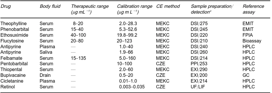
Table 1 Selected validated CZE/MEKCaassays for drugsb Drug Body fluid Therapeutic range
(g mL\1)
Calibration range (g m L\1)
CE method Sample preparation/ detectionc
Reference assay
Theophylline Serum 8}20 2.0}28.3 MEKC DSI/275 EMIT
Phenobarbital Serum 15}40 5.3}52.6 MEKC DSI/245 EMIT
Ethosuximide Serum 40}100 19.8}99.2 MEKC DSI/220 FPIA
Flucytosine Serum 20}80 20}123 MEKC DSI/210 Bioassay
Antipyrine Plasma * 1.0}40 MEKC DSI/240 HPLC
Antipyrine Saliva * 1.9}66 MEKC DSI/260 HPLC
Felbamate Serum 15}135 5.0}160 MEKC DSI/214 HPLC
Pentobarbital Serum * 10}100 CZE PPI/253 HPLC
Thiopental Serum * 2.0}60 MEKC EXI/290 HPLC
Bupivacaine Drain * 0.5}20 CZE EXI/200 GC
Cicletanine Plasma * 0.01}1.0 MEKC EXI/214 HPLC
Retinol Serum * 0.003}0.035 CZE UF/LIF HPLC
aMEKC, micellar electrokinetic capillary chromatography; CZE, capillary zone electrophoresis; DSI, direct sample injection; EXI, extract injection; PPI, injection of supernatant after protein precipitation; EMIT, enzyme-multiplied immunoassay technique; FPIA, fluores-cence polarization immunoassay; GC, gas chromatography; HPLC, high-performance liquid chromatography; UF, ultrafiltration; LIF, laser-induced fluorescence detection with 325 nm excitation and 465 nm emission.
bReprinted from Thormann W, Zhang CX and Schmutz A (1996)Therapeutic Drug Monitor 18, 506}520, with permission. cThe number represents the detection wavelength.
DNA for molecular diagnostics), Jellum et al. (1996, analysis of urinary diagnostic metabolites and serum proteins), Lazaruk et al. (1998; genotyping of forensic short tandem repeat systems).
CZE:
Some Basic Concepts Related to
Clinical Chemistry
An important aspect of CZE relevant to clinical chemistry is the paradox by which one of the noted advantages of CZE, namely the small volume of the capillary, also leads to a signiRcant drawback, i.e. the minute amount (3}10 nL) of sample introduced, which results in poor concentration limits of detec-tion, one to two orders of magnitude lower than in high-performance liquid chromatography (HPLC). Thus, preconcentration techniques are often required for compounds present in very low concentrations in biological Suids. These include transient isotacho-phoresis, analyte stacking, Reld-ampliRed sample injection (all on-capillary techniques) or off-column preconcentration techniques, such as liquid} liquid or liquid}solid extraction. Alternative on-column methods include miniaturized solid-phase extraction with cartridges containing reversed-phase HPLC packing materials or with impregnated mem-branes. When analysing small analytes or drugs in sera, it is often necessary to deproteinize the sample. This can be efRciently achieved by extracting sera with 60% acetonitrile, which accomplishes two tasks: protein precipitation and analyte stacking upon electrokinetic injection because of the very low
con-ductivity of such a solution. Some areas or interest where CZE offers unique resolution and sample quantiRcation are discussed below.
Drug Analysis
With the more efRcient therapeutic application of various drugs and the necessity for screening and conRrmation of drugs in body Suids for diagnostic and research purposes, there has evolved a need for reliable analytical procedures. CZE is becoming the method of choice for drug monitoring in bodySuids, including plasma, serum, saliva and urine. Some relevant data are given inTables 1+3.
Pro\ling Clinically Important Metabolites in Urines
Table 2 Selected CZE/MEKCascreening confirmation assays for illicit, abused and banned drugsb
Drugs, drug classes Body fluid, tissue CZE method Sample preparation Reference assay
Barbiturates Urine, serum MEKC DSI, EXI EMIT
Salicylate, paracetamol, antiepileptics
Urine, serum MEKC, CZE DSI, EXI FPIA, EMIT, UF
11-Nor--tetrahydro
cannabinol-9-carboxylic acid
Urine MEKC EXI FPIA
Methadone and its primary metabolite
Urine CZE DSI, EXI EMIT, GC-MS
Benzodiazepines Urine MEKC EXI EMIT, GC-MS
Benzoylecgonine, opioids, methaqualone, amphetamines
Urine MEKC EXI EMIT
Cocaine and all above mentioned classes
Urine MEKC, CZE EXI EMIT, FPIA, GC-MS
Cocaine, morphine Hair MEKC EXI HPLC
-Blockers Serum MEKC EXI *
Diuretics Urine, serum CZE EXI GC-MS
aFor abbreviations, see Table 1.
bReprinted from Thormann W, Zhang CX and Schmutz A (1996)Therapeutic Drug Moniter 18, 506}520, with permission.
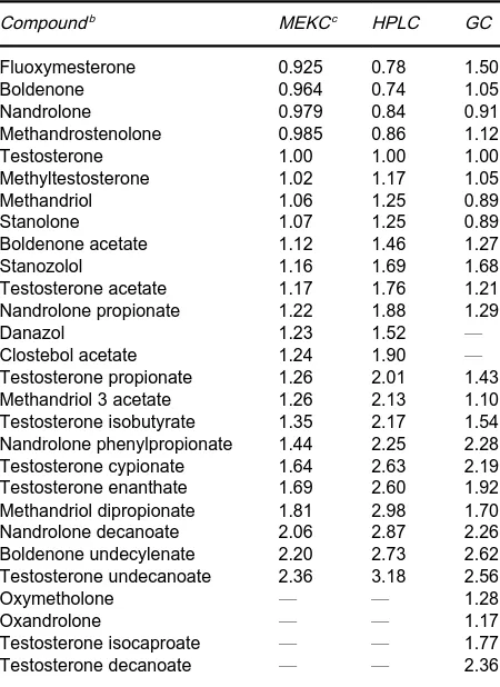
Table 3 Retention of anabolic steroids relative to testosteronea
Compoundb MEKCc HPLC GC
Fluoxymesterone 0.925 0.78 1.50
Boldenone 0.964 0.74 1.05
Nandrolone 0.979 0.84 0.91
Methandrostenolone 0.985 0.86 1.12
Testosterone 1.00 1.00 1.00
Methyltestosterone 1.02 1.17 1.05
Methandriol 1.06 1.25 0.89
Stanolone 1.07 1.25 0.89
Boldenone acetate 1.12 1.46 1.27
Stanozolol 1.16 1.69 1.68
Testosterone acetate 1.17 1.76 1.21
Nandrolone propionate 1.22 1.88 1.29
Danazol 1.23 1.52 *
Clostebol acetate 1.24 1.90 *
Testosterone propionate 1.26 2.01 1.43
Methandriol 3 acetate 1.26 2.13 1.10
Testosterone isobutyrate 1.35 2.17 1.54 Nandrolone phenylpropionate 1.44 2.25 2.28
Testosterone cypionate 1.64 2.63 2.19
Testosterone enanthate 1.69 2.60 1.92
Methandriol dipropionate 1.81 2.98 1.70
Nandrolone decanoate 2.06 2.87 2.26
Boldenone undecylenate 2.20 2.73 2.62
Testosterone undecanoate 2.36 3.18 2.56
Oxymetholone * * 1.28
Oxandrolone * * 1.17
Testosterone isocaproate * * 1.77
Testosterone decanoate * * 2.36
aReprinted from Lurie IS (1996) International Laboratory, with permission.
bIn order of increasing retention times. cFor abbreviations, see Table 1.
directly on urine without extensive sample clean-up or analyte derivatization. Some examples are given in
Table 4. Other interesting data on proRling the
following metabolites: orotic acid, pyroglutamate, adenylosuccinate and propionic acid, for the follow-ing diseases: HHH-syndrome (hyperornithinemia-hyperammonemia-homocitrullinuria), glutathione deRciency, adenylosuccinase deRciency and propionyl CoA carboxylase deRciency, respectively, can be found in Jellumet al. (1997).
Pro\ling Proteins in Biological Matrices
Separation and quantiRcation of distinct proteins from biological matrices is another goal now being accomplished by CZE. A list of some major proteins of importance for clinical diagnosis is given in
Table 5. In some cases, immunosubtraction can be an efRcient way of quantifying some protein families by CZE. A typical example is the quantiRcation of speci-Rc immunoglobulin subclasses by sequential immunosubtraction. The sample is exposed to Rve different Sepharose supports, each containing an im-munoglobulin-speciRc binder. Three of them are spe-ciRc for the heavy chains IgG, IgA and IgM and two are speciRc for the light chainsor. After incuba-tion and sedimentaincuba-tion, the treated samples and an untreated control are separated by CZE. Six elec-tropherograms are generated per sample. The class and type of monoclonal component can be deter-mined by overlaying electropherograms from before and after immunosubtraction.
CZE Separations of Clinically Relevant
Diagnostic DNA
[image:3.568.51.276.358.667.2]Table 4 Normal constituents of human urine and expected response for CZE at a Cu electrodea Compound Related metabolic
disorder
Normal concentrations (M)b Migration time in 0.1 N NaOH (min)
Detection limit (M)
Alditols
Erythritol * 608 12.6 0.5
Inositol Diabetes, renal failure 357 12.6 0.3
Ribitol * 35 12.8 *
Xylitol * 35 12.8 *
Arabitol * 195 12.8 *
Glucitol * 35 12.9 0.5
Mannitol Diabetes 104 14.6 0.5
Carbohydrates
Sucrose * 43 16.9 1
Lactose * 15 19.6 1
Fucose * 97 19.6 *
Galactose Galactosaemia 29 21.3 1
Glucose Diabetes 262 22.4 1
Rhamnose * 115 22.8 *
Arabinose Pentosuria 89 23.4 *
Fructose Fructosuria 72 23.4 1
Xylose Pentosuria 53 25.4 *
Ribose Pentosuria 31 26.2 1
Amino acids
Lysine Hyperlysinaemia 328 31.6 4
Threonine Aminoaciduria 183 35.4 2
Histidine Histidinaemia 860 43.1 1
Others
Creatinine Muscle and renal disease 8800 12.8 80
Uric acid Gout 2093 '60 1.6
aReprinted from Hong J and Baldwin RP (1997)Journal of Capillary Electrophoresis, 4, 65}71, with permission. bCalculated assuming that the average urine volume for a 24-h period is 1.5 L.
Table 5 Survey of CZE separations of proteins in biological matricesa
Proteins Matrix CZEbmode Detection mode
Immunoglobulin G, transferrin, albumin, prealbumin,-trace proteins
CSF CZE UV 185 nm
Apolipoprotein A-I, A-II, B100, B48, C-III and E
Serum MECC UV 190 nm
Lipoprotein subfractions (HDL, VLDL, IDL, LDL)
Serum CITP Vis 570 nm
Leucine aminopeptidase Serum, urine CZE LIF
Cerebrospinal fluid proteins CSF CZE UF 200 nm
Myoglobin Urine, tissue CZE Vis 405 nm
Albumin,1-acidic glycoprotein, transferrin,
-microglobulin, immunoglobulin light chains
Urine CZE UV 200 nm
Monoclonal antibodies (anti-TNF, anti-CEA) Serum containing culture medium CZE, CIEF, CGE UV 200 nm
Imidodipeptides (prolidase deficiency) Urine CZE UV 269 nm
Cathepsin D Breast tissue CZE UV 214 nm
Hemoglobin variants Plasma CZE, CIEF UV 210 nm
36 low side Mrproteins, cut-off 30 and 5 kDa Seminal, vaginal fluids, serum, saliva CZE UV 214 nm aReprinted from Lehmann R, Voelter W and Liebich HM (1997)Journal of Chromatography B 697, 37}66, with permission. bAbbreviations: CIEF, capillary isoelectric focusing; CGE, capillary gel electrophoresis; CITP, capillary isotachophoresis; CSF, cerebrospinal fluid; CEA, carcino-embryonic antigen; TNF, tumour necrosis factor, HDL, high density lipoproteins; LDL, low density lipoproteins; VLDL, very low density lipoproteins.
celluloses) for the analysis of polymerase chain reac-tion (PCR) products of clinically relevant, diagnostic DNA have been reported. Table 6lists some major
[image:4.568.49.518.475.667.2]Table 6 Survey of selected capillary gel electrophoretic separations of clinically relevant diagnostic DNAa
Disease DNA amplification Sieving polymers Detection
Human genetics
Cystic fibrosis Allele specific PCR and restriction digest of PCR products (deletion)
6%linear PAAb UV 254 nm
Cystic fibrosis PCR,F508 6%linear PAA UV 254 nm
Cystic fibrosis PCR/GATT microsatellites 6%linear PAA UV 254 nm
Cystic fibrosis Point mutants, TGCE 8%linear poly(AAEE) UV 254 nm
Duchenne/Becker muscular dystrophy PCR multiplex reaction 6}10%linear PAA UV 260 nm
Dystrophin gene RFLP 0.5%HPMC UV 254 nm
Thalassaemia Point mutants, TGCE 4%poly(AAP), 1.5%HEC UV 254 nm
Congenital adrenal hyperplasia PCR deletion 6%linear PAA UV 254 nm
Androgen insensitivity syndrome CAG triplet analysis 6%linear PAA UV 254 nm
Kennedy’s disease CAG triplet expansion 8%poly(AAEE) UV 254 nm
N-ras gene (human cancer) Point mutants SSCP 8%PAA UV 260 nm
ERBB2 oncogene RFLP 0.5%HPMC UV 260 nm
TX gene PCR 3%PAA LIF
P-53 SSCP 4%PAA UV 260 nm
P-53 SSCP 2%PAA LIF
Cancer (microsatellites instability) PCR Bio-Rad sieving polymer UV 260 nm
Apolipoprotein B gene PCR/VNTR 0.7%MC UV 260 nm
Apolipoprotein E gene PCR/RFLP 3%T PAA UV 260 nm
Apolipoprotein E gene PCR/RFLP Beckman e/CAP LIF
Medium chain AcylCoA dehydrogenase deficiency
PCR/allele specific Polyacrylamide gel LIF
von Willebrand Factor gene PCR/VNTR 1%HEC LIF
Fetal DNA (Y-chromosome) PCR Beckman dsDNA 1000 gel buffer LIF
Quantitative gene dosage
Down’s Syndrome Quantitative PCR 8%PAA UV 254 nm
Rh D/d genotyping Quantitative PCR 8%PAA UV 254 nm
Follicular lymphomas Competitive PCR 4%PAA UV 260 nm
Basic fibroblast growth factor Competitive RT-PCR 6%PAA UV 254 nm
Microbiology/virology
Mycobacterium tuberculosis SSCP and ddF 1%HEC or 3%T, 0.5%C PAA gel LIF
Hepatitis C virus RT-PCR 1%HEC LIF
Polio Virus RT-PCR 3%T linear PAA UV 254 nm
HIV-1 RT-PCR 3%linear PAA LIF
Mitochondrial DNA PCR 1%HEC LIF
Mitochondrial DNA PCR 0.5%MC LIF
VNTRs at locus D1S80 PCR/VNTR 0.5%HEC LIF
VNTRs at locus D1S80 PCR/VNTR 0.5%MC LIF
VNTRs at locus HUMTH01 PCR/VNTR 1%HEC LIF
VNTRs at locus HUMTH01 PCR/VNTR 3%T, 3%C gel UV 260 nm
Therapeutic DNA
Antisense oligonucleotides 18%PAA MALDI-MS LIF
Antisense oligonucleotides 10%PAA, isoelectric His UV 254 nm
Antisense oligonucleotides 10%T PAA, pH gradient UV 254 nm
aReprinted from Righetti PG and Gelfi C (1997), with permission.
bAbbreviations: PAA, polyacrylamide; VNMTR, variable number of tandem repeats; HEC, hydroxyethyl cellulose; MC, methyl cellulose; SSCP, single strand chain polymorphism; RT, reverse transcription; HPMS, hydroxypropyl methyl cellulose; AAP, acryloyl amino propanol; AAEE, acryloyl amino ethoxy ethanol, TGCE, temperature gradient capillary electrophoresis; RFLP, restriction fragment length polymorphism; LIF, laser induced fluorescence.
Examples of Some Separations
It is quite difRcult to compress in such a few pages the vast literature in theReld of clinico-chemical
Figure 1 Simultaneous detection ofF508, G542X, N1303K and 1717-1GPA mutations in cystic fibrosis by CZE in polymer networks. Traces: A, patient carrying the mutations 1717-1GPA andF508; B, patient affected by the G542X/N1303K mutations; A#B, artificial mixture of the amplified DNA fragments of patients A and B. Conditions: 100m ID, 37 cm long capillary, filled with a viscous solution of linear 6%T polyacrylamide in 100 mmol L\1TBE (Tris-borate-EDTA) buffer, 10mol L\1ethidium bromide, pH 8.3; Run at 165 V cm\1with detection at 254 nm; electrophoretic sample injection at 165 V cm\1for 8 s. Reprinted from Gelfi C, Righetti PG, Magnani C, Cremonesi L and Ferrari M (1994)Clinica Chimica Acta 229, 181}189, with permission from Elsevier Science.
simultaneous detection of four mutations, F508, G542X, N1303K and 1717-1GPA in cysticRbrosis (CF). This is an interesting example, in that it offers a fast and reliable method for the simultaneous detec-tion of mutadetec-tions which are predominant in a given population. In Italy, a survey of 391 CF patients, originating from all geographical regions, revealed that F508 (53% of CF chromosomes), G542X (4%), 1717-1GPA (4%) and N1303K (4%) are the most frequent mutations, accounting for 65% of all molecular defects pertaining to CF.Figure 2gives the CZE analysis for the Duchenne (DMD) and Becker (BMD) muscular dystrophies, which represent the two most common myopathies described to date. They are given the names of Chamberlain and Beggs since these two scientists proposed two PCR assays (each based on co-ampliRcation of nine dystrophin gene exons) allowing for the detection of over 98% DMD/BMD deletions. Thus, a method attempting simultaneous analysis of DMD/BMD should offer unambiguous resolution and identiRcation of 18 frag-ments ranging in size fromc. 100}500 bp. This is in fact achieved in the lower trace ofFigure 2 (repres-enting a healthy individual). The upper trace shows a patient affected by muscular dystrophy, in which four fragments (196, 202, 331 and 357 bp) are miss-ing. We hope that these two examples, albeit limited,
provide an insight on the unique resolving power and capability of CZE as applied to problem solving in clinical chemistry.
Conclusions and Future Horizons
Figure 2 Screening for Duchenne (DMD) and Becker (BMD) muscular dystrophies by CZE in sieving liquid polymers. The upper trace represents the separation of 14 exons of modified deleted Chamberlains’ and Beggs’ mixed multiplex. The lower electrophero-gram shows the separation of 18 exons of modified nondeleted Chamberlains’ and Beggs’ multiplex. All runs were in a 32 cm long, 75m internal diameter capillary, filled with short-chain polyacrylamide, obtained by chain transfer at 703C, in 89 mMTBE buffer, pH 8.3. Run: 165 V cm\1with detection at 254 nm. Sample injection: 100 V cm\1for 25 s. Reprinted from Gelfi C, Orsi A, Leoncini F, Righetti PG, Spiga I, Carrera P and Ferrari M (1995)BioTechniques 19, 254}263, with permission from Elsevier Science.
by photolithographic masking and chemical etching techniques and formed by bonding the etched sub-strate to a plain glass plate. Capillaries 30}70m wide, about 10m high and a few centimetres long have been shown to provide analytical runs in a few seconds.
Further Reading
Guzman NA, Park SS, Schaufelberger D, Hernandez L, Paez X, Rada P, Tomlison AJ and Naylor S (1997) New approaches in clinical chemistry: on-line analyte concen-tration and microreaction capillary electrophoresis for determination of drugs, metabolic intermediates and biopolymers in biologicalSuids.Journal of Chromato-graphy B697: 37}66.
Hong J and Baldwin RP (1997) ProRling clinically impor-tant metabolites in human urine by capillary elec-trophoresis and electrochemical detection. Journal of Capillary Electrophoresis4: 65}71.
Jellum E, Dollekamp H and Blessum C (1997) Capillary electrophoresis for clinical problem solving: analysis of urinary diagnostic metabolites and serum proteins.
Journal of Chromatography B683: 55}65.
Krstulovic AM (guest ed.) (1997) Capillary electrophoresis in the clinical sciences. Journal of Chromatography B697: 1}289.
Landers JP (guest ed.) (1997) Capillary electrophoresis in the clinical sciences.Electrophoresis18: 1707}1906. Lazaruk K, Walsh PS, Oaks F, Gilbert D, Rosenblum BB,
Menchen S, Scheibler D, Wenz HM, Holt C and Wallin J (1998) Genotyping of forensic short tandem repeat (STR) systems based on sizing precision in a capil-lary electrophoresis instrument. Electrophoresis 19: 86}93.
Lehmann R, Voelter W and Liebich HM (1997) Capillary zone electrophoresis in clinical chemistry. Journal of
Chromatography B697: 37}66.
Lurie IS (1996) Applications of capillary zone electrophor-esis to the analysis of seized drugs.International Labor-atory24: 21}29.
McCord BR (1998) Capillary Electrophoresis in Forensic Science.Electrophoresis19: 1}126.
Perrett D (1999) Capillary zone electrophoresis in clinical chemistry. Annals of Clinical Biochemistry 36: 133}150.
Righetti PG and GelR C (1997b) Non-isocratic capillary electrophoresis for detection of DNA point mutations.
Journal of Chromatography B697: 195}205.
Righetti PG and GelR C (1998) Analysis of clinically-relevant, diagnostic DNA by capillary zone and double-gradient gel slab electrophoresis. Journal of
Chromatography A806: 97}112.
Thormann W (1997) Drug monitoring by capillary elec-trophoresis. In Wong SHY, Sunshine I (Eds)Analytical Therapeutic Drug Monitoring, pp. 1}19. Boca Raton: CRC Press.
Thormann W, Zhang CX and Schmutz A (1996) Capillary zone electrophoresis for drug analysis in bodySuids.
Therapeutic Drug Monitor18: 506}520.
Electrophoresis
J.-D. Tissot, A. Layer and P. Schneider,
Fondation CRS,Lausanne, Switzerland
H. Henry, Centre Hospitalier Universitaire Vaudois,
Lausanne, Switzerland
Copyright^ 2000 Academic Press
Introduction
It is somewhat arbitrary to outline electrophoretic applications that are used in a routine clinical labor-atory because they are highly dependent on the speci-Rcity of each application. Nowadays, a multitude of electrophoretic methods are routinely used in various clinical laboratories, according to the speciRc re-search being developed. More and more sophisticated methods are needed to resolve speciRc clinical prob-lems, the reason why specialization of laboratories is mandatory in order to provide accurate results at the lowest possible cost. In addition, quality control is of major importance. Therefore, automation is pro-gressively introduced in all the steps involved in the analytical process, and only a few manual methods will survive in the future. However, highly sophisticated electrophoretic techniques should be maintained and developed in a limited number of specialized laboratories, in order to resolve the differ-ent problems that are encountered in clinical medicine. Here, we present selected examples of electrophoretic techniques that are employed in the clinical laboratory.
Serum Protein Electrophoresis
Electrophoretic techniques for the separation of human serum proteins have been used forRfty years. Resolution has been improved by the use of support media such as paper, starch gel, cellulose acetate, agarose, and polyacrylamide gels, which have ren-dered electrophoretic methods very popular in the diagnostic area. However, many of those methods have remained labour intensive, being difRcult to
automate. Serum protein electrophoresis is widely used in clinical laboratories, especially for the evalu-ation of changes in proteins associated with inS am-mation, liver or kidney diseases as well as for the detection and identiRcation of paraproteins. Tradi-tional clinical electrophoretic procedures are manual methods that use agarose gels or cellulose acetate membranes as the separation bed. Quantitation of the Rve major serum fractions is done by densitometric scanning of the gel or the membrane. Clinical inter-pretation is based on the alteration of the content of one or more of theRve fractions. Agarose, as support-ing medium for protein electrophoresis, has been re-ported to give better resolution as well as to allow better detection of paraproteins than is cellulose acetate. Semiautomated agarose electrophoresis and immunoRxation can be performed with various commercially available systems. No differences be-tween manual and semiautomated methods have been seen with respect to paraprotein identiRcation.