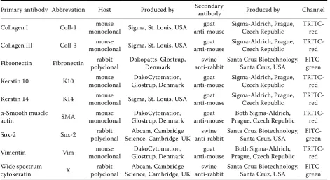
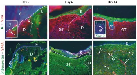
Immunohistological changes in skin wounds during the early periods of healing in a rat model
Full text
Figure

Related documents
evelopment of the Patients’ Expectations Questionnaire (PEQ) and examination of it encompasses by reviewing surveys of primary care and hospital outpatients before and Three
The objective of this research was to determine the impact of four winter cover crop treatments (no cover crop, hairy vetch, winter wheat, and crimson clover) and two
Akhavan A, Keith JD, Bastacky SI, Cai C, Wang Y, Nelson JB: The proportion of cores with high-grade prostatic intraepithelial neoplasia on extended-pattern needle biopsy
Thus, reducing the rate of redundant admissions by using external information produced larger odds ratios (of the β coefficients; e.g. viewing external information on patients
reported the first published case of an adult toxic epidermal necrolysis patient with ureteropelvic mu- cosa damage, haematuria, extensive ureteral mucosal sloughing, and acute
As Eisenberg (1998) emphasized in her study that Big6 is an inquiry process which is very critical to students’ learning in all areas like social studies (research process),
The scattergram represents the distribution with age of 69 determinations of concentration of potassium in serum of 39 premature infants with respiratory distress syndrome (Table
