The effects of inhalation salbutamol administration
on systemic and pulmonary hemodynamic, pulmonary
mechanics and oxygen balance during general
anaesthesia in the horse
M. Patschova, R. Kabes, S. Krisova
Faculty of Veterinary Medicine, University of Veterinary and Pharmaceutical Sciences, Brno, Czech Republic
ABSTRACT: This research aimed to determine the effect of aerosolized salbutamol administration on systemic
and pulmonary hemodynamic, pulmonary mechanics and oxygen balance in healthy horses during general anaes-thesia. Six healthy Thoroughbreds (body weight range 471–587 kg) underwent two general anaesthesias in dorsal recumbency with and without aerosolized salbutamol administration in randomized order with a one month washout period. The anaesthesia was induced by 1.1 mg/kg of xylazine, 0.02 mg/kg of diazepam and 2.2 mg/kg of ketamine, maintained with isoflurane in oxygen and air and horses were mechanically ventilated. Measure-ment of arterial and pulmonary arterial blood pressures, cardiac output and arterial and mixed venous blood gas analysis was carried out. Spirometry was performed using a Horse-lite. After achieving a steady state, baseline (T0) values of cardiac output, systemic and pulmonary arterial blood pressures, heart rate, dynamic compliance, airway resistance and arterial and mixed venous blood gas values and pH were recorded in both groups. In the S-group (salbutamol), 2 µg/kg of aerosolized salbutamol were administered synchronously with inspirium into the tracheal tube. In both groups data were recorded at 15, 30, 45 and 60 min (T15, T30, T45, T60) after the baseline. PaO2/FiO2 ratio, oxygen consumption (VO2), oxygen delivery (DO2), pulmonary shunt values were calculated. Data were tested for normality and compared within each group: T0 value with T15, T30, T45, T60 values using Wilcoxon’s test with Bonferoni correction (significance level 0.0125). For each time point, comparisons were made between the S- and C-groups (control) using Wilcoxon’s test. In the S-group, there was a significant increase in values (mean ± SD) of cardiac output (l/min), T0 (38 ± 7), a peak at T15 (64 ± 25.5), significantly higher values persisted throughout the period of anaesthesia; heart rate (beats/min), T0 (32 ± 2), T15 (40 ± 6), T30 (38 ± 5); DO2 (l/min), T0 (5.8 ± 0.8), a peak at T15 (9.6 ± 3.2), significantly higher values persisted until the end of anaesthesia and VO2 (l/min), T0 (1.1 ± 0.5), T30 (1.6 ± 0.7) and T45 (1.8 ± 0.5). In the C-group, there was a significant decrease in values of PaO2/FiO2 ratio from T0 (176 ± 67) to a minimum at T60 (114 ± 36) and in DO2 from T0 (6 ± 2.3) to a minimum at T60 (4.3 ± 1.2). A comparison of the S- and C-groups did not reveal any difference in the baseline data. Subsequently, significantly higher values of cardiac output, heart rate, DO2, and the PaO2/FiO2 ratio were found in the S-group compared to the C-group. Pulmonary arterial blood pressure was significantly lower in the S-group. Aerosolized salbutamol administration in healthy horses during general anaesthesia caused hemodynamic changes which resulted in an elevation of oxygen delivery. It can have a positive effect on arterial oxygenation, but the effect varies between individuals.
Keywords: horse; hypoxaemia; salbutamol; general anaesthesia; arterial oxygenation
General anaesthesia in the horse is accompanied by gas exchange impairment and development of hypoxaemia (Hall et al., 1968; Gillespie et al., 1969; Steffey et al., 1977; Trim and Wan, 1990; Whitehair and Willits, 1999). A decrease in PaO2 and elevation of PaCO2 is greater during spontaneous ventilation compared to mechanical ventilation (Hall et al., 1968; Gillespie et al., 1969).
Alteration of blood gas values and an increased alveolar-to-arterial oxygen gradient are the results of a ventilation and perfusion (V/Q) mismatch. This is a consequence of redistribution of perfusion caused by the force of gravity, redistribution of in-spired gas and hypoventilation of the dependent lung, a decrease in cardiac output, development of atelectasis and an increase in pulmonary shunt (Hall et al., 1968; Gillespie et al., 1969; Nyman et al., 1990; Trim and Wan, 1990; Moens et al., 1995, 1998). Atelectasis formation in dependent lung re-gions is considered to be the main cause of impaired arterial oxygenation (Nyman et al., 1990; Moens et al., 1995). The extent of hypoxaemia is also influ-enced by body position during anaesthesia, age, body weight, shape of the abdominal contour and amount of abdominal contents (Steffey et al., 1977; Moens et al., 1995; Whitehair and Willits, 1999).
Intermittent positive pressure ventilation (IPPV) with the addition of positive end expiratory pressure (PEEP) or a combination of differential ventilation with selective PEEP of the dependent lung is effec-tive in improving arterial oxygenation (Moens et al., 1994, 1998). The disadvantage of the latter technique is the technical difficulty involved. Implementation of a “recruitment manoeuvre” during IPPV allows opening of the collapsed lung areas and results in a significant increase in PaO2 and decrease in pulmo-nary shunt (Wettstein et al., 2006). However, high inspiratory pressure and PEEP are accompanied by a depression of hemodynamic and cardiac output (Moens et al., 1998; Wettstein et al., 2006).
Pulsed delivery of nitric oxide synchronized with inspirium was shown to successfully increase PaO2 values and decreased the amount of pulmonary shunt (Heinonen et al., 2001).
Studies concerned with the systemic or inha-lant use of β2 adrenergic agonists in hypoxaemic anaesthetized horses have reported controversial results. During inhalation anaesthesia, intravenous administration of clenbuterol was described to elicit an elevation in PaO2. Adverse effects were also ob-served; tachycardia and profuse sweating (Gleed and Dobson, 1990; Keegan et al., 1991). Dodam et al.
(1993) found no improvement in PaO2 values after intravenous administration of clenbuterol in horses during total intravenous anaesthesia. Conversely, PaO2 decreased as a result of β2 receptor stimula-tion, vasodilatastimula-tion, and an increase in heart rate and oxygen consumption. In the experimental study of Lee et al. (1998), intravenous administration of clenbuterol failed to produce any improvement in PaO2 and caused a temporary increase in heart rate, cardiac output and muscle perfusion in horses dur-ing inhalation anaesthesia. Sweatdur-ing also occurred. The sympathomimetic effects of clenbuterol, led to an increase in oxygen consumption.
Salbutamol is a selective β2 adrenergic agonist; its inhalant administration is used in human medicine in patients suffering from primary pulmonary hy-pertension (Spiekerkoetter et al., 2002) or asthma (Wong et al., 1990).
A clinical study by Robertson and Bailey (2002), showed that administration of aerosolized salbutamol in horses under inhalation anaesthesia with PaO2 val-ues less than 9.3 kPa, led to an increase in these valval-ues within 20 min of treatment. The exact mechanism of action was not determined in that study.
The aim of our experimental study was to deter-mine the effect of inhalation salbutamol adminis-tration on systemic and pulmonary hemodynamics, oxygen balance and pulmonary mechanics during general anaesthesia in healthy horses.
MATERIAL AND METHODS
The project for this experimental study (No. 1/2008) was approved by the Ethical Committee of the University of Veterinary and Pharmaceutical Sciences Brno and by the Ministry of Education, Youth and Sports of the Czech Republic.
Experimental animals
Anaesthetic protocol and instrumentation
Each horse underwent two general anaesthesias in dorsal recumbency, with and without aerosolized salbutamol administration (Ventolin; Glaxo Group Ltd, Greenford, Great Britain), in randomized or-der, with a one month washout period.
A venous catheter (Secalon T; Becton Dickinson Critical Care Systems Pte Ltd, Singapore) was placed into the right jugular vein before the induction of anaesthesia. Horses were sedated with 1.1 mg/kg of xylazine (Xylapan 2%; Vetoquinol Biowet, Gorzow Wlkp, Poland) intravenously and anaesthesia was induced with 0.02 mg/kg of diazepam (Apaurin; Krka, Novo Mesto, Slovenia) and 2.2 mg/kg of keta-mine (Narketan 10%; Vetoquinol SA, Lure Cedex, France) intravenously. After orotracheal intuba-tion with an endotracheal tube (Smiths Medical Pm Inc, Waukesha, USA) of suitable diameter, horses were transported to the operating theatre, placed in dorsal recumbency and connected to a large ani-mal circle system (Stephan GmbH, Gackenbach, Germany). IPPV in a control pressure setup with peak inspiratory pressure 20–25 cm H2O was used. Maintenance of anaesthesia was achieved with iso-flurane (Aerrane; Baxter SA, Lessines, Belgium) in oxygen and air. The aim was to reach a fraction of inspired oxygen (FiO2) of 0.60 but this param-eter varied between 0.53 and 0.60. The actually reached values were noted and used for calcula-tions. Ventilation was set to attain end tidal values of CO2 (Et CO2) between 5 kPa and 6 kPa and an Et value of isoflurane at 1.4%. At the beginning of anaesthesia, horses received a bolus of 1 l of colloids (Voluven; Fresenius Kabi, Bad Homburg, Germany) and supportive therapy continued by a constant rate infusion of crystalloids (Infusio Ringeri Mediekos; In Mediec, Luhacovice, Czech Republic) at a rate of 10 ml/kg/h.
An arterial catheter (Surflo 22 G; Terumo, Leuwen, Belgium) was placed into the right arte-ria facialis for artearte-rial blood sample withdrawal which was performed anaerobically using special syringes (Monovette 2 ml LH; Sarstedt, Nümbrecht, Germany). A second arterial catheter (BD Arterial Cannula with FloSwitch; BD, Swindon, Great Britain) was placed into the raised left arteria carotis communis for systemic blood pressure and lithium dilution cardiac output measurements. An arterial catheter was connected to a pressure transducer, which was zeroed at the level of the right atrium.
An 8.5 F introducer (Intro-Flex; Baxter Healthcare Co, Irvine, USA) was placed into the right jugular vein, near the apertura thoracis cranialis for the in-troduction of a 7 F Swan-Ganz catheter (Swan-Ganz; Edwards Lifesciences, Irvine, USA) for pulmonary arterial blood pressure measurement and blood sample withdrawal. The distal port of the catheter was placed in the pulmonary artery and its position was confirmed by the characteristic shape of the pressure curve on a Datex-Ohmeda monitor (Datex-Ohmeda S/5; Datex-(Datex-Ohmeda Inc, Madison, USA). The introducer was placed according to Seldinger’s method (Seldinger, 1953). A pressure transducer for pulmonary arterial blood pressure measurement was set at the level of the right atrium.
Lithium dilution cardiac output measurements were performed using a commercial machine (LiDCOplus; LiDCO Ltd, London, Great Britain) and lithium chloride (LiCl; LiDCO Ltd, London, Great Britain) at a concentration of 0.15 mmol/ml was used at a dose of 0.03 mg/kg and administered intravenously. A calculated bolus of LiCl increased by the dead space value of the catheter was deliv-ered through the jugular catheter. The measure-ment procedure was performed according to the manufacturer’s instructions.
Blood gas analysis of arterial and mixed venous blood samples was performed using a Blood Gas analyzer with the CO-Ox module (Rapidlab 855; Bayer, Germany).
Monitoring
The following parameters were monitored con-tinuously during anaesthesia: heart rate, ECG, SpO2, systemic and pulmonary arterial blood pressures, Fi and Et of O2, CO2 and isoflurane, capnography and respiratory rate. Spirometric measurements of respiratory pressures and volumes were performed using Horse-lite, a Pitot-based flow meter (Moens et al., 2009) and dynamic compliance and airway resistance were calculated.
Experimental protocol
blood pressures, dynamic compliance, airway resist-ance and cardiac output. From this data, the following values were calculated according to the formulas be-low: PaO2/FiO2 ratio, oxygen delivery (DO2), oxygen consumption (VO2) and pulmonary shunt (Qs/Qt). PaO2/FiO2 ratio = PaO2/FiO2/0,133
DO2 = ctO2(a) × Qt VO2 = ctO2(a–v) × Qt
Qs/Qt = ctO2(c) – ctO2(a)/ctO2(c) – ctO2(v) where:
ctO2(a) = arterial oxygen content ctO2(v) = mixed venous oxygen content
ctO2(a–v) = arterial-mixed venous oxygen content differ-ence
Qt = cardiac output
0.133 = conversion factor kPa to mmHg
In the salbutamol group, 2 µg/kg of aerosolized salbutamol were administered synchronously with inspirium into the tracheal tube using a microdose inhaler (MDI) adapter with a spacer (AeroChamber; Trudell Medical International, Ontario, Canada). The MDI adapter was connected between the trache-al tube and the Horse-lite which was attached to the Y-piece of the anaesthetic circle system. Salbutamol was administered at the onset of inspirium. The salb-utamol inhaler was pressed 2–3 times during each inspirium, one press for each 50 kg of body weight. This step was omitted in the control group.
In both groups all data mentioned above were recorded at 15, 30, 45 and 60 minutes (T15, T30, T45, T60) after the baseline.
Statistical analysis
The following parameters were statistically ana-lysed: cardiac output, heart rate, systemic and pulmonary arterial blood pressure (systolic, diasto-lic and mean), PaO2/FiO2 ratio, oxygen delivery, oxygen consumption, pulmonary shunt, dynamic compliance and airway resistance. Data were tested for normality and compared within each group: T0 value with T15, T30, T45 and T60 values using Wilcoxon’s test with Bonferoni correction (signifi-cance level 0.0125). For each time point (T0, T15, T30, T45, T60), comparisons were made between the salbutamol and the control group using Wilcoxon’s test in the PC programme KyPlot.
RESULTS
Results are presented in Tables 1 to 5 as means ± SD.
In the salbutamol group, a statistically significant difference (P < 0.05) was found between T0 and T15, T30, T45, T60 values for the following
param-Table 1. Hemodynamic parameters (cardiac output, heart rate, pulmonary and systemic arterial blood pressure) before (T0) and after (T15, T30, T45, T60) salbutamol administration (salbutamol group) – comparison of T0 value with T15, T30, T45, T60 values
Parameter T0 T15 T30 T45 T60
Cardiac output (l/min) 38 ± 7 P64 ± 25.5 = 0.031 59.8 ± 23.4 P = 0.031 P55.3 ± 19 = 0.031 50.4 ± 9.4 P = 0.046
Heart rate (beats/min) 32 ± 2 P = 0.04640 ± 6 P = 0.04638 ± 5 34 ± 2 N.S. 32 ± 1 N.S.
Systolic pulmonary arterial blood
pressure (mmHg) 26 ± 4 26 ± 2 N.S. 25 ± 2 N.S. 25 ± 3 N.S. 24 ± 2 N.S. Diastolic pulmonary arterial blood
pressure (mmHg) 12 ± 3 9 ± 1 N.S. 7 ± 4 N.S. 8 ± 3 N.S. P = 0.0317 ± 2 Mean pulmonary arterial blood
pressure (mmHg) 16 ± 3 15 ± 2 N.S. 13 ± 3 N.S. 13 ± 2 N.S. P = 0.04912 ± 2 Systolic arterial blood pressure
(mmHg) 80 ± 13 90 ± 13 N.S. 94 ± 13 N.S. 94 ± 14 N.S. 93 ± 10 N.S. Diastolic arterial blood pressure
(mmHg) 46 ± 10 56 ± 14 N.S. 60 ± 14 N.S. 58 ± 12 N.S. P = 0.04658 ± 7 Mean arterial blood pressure
(mmHg) 60 ± 12 70 ± 16 N.S. 74 ± 15 N.S. 73 ± 14 N.S. 73 ± 9 N.S.
[image:4.595.63.534.516.741.2]eters. Cardiac output significantly increased from the baseline to a maximum value at T15 followed by a moderate decrease, but with significantly higher values at T30, T45 and T60 when compared to the baseline. A transient increase in heart rate was noted at T15 and T30; the following values at T45 and T60 were without any significant difference from the baseline. Diastolic and mean pulmonary arterial blood pressure decreased after salbutamol administration but a significant decrease was only recorded at T60 (Table 1). Oxygen delivery signifi-cantly increased from T0 to a maximum value at T15, subsequent values at T30, T45 and T60 slightly decreased but were still significantly higher than the baseline. There was a significant increase in oxygen consumption at T30 and T45 compared to the baseline. Other values of VO2 were not sig-nificantly different from T0 despite their apparent increase (Table 3).
In the control group, a statistically significant dif-ference was recorded between T0 and T15, T30, T45, T60 values for the following parameters. Systemic arterial blood pressure significantly increased at T30, T45 and T60 when compared to the baseline (Table 2). The PaO2/FiO2 ratio significantly de-creased between T0 and all subsequent time points, with the minimum value at T60. Oxygen delivery decreased, with significantly lower values at T30, T45 and a minimum value at T60 when compared to the baseline (Table 3).
In a comparison between the salbutamol and control groups (Table 5), there was no signifi-cant difference in any parameter at the baseline. Subsequently, a statistically significant difference was found between the salbutamol and control group in the following parameters. There were sig-nificantly higher values for cardiac output and oxy-gen delivery after salbutamol administration than in the control group at T30, T45 and T60. Heart rate was significantly higher at T15 and T30 after salb-utamol administration. The PaO2/FiO2 ratio was significantly higher after salbutamol administration than in the control group at T15, T45 and T60, while there was no significant difference at T30 despite a higher value in the salbutamol group. Diastolic and mean pulmonary arterial blood pressure was significantly lower after salbutamol administration than in the control group at T15, T30, T45 and T60.
DISCUSSION
[image:5.595.64.533.113.341.2]A statistically significant increase in cardiac output following salbutamol administration was recorded, reaching a maximum value fifteen min-utes after administration. The increased cardiac output lasted for the whole monitoring period. At the same time, a transitory increase in heart rate oc-curred following salbutamol administration at T15 and T30 while heart rate was stable in the control
Table 2. Hemodynamic parameters (cardiac output, heart rate, pulmonary and systemic arterial blood pressure) in the control group – comparison of T0 value with T15, T30, T45, T60 values
Parameter T0 T15 T30 T45 T60
Cardiac output (l/min) 37.4 ± 12.7 35.4 ± 10.1 N.S. 30.1 ± 5.4 N.S. 30 ± 5.9 N.S. 29.4 ± 3.7 N.S.
Heart rate (beats/min) 30 ± 2 30 ± 2 N.S. 30 ± 1 N.S. 31 ± 4 N.S. 31 ± 3 N.S.
Systolic pulmonary arterial blood
pressure (mmHg) 28 ± 5 28 ± 4 N.S. 27 ± 3 N.S. 27 ± 4 N.S. 27 ± 4 N.S. Diastolic pulmonary arterial blood
pressure (mmHg) 13 ± 4 13 ± 2 N.S. 15 ± 2 N.S. P = 0.03917 ± 4 16 ± 5 N.S. Mean pulmonary arterial blood pressure
(mmHg) 20 ± 4 20 ± 3 N.S. 21 ± 3 N.S. 22 ± 4 N.S. 22 ± 4 N.S. Systolic arterial blood pressure
(mmHg) 77 ± 11 76 ± 13 N.S. P86 ± 14 = 0.046 P88 ± 12 = 0.045 P87 ± 12 = 0.045
Diastolic arterial blood pressure (mmHg) 45 ± 11 45 ± 9 N.S. P56 ± 14 = 0.046 P59 ± 12 = 0.045 P58 ± 11 = 0.049
Mean arterial blood pressure (mmHg) 58 ± 11 58 ± 10 N.S. P68 ± 14 = 0.045 P72 ± 13 = 0.046 P70 ± 12 = 0.045
Ta ble 3. O xy gen bal anc e (P aO 2 /F iO 2 ra tio, D O 2 , VO 2 , Qs/Q t) in the salbut amol and con tr ol gr oup s – com par is on of T 0 v alue w ith T 15 , T 30 , T 45 , T 60
v alue s w ithin eac h gr oup Parame ter T0 T15 T30 T45 T60 salbut amol con tr ol salbut amol con tr ol salbut amol con tr ol salbut amol con tr ol salbut amol con tr ol Pa O 2 /F iO 2
158 ± 32
176 ± 67
178 ± 52 N.
S.
151 ± 56 P = 0.031
172 ± 37 N.
S.
138 ± 56 P = 0.031
180 ± 43 N.
S.
121 ± 41 P = 0.031
181 ± 47 N.
S.
114 ± 36 P = 0.031
DO
2
(l/min)
5.8 ± 0.8
6.0 ± 2.3
9.6 ± 3.2 P = 0.031
5.4 ± 1.8 N.
S.
8.9 ± 2.5 P = 0.031
4.4 ± 1.0 P =
0.046
8.3 ± 1.9 P = 0.031
4.3 ± 1.1 P =
0.046 7. 6 ± 0. 8 P = 0.031
4.3 ± 1.2 P =
0.046
VO
2
(l/min)
1.1 ± 0.5
1.4 ± 0.4
1.7 ± 0.6 N.
S.
1.3 ± 0.2 N.
S.
1.6 ± 0.7 P = 0.031
1.2 ± 0.2 N.
S.
1.8 ± 0.5 P = 0.046
1.3 ± 0.2 N.
S.
1.5 ± 0.5 N.
S
1.4 ± 0.3 N.
S.
Qs/Q
t (%)
23 ± 4
19 ± 4
25 ± 4 N.
S.
23 ± 4 N.
S.
25 ± 7 N.
S.
22 ± 4 N.
S.
20 ± 3 N.
S.
23 ± 6 N.
S.
23 ± 5 N.
S.
25 ± 8 N.
S.
N
.S
. = not sig
nific an t Ta ble 4. Spir ome tr ic me asur emen ts (dy namic com pli anc e and air w ay re si st anc e) in the salbut amol and con tr ol gr oup s – com par is on of T0 v alue w ith T15 , T30 , T45 , T60
value s w ithin e ac h g roup Parame ter T 0 T 15 T 30 T 45 T 60 salbut amol con tr ol salbut amol con tr ol salbut amol con tr ol salbut amol con tr ol salbut amol con tr ol D ynamic c om pli anc e (l/kP a)
3.6 ± 0.4
3.5 ± 0.4
3.5 ± 0.4 N.
S.
3.3 ± 0.4 N.
S.
3.4 ± 0.4 N.
S.
3.4 ± 0.3 N.
S.
3.2 ± 0.3 N.
S.
3.3 ± 0.5 N.
S.
3.2 ± 0.3 N.
S.
3.5 ± 0.5 N.
S. Air w ay r esi st anc e (kP a/l/s)
0.36 ± 0.05
0.38 ± 0.07
0.38 ± 0.07
N.
S.
0.39 ± 0.14
N.
S.
0.43 ± 0.11
N.
S.
0.4 ± 0.15 N.
S.
0.41 ± 0.13
N.
S.
0.42 ± 0.13
N.
S.
0.38 ± 0.13
N.
S.
0.36 ± 0.13
N.
S.
N
.S
. = not sig
nific
an
[image:6.595.73.431.65.773.2] [image:6.595.298.413.98.757.2]Ta
ble 5. Com
par is on b etwe en t he salbut
amol and c
on tr ol g roup a t e ac
h time p
oin t (hemo dy namic p arame ters , o xy gen b al anc
e and spir
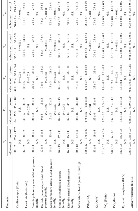
ome tr ic me asur emen ts) Parame ter T0 T15 T30 T45 T60 salbut amol con tr ol salbut amol con tr ol salbut amol con tr ol salbut amol con tr ol salbut amol con tr ol C ar di ac out put (l/min)
38 ± 7
37.4 ± 12.7
64 ± 25.5
35.4 ± 10.1
59.8 ± 23.4
30.1 ± 5.4
55.3 ± 19
30 ± 5.9
50.4 ± 9.4
29.4 ± 3.7
N. S. N. S. P = 0.031 P = 0.031 P = 0.031 H ear t ra te ( be at s/min)
32 ± 2
30 ± 2
40 ± 6
30 ± 2
38 ± 5
30 ± 1
31 ± 4
34 ± 2
31 ± 3
32 ± 1
N. S. P = 0.031 P = 0.029 N. S. N. S. Sy st olic pulmonar y ar ter ial blo od pr essur e (mmHg)
26 ± 4
28 ± 5
26 ± 2
28 ± 4
25 ± 2
27 ± 3
25 ± 3
27 ± 4
24 ± 2
27 ± 4
N. S. N. S. N. S. N. S. N. S. D ia st ol ic p ul m on ar y ar te ri al b lo od pr essur e (mmHg)
12 ± 3
13 ± 4
9 ± 1
13 ± 2
7 ± 4
15 ± 2
8 ± 3
17 ± 4
7 ± 2
16 ± 5
N. S. P = 0.029 P = 0.031 P = 0.031 P = 0.030 M ea n pu lm on ar y ar te ri al b lo od p re ss ur e (mmHg)
16 ± 3
20 ± 4
15 ± 2
20 ± 3
13 ± 3
21 ± 3
13 ± 2
22 ± 4
12 ± 2
22 ± 4
N. S. P = 0.030 P = 0.031 P = 0.031 P = 0.031 Sy st ol ic a rt er ia l b lo od p re ss ur e (mmHg)
80 ± 13
77 ± 11
90 ± 13
76 ± 13
94 ± 13
86 ± 14
94 ± 14
88 ± 12
93 ± 10
87 ± 12
N. S. P = 0.045 N. S. N. S. N. S. D ia st ol ic a rt er ia l b lo od p re ss ur e (mmHg)
46 ± 10
45 ± 11
56 ± 14
45 ± 9
60 ± 14
56 ± 14
58 ± 12
59 ± 12
58 ± 7
58 ± 11
N. S. N. S. N. S. N. S. N. S. M ean ar ter ial blo od pr essur e (mmHg)
60 ± 12
58 ±11
70 ± 16
58 ± 10
74 ± 15
68 ± 14
73 ± 14
72 ± 13
73 ± 9
70 ± 12
N. S. N. S. N. S. N. S. N. S. Pa O 2 /F iO 2
158 ± 32
176 ± 67
178 ± 52
151 ± 56
172 ± 37
138 ± 56
180 ± 43
121 ± 41
181 ± 47
114 ± 36
N. S. P = 0.031 N. S. P = 0.031 P = 0.031 Qs/Q t (%)
23 ± 4
19 ± 4
25 ± 4
23 ± 4
25 ± 7
22 ± 4
20 ± 3
23 ± 6
23 ± 5
25 ± 8
N. S. N. S. N. S. N. S. N. S. VO 2 (l/min)
1.1 ± 0.5
1.4 ± 0.4
1.7 ± 0.6
1.3 ± 0.2
1.6 ± 0.7
1.2 ± 0.2
1.8 ± 0.5
1.3 ± 0.2
1.5 ± 0.5
1.4 ± 0.3
N. S. N. S. N. S. N. S. N. S. DO 2 (l/min)
5.8 ± 0.8
6 ± 2.3
9.6 ± 3.2
5.4 ± 1.8
8.9 ± 2.5
4.4 ± 1
8.3 ± 1.9
4.3 ± 1.1
7.6 ± 0.8
4.3 ± 1.2
N. S. N. S. P = 0.031 P = 0.031 P = 0.031 D ynamic c om pli anc e (l/kP a)
3.6 ± 0.4
3.5 ± 0.4
3.5 ± 0.4
3.3 ± 0.4
3.4 ± 0.4
3.4 ± 0.3
3.2 ± 0.3
3.3 ± 0.5
3.2 ± 0.3
3.5 ± 0.5
N. S. N. S. N. S. N. S. N. S. Air w ay r esi st anc e (kP a/l/s)
0.36 ± 0.05
0.38 ± 0.07
0.38 ± 0.07
0.39 ± 0.14
0.43 ± 0.11
0.4 ± 0.15
0.41 ± 0.13
0.42 ± 0.13
0.38 ± 0.13
0.36 ± 0.13
N. S. N. S. N. S. N. S. N. S. N .S
. = not sig
nific
an
group during the whole monitoring period. Based on the results and the fact that in our study we have excluded hemodynamic medication support, we assume that inhalation salbutamol administration had a positive influence on heart activity through chronotropic and inotropic action with the result being an increase in cardiac output. The chrono-tropic action of salbutamol was recorded in the first half of the monitoring period while the ino-tropic action of salbutamol persisted throughout the whole monitoring period. This is proved by the fact that heart rate returned to basal values but cardiac output remained at an increased level for the whole period of anaesthesia. The inotropic mechanism for an increase in cardiac output fol-lowing inhalation salbutamol administration is de-scribed by Spiekerkoetter et al. (2002) in human patients with primary pulmonary hypertension. An increase in the inotropic state following intrave-nous salbutamol administration was also recorded by Insulander et al. (2004). The chronotropic action of salbutamol is derived from direct stimulation of β2 receptors in the heart and from the baroreceptor reflex that results from peripheral vasodilatation caused by the influence on β2 vascular receptors (Insulander et al., 2004). Studies in human medicine (Wong et al., 1990; Bennet et al., 1994; Insulander et al., 2004) describe the action of inhalation or intravenous salbutamol administration similarly: heart rate increase, peripheral vasodilatation and a decrease in diastolic arterial blood pressure caused by the above mentioned mechanisms.
In our study, systemic arterial blood pressure was stable following salbutamol administration which corresponds with a clinical study conducted by Robertson and Bailey (2002). When compared to the baseline we recorded a slight increase that was not statistically significant. This stability of sys-temic arterial blood pressure following salbutamol administration, despite the increased cardiac out-put, can be attributed to changes in systemic vascu-lar resistance. We did not measure central venous pressure and therefore calculate systemic vascular resistance, but if cardiac output, systemic arterial blood pressure and systemic vascular resistance are interrelated, then an increase in cardiac output at stable, or in our study, slightly increased, blood pressure could be accompanied by some degree of decrease in systemic vascular resistance. In the control group of horses, the low cardiac output was compensated for by an increase in systemic arterial blood pressure, possibly caused by an increased
systemic vascular resistance. We did not record any decrease in systemic arterial blood pressure in our experimental horses following salbutamol administration which was however observed in hu-man patients in studies conducted by Wong et al. (1990), Bennet et al. (1994) and Insulander et al. (2004). In a study on horses, Lee et al. (1998) also recorded a short-term decrease in systemic arte-rial blood pressure following intravenous adminis-tration of clenbuterol which lasted on average for 5 min. Dodam et al. (1993) recorded a decrease in mean arterial blood pressure as well as a decrease in mean pulmonary arterial blood pressure plus peripheral and pulmonary vascular resistance fol-lowing intravenous administration of clenbuterol to horses undergoing total intravenous anaesthesia. We assume that in our study a decrease in systemic vascular resistance and peripheral vasodilatation occurred, but the absence of any decrease in sys-temic arterial blood pressure may be explained by compensation due to high cardiac output.
be a result of vasodilatation of pulmonary vessels which is caused by the effect on β2 receptors in combination with a reaction to high cardiac output. The presence of β2 receptors in pulmonary ves-sels in mice and rats and their contribution to the mechanism of vasodilatation was proven in studies conducted by Leblais et al. (2008) and Pourageaud et al. (2005).
Cardiovascular changes observed following inha-lation salbutamol administration support the idea that salbutamol is absorbed in the systemic blood circulation and influences heart activity and hemo-dynamics. This is further supported by the sweating which occurred in all horses following salbutamol administration and is in contrast with the control group where no such sweating was recorded. The idea of systemic absorption is also mentioned in a report of Robertson and Bailey (2002). The dose and method of administration of salbutamol in our study correspond to the dose and method of ad-ministration in the study conducted by Robertson and Bailey (2002), but they recorded sweating only in some patients (approximately 10%) which is in contrast with our experimental horses. Sweating in horses as a result of administration of β2 agonists is also described in studies by Keegan et al. (1991) and Lee et al. (1998).
The PaO2/FiO2 ratio increased insignificantly fol-lowing salbutamol administration but comparison of the two groups revealed significantly higher val-ues in the salbutamol group than in the control group. The results clearly indicate a positive in-fluence of salbutamol on the PaO2/FiO2 ratio. In the clinical study of Robertson and Bailey (2002) a more distinct improvement of arterial oxygena-tion was recorded following inhalaoxygena-tion salbuta-mol administration, almost double the basal PaO2 values were recorded. Based on the results of this study, the PaO2/FiO2 ratio increased from 66 to 127, which represents a 92% improvement, while the improvement in our study was only 12%. This can be explained by the fact that in our study we maintained constant ventilation parameters and ex-cluded medication for hemodynamic support while in the study of Robertson and Bailey (2002) the goal to increase PaO2 was achieved by increased inten-sity of ventilation and in cases of low blood pres-sure, by inotropic support with dobutamin prior to salbutamol administration. The increase in cardiac output caused by the effect of dobutamin did not improve arterial oxygenation (Swanson and Muir, 1986) but the combination with increased
ventila-tion and salbutamol might intensify the effect of salbutamol. Robertson and Bailey (2002) assume that the mechanism for improvement in PaO2 val-ues includes bronchodilatation of the small bron-chioles in the perfused lung areas in combination with increased cardiac output. The bronchodilata-tion acbronchodilata-tion of salbutamol is known from studies in human patients (Wong et al., 1990; Bennet et al., 1994) and horses (Derksen et al., 1999). However, bronchodilatation in these cases is preceded by bronchoconstriction. Robertson and Bailey (2002) do not assume that cardiovascular factors contrib-uted significantly to the improvement in arterial oxygenation, because no changes in heart rate and mean arterial blood pressure were observed either before or after salbutamol administration. They admit the possibility of a transitory improvement in pulmonary perfusion even though they did not measure cardiac output and the distribution of ventilation and perfusion. We assume that in our study salbutamol had a positive effect on hemo-dynamics and heart activity that led to increased cardiac output. This increase caused higher per-fusion of the lungs and the decrease in pulmonary arterial blood pressure suggests that vasodilatation occurred in the pulmonary vessels. It is difficult to determine if the increase in the PaO2/FiO2 ra-tio occurred as a result of an improved V/Q rara-tio caused by salbutamol or only through an increase in cardiac output and pulmonary perfusion. As the pulmonary shunt values remained constant, we as-sume that there was no effect on the V/Q ratio and that improved pulmonary perfusion, in connection with a decrease in pulmonary arterial blood pres-sure, resulted in improved arterial oxygenation. The insignificant increase in the PaO2/FiO2 ratio following salbutamol administration could also be attributed to an insufficient number of values as the number of horses in both groups was the minimum required for statistical calculations. Other factors related to the effect of salbutamol, especially in-dividual variability, might also contribute to the overall result.
output and, to a lesser extent, as a result of the improved PaO2/FiO2 ratio. The decrease in oxygen delivery in the control group could be related to low cardiac output and the level of arterial oxygenation which could also be observed as decreasing PaO2/ FiO2 ratio values.
Oxygen consumption increased following salb-utamol administration, whereas in the control group oxygen consumption was constant. However, a comparison between the groups revealed no sig-nificant difference which suggests that the extent of increase in oxygen consumption following salbuta-mol administration is not relevant and the resultant effect of its administration is positive. An increase in oxygen consumption following intravenous ad-ministration of clenbuterol was also recorded in studies conducted by Dodam et al. (1993) and Lee et al. (1998).
No statistically significant change in the amount of pulmonary shunt was recorded in either group. In contrast to this in a study conducted by Dodam et al. (1993), administration of clenbuterol caused an increase in pulmonary shunt and at the same time an increase in the proportion of dead space ventilation. Increased pulmonary shunt resulted in a decrease in PaO2. In our study, we did not have at our disposal any device for measuring the ven-tilation-perfusion ratio, we were able to determine only the perfusion component and the ventilation component is missing. Therefore we could not de-termine the changes in the ventilation-perfusion ratio after salbutamol administration. But the pos-sible causes of different results in pulmonary shunt in our study include the type of anaesthesia: inha-lation anaesthesia vs. total intravenous anaesthe-sia, method of drug administration: inhalation vs. intravenous and different β2 agonist: salbutamol vs. clenbuterol.
The results of spirometric measurements per-formed in our study did not reveal any changes in either the salbutamol group or the control group. The values for dynamic compliance and airway resistance were stable for both groups. These re-sults are limited by the fact that they were ob-tained from horses with no muscle relaxation. Even though total compliance is dependent on the compliance of the chest, diaphragm, abdominal wall and lungs, muscle relaxation is required for monitoring changes in compliance of the lungs which limits the influence of other components. However, provided that the values obtained for the salbutamol and control group did not differ, we
may assume that inhalation salbutamol adminis-tration does not have any influence on ventilation parameters.
The results of our study confirm the onset and duration of salbutamol action observed in the study by Robertson and Bailey (2002). However, we as-sume that the primary effect of salbutamol was seen in hemodynamics but based on our results, the mechanism of bronchodilatation or opening of collapsed lung areas is unlikely as this effect would be accompanied by a decrease in pulmonary shunt and an increase in dynamic compliance.
In our study, we recorded individual variability in response to inhalation salbutamol administra-tion which was reflected in the values recorded for cardiac output, pulmonary arterial blood pres-sure, oxygen delivery, oxygen consumption, PaO2/ FiO2 ratio and systemic arterial blood pressure. This fact may be possibly attributed to individual differences in β2 receptor presence which are de-scribed in a study on horses conducted by Torneke (1999). Significant individual differences were also recorded in responses to β-adrenergic agonist ad-ministration (Torneke et al., 1998).
Inhalation salbutamol administration to healthy horses under general anaesthesia, which were arti-ficially ventilated, resulted in an increase in cardiac output and oxygen delivery. Oxygen consumption was also slightly increased, but not to an extent that should overweigh the positive actions of salbuta-mol. Individual variations were large. It is necessary to assess the V/Q ratio and its changes following salbutamol administration in order to determine the exact mechanism of salbutamol action. Further clinical studies are warranted to study the mecha-nism of action in compromised patients.
REFERENCES
Bennett JA, Smyth ET, Pavord ID, Wilding JP, Tattersfield AE (1994): Systemic effects of salbutamol and salm-eterol in patients with asthma. Thorax 49, 771–774. Derksen FJ, Olszewski MA, Robinson NE, Berney C,
Hakala JE, Matson CJ, Ruth DT (1999): Aerosolized albuterol sulfate used as a bronchodilator in horses with recurrent airway obstruction. American Journal of Veterinary Research 60, 689–693.
anesthe-tized horses. American Journal of Veterinary Research 54, 776–782.
Gillespie JR, Tyler WS, Hall LW (1969): Cardiopulmo-nary Dysfunction in anesthetized, laterally recumbent horses. American Journal of Veterinary Research 30, 61–72.
Gleed RD, Dobson A (1990): Effect of clenbuterol on arterial oxygen tension in the anaesthetised horse. Research in Veterinary Science 48, 331–337.
Hall LW, Gillespie JR, Tyler WS (1968): Alveolar-arterial oxygen tension differences in anaesthetized horses. British Journal of Anaesthesia 40, 560–568.
Heinonen E, Hedenstierna G, Merilainen P, Hogman M, Nyman G (2001): Pulsed delivery of nitric oxide coun-teracts hypoxaemia in the anaesthetized horse. Vet-erinary Anaesthesia and Analgesia 28, 3–11.
Insulander P, Juhlin-Dannfelt A, Freyschuss U, Vallin H (2004): Electrophysiologic effects of salbutamol, a β2 -selective agonist. Journal of Cardiovascular Electro-physiology 15, 316–322.
Keegan RD, Gleed RD, Sanders EA, Seaman GC, Wertz EM, Short CE (1991): Treatment of low arterial oxygen tension in anesthetized horses with clenbuterol. Vet-erinary Surgery 20, 148–152.
Leblais V, Delannoy E, Fresquet F, Begueret H, Bellance N, Banquet S, Allieres C, Leroux L, Desgranges C, Gadeau A, Muller B (2008): β-adrenergic relaxation in pulmonary arteries: preservation of the endothelial nitric oxide-dependent β2 component in pulmonary hypertension. Cardiovascular Research 77, 202– 210.
Lee YHL, Clarke KW, Alibhai HIK (1998): The cardiop-ulmonary effects of clenbuterol when administered to dorsally recumbent halothane-anaesthetised ponies – failure to increase arterial oxygenation. Research in Veterinary Science 65, 227–232.
Moens Y, Lagerweij E, Gootjes P, Poortman J (1994): Differential artificial ventilation in anesthetized horses positioned in lateral recumbency. American Journal of Veterinary Research 55, 1319–1326.
Moens Y, Lagerweij E, Gootjes P, Poortman J (1995): Distribution of inspired gas to each lung in the anaes-thetised horse and influence of body shape. Equine Veterinary Journal 27, 110–116.
Moens Y, Lagerweij E, Gootjes P, Poortman J (1998): Influence of tidal volume and positive end-expiratory pressure on inspiratory gas distribution and gas ex-change during mechanical ventilation in horses posi-tioned in lateral recumbency. American Journal of Veterinary Research 59, 307–312.
Moens Y, Gootjes P, Ionita JC, Heinonen E, Schatzmann U (2009): In vitro validation of a Pitot-based flow
me-ter for the measurement of respiratory volume and flow in large animal anaesthesia. Veterinary Anaes-thesia and Analgesia 36, 209–219.
Nyman G, Funkquist B, Kvart C, Frostell C, Tokics L, Strandberg A, Lundquist H, Lundh B, Brismar B, Hedenstierna G (1990): Atelectasis causes gas ex-change impairment in the anaesthetised horse. Equine Veterinary Journal 22, 317–324.
Pourageaud F, Leblais V, Bellance N, Marthan R, Muller B (2005): Role of β2-adrenoceptors (β-AR), but not β1-, β3-AR and endothelial nitric oxide, in β-AR-mediated relaxation of rat intrapulmonary artery. Naunyn-Schmiedeberg’s Archives of Pharmacology 372, 14–23. Robertson SA, Bailey JE (2002): Aerosolized salbutamol
(albuterol) improves PaO2 in hypoxaemic anaesthe-tized horses – a prospective clinical trial in 81 horses. Veterinary Anaesthesia and Analgesia 29, 212–218. Seldinger SI (1953): Catheter replacement of the needle
in percutaneous arteriography. A new technique. Acta Radiologica 39, 368–376.
Spiekerkoetter E, Fabel H, Hoeper MM (2002): Effects of inhaled salbutamol in primary pulmonary hyperten-sion. European Respiratory Journal 20, 524–528. Steffey EP, Wheat JD, Meagher DM, Norrie RD, McKee
J, Brown M, Arnold J (1977): Body position and mode of ventilation influences arterial pH, oxygen, and car-bon dioxide tensions in halothane-anesthetized horses. American Journal of Veterinary Research 38, 379– 382.
Swanson CR, Muir WW (1986): Dobutamine-induced augmentation of cardiac output does not enhance res-piratory gas exchange in anesthetized recumbent healthy horses. American Journal of Veterinary Re-search 47, 1573–1576.
Torneke K (1999): β-Adrenoceptors in equine trachea and heart. Veterinary Research Communications 23, 41–51.
Torneke K, Ingvast Larsson C, Appelgren LE (1998): A comparison between clenbuterol, salbutamol and terb-utaline in relation to receptor binding and in vitro re-laxation of equine tracheal muscle. Journal of Veterinary Pharmacology and Therapeurics 21, 388–392.
Trim CM, Wan PY (1990): Hypoxaemia during anaes-thesia in seven horses with colic. Journal of the As-sociation of Veterinary Anaesthetists of Great Britain and Ireland 17, 45–49.
Whitehair KJ, Willits NH (1999): Predictors of arterial oxygen tension in anesthetized horses: 1610 cases (1992–1994). Journal of the American Veterinary Medical Association 215, 978–981.
Wong CS, Pavord ID, Williams J, Britton JR, Tattersfield AE (1990): Bronchodilator, cardiovascular, and
hy-pokalaemic effects of fenoterol, salbutamol, and terb-utaline in asthma. Lancet 336, 1396–1399.
Received: 2010–01–08 Accepted after corrections: 2010–09–28
Corresponding Author:
MVDr. Maria Patschova, University of Veterinary and Pharmaceutical Sciences, Faculty of Veterinary Medicine, Equine Clinic, Palackeho 1–3, 612 42 Brno, Czech Republic