2-[2-(3-Methoxyphenyl)-2-oxoethyl]-1,2-benzisothiazol-3(2
H
)-one 1,1-dioxide
Salman Gul,aHamid Latif Siddiqui,a* Matloob Ahmad,a Muhammad Azamband Masood Parvezc
aInstitute of Chemistry, University of the Punjab, Lahore, Pakistan,bInstitute of Biochemistry, University of Baluchistan, Quetta 8700, Pakistan, andcDepartment of Chemistry, University of Calgary, 2500 University Drive NW, Calgary, Alberta, Canada T2N 1N4
Correspondence e-mail: drhamidlatif@yahoo.com
Received 29 January 2010; accepted 9 February 2010
Key indicators: single-crystal X-ray study;T= 173 K; mean(C–C) = 0.003 A˚; Rfactor = 0.046;wRfactor = 0.136; data-to-parameter ratio = 16.3.
In the title compound, C16H13NO5S, the benzothiazole unit is essentially planar [maximum deviation = 0.0501 (10) A˚ for the S atom] and is oriented at a dihedral angle of 67.85 (5)with
respect to the methoxy-substituted benzene ring. The mean plane of the methoxy group is oriented at 14.3 (3) with
respect to the benzene ring to which it is attached. In the crystal structure, weak C—H O hydrogen bonds form macrocyclic rings withR2
2
(10) andR2 2
(12) motifs.
Related literature
For the use of 1,2-benzisothiazoline-3-one 1,1-dioxide (saccharine) as an intermediate in the preparation of medic-inally important molecules, see: Siddiquiet al.(2006); Zia-ur-Rehman et al. (2005, 2009). For the biological activity of saccharine, see: Singh et al. (2007); Vaccarino et al. (2007); Kapui et al.(2003). For related structures, see: Ahmadet al. (2008, 2009). For hydrogen-bonding motifs, see: Bernstein et al.(1995).
Experimental
Crystal data
C16H13NO5S Mr= 331.33 Monoclinic,P21=n a= 8.9824 (3) A˚
b= 8.5801 (4) A˚
c= 19.5645 (7) A˚ = 97.942 (2)
V= 1493.37 (10) A˚3
Z= 4
MoKradiation = 0.24 mm1 T= 173 K
0.140.120.10 mm
Data collection
Nonius diffractometer with Bruker APEXII CCD
Absorption correction: multi-scan (SORTAV; Blessing, 1997)
Tmin= 0.967,Tmax= 0.976
15084 measured reflections 3399 independent reflections 2897 reflections withI> 2(I)
Rint= 0.027
Refinement
R[F2> 2(F2)] = 0.046 wR(F2) = 0.136
S= 1.06 3399 reflections
209 parameters
H-atom parameters constrained
max= 0.34 e A˚
3
min=0.37 e A˚
3
Table 1
Hydrogen-bond geometry (A˚ ,).
D—H A D—H H A D A D—H A
C5—H5 O5i
0.95 2.53 3.404 (3) 153
C8—H8B O1ii
0.99 2.42 3.318 (3) 150
C8—H8A O2i
0.99 2.51 3.301 (3) 137
Symmetry codes: (i)xþ1 2;y
1 2;zþ
1 2; (ii)xþ
1 2;yþ
1 2;zþ
1 2.
Data collection:COLLECT(Nonius, 1998); cell refinement:HKL DENZO (Otwinowski & Minor, 1997); data reduction: SCALE-PACK (Otwinowski & Minor, 1997); program(s) used to solve structure:SHELXS97(Sheldrick, 2008); program(s) used to refine structure: SHELXL97 (Sheldrick, 2008); molecular graphics: ORTEP-3 for Windows(Farrugia, 1997); software used to prepare material for publication:SHELXL97.
The authors thank the Higher Education Commission of Pakistan for financial support of this research.
Supplementary data and figures for this paper are available from the IUCr electronic archives (Reference: LH2991).
References
Ahmad, M., Siddiqui, H. L., Azam, M., Siddiqui, W. A. & Parvez, M. (2009).
Acta Cryst.E65, o2185.
Ahmad, M., Siddiqui, H. L., Zia-ur-Rehman, M., Ashiq, M. I. & Tizzard, G. J. (2008).Acta Cryst.E64, o788.
Bernstein, J., Davis, R. E., Shimoni, L. & Chang, N.-L. (1995).Angew. Chem. Int. Ed. Engl.34, 1555–1573.
Blessing, R. H. (1997).J. Appl. Cryst.30, 421–426. Farrugia, L. J. (1997).J. Appl. Cryst.30, 565.
Kapui, Z., Varga, M., Urban-Szabo, K., Mikus, E., Szabo, T., Szeredi, J., Batori, S., Finance, O. & Aranyi, P. (2003).J. Pharmacol. Exp. Ther.305, 451–459. Nonius (1998).COLLECT. Nonius BV, Delft, The Netherlands.
Otwinowski, Z. & Minor, W. (1997). Methods in Enzymology, Vol. 276,
Macromolecular Crystallography, Part A, edited by C. W. Carter Jr & R. M. Sweet, pp. 307–326. New York: Academic Press.
Sheldrick, G. M. (2008).Acta Cryst.A64, 112–122.
Siddiqui, W. A., Ahmad, S., Ullah, I. & Malik, A. (2006).J. Chem. Soc. Pak.28, 583–589.
Singh, S. K., Shivaramakrishna, S., Saibaba, V., Rao, K. S., Ganesh, K. R., Vasudev, R., Kumar, P. P., Babu, J. M., Vyas, K., Rao, Y. K. & Iqbal, J. (2007).
Eur. J. Med. Chem.42, 456–462.
Vaccarino, A. L., Paul, D., Mukherjee, P. K., de Turco, E. B. R., Marcheselli, V. L., Xu, L., Trudell, M. L., Minguez, J. M., Matia, M. P., Sunkel, C., Alvarez-Builla, J. & Bazan, N. G. (2007).Bioorg. Med. Chem.15, 2206–2215. Zia-ur-Rehman, M. Z., Choudary, J. A. & Ahmad, S. (2005).Bull. Korean
Chem. Soc.26, 1771–1175.
Zia-ur-Rehman, M., Choudary, J. A., Elsegood, M. R. J., Siddiqui, H. L. & Khan, K. M. (2009).Eur. J. Med. Chem.44, 1311–1316.
Acta Crystallographica Section E Structure Reports
Online
supporting information
Acta Cryst. (2010). E66, o618 [doi:10.1107/S160053681000543X]
2-[2-(3-Methoxyphenyl)-2-oxoethyl]-1,2-benzisothiazol-3(2
H
)-one 1,1-dioxide
Salman Gul, Hamid Latif Siddiqui, Matloob Ahmad, Muhammad Azam and Masood Parvez
S1. Comment
1,2-Benzisothiazoline-3-one 1,1-dioxide (saccharine) is an important starting material for the synthesis of different
heterocyclic compounds and plays a role as an intermediate for the preparation of medicinally important molecules
(Siddiqui et al., 2006; Zia-ur-Rehman et al., 2009). Various derivatives of saccharin are known to be cyclooxygenase-2
(COX-2) inhibitors (Singh et al., 2007), analgesic (Vaccarino et al., 2007), human leucocyte elastase (HLE) inhibitors
(Kapui et al., 2003) etc. In continuation of our research on the synthesis of potential biologically active derivatives of
benzothiazines (Ahmad et al., 2008; Ahmad et al., 2009), we herein report the crystal structure of the title compound,
N-(3-methoxyphenacyl)saccharin, (I).
The structure of (I) contains discrete molecules separated by normal van der Waals distances (Fig. 1). The benzothiazole
moiety (S1/N1/C1–C7) is essentially planar (maximum deviation = 0.0501 (10) Å for atom S1) and lies at an angle
67.85 (5)° with respect to the benzene ring ). The methoxy group is oriented at 14.3 (3)° with respect to the benzene ring
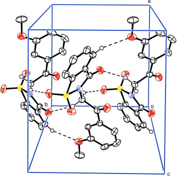
(C10–C15). The structure is devoid of any classical hydrogen bonds. However, non-classical hydrogen bonding
interactions of the type C—H···O are present in the crystal structure resulting in ten and twelve membered macrocyclic
rings in R22(10) and R22(12) motifs (Bernstein et al., 1995) (Fig. 2 and Table 1).
S2. Experimental
3-Methoxy phenacyl bromide (5.49 g, 0.024 mol) was slowly added to a suspension of sodium saccharine (5 g, 0.024
mol) in dimethylformamide (15 ml) and the mixture was stirred at 383 K for 3.0 hours under anhydrous conditions. On
completion of reaction (indicated by tlc), the mixture was poured on crushed ice and the precipitates formed were filtered
and washed with an excess of distilled water and cold ethanol respectively. Crystals suitable for diffraction were grown
from a solution of (I) in chloroform–methanol (3:1).
S3. Refinement
All H atoms were located from the difference Fourier maps and were included in the refinements at geometrically
idealized positions with C—H distances = 0.95, 0.98 and 0.99 Å for aryl, methyl and methylene H atoms, respectively,
and Uiso = 1.2 times Ueq of the C atoms to which they were bonded. The final difference map was free of chemically
Figure 1
ORTEP-3 (Farrugia, 1997) drawing of (I) with displacement ellipsoids plotted at 50% probability level.
Figure 2
Unit cell packing of (I) showing non-classical hydrogen bonding interactions with dashed lines; H atoms not involved in
[image:3.610.128.483.262.622.2]2-[2-(3-Methoxyphenyl)-2-oxoethyl]-1,2-benzisothiazol-3(2H)-one 1,1-dioxide
Crystal data
C16H13NO5S
Mr = 331.33
Monoclinic, P21/n
Hall symbol: -P 2yn
a = 8.9824 (3) Å
b = 8.5801 (4) Å
c = 19.5645 (7) Å
β = 97.942 (2)°
V = 1493.37 (10) Å3
Z = 4
F(000) = 688
Dx = 1.474 Mg m−3
Melting point: 446 K
Mo Kα radiation, λ = 0.71073 Å Cell parameters from 3447 reflections
θ = 1.0–27.5°
µ = 0.24 mm−1
T = 173 K Prism, white
0.14 × 0.12 × 0.10 mm
Data collection
Nonius APEX2 CCD diffractometer
Radiation source: fine-focus sealed tube Graphite monochromator
φ and ω scans
Absorption correction: multi-scan (SORTAV; Blessing, 1997)
Tmin = 0.967, Tmax = 0.976
15084 measured reflections 3399 independent reflections 2897 reflections with I > 2σ(I)
Rint = 0.027
θmax = 27.5°, θmin = 2.6°
h = −11→11
k = −11→11
l = −25→25
Refinement
Refinement on F2
Least-squares matrix: full
R[F2 > 2σ(F2)] = 0.046
wR(F2) = 0.136
S = 1.06 3399 reflections 209 parameters 0 restraints
Primary atom site location: structure-invariant direct methods
Secondary atom site location: difference Fourier map
Hydrogen site location: inferred from neighbouring sites
H-atom parameters constrained
w = 1/[σ2(F
o2) + (0.0682P)2 + 1.0411P]
where P = (Fo2 + 2Fc2)/3
(Δ/σ)max = 0.001
Δρmax = 0.34 e Å−3
Δρmin = −0.37 e Å−3
Special details
Geometry. All esds (except the esd in the dihedral angle between two l.s. planes) are estimated using the full covariance
matrix. The cell esds are taken into account individually in the estimation of esds in distances, angles and torsion angles; correlations between esds in cell parameters are only used when they are defined by crystal symmetry. An approximate (isotropic) treatment of cell esds is used for estimating esds involving l.s. planes.
Fractional atomic coordinates and isotropic or equivalent isotropic displacement parameters (Å2)
x y z Uiso*/Ueq
C2 −0.0056 (3) 0.2914 (3) −0.00668 (11) 0.0332 (5)
H2 0.0427 0.3702 −0.0297 0.040*
C3 −0.1387 (3) 0.2208 (3) −0.03674 (12) 0.0389 (5)
H3 −0.1832 0.2531 −0.0814 0.047*
C4 −0.2074 (2) 0.1051 (3) −0.00311 (12) 0.0398 (5)
H4 −0.2966 0.0577 −0.0256 0.048*
C5 −0.1486 (2) 0.0565 (3) 0.06313 (12) 0.0355 (5)
H5 −0.1971 −0.0217 0.0865 0.043*
C6 −0.0165 (2) 0.1264 (2) 0.09381 (10) 0.0283 (4) C7 0.0673 (2) 0.0893 (2) 0.16294 (10) 0.0285 (4) C8 0.3125 (2) 0.1633 (2) 0.23198 (10) 0.0283 (4)
H8A 0.2684 0.1527 0.2754 0.034*
H8B 0.3787 0.2561 0.2363 0.034*
C9 0.4060 (2) 0.0187 (2) 0.22173 (10) 0.0288 (4) C10 0.5481 (2) −0.0057 (2) 0.27013 (10) 0.0264 (4) C11 0.6073 (2) 0.1091 (2) 0.31602 (10) 0.0282 (4)
H11 0.5553 0.2048 0.3187 0.034*
C12 0.7438 (2) 0.0836 (3) 0.35828 (10) 0.0313 (4) C13 0.8174 (2) −0.0582 (3) 0.35630 (11) 0.0403 (6)
H13 0.9093 −0.0765 0.3856 0.048*
C14 0.7554 (3) −0.1730 (3) 0.31103 (12) 0.0422 (6)
H14 0.8052 −0.2705 0.3101 0.051*
C15 0.6230 (3) −0.1486 (3) 0.26732 (11) 0.0353 (5)
H15 0.5833 −0.2273 0.2359 0.042*
C16 0.9474 (3) 0.2006 (4) 0.43204 (13) 0.0506 (7)
H16A 0.9728 0.2990 0.4563 0.061*
H16B 1.0139 0.1849 0.3969 0.061*
H16C 0.9601 0.1143 0.4652 0.061*
Atomic displacement parameters (Å2)
U11 U22 U33 U12 U13 U23
C10 0.0266 (9) 0.0270 (10) 0.0254 (9) 0.0008 (7) 0.0031 (7) 0.0019 (7) C11 0.0238 (9) 0.0302 (11) 0.0299 (9) 0.0026 (8) 0.0007 (7) 0.0010 (8) C12 0.0236 (9) 0.0417 (12) 0.0280 (9) 0.0020 (8) 0.0011 (8) 0.0021 (9) C13 0.0283 (10) 0.0580 (15) 0.0335 (11) 0.0155 (10) 0.0005 (9) 0.0051 (10) C14 0.0399 (12) 0.0450 (14) 0.0416 (12) 0.0209 (11) 0.0051 (10) 0.0012 (10) C15 0.0396 (11) 0.0324 (11) 0.0341 (10) 0.0086 (9) 0.0053 (9) −0.0009 (9) C16 0.0268 (11) 0.080 (2) 0.0413 (12) −0.0054 (11) −0.0093 (9) 0.0013 (13)
Geometric parameters (Å, º)
S1—O2 1.4286 (16) C5—H5 0.9500
S1—O3 1.4291 (16) C6—C7 1.489 (3)
S1—N1 1.6701 (17) C8—C9 1.527 (3)
S1—C1 1.7555 (19) C8—H8A 0.9900
O1—C7 1.212 (2) C8—H8B 0.9900
O4—C9 1.215 (2) C9—C10 1.496 (3)
O5—C12 1.364 (3) C10—C11 1.389 (3)
O5—C16 1.427 (3) C10—C15 1.403 (3)
N1—C7 1.390 (3) C11—C12 1.398 (3)
N1—C8 1.455 (2) C11—H11 0.9500
C1—C6 1.379 (3) C12—C13 1.388 (3)
C1—C2 1.391 (3) C13—C14 1.388 (4)
C2—C3 1.396 (3) C13—H13 0.9500
C2—H2 0.9500 C14—C15 1.382 (3)
C3—C4 1.383 (4) C14—H14 0.9500
C3—H3 0.9500 C15—H15 0.9500
C4—C5 1.394 (3) C16—H16A 0.9800
C4—H4 0.9500 C16—H16B 0.9800
C5—C6 1.390 (3) C16—H16C 0.9800
O2—S1—O3 117.07 (10) N1—C8—H8A 109.3
O2—S1—N1 109.91 (9) C9—C8—H8A 109.3
O3—S1—N1 109.52 (9) N1—C8—H8B 109.3
O2—S1—C1 112.10 (9) C9—C8—H8B 109.3
O3—S1—C1 112.87 (9) H8A—C8—H8B 107.9
N1—S1—C1 92.63 (9) O4—C9—C10 121.86 (19)
C12—O5—C16 117.68 (19) O4—C9—C8 120.25 (17)
C7—N1—C8 123.05 (17) C10—C9—C8 117.87 (16)
C7—N1—S1 115.04 (13) C11—C10—C15 120.17 (19)
C8—N1—S1 119.92 (14) C11—C10—C9 121.80 (18)
C6—C1—C2 123.07 (19) C15—C10—C9 118.02 (18)
C6—C1—S1 110.15 (14) C10—C11—C12 119.80 (19)
C2—C1—S1 126.75 (17) C10—C11—H11 120.1
C1—C2—C3 116.1 (2) C12—C11—H11 120.1
C1—C2—H2 122.0 O5—C12—C13 124.50 (19)
C3—C2—H2 122.0 O5—C12—C11 115.27 (19)
C4—C3—C2 121.6 (2) C13—C12—C11 120.2 (2)
C2—C3—H3 119.2 C12—C13—H13 120.4
C3—C4—C5 121.4 (2) C14—C13—H13 120.4
C3—C4—H4 119.3 C15—C14—C13 121.5 (2)
C5—C4—H4 119.3 C15—C14—H14 119.2
C6—C5—C4 117.6 (2) C13—C14—H14 119.2
C6—C5—H5 121.2 C14—C15—C10 119.0 (2)
C4—C5—H5 121.2 C14—C15—H15 120.5
C1—C6—C5 120.31 (19) C10—C15—H15 120.5
C1—C6—C7 113.22 (17) O5—C16—H16A 109.5
C5—C6—C7 126.45 (19) O5—C16—H16B 109.5
O1—C7—N1 123.93 (18) H16A—C16—H16B 109.5
O1—C7—C6 127.41 (18) O5—C16—H16C 109.5
N1—C7—C6 108.65 (17) H16A—C16—H16C 109.5
N1—C8—C9 111.66 (16) H16B—C16—H16C 109.5
O2—S1—N1—C7 120.14 (16) S1—N1—C7—C6 −5.4 (2)
O3—S1—N1—C7 −109.92 (16) C1—C6—C7—O1 −178.6 (2)
C1—S1—N1—C7 5.48 (16) C5—C6—C7—O1 −0.4 (4)
O2—S1—N1—C8 −75.42 (17) C1—C6—C7—N1 2.3 (2)
O3—S1—N1—C8 54.52 (17) C5—C6—C7—N1 −179.5 (2)
C1—S1—N1—C8 169.92 (16) C7—N1—C8—C9 70.4 (2)
O2—S1—C1—C6 −116.57 (15) S1—N1—C8—C9 −92.72 (19)
O3—S1—C1—C6 108.65 (16) N1—C8—C9—O4 −11.3 (3)
N1—S1—C1—C6 −3.83 (16) N1—C8—C9—C10 170.25 (17)
O2—S1—C1—C2 65.5 (2) O4—C9—C10—C11 171.5 (2)
O3—S1—C1—C2 −69.3 (2) C8—C9—C10—C11 −10.0 (3)
N1—S1—C1—C2 178.3 (2) O4—C9—C10—C15 −7.4 (3)
C6—C1—C2—C3 −0.7 (3) C8—C9—C10—C15 171.06 (19)
S1—C1—C2—C3 176.93 (17) C15—C10—C11—C12 1.6 (3)
C1—C2—C3—C4 −0.6 (3) C9—C10—C11—C12 −177.32 (18)
C2—C3—C4—C5 1.6 (4) C16—O5—C12—C13 13.8 (3)
C3—C4—C5—C6 −1.2 (3) C16—O5—C12—C11 −166.2 (2)
C2—C1—C6—C5 1.1 (3) C10—C11—C12—O5 177.70 (18)
S1—C1—C6—C5 −176.85 (17) C10—C11—C12—C13 −2.3 (3)
C2—C1—C6—C7 179.45 (19) O5—C12—C13—C14 −178.9 (2)
S1—C1—C6—C7 1.4 (2) C11—C12—C13—C14 1.2 (3)
C4—C5—C6—C1 −0.2 (3) C12—C13—C14—C15 0.8 (4)
C4—C5—C6—C7 −178.2 (2) C13—C14—C15—C10 −1.5 (4)
C8—N1—C7—O1 11.6 (3) C11—C10—C15—C14 0.3 (3)
S1—N1—C7—O1 175.52 (17) C9—C10—C15—C14 179.3 (2)
C8—N1—C7—C6 −169.29 (17)
Hydrogen-bond geometry (Å, º)
D—H···A D—H H···A D···A D—H···A
C8—H8B···O1ii 0.99 2.42 3.318 (3) 150
C8—H8A···O2i 0.99 2.51 3.301 (3) 137