organic papers
Acta Cryst.(2007). E63, o1747–o1748 doi:10.1107/S1600536807010951 Robeynset al. C
12H11NO4
o1747
Acta Crystallographica Section E Structure Reports
Online
ISSN 1600-5368
Ethyl 2-(2,3-dioxoindolin-1-yl)acetate
Koen Robeyns,aTaoufik Rohand,bRachid Bouhfid,c EL Mokhtar Essassicand Luc Van Meervelta*
aKatholieke Universiteit Leuven, Department of
Chemistry, Biomolecular Architecture, Celestijnenlaan 200F, B-3001 Leuven, Belgium,
bKatholieke Universiteit Leuven, Department of
Chemistry, Molecular Design and Synthesis, Celestijnenlaan 200F, B-3001 Leuven, Belgium, andcUniversite´ Mohammed V-Agdal,
Laboratoire de Chimie Organique
He´te´rocyclique, BP: 1014 Avenue Ibn Batouta, Rabat, Morocco
Correspondence e-mail:
Key indicators
Single-crystal X-ray study
T= 100 K
Mean(C–C) = 0.002 A˚
Rfactor = 0.040
wRfactor = 0.104
Data-to-parameter ratio = 12.9
For details of how these key indicators were automatically derived from the article, see http://journals.iucr.org/e.
Received 5 March 2007 Accepted 8 March 2007
#2007 International Union of Crystallography All rights reserved
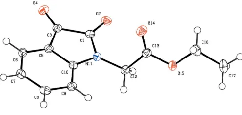
The molecular structure of the title compound, C12H11NO4, at 100 (2) K is characterized by a planar indole ring system, containing a long C—C bond in the 1,2-diketone unit.
Comment
Isatin (1H-indole-2,3-dione) is a natural product found in certain plants of the Isatis genus. It is also found in the human body, as a metabolic derivative of adrenalin. Several deriva-tives of isatin exhibit a wide range of pharmacological activity (Pandeya et al., 2005). Furthermore, it has been shown that isatin and its derivatives are active with respect to the central nervous system (Geronikaki et al., 2004) and have anti-microbial (Rajet al., 2003), anticancerous (Eshba & Salama, 1985) and anti-HIV (Pandeya et al., 1999) properties. Isatin has also proved to be an interesting precursor for the synthesis of new heterocyclic systems endowed with potential biological activity (Bouhfidet al., 2005).
The synthesis of the title compound, (I), is shown in the scheme above. The 1H-indoline ring system is planar, with a maximum deviation of 0.042 (2) A˚ for atom C1, and makes an angle of 79.04 (8) with the ester unit. Isatin derivatives are
characterized by a rather long bond between the carbon atoms of the diketone group. A search of the Cambridge Structural Database (Version 5.28; Allen, 2002) revealed that the average bond distance for 23 entries is 1.56 (3) A˚ , which is longer than a normal Csp2—Csp2bond due to steric repulsion between the ketone functions. The value of C1—C3 in (I) is 1.558 (2) A˚ .
Experimental
tetrabutylammonium bromide (1 mmol) were added. The reaction mixture was stirred at room temperature for 24 h. After removal of the salts by filtration, the solvent was removed under reduced pres-sure and the resulting residue was treated with dichloromethane (20 ml). The pure compound was isolated after a second filtration and recrystallized from methanol (yield 1.77 g, 76%; m.p. 401–403 K).
Crystal data
C12H11NO4
Mr= 233.22
Monoclinic,P21=c a= 13.6433 (6) A˚
b= 4.9505 (3) A˚
c= 16.2899 (8) A˚
= 104.246 (2)
V= 1066.40 (10) A˚3
Z= 4
CuKradiation
= 0.93 mm 1
T= 100 (2) K 0.50.20.2 mm
Data collection
Bruker SMART 6000 diffractometer
Absorption correction: multi-scan (SADABS; Bruker, 1997)
Tmin= 0.594,Tmax= 0.831
10352 measured reflections 2000 independent reflections 1725 reflections withI> 2(I)
Rint= 0.048
Refinement
R[F2> 2(F2)] = 0.040
wR(F2) = 0.104
S= 1.06 2000 reflections
155 parameters
H-atom parameters constrained
max= 0.33 e A˚ 3
min= 0.21 e A˚ 3
H atoms were positioned geometrically and refined as riding, with C—H = 0.95–0.99 A˚ andUiso(H) =xUeq(C), wherex= 1.5 for methyl and 1.2 for all other H atoms.
Data collection:SMART(Bruker, 1997); cell refinement:SAINT
(Bruker, 1997); data reduction: SAINT; program(s) used to solve structure: SHELXS97(Sheldrick, 1997); program(s) used to refine
structure: SHELXL97 (Sheldrick, 1997); molecular graphics:
PLATON (Spek, 2003); software used to prepare material for publication:PLATON.
The K. U. Leuven is gratefully acknowledged for financial support. The experimental part was carried out as part of the Poˆle de Compe´tences Pharmacochimie.
References
Allen, F. H. (2002).Acta Cryst.B58, 380–388.
Bouhfid, R., Joly, N., Massoui, M., Ceechelli, R., Lequart, V., Martin, P. & Essassi, E. M. (2005).Heterocycles,56, 2949–2955.
Bruker (1997).SADABS(Version 2.10),SMART(Version 5.625) andSAINT
(Version 5/6.0). Bruker AXS Inc., Madison, Wisconsin, USA. Eshba, N. H. & Salama, H. M. (1985).Pharmazie,40, 320–322.
Geronikaki, A., Babaev, E., Dearden, J., Dehaen, W., Filimonov, D., Galaeva, I., Krajneva, V., Lagunin, A., Macaev, F., Molodavkin, G., Poroikov, V., Pogrebnoi, S., Saloutin, V., Stepanchikova, A., Stingaci, E.et al.(2004).
Bioorg. Med. Chem.12, 6559–6568.
Pandeya, S. N., Smitha, S., Jyoti, M. & Sridhar, S. K. (2005).Acta Pharm.55, 27–46.
Pandeya, S. N., Sriram, D., Nath, G. & De Clercq, E. (1999).Pharm. Acta Helv.
74, 11–17.
Raj, A. A., Raghunathan, R., Sridevi Kumari, M. R. & Raman, N. (2003).
Bioorg. Med. Chem.11, 407–419.
Sheldrick, G. M. (1997). SHELXS97 and SHELXL97. University of Go¨ttingen, Germany.
[image:2.610.315.563.71.189.2]Spek, A. L. (2003).J. Appl. Cryst.36, 7–13.
Figure 1
supporting information
sup-1 Acta Cryst. (2007). E63, o1747–o1748
supporting information
Acta Cryst. (2007). E63, o1747–o1748 [https://doi.org/10.1107/S1600536807010951]
Ethyl 2-(2,3-dioxoindolin-1-yl)acetate
Koen Robeyns, Taoufik Rohand, Rachid Bouhfid, EL Mokhtar Essassi and Luc Van Meervelt
Ethyl 2-(2,3-dioxoindolin-1-yl)acetate
Crystal data C12H11NO4
Mr = 233.22 Monoclinic, P21/c
Hall symbol: -P 2ybc a = 13.6433 (6) Å b = 4.9505 (3) Å c = 16.2899 (8) Å β = 104.246 (2)° V = 1066.40 (10) Å3
Z = 4
F(000) = 488 Dx = 1.453 Mg m−3
Cu Kα radiation, λ = 1.54178 Å Cell parameters from 3255 reflections θ = 3.3–70.3°
µ = 0.93 mm−1
T = 100 K Block, orange 0.5 × 0.2 × 0.2 mm
Data collection Bruker SMART 6000
diffractometer
Radiation source: fine-focus sealed tube Crossed Goebel mirrors monochromator ω and φ scans
Absorption correction: multi-scan (SADABS; Bruker, 1997) Tmin = 0.594, Tmax = 0.831
10352 measured reflections 2000 independent reflections 1725 reflections with I > 2σ(I) Rint = 0.048
θmax = 70.3°, θmin = 3.3°
h = −16→16 k = −5→6 l = −19→19
Refinement Refinement on F2
Least-squares matrix: full R[F2 > 2σ(F2)] = 0.040
wR(F2) = 0.104
S = 1.06 2000 reflections 155 parameters 0 restraints
Primary atom site location: structure-invariant direct methods
Secondary atom site location: difference Fourier map
Hydrogen site location: inferred from neighbouring sites
H-atom parameters constrained w = 1/[σ2(F
o2) + (0.0564P)2 + 0.3661P]
where P = (Fo2 + 2Fc2)/3
(Δ/σ)max < 0.001
Δρmax = 0.33 e Å−3
Δρmin = −0.21 e Å−3
Special details
Refinement. Refinement of F2 against ALL reflections. The weighted R-factor wR and goodness of fit S are based on F2,
conventional R-factors R are based on F, with F set to zero for negative F2. The threshold expression of F2 > σ(F2) is used
only for calculating R-factors(gt) etc. and is not relevant to the choice of reflections for refinement. R-factors based on F2
are statistically about twice as large as those based on F, and R- factors based on ALL data will be even larger.
Fractional atomic coordinates and isotropic or equivalent isotropic displacement parameters (Å2)
x y z Uiso*/Ueq
C1 0.60794 (11) 0.8944 (3) 0.78627 (9) 0.0168 (3)
O2 0.58879 (8) 1.0495 (2) 0.72671 (7) 0.0209 (3)
C3 0.56599 (11) 0.9004 (3) 0.86695 (9) 0.0166 (3)
O4 0.50308 (8) 1.0589 (2) 0.87787 (7) 0.0211 (3)
C5 0.62080 (10) 0.6848 (3) 0.92122 (9) 0.0163 (3)
C6 0.61795 (11) 0.6038 (3) 1.00197 (9) 0.0189 (3)
H6 0.5748 0.6910 1.0315 0.023*
C7 0.68022 (12) 0.3908 (3) 1.03865 (9) 0.0209 (3)
H7 0.6797 0.3303 1.0939 0.025*
C8 0.74314 (11) 0.2664 (3) 0.99462 (10) 0.0206 (3)
H8 0.7851 0.1217 1.0208 0.025*
C9 0.74672 (11) 0.3474 (3) 0.91294 (9) 0.0181 (3)
H9 0.7900 0.2609 0.8834 0.022*
C10 0.68436 (10) 0.5588 (3) 0.87729 (9) 0.0155 (3)
N11 0.67385 (9) 0.6806 (2) 0.79704 (7) 0.0165 (3)
C12 0.72442 (11) 0.5980 (3) 0.73313 (9) 0.0172 (3)
H12A 0.6761 0.6084 0.6769 0.021*
H12B 0.7458 0.4073 0.7432 0.021*
C13 0.81627 (11) 0.7699 (3) 0.73224 (9) 0.0176 (3)
O14 0.84687 (8) 0.9564 (2) 0.77891 (7) 0.0266 (3)
O15 0.85739 (8) 0.6869 (2) 0.67021 (7) 0.0198 (3)
C16 0.94114 (11) 0.8490 (3) 0.65638 (10) 0.0231 (4)
H16A 1.0004 0.8333 0.7055 0.028*
H16B 0.9213 1.0415 0.6490 0.028*
C17 0.96670 (12) 0.7429 (4) 0.57764 (11) 0.0276 (4)
H17A 0.9854 0.5518 0.5855 0.041*
H17B 1.0235 0.8458 0.5667 0.041*
H17C 0.9078 0.7620 0.5294 0.041*
Atomic displacement parameters (Å2)
U11 U22 U33 U12 U13 U23
C1 0.0177 (7) 0.0136 (7) 0.0182 (7) −0.0019 (5) 0.0028 (5) −0.0020 (5)
O2 0.0255 (6) 0.0177 (5) 0.0185 (6) −0.0004 (4) 0.0037 (4) 0.0024 (4)
C3 0.0157 (6) 0.0147 (7) 0.0188 (7) −0.0029 (5) 0.0030 (5) −0.0034 (5)
O4 0.0188 (5) 0.0200 (5) 0.0243 (6) 0.0028 (4) 0.0047 (4) −0.0045 (4)
C5 0.0160 (6) 0.0149 (7) 0.0178 (7) −0.0022 (5) 0.0036 (5) −0.0021 (5)
C6 0.0206 (7) 0.0199 (7) 0.0171 (8) −0.0041 (6) 0.0063 (6) −0.0041 (6)
C7 0.0266 (8) 0.0202 (7) 0.0146 (7) −0.0050 (6) 0.0027 (6) 0.0001 (5)
supporting information
sup-3 Acta Cryst. (2007). E63, o1747–o1748
C9 0.0165 (7) 0.0175 (7) 0.0200 (8) −0.0008 (5) 0.0038 (5) −0.0034 (6)
C10 0.0150 (6) 0.0153 (7) 0.0159 (7) −0.0053 (5) 0.0031 (5) −0.0028 (5)
N11 0.0185 (6) 0.0165 (6) 0.0155 (6) 0.0004 (5) 0.0059 (5) −0.0005 (4)
C12 0.0190 (7) 0.0174 (7) 0.0160 (7) −0.0004 (5) 0.0060 (5) −0.0022 (5)
C13 0.0177 (7) 0.0179 (7) 0.0171 (7) 0.0029 (6) 0.0039 (5) 0.0005 (5)
O14 0.0239 (6) 0.0272 (6) 0.0306 (7) −0.0066 (5) 0.0102 (5) −0.0115 (5)
O15 0.0199 (5) 0.0212 (5) 0.0207 (6) −0.0028 (4) 0.0095 (4) −0.0026 (4)
C16 0.0182 (7) 0.0259 (8) 0.0263 (8) −0.0035 (6) 0.0078 (6) 0.0028 (6)
C17 0.0218 (7) 0.0383 (9) 0.0249 (8) 0.0004 (7) 0.0101 (6) 0.0067 (7)
Geometric parameters (Å, º)
C1—O2 1.2143 (18) C10—N11 1.4146 (19)
C1—N11 1.3718 (19) N11—C12 1.4426 (18)
C1—C3 1.558 (2) C12—C13 1.518 (2)
C3—O4 1.2078 (18) C12—H12A 0.9900
C3—C5 1.468 (2) C12—H12B 0.9900
C5—C6 1.385 (2) C13—O14 1.2036 (18)
C5—C10 1.400 (2) C13—O15 1.3354 (18)
C6—C7 1.393 (2) O15—C16 1.4584 (18)
C6—H6 0.9500 C16—C17 1.504 (2)
C7—C8 1.390 (2) C16—H16A 0.9900
C7—H7 0.9500 C16—H16B 0.9900
C8—C9 1.402 (2) C17—H17A 0.9800
C8—H8 0.9500 C17—H17B 0.9800
C9—C10 1.383 (2) C17—H17C 0.9800
C9—H9 0.9500
O2—C1—N11 126.56 (14) C1—N11—C12 122.77 (12)
O2—C1—C3 127.63 (13) C10—N11—C12 126.04 (12)
N11—C1—C3 105.79 (12) N11—C12—C13 112.98 (12)
O4—C3—C5 131.21 (14) N11—C12—H12A 109.0
O4—C3—C1 123.97 (13) C13—C12—H12A 109.0
C5—C3—C1 104.82 (11) N11—C12—H12B 109.0
C6—C5—C10 121.49 (13) C13—C12—H12B 109.0
C6—C5—C3 130.79 (13) H12A—C12—H12B 107.8
C10—C5—C3 107.71 (12) O14—C13—O15 124.66 (14)
C5—C6—C7 118.02 (14) O14—C13—C12 125.66 (14)
C5—C6—H6 121.0 O15—C13—C12 109.66 (12)
C7—C6—H6 121.0 C13—O15—C16 116.03 (12)
C8—C7—C6 120.17 (14) O15—C16—C17 107.12 (13)
C8—C7—H7 119.9 O15—C16—H16A 110.3
C6—C7—H7 119.9 C17—C16—H16A 110.3
C7—C8—C9 122.23 (14) O15—C16—H16B 110.3
C7—C8—H8 118.9 C17—C16—H16B 110.3
C9—C8—H8 118.9 H16A—C16—H16B 108.5
C10—C9—C8 116.95 (14) C16—C17—H17A 109.5
C8—C9—H9 121.5 H17A—C17—H17B 109.5
C9—C10—C5 121.14 (14) C16—C17—H17C 109.5
C9—C10—N11 128.51 (13) H17A—C17—H17C 109.5
C5—C10—N11 110.35 (12) H17B—C17—H17C 109.5