(4-Chloro-2-{[(pyridin-2-ylmethyl)-
imino]methyl}phenolato)iodido-(methanol)zinc(II)
Hong-Wei Huang
College of Chemistry and Biology Engineering, Yichun University, Yichun 336000, People’s Republic of China
Correspondence e-mail: huanghongwei_ycu@126.com
Received 24 January 2011; accepted 3 February 2011
Key indicators: single-crystal X-ray study;T= 298 K; mean(C–C) = 0.004 A˚; Rfactor = 0.025;wRfactor = 0.058; data-to-parameter ratio = 18.1.
The title Schiff base zinc(II) complex, [Zn(C13H10ClN2 O)I-(CH3OH)], was synthesized by the reaction of 5-chlorosalicyl-aldehyde, 2-aminomethylpyridine and zinc iodide in methanol. The ZnIIatom is five-coordinated by one phenolate O atom, one imine and one pyridine N atom of the Schiff base ligand, one methanol O atom and one I atom, forming a distorted square-pyramidal geometry, with the I atom at the apical site. The dihedral angle between the benzene and pyridine rings is 22.9 (2). In the crystal, centrosymmetrically related molecules are linked through intermolecular O—H O hydrogen bonds, forming dimers.
Related literature
For the structures of Schiff bases and their complexes, see: Ali et al.(2008); Eltayebet al.(2007); Dattaet al.(2009); Zhaoet al.(2010); Temelet al.(2010); Naveenkumaret al.(2010).
Experimental
Crystal data
[Zn(C13H10ClN2O)I(CH4O)] Mr= 469.99
Monoclinic,P21=c
a= 7.0769 (9) A˚ b= 12.7212 (16) A˚ c= 18.225 (2) A˚
= 98.994 (1) V= 1620.5 (3) A˚3
Z= 4
MoKradiation
= 3.59 mm1 T= 298 K
0.200.200.18 mm
Data collection
Bruker SMART CCD area-detector diffractometer
Absorption correction: multi-scan (SADABS; Sheldrick, 1996) Tmin= 0.534,Tmax= 0.564
9273 measured reflections 3522 independent reflections 2947 reflections withI> 2(I) Rint= 0.021
Refinement
R[F2> 2(F2)] = 0.025
wR(F2) = 0.058 S= 1.04 3522 reflections 195 parameters 1 restraint
H atoms treated by a mixture of independent and constrained refinement
max= 0.37 e A˚
3 min=0.91 e A˚
3
Table 1
Hydrogen-bond geometry (A˚ ,).
D—H A D—H H A D A D—H A O2—H2 O1i 0.86 (3) 1.79 (3) 2.643 (3) 176 (3)
Symmetry code: (i)xþ1;yþ2;zþ1.
Data collection:SMART(Bruker, 1998); cell refinement:SAINT
(Bruker, 1998); data reduction:SAINT; program(s) used to solve structure:SHELXS97(Sheldrick, 2008); program(s) used to refine structure: SHELXL97 (Sheldrick, 2008); molecular graphics:
SHELXTL(Sheldrick, 2008); software used to prepare material for publication:SHELXTL.
This work was supported by Yichun University.
Supplementary data and figures for this paper are available from the IUCr electronic archives (Reference: RZ2553).
References
Ali, H. M., Mohamed Mustafa, M. I., Rizal, M. R. & Ng, S. W. (2008).Acta Cryst.E64, m718–m719.
Bruker (1998).SMARTandSAINT. Bruker AXS Inc., Madison, Wisconsin, USA.
Datta, A., Chuang, N.-T., Huang, J.-H. & Lee, H. M. (2009).Acta Cryst.E65, m964.
Eltayeb, N. E., Teoh, S. G., Chantrapromma, S., Fun, H.-K. & Ibrahim, K. (2007).Acta Cryst.E63, m1672–m1673.
Naveenkumar, H. S., Sadikun, A., Ibrahim, P., Yeap, C. S. & Fun, H.-K. (2010). Acta Cryst.E66, o1918–o1919.
Sheldrick, G. M. (1996).SADABS. University of Go¨ttingen, Germany. Sheldrick, G. M. (2008).Acta Cryst.A64, 112–122.
Temel, E., Ag˘ar, E. & Bu¨yu¨kgu¨ngo¨r, O. (2010).Acta Cryst.E66, o1131. Zhao, L., Cao, D. & Cui, J. (2010).Acta Cryst.E66, o2204.
Acta Crystallographica Section E
Structure Reports
Online
supporting information
Acta Cryst. (2011). E67, m313 [doi:10.1107/S160053681100417X]
(4-Chloro-2-{[(pyridin-2-ylmethyl)imino]methyl}-phenolato)iodido(methanol)zinc(II)
Hong-Wei Huang
S1. Comment
Schiff bases and their complexes have attracted much attention for their interesting structures (Ali et al., 2008; Eltayeb et
al., 2007; Datta et al., 2009; Zhao et al., 2010; Temel et al., 2010; Naveenkumar et al., 2010). In this paper, the title new
Schiff base zinc(II) complex, Fig. 1, is reported.
The Zn atom in the complex is five-coordinated by one phenolate O atom, one imine and one pyridine N atoms of the
Schiff base ligand, one methanol O atom, and one iodide atom to form a distorted square pyramidal geometry. The
dihedral angle between the benzene and the pyridine rings is 22.9 (2)°. In the crystal structure (Fig. 2),
centrosymmetrically related molecules are linked through intermolecular O—H···N hydrogen bonds (Table 1) to form
dimers.
S2. Experimental
Equimolar quantities (0.1 mmol each) of 5-chlorosalicylaldehyde, 2-aminomethylpyridine, and zinc iodide were mixed
and stirred in methanol for 30 min at reflux. After keeping the filtrate in air for a few days, colourless block crystals
suitable for X-ray analysis were formed.
S3. Refinement
H2 attached to O2 was located from a difference Fourier map, and refined with the O–H distance restrained to 0.85 (1) Å,
and with Uiso restrained to 0.08 Å2. The remaining H atoms were placed in calculated positions and constrained to ride on
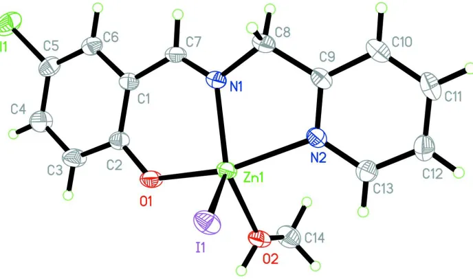
Figure 1
[image:3.610.135.479.71.274.2]The molecular structure of the title compound, with 30% displacements ellipsoids.
Figure 2
The molecular packing of the title compound, viewed along the c axis. Hydrogen atoms not involved in hydrogen bonds
(dashed lines) are omitted for clarity.
(4-Chloro-2-{[(pyridin-2- ylmethyl)imino]methyl}phenolato)iodido(methanol)zinc(II)
Crystal data
[Zn(C13H10ClN2O)I(CH4O)]
Mr = 469.99 Monoclinic, P21/c
Hall symbol: -P 2ybc
a = 7.0769 (9) Å
b = 12.7212 (16) Å
c = 18.225 (2) Å
β = 98.994 (1)°
V = 1620.5 (3) Å3
Z = 4
F(000) = 912
Dx = 1.926 Mg m−3
Mo Kα radiation, λ = 0.71073 Å Cell parameters from 3746 reflections
θ = 2.7–27.8°
µ = 3.59 mm−1
T = 298 K Block, colorless 0.20 × 0.20 × 0.18 mm
Data collection
Bruker SMART CCD area-detector diffractometer
Radiation source: fine-focus sealed tube Graphite monochromator
ω scans
Absorption correction: multi-scan (SADABS; Sheldrick, 1996)
9273 measured reflections 3522 independent reflections 2947 reflections with I > 2σ(I)
Rint = 0.021
θmax = 27.0°, θmin = 2.0°
h = −9→8
k = −16→15
l = −23→17
Refinement
Refinement on F2
Least-squares matrix: full
R[F2 > 2σ(F2)] = 0.025
wR(F2) = 0.058
S = 1.04 3522 reflections 195 parameters 1 restraint
Primary atom site location: structure-invariant direct methods
Secondary atom site location: difference Fourier map
Hydrogen site location: inferred from neighbouring sites
H atoms treated by a mixture of independent and constrained refinement
w = 1/[σ2(F
o2) + (0.0266P)2 + 0.3654P]
where P = (Fo2 + 2Fc2)/3
(Δ/σ)max = 0.003
Δρmax = 0.37 e Å−3
Δρmin = −0.91 e Å−3
Special details
Geometry. All e.s.d.'s (except the e.s.d. in the dihedral angle between two l.s. planes) are estimated using the full covariance matrix. The cell e.s.d.'s are taken into account individually in the estimation of e.s.d.'s in distances, angles and torsion angles; correlations between e.s.d.'s in cell parameters are only used when they are defined by crystal symmetry. An approximate (isotropic) treatment of cell e.s.d.'s is used for estimating e.s.d.'s involving l.s. planes.
Refinement. Refinement of F2 against ALL reflections. The weighted R-factor wR and goodness of fit S are based on F2,
conventional R-factors R are based on F, with F set to zero for negative F2. The threshold expression of F2 > σ(F2) is used
only for calculating R-factors(gt) etc. and is not relevant to the choice of reflections for refinement. R-factors based on F2
are statistically about twice as large as those based on F, and R- factors based on ALL data will be even larger.
Fractional atomic coordinates and isotropic or equivalent isotropic displacement parameters (Å2)
x y z Uiso*/Ueq
Zn1 0.56550 (5) 1.00055 (2) 0.370141 (16) 0.03411 (9) Cl1 0.80941 (15) 0.43991 (6) 0.42899 (5) 0.0673 (3) I1 0.20664 (3) 1.054046 (16) 0.337917 (10) 0.04454 (7) O1 0.5675 (3) 0.87992 (14) 0.44157 (10) 0.0428 (5) O2 0.6619 (3) 1.10027 (17) 0.45789 (11) 0.0461 (5) N1 0.6673 (3) 0.89862 (17) 0.29675 (11) 0.0343 (5) N2 0.6831 (3) 1.10583 (17) 0.29614 (12) 0.0353 (5) C1 0.6966 (4) 0.7424 (2) 0.37342 (14) 0.0331 (6) C2 0.6271 (4) 0.7827 (2) 0.43675 (14) 0.0350 (6) C3 0.6226 (5) 0.7138 (2) 0.49658 (16) 0.0502 (8)
H3 0.5819 0.7388 0.5394 0.060*
C4 0.6768 (5) 0.6104 (2) 0.49358 (17) 0.0517 (8)
H4 0.6702 0.5664 0.5338 0.062*
C5 0.7407 (5) 0.5715 (2) 0.43160 (17) 0.0443 (7) C6 0.7511 (4) 0.6356 (2) 0.37262 (16) 0.0394 (6)
H6 0.7951 0.6086 0.3310 0.047*
C7 0.7123 (4) 0.8024 (2) 0.30781 (15) 0.0357 (6)
H7 0.7599 0.7676 0.2697 0.043*
C8 0.6893 (5) 0.9462 (2) 0.22567 (15) 0.0450 (7)
H8B 0.7983 0.9150 0.2076 0.054* C9 0.7177 (4) 1.0624 (2) 0.23317 (15) 0.0364 (6) C10 0.7742 (4) 1.1230 (3) 0.17688 (15) 0.0451 (7)
H10 0.7966 1.0917 0.1329 0.054*
C11 0.7966 (4) 1.2294 (3) 0.18702 (17) 0.0492 (8)
H11 0.8336 1.2712 0.1499 0.059*
C12 0.7637 (4) 1.2735 (2) 0.25260 (17) 0.0480 (7)
H12 0.7803 1.3452 0.2610 0.058*
C13 0.7061 (4) 1.2099 (2) 0.30520 (17) 0.0437 (7)
H13 0.6817 1.2400 0.3493 0.052*
C14 0.8502 (5) 1.1310 (3) 0.48795 (17) 0.0548 (8)
H14A 0.8972 1.0869 0.5296 0.082*
H14B 0.8498 1.2029 0.5040 0.082*
H14C 0.9315 1.1240 0.4507 0.082*
H2 0.585 (4) 1.104 (2) 0.4897 (14) 0.055 (9)*
Atomic displacement parameters (Å2)
U11 U22 U33 U12 U13 U23
Zn1 0.03959 (19) 0.03446 (17) 0.03043 (16) 0.00566 (13) 0.01222 (13) 0.00098 (12) Cl1 0.0921 (7) 0.0349 (4) 0.0725 (6) 0.0165 (4) 0.0054 (5) −0.0032 (4) I1 0.03836 (12) 0.05586 (13) 0.04069 (12) 0.01147 (9) 0.01019 (8) 0.00744 (8) O1 0.0617 (13) 0.0352 (10) 0.0351 (10) 0.0147 (9) 0.0191 (9) 0.0028 (8) O2 0.0553 (14) 0.0511 (12) 0.0363 (11) −0.0032 (10) 0.0210 (10) −0.0105 (9) N1 0.0387 (13) 0.0371 (12) 0.0285 (11) 0.0009 (10) 0.0102 (9) −0.0023 (9) N2 0.0354 (13) 0.0385 (12) 0.0333 (12) 0.0014 (10) 0.0096 (10) 0.0029 (10) C1 0.0311 (14) 0.0365 (14) 0.0320 (13) 0.0031 (11) 0.0055 (11) −0.0022 (11) C2 0.0359 (15) 0.0360 (14) 0.0327 (14) 0.0056 (11) 0.0045 (11) −0.0002 (11) C3 0.072 (2) 0.0460 (17) 0.0355 (15) 0.0140 (16) 0.0157 (15) 0.0035 (13) C4 0.072 (2) 0.0411 (16) 0.0431 (17) 0.0130 (16) 0.0112 (15) 0.0108 (14) C5 0.0494 (18) 0.0337 (14) 0.0481 (17) 0.0080 (13) 0.0023 (14) −0.0022 (12) C6 0.0381 (16) 0.0379 (15) 0.0421 (16) 0.0038 (12) 0.0063 (12) −0.0065 (12) C7 0.0356 (15) 0.0401 (15) 0.0330 (14) 0.0003 (12) 0.0104 (11) −0.0102 (12) C8 0.061 (2) 0.0469 (17) 0.0299 (14) 0.0002 (14) 0.0149 (14) −0.0022 (12) C9 0.0304 (15) 0.0473 (16) 0.0323 (14) 0.0017 (12) 0.0079 (11) 0.0051 (12) C10 0.0418 (17) 0.061 (2) 0.0338 (15) 0.0001 (14) 0.0104 (13) 0.0063 (13) C11 0.0449 (18) 0.0580 (19) 0.0461 (17) −0.0019 (14) 0.0111 (14) 0.0198 (15) C12 0.0471 (18) 0.0418 (16) 0.0557 (19) 0.0003 (14) 0.0101 (15) 0.0103 (14) C13 0.0485 (18) 0.0404 (16) 0.0433 (16) 0.0046 (13) 0.0108 (13) 0.0030 (13) C14 0.057 (2) 0.066 (2) 0.0415 (17) −0.0016 (17) 0.0095 (15) 0.0005 (15)
Geometric parameters (Å, º)
Zn1—O1 2.0111 (18) C4—C5 1.373 (4)
Zn1—N1 2.071 (2) C4—H4 0.9300
Zn1—O2 2.071 (2) C5—C6 1.361 (4)
Zn1—N2 2.158 (2) C6—H6 0.9300
Cl1—C5 1.746 (3) C8—C9 1.496 (4)
O1—C2 1.314 (3) C8—H8A 0.9700
O2—C14 1.415 (4) C8—H8B 0.9700
O2—H2 0.86 (3) C9—C10 1.391 (4)
N1—C7 1.272 (3) C10—C11 1.372 (4)
N1—C8 1.460 (3) C10—H10 0.9300
N2—C9 1.330 (3) C11—C12 1.372 (4)
N2—C13 1.340 (3) C11—H11 0.9300
C1—C6 1.412 (4) C12—C13 1.365 (4)
C1—C2 1.419 (3) C12—H12 0.9300
C1—C7 1.438 (4) C13—H13 0.9300
C2—C3 1.403 (4) C14—H14A 0.9600
C3—C4 1.373 (4) C14—H14B 0.9600
C3—H3 0.9300 C14—H14C 0.9600
O1—Zn1—N1 88.42 (8) C4—C5—Cl1 119.8 (2)
O1—Zn1—O2 89.96 (8) C5—C6—C1 121.3 (3)
N1—Zn1—O2 140.91 (9) C5—C6—H6 119.4
O1—Zn1—N2 156.05 (8) C1—C6—H6 119.4
N1—Zn1—N2 77.17 (8) N1—C7—C1 126.4 (2)
O2—Zn1—N2 89.42 (8) N1—C7—H7 116.8
O1—Zn1—I1 104.65 (6) C1—C7—H7 116.8
N1—Zn1—I1 116.29 (6) N1—C8—C9 111.1 (2)
O2—Zn1—I1 101.87 (6) N1—C8—H8A 109.4
N2—Zn1—I1 98.90 (6) C9—C8—H8A 109.4
C2—O1—Zn1 130.13 (16) N1—C8—H8B 109.4
C14—O2—Zn1 130.14 (18) C9—C8—H8B 109.4
C14—O2—H2 112 (2) H8A—C8—H8B 108.0
Zn1—O2—H2 113 (2) N2—C9—C10 121.3 (3)
C7—N1—C8 118.7 (2) N2—C9—C8 116.7 (2)
C7—N1—Zn1 127.11 (18) C10—C9—C8 122.0 (2)
C8—N1—Zn1 114.18 (16) C11—C10—C9 119.2 (3)
C9—N2—C13 118.8 (2) C11—C10—H10 120.4
C9—N2—Zn1 115.00 (17) C9—C10—H10 120.4
C13—N2—Zn1 125.93 (18) C12—C11—C10 119.2 (3)
C6—C1—C2 119.1 (2) C12—C11—H11 120.4
C6—C1—C7 116.5 (2) C10—C11—H11 120.4
C2—C1—C7 124.4 (2) C13—C12—C11 118.7 (3)
O1—C2—C3 119.3 (2) C13—C12—H12 120.7
O1—C2—C1 123.4 (2) C11—C12—H12 120.7
C3—C2—C1 117.3 (2) N2—C13—C12 122.9 (3)
C4—C3—C2 121.8 (3) N2—C13—H13 118.6
C4—C3—H3 119.1 C12—C13—H13 118.6
C2—C3—H3 119.1 O2—C14—H14A 109.5
C5—C4—C3 120.5 (3) O2—C14—H14B 109.5
C5—C4—H4 119.7 H14A—C14—H14B 109.5
C3—C4—H4 119.7 O2—C14—H14C 109.5
C6—C5—Cl1 120.2 (2) H14B—C14—H14C 109.5
Hydrogen-bond geometry (Å, º)
D—H···A D—H H···A D···A D—H···A
O2—H2···O1i 0.86 (3) 1.79 (3) 2.643 (3) 176 (3)