Original Article
Serum SQSTM1 is a potential predictor for
chemotherapeutic efficacy against
non-small cell lung cancer
Xiaohui Miao*, Zhifang Zhuang*, Yao Zhou,Yanjuan Zhou, Jiao Xu, Yafang Liu
Department of Respiratory, Affiliated Wujin Hospital of Jiangsu University, Changzhou 213017, Jiangsu, PR China.
*Equal contributors and co-first authors.
Received June 6, 2017; Accepted January 25, 2018; Epub June 15, 2018; Published June 30, 2018
Abstract: This study was aimed to evaluate the predictive value of serum SQSTM1 for routine chemotherapy against non-small cell lung cancer (NSCLC). Lung tumor tissues and the paired normal lung tissues from 40 NSCLC patients undergoing primary surgical, and then SQSTM1 expressions in tumor tissues or normal lung tissues were detect-ed by using immunohistochemistry (IHC). Furthermore, serum levels of SQSTM1 were investigatdetect-ed in 80 patients with advanced NSCLC receiving chemotherapy, and other 40 patients with benign pulmonary diseases (BPD) were served as the control. Our results indicated that SQSTM1 is significantly up-regulated in NSCLC tumor tissues, and however, it has no correlation to the clinical outcomes of NSCLC. The serum level of SQSTM1 elevated in 78.75% of the patients prior treatment, and in such individuals were less likely to obtain complete remission (CR) or partial remission (PR) than patients with normal or moderately elevated serum SQSTM1 (p < 0.05). After two chemotherapy cycles, 38 of the 47 (80.85%) patients with CR or PR had a SQSTM1 serum level below the cut-off point versus, and there are only four of the 33 (12.12%) patients in the group with stable disease (SD) and progressive disease (PD), and the decreases of SQSTM1 were statistically significant differences in serum between the effective group and ineffective group (p < 0.01). These results were validated by western blotting analysis. Overall, the SQSTM1 could be served as a useful diagnosis factor for NSCLC, and our results also support the link between serum level of SQSTM1 and chemotherapeutic efficacy against non-small cell lung cancer.
Keywords: Serum SQSTM1, non-small cell lung cancer, predictor, chemotherapeutic efficacy
Introduction
Currently, cancer is the leading cause of death around the world, accounting for about 27% of all cancer-related deaths [1]. Especially in China, the incidence of lung cancer leads indi-vidual cancers in male [2]. Among the two main histological types of lung cancer, non-small cell lung cancer (NSCLC) accounts for approximate-ly 85% and mostapproximate-ly has been localapproximate-ly advanced or metastatic at the time of diagnosis [3, 4]. With the appeal of precision medicine, lung cancer immunotherapy and targeted therapy
are thriving fields, especially in NSCLC, but che -motherapy still occupies an important position in all tumor treatments, and more and more attention to precision chemotherapy [5]. Liquid
biopsy is defined as a non-invasive procedure
for obtaining the plasma or serum [6], which
may significantly improve the cancer diagnosis, prognosis and therapeutic efficacy evaluation.
For instance, CECs based counts can predict
prognosis [7], and CTCs has high specificity in
detecting the EGFR mutation status in NSCLC patients [8]. Therefore, serum markers are also suitable for predicting and monitoring the cura-tive effects, and there is an urgent need to seek more effective serum markers.
survival, and SQSTM1 participates in cell
autophagy through specifically associates with
mTOR [11]. The loss of SQSTM1 leads to an increase in cell apoptosis through the accumu-lation of Ras-induced ROS [12], and SQSTM1 regulates mitosis via de-gradating Cyclin B1 protein [13]. With respect to cancer, SQSTM1 is also related to tumorigenesis signaling path-ways. A large number of studies have shown that SQSTM1 is over-expressed in breast can-cer, gastrointestinal tumor and genital neo-plasms [14-17]. Of course, SQSTM1 is also associated with the occurrence and develop-ment of lung cancer [18]. Most of the research-es only focused on the exprresearch-essions of SQSTM1 in tumor tissues, and it’s a pity that the serum level of SQSTM1 has rarely been studied so far. In order to better implement the principle of precise chemotherapy, we designed this study to evaluate the potential roles of serum SQSTM1 in the development of NSCLC, and our investigation also explored the correla- tion between SQSTM1 and chemotherapeutic
efficacy.
Materials and methods
Clinical samples collection
Tumor tissue and their matched normal lung tissues were obtained from 40 underwent sur-gical resection early stage NSCLC patients without any preoperative chemotherapy in the period 2014.01-2015.09. All resected tissues
were evaluated by a pathologist to confirm the
diagnosis and were graded using the 7th Edition
TNM classification criteria (International Union
Against Cancer).
Eighty patients with advanced NSCLC recei- ving sequential chemotherapy were studied between January 2014 and September 2016. The age, sex, pathological type and the basic values of carcinoembryonic antigen (CEA) were recorded. All patients had a performance score of 0, 1 or 2 (ECOG), and would receive up to at least three 21-day cycles of cisplatinbased che-motherapy (cisplatin [75 mg/m2, day 1] + either
docetaxel [75 mg/m2, day 1] or gemcitabine
[1250 mg/m2, days 1, 8], or pemetrexed [500
mg/m2, day 1; nonsquamous NSCLC only].
Before the first and the third cycle of chemo -therapy, general assessment was evaluated with RECIST1.1 standard, the complete
remis-sion (CR) and partial remisremis-sion (PR) were regarded as the effective group, while the sta-ble disease (SD) and progressive disease (PD)
were classified as invalid group. Serum sam -ples of these participants were taken to de- tect SQSTM1 levels. Additionally, 40 serum sa- mples of patients with BPD were acquired as controls.
All tumor tissues and serum samples acquisi-tion were carried out in accordance with the institutional guidelines, and all the samples were collected from the Wujin People’s Hospital (Changzhou, China). The present study was approved by the ethics board of the Wujin People’s Hospital (No. 2014 EB-021a). All par-ticipants signed written informed consent, and these consent procedures were approved by this ethics board of the Wujin People’s Hospital (Changzhou, China).
Immunohistochemistry analysis
All the tissues samples were de-paraffinized in
xylene and de-hydrated through graded ethanol
after formalin-fixed and paraffin embedded,
and then antigen retrieval was accomplished by using the boiling citric acid buffer (pH = 6.0) in a water bath for 20 min; Next sections were incubated with 3% hydrogen peroxide to block endogenous peroxidase activity, and rinsed with 5% bovine serum albumin (BSA) to prevent
non-specific straining; Again, the slides were
incubated with rabbit anti SQSTM1 (PM045, 1:1000; MBL, Nagoya, Japan) overnight at 4°C, followed by three washes with phosphate-buff-ered saline (PBS); Thereafter, the sections were incubated with biotin-labeled anti-rabbit sec-ondary antibodies at room temperature for 30 min followed by three washes with PBS, and detecting with 3,3-diaminobenzidine (DAB). Finally, the slides were de-hydrated and mount-ed with coverslipsafter counterstaining with hematoxylin, and subsequently examined using light microscopy.
Immunohistochemical determination
The intensity for SQSTM1 immunostaining was judged on a scale as follows: 0) negative (0%); 1) weak positive (< 25%); 2) moderate positive
(< 75%); 3) strong positive (≥ 75%). For simple
and 3). All of the immune-staining results were evaluated by two senior pathologists and inter-preted by the chief physician of pathology department if there was ambiguity.
Enzyme-linked immunosorbent assay (ELISA)
Recombinant IMP2/SQSTM1, which is same to the IGF2BP2 isoform, has been expressed from a clone isolated from cDNA expression library by immunoscreening with serum antibody from
a patient with HCC [18]. Purified recombinant protein was diluted in PBS to a final concentra
-tion of 0.5 μg/mL, and 100 μl were added into
each well to coat microtitre plates (Fisher
Scientific, Houston, TX, USA) overnight at 4°C.
Plates were blocked with gelatin post-coating solution for 2 h at room temperature. The human serum samples were diluted at 1:200, incubated with the antigen-coated wells at 37°C for 90 min followed by washing with PBS containing 0.05% Tween 20 (PBST), then incu-bated with goat antihuman IgG-HRP (Invitrogen, NY) as a secondary antibody diluted 1:4000 for 90 min followed by washing with PBST. The solution of 2,2’-azino-bis-3-ethylbenzo-thiazo-line-6-sulfonic acid (ABTS, Invitrogen) was used as detecting agent. The optical density (OD)
value of each well was read at 450 nm. Each sample was tested in duplicate. The cut-off value for determining a positive reaction was designated as the mean OD value of the 40 control human sera plus 2 standard deviations (mean + 2SD) [19].
Western blot analysis
The protein was extracted from tumor tissues
and 50 μg total proteins were separated using
10% sodium dodecyl sulfate polyacrylamide gel electrophoresis (SDS-PAGE) and blotted onto
polyvinylidene difluoride (PVDF) membranes.
The membranes were blocked with 5% non-fat milk and subsequently incubated with the cor-responding primary antibodies overnight at 4°C. After incubation with horseradish peroxi-dase-conjugated goat anti-mouse secondary antibodies, protein bands were visualized us- ing chemiluminescence peroxidase reagents.
β-actin was used to measure protein loading.
Statistical analysis
Statistical analysis was performed using SPSS 20.0, and the experimental data were ex- pressed as Mean ± SD. The paired t test was used to detect the change of serum levels of SQSTM1 before and after treatment, and the Mann-Whitney test was detect the alteration of serum SQSTM1 in different therapeutic effect groups. Furthermore, bivariate correlation an- alysis was used to analyze the relationship between serum SQSTM1 and carcinoembryon-ic antigen (CEA). Moreover, count data were compared using the Chi square test or Fisher exact probability method. A value of p < 0.05
was considered statistically significant.
Results
The SQSTM1 overexpressed in tumor tissues of NSCLC patients
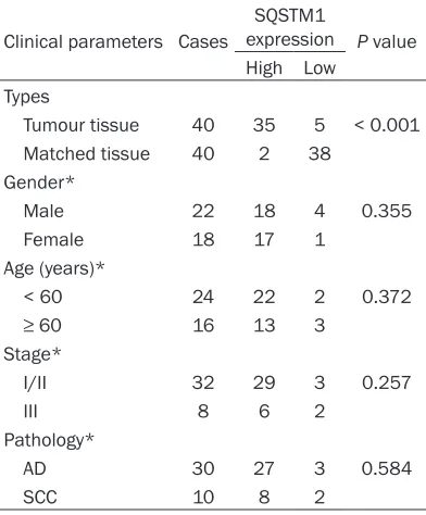
[image:3.612.91.287.97.334.2]We collected the general clinical characteris-tics and analyzed the SQSTM1 protein expres-sions in 40 NSCLC tumor tissues and paired normal lung tissues by IHC assay. Table 1 showed the gender, age, surgery staging and histopathologic types. The average age was 54 years (range 40-76 years). High immune-reac-tive group was more frequently found in NSCLC tumor tissues than in normal lung tissues (Figure 1A-C), and the overexpressed SQSTM1 Table 1. Clinical parameters and SQSTM1
expression by IHC
Clinical parameters Cases
SQSTM1
expression P value High Low
Types
Tumour tissue 40 35 5 < 0.001 Matched tissue 40 2 38
Gender*
Male 22 18 4 0.355
Female 18 17 1
Age (years)*
< 60 24 22 2 0.372
≥ 60 16 13 3
Stage*
I/II 32 29 3 0.257
III 8 6 2
Pathology*
AD 30 27 3 0.584
SCC 10 8 2
had no relation with age, sex and stage in NSCLC (Table 1).
The high-expressed SQSTM1 in tumor tissues was not associated with prognosis
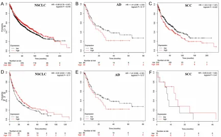
In order to validate the relationship between SQSTM1 expression and prognosis of NSCLC, we mined the data from the publicly available Kaplan-Meier plot (http://kmplot.com/analy-sis/index. php?p=background) database. The browser indicated that the patients with
[image:4.612.90.524.73.178.2]SQSTM1 mRNA high-expression subgroup (red line) had favorable overall survival (OS) in NSCLC; in contrast, the SQSTM1 mRNA high-expression was associated with poor prognosis of squamous cell carcinoma (SCC) while its high-expression was not related to the progno-sis of adenocarcinoma (AD). In addition, as shown in Figure 2, there was no correlation between SQSTM1 mRNA expression and pro-gression-free survival (PFS) in patients with NSCLC, AD or SCC.
Figure 1. SQSTM1 protein expression in NSCLC tissues and matched normal tissues using IHC. A. Staining in adeno-carcinoma tissues; B. Staining in squamous cell adeno-carcinoma tissues; C. Staining in paired normal tissues.
[image:4.612.91.524.227.495.2]Serum SQSTM1 levels in NSCLC patients were elevated
The mean value of serum SQSTM1 of 40 patients with BPD was 89.23 ± 14.8 ng/L, therefore the cutoff value was 118.83 ng/L.
Under the cutoff standard, serum SQSTM1 lev -els were elevated in 63 of the 80 (78.75%) NSCLC patients before treatment, but were within the normal range in 38 of the 40 BPD
patients. There was significant difference
two chemotherapy cycles. The average of serum SQSTM1 in 80 NSCLC patients de- creased from 191.08 ng/L (19.93-387.04 ng/L) to 124.91 ng/L (14.84-300.98 ng/L), the SD changed from 68.34 ng/L to 59.96 ng/L.
There was significant statistical difference, p < 0.001 (Table 3). The results of western blotting analysis also indicated SQSTM1 expression was decreased after the treatments (Figure 4). Therapeutic effects were evaluated after two treatment cycles, 47 of the 80 (58.75%) patients were effective (CR + PR), the remain-ing 33 (41.25%) patients were invalid (SD + PD). Further subgroup analysis, the decreased
amplitude of the effective group was signifi -cantly higher than that of the invalid group, and the mean values of decline were 87.94 ng/L and 35.18 ng/L in two subgroup, respectively. The result of the Mann-Whitney test showed that the p value was less than 0.001 (Table 4).
Initial SQSTM1 serum concentration could predict the chemotherapeutic efficacy
Serum SQSTM1 values were divided into nor-mal levels (< 118.83 ng/L) and high levels (>
118.83 ng/L). Using this classification, a favor -able outcome (CR or PR) was established in the patients with normal SQSTM1 levels, but not the high levels group (p = 0.026). Besides, the initial SQSTM1 serum concentration had no relationship with the age and gender (Table 5).
Pretreatment CEA level was positively corre-lated with the SQSTM1 level
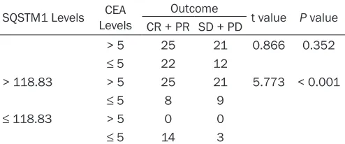
The normal range of CEA level was 0-5 ng/mL in our hospital. There was a positive correlation between initial SQSTM1 serum concentration and pretreatment CEA level (r = 0.819, p < 0.001, Table 5 & Figure 3). A simpler classifica -tion, in which CEA levels were divided into > 5
ng/mL and ≤ 5 ng/ml. CEA levels could not pre
-dict the efficacy of chemotherapy (p = 0.352, Table 6), while combining SQSTM1 data would Table 2. Serum levels of SQSTM1 in NSCLC patients
and BPD patients
Groups Cases Serum SQSTM1 (ng/L) t value P value > 118.83 < 118.83
NSCLC 80 63 17 58.422 < 0.001
BPD 40 2 38
NSCLC-non-small cell lung cancer; BPD-benign pulmonary diseases; P < 0.05 was considered statistically significant.
Figure 3. Correlation between baseline SQSTM1 and CEA levels in 80 patients. The dashed lines repre-sent the cut-off values of 118.83 ng/L for SQSTM1 and 5 ng/mL for CEA.
between the two groups (Table 2). The western blotting also showed the SQSTM1 expression in the NSCLC patients was higher than that of BDP patients (Figure 4).
Serum SQSTM1 levels declined signifi-cantly after two treatment cycles
A significant decline from the initial serum
SQSTM1 concentrations was seen after
increase the power to predict
chemotherapeu-tic efficacy (p < 0.001, Table 6). Discussion
Our study identifies SQSTM1 is expressed in
NSCLC tumor tissues, but not in adjacent non-malignant cells. This is consistent with the pre-vious report [20], who showed that high-expressed SQSTM1 was detected in 49.03%
power of cytoplasmic SQSTM1 depends on the mutation status of KRAS in colorectal cancer [23]. Based on diverse results, the following
reasons are considered: first, tumor heteroge -neity might be cause this phenomenon; then, racial disparities in participants and different
sample size are inevitable existence; finally,
mRNA levels and protein levels are not com-pletely consistent.
SQSTM1 is a multifunctional adapter protein implicated in selective autophagy [24], and the overexpression of SQSTM1 in tumor tissues is not in doubt. High levels of SQSTM1 autoanti-body in several cancer patients’ sera had also
been confirmed in recent 5 years [19, 25, 26].
Our data support the working hypothesis that autoantibody against SQSTM1 may be a useful serum biomarker for early stage NSCLC cancer screening, and SQSTM1 can induce relatively higher frequency of autoantibody responses in patients with NSCLC (78.75%). Liquid markers have also been detected in NSCLC [27-29]. Besides, commonly used serum markers of lung cancer that are currently recommended by multiple guidelines on the diagnosis of lung
cancer [30], include CEA, neuron-specific eno -lase (NSE), cytokeratin fragment (Cyfra21-1), pro-gastrin-releasing peptide (ProGRP) and squamous cell carcinoma antigen (SCC). How-
ever, their specificity and sensitivity are rela -tively scarce, and thus the combined detection of them was usually performed in clinical prac-Table 3. The concentration of serum SQSTM1 before
and after treatment in NSCLC patients
Groups Cases Serum SQSTM1 (ng/L) t value P value
Mean Range
Before 80 191.08 19.93~387.04 11.791 < 0.001 After 80 124.91 14.84~300.98
[image:6.612.91.324.98.150.2]Before-before chemotherapy; After-after two chemotherapy cycles; P < 0.05 was considered statistically significant.
Table 4. The decreased amplitude of serum SQSTM1
in different efficacy subgroups
Groups Case ΔSerum SQSTM1 (ng/L) U value P value
Mean Range
CR + PR 47 87.94 -3.55~97.03 307 < 0.001 SD + PD 33 35.18 5.09~178.28
CR-complete remission; PR-partial remission; SD-stable disease; PD-progressive disease; Δsymbols indicates the decreased ampli -tude; P < 0.05 was considered statistically significant.
Table 5. Relationship of initial SQSTM1 levels with clinical characteristics and outcome after therapy in NSCLC patients
Clinical
parameters Cases
Serum SQSTM1 (ng/L)
P value > 118.83 < 118.83
Gender
Male 21 16 5 0.738
Female 59 47 12
Age (years)
< 60 28 21 7 0.547
≥ 60 52 42 10
Response
CR + PR 47 33 14 0.026
SD + PD 33 30 3
CEA (ng/mL)
> 5 46 46 0 < 0.001
≤ 5 34 17 17
CR-complete remission; PR-partial remission; SD-stable disease; PD-progressive disease; P < 0.05 was considered statistically significant.
(51 of 104) of NSCLC tumor tissues, and the SQSTM1 expression was correlated
significantly with TNM stage and lymph
node metastasis, suggesting that SQSTM1
expression is of significance in diagnosing
NSCLC.
Previous studies have showed that SQSTM1 was an independent risk factor related to OS for NSCLC patients, and the prognostic impact of SQSTM1 status was pronounced in adenocarcinoma patients [20-22]; Furthermore, Anna et al pointed out that general SQSTM1 high-expression
[image:6.612.90.325.220.272.2] [image:6.612.91.300.364.548.2]tice. In such circumstances, SQSTM1 appears add brilliance to the diagnosis of lung cancer. Serum markers can be used not only for diag-nosis but also for therapeutic monitoring. A recent Meta-analysis [31] showed that the baseline Cyfra21-1 level is a better factor in
predicting treatment benefit, and Cyfra21-1
and CEA exhibited the good performance in forecasting objective response during treat-ment, and more, the decline of Cyfra21-1 levels during treatment is highly indicative for
objec-tive response. Our study identifies serum accu -mulation of SQSTM1 as a strong predictor of an ineffective treatment behavior of NSCLC, and substantial decline of SQSTM1 in plasma is correlated with effective treatment. In addi- tion, combination of basic CEA level resulted in increased predictive power of the assay sug-gesting that serum SQSTM1 level could
poten-tially be valuable chemotherapy efficacy mark
-ers. Confirmed by experiments, high expression
of SQSTM1 could prevent ROS stress-induced apoptosis or ER stress-induced apoptosis by regulating the Keap1-Nrf2-ARE signaling path-way, results in cisplatin resistance in human ovarian cancer cells [32, 33]. The same result, the activation of Nrf2 and SQSTM1 was associ-ated with doxorubicin resistance in breast can-cer [34]. Our data also suggested that a high level of SQSTM1 was not sensitive to conven-tional chemotherapy in NSCLC patients, and it is conceivable that high SQSTM1 level inhibits apoptosis or autophagy.
Autophagy is a cellular degrading process that regulates degradation cytoplasmic macromol-ecules and organelles by transportating to lyso-somes [35]. Autophagy is involved in both
ways [37]. Distinguishing the role of SQSTM1 in cacner might help in identifying high risk
patients who would benefit from autophagy
related anticancer drugs. The exciting recent investigations formed SQSTM1 as a potential therapeutic target in some types of cancer. As Chang et al said [38], SQSTM1 may be a ratio-nal target for bone metastatic in prostate cancer. Furthermore, some traditional herbal medicines have been used in the treatment or prevention of cancer via regulating SQS- TM1 mediated signaling pathway [39-42], and SQSTM1 vaccine has proven strong antitumor activity in solid tumors (lung and breast carci-nomas, melanoma and sarcoma) in hundreds of animals [43].
Our study was the first research to determine
the serum SQSTM1 level in NSCLC patients,
and it is the first evidence that SQSTM1 can predict the efficacy of conventional chemother -apy, in particularly after the combination of CEA. But there still has several limitations. First, it belongs to a single center study,
insuf-ficient sample size may cause an error that can -not be ignored; thus, multicenter studies are needed. Second, we only concluded that high serum SQSTM1 levels were more likely to occur in the resistance group, further mechanisms need to be explored.
Conclusions
In summary, our findings clearly show that
SQSTM1 over-expression has been implicated in NSCLC development, and serum SQSTM1 level is valuable for predicting the
chemothera-peutic efficacy. Thus, serum SQSTM1 can be
[image:7.612.91.338.98.201.2]used as an early diagnostic and therapeutic management biomaker.
Table 6. Outcome after therapy according to initial CEA and SQSTM1 levels
SQSTM1 Levels LevelsCEA Outcome t value P value CR + PR SD + PD
> 5 25 21 0.866 0.352
≤ 5 22 12
> 118.83 > 5 25 21 5.773 < 0.001
≤ 5 8 9
≤ 118.83 > 5 0 0
≤ 5 14 3
CR-complete remission; PR-partial remission; SD-stable disease; PD- progressive disease; P < 0.05 was considered statistically significant.
tumor formation and tumour suppres-sion [24]. SQSTM1 is an autophagic substrate, and it confers a highly selec-tive between cancer and autophagy through the degradation of important signaling molecules. Although SQSTM1 carcinogenic effects have been dem-onstrated by most of the relevant experiments (including this study), SQSTM1 also acts as a tumor suppres-sor through applying to tumor-associat-ed macrophages (TAM) in the tumor microenvironment [36]. SQSTM1 ca- uses cancer not only through
-Acknowledgements
This research was supported by the Scientific Research Program of Jiangsu University
(JLY-20140041), and the Science and Technology Development Project of Wujin (WS201401). Disclosure of conflict of interest
None.
Address correspondence to: Zhifang Zhuang, De- partment of Respiratory, Affiliated Wujin Hospital of Jiangsu University, No. 2 Yongning North Road, Changzhou 213017, PR China. Tel: +86-0519-85336190; E-mail:[email protected]
References
[1] Siegel RL, Miller KD, Jemal A. Cancer statistics. CA Cancer J Clin 2016; 66: 7-30.
[2] Chen W, Zheng R, Baade PD, Zhang S, Zeng H, Bray F, Jemal A, Yu XQ, He J. Cancer statistics in China, 2015. CA Cancer J Clin 2016; 66: 115-132.
[3] Peng W, Wu JG, Jiang YB, Liu YJ, Sun T, Wu N, Wu CJ. Antitumor activity of 4-O-(2”-O-acetyl- 6”-O-p-coumaroyl-β-D-glucopyranosyl)-p-cou-maric acid against lung cancers via mitochon-drial-mediated apoptosis. Chem Biol Interact 2015; 233: 8-13.
[4] Herlst RS, Heymach JV, Lippman SM. Molecu -lar origins of cancer: lung cancer. N Engl J Med 2008; 359: 1367-1380.
[5] Molina JR, Yang P, Cassivi SD, Schild SE, Adjei AA. Non-small cell lung cancer: epidemiology, risk factors, treatment, and survivorship. Mayo Clin Proc 2008; 83: 584-594.
[6] Pérez-Ramírez C, Cañadas-Garre M, Robles AI, Molina MÁ, Faus-Dáder MJ, Calleja-Hernández MÁ. Liquid biopsy in early stage lung cancer. Transl Lung Cancer Res 2016; 5: 517.
[7] Liu Y, Yuan D, Ye W, Lv T, Song Y. Prognostic value of circulating endothelial cells in non-small cell lung cancer patients: a systematic review and meta-analysis. Transl Lung Cancer Res 2015; 4: 610-618.
[8] Liu Y, Xing Z, Zhan P, Liu H, Ye W, Lv T, Song Y. Is it feasible to detect epidermal growth factor receptor mutations in circulating tumor cells in nonsmall cell lung cancer?: a meta-analysis. Medicine (Baltimore) 2016; 95: e5115. [9] Sanchez P, De Carcer G, Sandoval IV, Moscat J,
Diaz-Meco MT. Localization of atypical protein kinase C isoforms into lysosome-targeted en-dosomes through interaction with p62. Mol Cell Biol 1998; 18: 3069-3080.
[10] Moscat J, Diaz-Meco MT. p62 at the cross-roads of autophagy, apoptosis, and cancer. Cell 2009; 137: 1001-1004.
[11] Duran A, Amanchy R, Linares JF, Joshi J, Abu-Baker S, Porollo A, Hansen M, Moscat J, Diaz-Meco MT. p62 is a key regulator of nutrient sensing in the mTORC1 pathway. Mol Cell Biol 2011; 44: 134-146.
[12] Duran A, Linares JF, Galvez AS, Wikenheiser K, Flores JM, Diaz-Meco MT, Moscat J. The signal-ing adaptor p62 is an important NF-κB media -tor in tumorigenesis. Cancer cell 2008; 13: 343-354.
[13] Linares JF, Amanchy R, Greis K, Diaz-Meco MT, Moscat J. Phosphorylation of p62 by cdk1 con-trols the timely transit of cells through mitosis and tumor cell proliferation. Mol Cell Biol 2011; 31: 105-117.
[14] Luo RZ, Yuan ZY, Li M, Xi SY, Fu J, He J. Accu- mulation of p62 is associated with poor prog-nosis in patients with triple-negative breast cancer. Onco Targets Ther 2013; 6: 883-888. [15] Masuda GO, Yashiro M, Kitayama K, Miki Y,
Kasashima H, Kinoshita H, Morisaki T, Fukuo-ka T, Hasegawa T, Sakurai K, ToyoFukuo-kawa T, Kubo N, Tanaka H, Muguruma K, Masaichi O, Hiraka-wa K. Clinicopathological correlations of au-tophagy-related proteins LC3, Beclin 1 and p62 in gastric cancer. Anticancer Res 2016; 36: 129-136.
[16] Iwadate R, Inoue J, Tsuda H, Takano M, Furuya K, Hirasawa A, Aoki D, Inazawa J. High expres-sion of SQSTM1/p62 protein is associated with poor prognosis in epithelial ovarian can-cer. Acta Histochem Cytochem 2014; 47: 295. [17] Bartsch G, Jennewein L, Harter PN, Antonietti
P, Blaheta RA, Kvasnicka HM, Kögel D, Hafer-kamp A, Mittelbronn M, Mani J. Autophagy- as-sociated proteins BAG3 and p62 in testicular cancer. Oncol Rep 2016; 35: 1629-1635. [18] Zhang JY, Chan EK, Peng XX, Tan EM. A novel
cytoplasmic protein with RNA-binding motifs is an autoantigen in human hepatocellular carci-noma. J Exp Med 1999; 189: 1101-1110. [19] Liu W, Li Z, Xu W, Yang S. Humoral
autoim-mune response to IGF2 mRNA-binding protein (IMP2/p62) and its tissue-specific expression in colon cancer. Scand J Immunol 2013; 77: 255-260.
[20] Wang X, Du Z, Li L, Shi M, Yu Y. Beclin 1 and p62 expression in non-small cell lung cancer: relation with malignant behaviors and clinical outcome. Int J Clin Exp Pathol 2015; 8: 10644. [21] Inoue D, Suzuki T, Mitsuishi Y, Miki Y, Suzuki S,
with poor prognosis in patients with lung ade-nocarcinoma. Cancer Sci 2012; 103: 760-766. [22] Schläfli AM, Adams O, Galván JA, Gugger M,
Savic S, Bubendorf L, Schmid RA, Becker KF, Tschan MP, Langer R, Berezowska S. Prognos-tic value of the autophagy markers LC3 and p62/SQSTM1 in early-stage non-small cell lung cancer. Oncotarget 2016; 7: 39544-39555.
[23] Schmitz KJ, Ademi C, Bertram S, Schmid KW, Baba HA. Prognostic relevance of autophagy-related markers LC3, p62/sequestosome 1, Beclin-1 and ULK1 in colorectal cancer pa -tients with respect to KRAS mutational status. World J Surg Oncol 2016; 14: 189.
[24] Nezis IP, Stenmark H. p62 at the interface of autophagy, oxidative stress signaling, and can-cer. Antioxid Redox Signal 2012; 17: 786-793. [25] Li Y, Francia G, Zhang JY. p62/IMP2 stimulates
cell migration and reduces cell adhesion in breast cancer. Oncotarget 2015; 6: 32656-68. [26] Liu X, Ye H, Li L, Li W, Zhang Y, Zhang JY. Hu-moral autoimmune responses to insulin-like growth factor II mRNA-binding proteins IMP1 and p62/IMP2 in ovarian cancer. J Immunol Res 2014; 2014: 326593.
[27] Molina R, Holdenrieder S, Auge JM, Schalhorn A, Hatz R, Stieber P. Diagnostic relevance of circulating biomarkers in patients with lung cancer. Cancer Biomark 2010; 6: 163-178. [28] Reitsma JB, Glas AS, Rutjes AW, Scholten RJ,
Bossuyt PM, Zwinderman AH. Bivariate analy-sis of sensitivity and specificity produces infor -mative summary measures in diagnostic re-views. J Clin Epidemiol 2005; 58: 982-990. [29] Salgia R, Harpole D, Herndon JE 2nd, Pisick E,
Elias A, Skarin AT. Role of serum tumor mark-ers CA 125 and CEA in non-small cell lung can-cer. Anticancer Res 2000; 21: 1241-1246. [30] Zhi XY, Yu JM, Shi YK. Chinese guidelines on
the diagnosis and treatment of primary lung cancer (2015 version). Cancer biomark 2015; 121: 3165-3181.
[31] Holdenrieder S, Wehnl B, Hettwer K, Simon K, Uhlig S, Dayyani F. Carcinoembryonic antigen and cytokeratin-19 fragments for assessment of therapy response in non-small cell lung can-cer: a systematic review and meta-analysis. Br J Cancer 2017; 116: 1037-1045.
[32] Xia M, Yu H, Gu S, Xu Y, Su J, Li H, Kang J, Cui M. p62/SQSTM1 is involved in cisplatin resis-tance in human ovarian cancer cells via the Keap1-Nrf2-ARE system. Int J Oncol 2014; 45: 2341-2348.
[33] Yu H, Su J, Xu Y, Kang J, Li H, Zhang L, Yi H, Xiang X, Liu F, Sun L. p62/SQSTM1 involved in cisplatin resistance in human ovarian cancer cells by clearing ubiquitinated proteins. Eur J Cancer 2011; 47: 1585-1594.
[34] Wei Y, Liu D, Jin X, Gao P, Wang Q, Zhang J, Zhang N. PA-MSHA inhibits the growth of doxo-rubicin-resistant MCF-7/ADR human breast cancer cells by downregulating Nrf2/p62. Can-cer Med 2016; 5: 3520-3531.
[35] Puissant A, Fenouille N, Auberger P. When au-tophagy meets cancer through p62/SQSTM1. Am J Cancer Res 2012; 2: 397-413.
[36] Grivennikov SI, Greten FR, Karin M. Immunity, inflammation, and cancer. Cell 2010; 140: 883-899.
[37] Zhong Z, Umemura A, Sanchez-Lopez E, Liang S, Shalapour S, Wong J, He F, Boassa D, Per-kins G, Ali SR, McGeough MD, Ellisman MH, Seki E, Gustafsson AB, Hoffman HM, Diaz-Me-co MT, Moscat J, Karin M. NF-κB restricts in -flammasome activation via elimination of dam -aged mitochondria. Cell 2016; 164: 896-910. [38] Chang MA, Morgado M, Warren CR, Hinton CV,
Farach-Carson MC, Delk NA. p62/SQSTM1 is required for cell survival of apoptosis-resistant bone metastatic prostate cancer cell lines. Prostate 2014; 74: 149-163.
[39] Choi YK, Cho SG, Choi YJ, Yun YJ, Lee KM, Lee K, Yoo HH, Shin YC, Ko SG. SH003 suppresses breast cancer growth by accumulating p62 in autolysosomes. Oncotarget 2016; 8: 88386-88400.
[40] Moscat J, Diaz-Meco MT. The atypical PKC scaffold protein P62 is a novel target for anti-inflammatory and anti-cancer therapies. Adv Enzyme Regul 2002; 42: 173-179.
[41] Lou JS, Yan L, Bi CW, Chan GK, Wu QY, Liu YL, Huang Y, Yao P, Du CY, Dong TT, Tsim KW. Yu Ping Feng San reverses cisplatin-induced multi-drug resistance in lung cancer cells via regulating drug transporters and p62/TRAF6 signalling. Sci Rep 2016; 6: 31926.
[42] Ishaq M, Khan MA, Sharma K, Sharma G, Dut-ta RK, Majumdar S. Gambogic acid induced oxidative stress dependent caspase activation regulates both apoptosis and autophagy by targeting various key molecules (NF-κB, Be -clin-1, p62 and NBR1) in human bladder can-cer cells. Biochim Biophys Acta 2014; 1840: 3374-3384.