Case Report
Primary hyperoxaluria type 1: a case report in a
7-month-old Chinese infant under biopsy and
light microscope diagnosis
Qiang Sun1,2, Liping Jiao1,2, Xiaochun Wang3, Hui Wang1,2, Xiaorong Liu1,2
1Department of Nephrology, Beijing Children’s Hospital, Affiliated to Capital Medical University, National Center for Children’s Health, Beijing 100045, China; 2Beijing Key Laboratory of Pediatric Chronic Kidney Disease and Blood Purification, Beijing 100045, China; 3Department of Nutrition, New Century International Children’s Hospital, Beijing 100045, China
Received August 30, 2017; Accepted January 4, 2018; Epub January 15, 2018; Published January 30, 2018
Abstract: Primary hyperoxaluria type one (PH1) is a rare inborn autosomal recessive monogenic metabolic disorder due to hepatic alanine-glyoxylate-aminotransferase deficiency which results in excessive synthesis and urinary ex-cretion of oxalate, inducing renal stone formation and deposition of calcium oxalate stones throughout the entire body. We present the clinical consequences in a 7-month-old male infant diagnosed with PH1. The patient was diagnosed by renal biopsy, which showed acute tubular injury with diffuse calcium oxalate crystals. The results of genomic DNA of AGXT gene were examined; the gene of the patient was similar to his mother, but different from his father and a healthy control. Mutation analysis of AGXT in the patient revealed the mutation, c.814T>GA, which was considered to be a silent mutation. During 22 days of hospitalization, supplementary pyridoxine was adminis-trated intravenously at 50 mg per day for 3 days, and then orally at 60 mg three times daily. Meanwhile the patient was treated with peritoneal dialysis and after 12 months of follow-up, he unfortunately died of refractory bacterial pneumonia.
Keywords: Primary hyperoxaluria type 1, alanine-glyoxylate-aminotransferase (AGT), oxalate, systemic oxalosis (SO)
Introduction
Human alanine glyoxylate aminotransferase (AGT; EC2.6.1.44), localized to liver peroxi-somes, has a broad substrate specificity that uses glyoxylate as the preferred amino accep-tor [1]. AGT catalyzes the formation of glycine and prevents the formation of oxalate from gl- yoxylate. In humans, hereditary deficiency of AGT results in rare clinical consequences in which excessive amounts of glyoxylate are con-verted to oxalate. This disorder, known as pri-mary hyperoxaluria type 1 (PH1; MIM259900), is characterized by hyperoxalemia and hype- roxaluria with massive calcium oxalate deposi-tion in the kidney, bone, myocardium, vessels, and throughout the entire body [2].
PH1 is a rare autosomal-recessive disorder caused by a deficiency of the liver-specific py- ridoxal-phosphate dependent enzyme AGT. The
AGT gene (AGXT) mutation that underlies this disorder has been reported [3], including stud-ies of PH1-specific mutations (G41R, F152I, G170R, and I244T) in European and North American patients [4], as well as a PH1-speci- fic mutation (S205P) in Japanese patients [5]. Our study investigated the genetic profile of both a 7-month-old infant with PH1 and his par-ents to understand how this disease gene was inherited in this family.
Case report
plicated neonatal course. The family history was negative with respect to renal and meta-bolic disease. The infant was well nourished on mixed feeding with normal growth and development.
Five months before admission, the infant had diarrhea about 5-6 times per day with unform- ed stools. Then he was given probiotic treat-ment in the hospital, but symptoms did not improve. His parents did not continue any me- dical treatment or investigate further exami- nation until the infant had decreased urine output to about 100 ml per day, with facial edema 4 days before admission.
The laboratory blood examination in our hos- pital found Hb 55 g/L, WBC 8.67×10⁹/L, PLT
253×109/L, Serum Potassium 2.93 mmol/L,
Sodium 116 mmol/L, Calcium 1.85 mmol/L, Phosphate 3.60 mmol/L, Parathyroid hormo- ne 231 pg/mL, BUN 30.93 mmol/L, Cr 662 mmol/L, Uric acid 650 mmol/L. Urine routine: pH6.5, SG 1.010, protein (-) - (+), WBC 1-3/HP. Blood gases: pH 7.30, HCO3- 17.8 mmol/L, BE-10.7 mmol/L. Urine Calcium/Cr: 0.37. Ul- trasound showed both kidneys were enlarged with severe parenchymal diffuse damage and enhanced echogenicity, with the left kidney sized at 7.3×3.7 cm and the right kidney siz- ed at 7.5×3.8 cm. There were small pieces of microlith shaped stones on the left collective system of size 0.2×0.1 cm, without ureteral
dilatation. Renal biopsy was performed and the pathological results indicated normal glo- meruli, but the renal tubular epithelial cells were disrupted. Large amounts of crystals could be seen within the lumen of the urinary cast. Infiltrating lymphocytes were observed in the renal interstitium (Figure 1). The treat-ment course was given without any com- plications.
Genome analysis
Genomic DNA of the infant and both his par-ents was extracted from peripheral blood sa- mples using MagExtractor-Genome (TOYOBO, Tokyo, Japan) according to the manufacturer’s instructions. The DNA encoding of AGXT gene was amplified as previously described [6]. The genomic DNA sequences were determined as follows: sense, 5’-GGGGAGAGAAAGGGGCACA- CAGAGT-3’, and antisense, 5’-TGGGGCTVTAGT- TGGGGTTCTTGAG-3’, using BigDye Terminator Cycle Sequencing Kit (Applied Biosystems, CA, USA).
The molecular basis of PH1 was examined by analyzing the entire coding sequence of
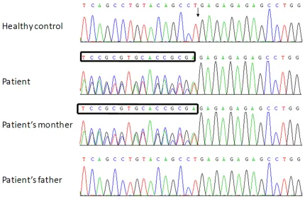
AGXT. Genetic mutation of GA heterozygous insertion was found in both the patient and his mother, but his father’s DNA profile was normal. (Results were shown in Figure 2). Treatment and follow up
The infant received anti-diarrheal treatment immediately after admission. Peritoneal dialy-sis (CAPD) was initiated on the second day af- ter admission and treated for 22 days. Py- ridoxine treatment was administrated intrave-nously at 50 mg per day for 3 days, and then orally at 60 mg per time three times daily. The patient’s infection was controlled with reduced symptoms of edema and diarrhea, and blood pressure was stably maintained in the 100-120/70-80 mm Hg. After 22 days, the patient was discharged, however the peritoneal dia lysis was continued. Unfortunately, the infant died of refractory bacterial pneumonia 12 months later.
Discussion
[image:2.612.89.290.72.223.2]Oxalate is a metabolic waste product excreted by the kidney. Hyperoxaluria is a common cau- se of kidney stones due to low solubility of
calcium oxalate salts. Severe hyperoxaluria may lead to loss of renal function and syste- mic oxalosis. Hyperoxaluria can be caused by genetic defects that lead to endogenous over-production or excessive oxalate absorption fr- om the diet [7].
The primary hyperoxalurias are a group of ra- re hereditary calcium oxalate kidney stone dis-eases, the most prominent of which are pri- mary hyperoxaluria type 1 (alanine/glyoxylate aminotransferase deficiency) and type 2 (gly- oxylate/hydroxypyruvate reductase deficiency). Insoluble calcium oxalate crystallizes out in the kidney and urinary tract to cause kidney dysfunction and eventually complete organ fai- lure. More than 100 mutations have been fo- und in PH1, but less than 20 in PH2. Screen- ing for the three most common mutations, c.33_34insC, c.508G>A, and c.731T>C, en- ables a molecular diagnosis in 34.5% cases [8].
The genomic DNA of AGXT gene was shown in Figure 2. Mutation analysis of the AGXT
revealed the mutation, c.814T>GA, which is considered to be a silent mutation. It is un- known whether the mother’s gene will confer disease in future generations or not. Other- wise, the abnormal AGXT gene was not the contributing factor for this patient. The patho-genesis of PH1 still needs further investi- gation.
Since AGT is expressed predominantly in the liver, definitive diagnosis of the disease requi- res liver biopsy to assess AGT activity [9]. Unfortunately, we could not perform liver biop-sy or exam AGT activity in our hospital.
The symptoms of the patient manifested as oliguria, mild leukocyturia, proteinuria, less mi- croliths in the kidney, and severe renal failure. Because of the lack of certain examination facilities, we were unable to perform blood and urine oxalate tests, as well as the assessment of the AGT activity. Nonetheless, we performed renal biopsy and found diffuse crystals within the lumen of urinary cast, while the glomeruli were almost normal. These crystals were
[image:3.612.90.520.71.352.2]acterized as highly transparent, colorless, sub angular,and amorphous. Some of the crystals were hexagonal or had eight surfaces, and were positively birefringent on polarized light [10].
We found that the crystals were oxalate crys- tallization and the patient was diagnosed with oxalate nephropathy by medical history, clini- cal signs, laboratory tests, and the diagnostic crystals.
Since the patient’s serum uric acidity was high, we should pay attention to urate nephropathy. However, there were two pieces of evidence that did not support this. First of all, the urate crystal was radiolytic and needle-like, which is different from the colorless, amorphous crys-tals of the infant. Secondly, the relatively high level of the patient’s serum uric acidity was not significantly elevated, which may be proportion-ally due to the increase serum creatinine level. Furthermore, by renal biopsy we found that another 10-month old infant had renal tubular melamine crystals, and this patient was com- plicated with oliguria, renal microliths, and pro-gressive decline of renal function. But the cry- stals’ characteristics of this case were different from the melamine crystals, which stained blue on the edge by HE stains [11]. On the basis of the renal pathology, considering the infant’s early onset of only 7 months old, without any family history, and accompanied by end stage renal failure, we suggest the diagnosis is pri-mary hyperoxaluria, and type 1 is the most like-ly type based on clinical signs and symptoms. Oxalate is an end product of human metabo-lism, produced in the liver and excreted primar-ily by the kidney. Oxalate is also absorbed from the diet so that renal excretion reflects a combi-nation of endogenous and exogenous oxalate loads. Since the child had chronic diarrhea, the hyperoxaluria could result from excess absor- ption from the intestinal tract, which was dam-aged by chronic diarrhea. After removing the allergen and relieving edema, the diarrhea im- proved, but the oliguria remained and aggra-vated renal function. This demonstrated that the hyperoxaluria might not be secondary to the diarrhea, which most likely was primary. Oxalosis could affect many other organs as well, including bone, retina, vessel,
myocardi-um, nerve, and joint damage, which results in poor quality of life or even death [12]. Howev- er, no cardiovascular or skeletal complications were found in this case. We considered that the reason could be due to the limited dura- tion of the disease. If the patient hadn’t be- en treated, diverse clinical manifestations ac- companied with multiple organ injury could appear after a couple of months.
To prevent exacerbating hyperoxaluria, the infant and his mother were instructed to avoid high oxalate foods, such as spinach, bran flakes, beets, potato chips, nuts, et al. The on- ly pharmacologic intervention known to lower urine oxalate in PH is pyridoxine supplemen- tation. Pyridoxine is a cofactor for AGT mediat-ed conversion of glyoxylate to glycine. Ap- proximately 25% to 30% of PH1 patients will reduce urine oxalate excretion significantly wi- th pyridoxine [13]. The patient was given oral pyridoxine treatment but was not effective at the end, which suggested that the patient’s disease was not caused by the deficiency of the pyridoxine.
The reported effective treatment is organ tr- ansplantation. Isolated kidney transplantation can reduce plasma oxalate levels, but disease recurrence often leads to poor graft survival. Liver transplantation completely corrects the enzyme defect. In Europe, with the poor results of isolated kidney transplantation reported 2 decades ago, combined or sequential liver- kidney transplantation has predominated the- reafter [14]. We observed regretfully that there was no successful case with PH in children undergoing combined liver and kidney trans-plantation as reported in China to date.
Under conditions of limited testing for gene- tics, serum oxalate, urine oxalate, and AGT en- zyme, we suggest that renal biopsy and light microscopic be performed together to confirm a diagnosis as soon as possible.
Acknowledgements
Disclosure of conflict of interest
None.
Address correspondence to: Dr. Xiaorong Liu, De- partment of Nephrology, Beijing Children’s Hospital, Affiliated to Capital Medical University, National Center for Children’s Health, Beijing 100045, China; Beijing Key Laboratory of Pediatric Chronic Kidney Disease and Blood Purification, Beijing 100045, China. Tel: 86-10-59616337; Fax: 86-10-59718700; E-mail: [email protected]
References
[1] Bradford MM. A rapid and sensitive method for the quantitation of microgram quantities of protein utilizing the principle of protein-dye binding. Anal Biochem 1976; 72: 248-254. [2] Danpure CJ, Jennings PR. Peroxisomal
ala-nine: glyoxylate aminotransferase deficiency in primary hyperoxaluria type I. FEBS Lett 1986; 201: 20-24.
[3] Lumb MJ, Danpure CJ. Functional synergism between the most common polymorphism in human alanine: glyoxylate aminotransferase and four of the most common disease-causing mutations. J Biol Chem 2000; 275: 36415-36422.
[4] Williams EL, Acquaviva C, Amoroso A, Cheva-lier F, Coulter-Mackie M, Monico CG, Giachino D, Owen T, Robbiano A, Salido E, Waterham H, Rumsby G. Primary herperoxaluria type 1: up-date and additional mutation analysis of the AGXT gene. Hum Mutat 2009; 30: 910-917. [5] Nishiyama K, Funai T, Katafuchi R, Hattori F,
Onoyama K, Ichiyama A. Primary hyperoxaluria type I due to a point mutation of T to C in the coding region of the serine: pyruvate amino-transferase gene. Biochem Biophys Res Com-mun 1991; 176: 1093-1099.
[6] Venter JC, Adams MD, Myers EW. The se-quence of the human genome. Science 2001; 291: 1304-1351.
[7] John RA. Hyperoxaluric calcium nephrolithia-sis. Endocrinol Metab Clin N Am 2002; 31: 927-949.
[8] Mbarek B, Abroug S, Omezzine A, Zellama D, Achour A, Harbi A, Bouslama1 A. Selected AGXT gene mutations analysis provides a ge-netic diagnosis in 28% of Tunisian patients with primary hyperoxaluria. BMC Nephrol 2011; 12: 25.
[9] Fargue S, Rumsby G, Danpure CJ. Multiple mechanisms of action of pyridoxine in primary hyperoxaluria type 1. Biochim Biophys Acta 2013; 1832: 1776-1783.
[10] Chintanaboina JK, Nadasdy T, Manahan F, Muppidi V. Acute oxalate nephropathy diag-nosed by renal biopsy. Intern Med 2012; 51 2249.
[11] Sun N, Shen Y, He LJ. Histopathological fea-tures of the kidney after acute renal failure from melamine. N Engl J Med 2010; 362: 662-4.
[12] Harambat J, van Stralen KJ, Espinosa L, Groot-hoff JW, Hulton SA, Cerkauskiene R, Schaefer F, Verrina E, Jager KJ, Cochat P; European Soci-ety for Pediatric Nephrology/European Renal Association-European Dialysis and Transplant Association (ESPN/ERA-EDTA) Registry. Char-acteristics and outcomes of children with pri-mary oxalosis requiring renal replacement therapy. Clin J Am Soc Nephrol 2012; 7: 458-65.
[13] Kopp N, Leumann E. Changing pattern of pri-mary hyperoxaluria in Switzerland. Nephrol Dial Transplant 1995; 10: 2224-7.