Inhibition of proteasome by bortezomib increase
chemosensitivity of bcr/abl positive human k562 chronic
myleoid leukemia cells to imatinib
Baran Yusuf1, Coskun Oztekin2, Bassoy Esen Yonca1
1Department of Molecular Biology and Genetics, Izmir Institute of Technology, Urla, Izmir, Turkey;yusufbaran@iyte.edu.tr 2Department of Family Medicine, Ankara Numune Education and Research Hospital, Çankaya, Ankara, Turkey
Received 18 July 2009; revised 28 July 2009; accepted 14 August 2009.
ABSTRACT
Chronic myeloid leukaemia (CML) results from a translocation between chromosomes 9 and 22 which generates the BCR/ABL fusion oncopro-tein. BCR/ABL has constitutively tyrosine kinase activity resulting in leukemogenesis. Imatinib, a competitive inhibitor of the BCR/ABL tyrosine kinase, is the common treatment of CML. De-spite the outstanding results of imatinib in the chronic phase of CML, cases of treatment fail-ure have been reported, resulting in heteroge-neous molecular response. Bortezomib is a re-versible inhibitor of the 26S proteasome induc-ing cell cycle arrest in G2/M phase, apoptosis by inhibition of NF-kB. In this study, we examined the possible synergistic apoptotic effects of the imatinib/bortezomib combination and the re-sponsible apoptotic mechanisms induced by this combination in K562 cells. The results of this study showed increased cytotoxicity by XTT assay in combination of imatinib and bor-tezomib as compared to any agent alone. On the other hand, synergistic apoptotic affects of combination of these agents were also con-firmed by changes in caspase-3 enzyme activity and mitochondrial membrane potential. Taking together, all the results, confirming each other, showed that the combination of the imatinib and bortezomib has considerable synergistic effects on the apoptosis through increase in caspase-3 enzyme activity and decrease in mitochondrial membrane potential in human K562 CML cells. Keywords:Imatinib; Bortezomib; Combination Therapy; CML, BCR/ABL
1. INTRODUCTION
Chronic myeloid leukemia (CML) is characterized by a reciprocal translocation between chromosomes 9 and 22
bringing together BCR and ABL genes to form BCR/ ABL fusion protein. BCR/ABL protein, having constitu-tive tyrosine kinase activity, is responsible for the pathogenesis of CML. Imatinib (imatinib mesylate, STI571, Gleevec) is a tyrosine kinase inhibitor with good efficacy and recently recommended as first line therapy for the chronic phase CML. BCR/ABL has a special ATP binding site close to the substrate proteins binding region. Imatinib works by binding for blocking function of BCR/ABL ATP binding site. When the ATP binding site is filled by imatinib, ATP cannot donate the phosphate and BCR/ABL can no longer activate down-stream signaling proteins that promote chronic myeloid leukemia [1,2,3,4,5]. Although imatinib has an out-standing outcomes in the chronic phase of CML, cases of treatment failure has been reported. Resistance to im-atinib is a major problem in CML patients in blast crisis phase and has been principally associated with BCR/ ABL tyrosine kinase domain point mutation decreasing the affinity of imatinib to BCR/ABL protein. In addition, amplification of the BCR/ABL oncogene, aberrant ce-ramide metabolism, inhibition of apoptotic pathways, and reduction in effective intracellular concentrations of the drug by altered drug efflux or influx are responsible for imatinib resistance [6,7]. As a result of disease per-sistence and resistance imatinib monotherapy cannot be referred to a cure for the majority of patients with CML [8,9,10,11,12]. The primary and acquired resistance to imatinib is the developing problem in current CML treatment and has various underlying mechanisms [1]. On the other hand, the combination of imatinib with anti-CML agents, may be able to kill drug resistant cells as highly as drug-sensitive parental BCR/ABL+
leukae-mic cells, unless the completely refractory to the partner drugs is developed by leukaemic cells.
has been proved in myeloma, myeloid leukemia, lym-phoma, prostate, breast, colon, and lung cancers. For patients with myeloma and/or relapsed and refractory diseases bortezomib shows promising results [17]. Bor-tezomib induces cell cycle arrest in G2M phase and apoptosis by inhibition of NF-kB [14,18,19,20]. Studies strongly implicate the activity of the transcription factor NF-κB in promoting chemoresistance, cytokine-medi- ated proliferation, tumor metastasis, and angiogenesis. Bortezomib, as a proteosome inhibitor, also enhances the sensitivity of tumor cells to chemotherapy and radiation and reverses chemoresistance. Induction of apoptosis involves increased Bcl-2 phosphorylation and cleavage, accumulation of cyclins A and B1 and increased stability of p53 and up-regulated p53-induced gene expression [21].
In this study we describe synergistic cytotoxicity when cells are treated with the imatinib in combination with bortezomib and responsible signaling pathways that induce apoptosis in human K562 CML cells.
2. METHODS
Cell lines, Growth Conditions, and Drugs. Human K562 CML cells were obtained from German Collection of Microorganisms and Cell Cultures. Human K562 CML cells were maintained in RPMI 1640 growth me-dium containing 10% fetal bovine serum and 1% peni-cillin-streptomycin at 37℃ in 5% CO2. Imatinib was a
generous gift from Novartis (USA) and bortezomib was from Gulhane Medical School, Department of Hematol-ogy. Imatinib and bortezomib were dissolved in distilled water and 10 mM stock solutions were prepared.
Measurement of growth by XTT. The IC50 values of imatinib and bortezomib that inhibited cell growth by 50% were determined from cell proliferation plots ob-tained by XTT as described previously [7]. Briefly, 2x10⁴ cells were seeded into the 96-well plates that
con-tain 200 µl growth medium and the absence or presence of increasing concentrations of bortezomib and imatinib, and incubated at 37˚C in 5% CO2 for 72 hours.
After-wards, they were treated with 40 µl XTT reagent for 4 hours. Then, the absorbances of the samples under 490 nm wavelength were measured by Elisa reader (Thermo Electron Corporation Multiskan Spectrum, Finland). At the end, IC50 values were calculated to cell survival plots.
Measurement of caspase-3 activity. Caspase-3 activ-ity was determined using the caspase-3 colorimetric as-say (R&D Systems, USA). To start with, the cells that treated with bortezomib and imatinib for 72 hours were collected by centrifugation at 1000 rpm for 10 minutes. Afterwards, pellets were treated with 100 µl of cold lysis buffer (1X) in order to obtain cell lysate. Then cell lys-ates were incubated on ice for 10 minutes and they were
centrifuged at 14000 rpm for 1 minute. After that, su-pernatants were transferred to new eppendorf tubes. For measuring caspase activity, reaction mixture that include 20 µl assay buffer (5X), 25 µl of sample, 50 µl of steril-ized water and 5 µl of caspase-3 colorimetric substrate was prepared in 96-well plates adding and incubated for 2 hours incubation at 37˚C. samples was read under 405 nm wavelength by Elisa reader (Thermo Electron Cor-poration Multiskan Spectrum, Finland). Absorbances were normalized to protein levels as determined by Bradford Assay.
Detection of the loss of mitochondrial membrane potential (MMP). The loss of mitochondrial membrane potential was detected by JC-1 mitochondrial membrane potential (MMP) kit (Cell Technology, USA). The loss of mitochondrial membrane potential is a hallmark for apoptosis. Firstly, the cells treated with imatinib and bortezomib were collected by centrifugation at 1000 rpm for 10 minutes. Supernatants were discarded and 500 µl of JC-1 dye was added onto pellets and incubated at 37˚C in 5% CO2 for 15 minutes. Then samples were
centrifuged at 1000 rpm for 5 minutes and 2 ml of assay buffer was added onto the pellets and they were centri-fuged for 5 minutes at 1000 rpm. All pellets were resus-pended with 500 µl assay buffer and 150 µl from each of them was added into the 96-well plate. The aggregate red form has absorption/emission maxima of 585/590 nm and the green monomeric form has absorption/ emis-sion maxima of 510/527 nm. The plate was read in these wavelengths by fluorescence Elisa reader (Thermo Vari-oskan Spectrum, Finland).
3. RESULTS
The cytotoxic effects of imatinib or bortezomib alone or in combination on K562 cells. IC50 values of imati-nib and bortezomib were found to be 267 nM (Figure 1) and 65 nM (Figure 2) in K562 cells exposed to increas-ing concentrations of imatinib and bortezomib for 72 hours, respectively. There were decreases (from 8 to 82%) in proliferation of K562 cells exposed to increas-ing nanomolar concentrations of imatinib (from 0,2 nM to 600 nM) for 72 hours as compared to untreated con-trols (Figure 1). On the other hand 10-, 50-, and 100 nM bortezomib resulted in 5-, 36-, and 70% decreases in cell proliferation (Figure 2), respectively. The same concen-trations of imatinib with a combination of 65 nM borte-zomib decreased cellular proliferation significantly (from 82 to 90%), as compared to untreated controls (Figure 3a). There were 50% decrease in proliferation of K562 cells in response to 65 nM bortezomib (Figure 3b).
and 1.29-fold increase in 0.5- and 5 nM imatinib alone applied K562 cells, respectively while 65 nM bortezo- mib increased the caspase-3 enzyme activity 1.24-fold increase by itself (Figure 4). On the other hand, the same concentrations of imatinib in combination with 65 nM bortezomib increased the enzyme activity 2.4- and 2.85-fold, respectively (Figure 4).
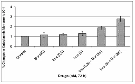
[image:3.595.310.538.163.423.2]The effects of combination of Imatinib/Bortezomib on mitochondrial membrane potential. Treatment with imatinib/Bortezomib caused a significant loss of MMP, as measured by increased accumulation of of cytoplas-mic/mitochondrial form of JC-1, in K562 cells as com-pared any agent alone. There were 1.19- and 1.31-fold increase in cytoplasmic/mitochondrial JC-1 in 0.5- and 5 nM imatinib applied K562 cells, respectively, while 65 nM bortezomib decreased MMP 1.16 fold by itself. On the other hand, the same concentrations of imatinib
[image:3.595.62.283.284.408.2]Figure 1. Effects of imatinib on the growth of K562 cells. The IC50 concentration of imatinib was calculated from cell proliferation plots. The XTT assays were performed using triplicate samples in at least two independent ex-periments. Statistical significance was determined using two-way analysis of variance, and (P < 0,05) was consid-ered significant.
Figure 2. Effects of bortezomib on the growth of K562 cells. The IC50 concentration of bortezomib was deter-mined by XTT assay for each cell line as described. The XTT assays were performed using triplicate samples in at least two independent experiments. Statistical significance was determined using two-way analysis of variance, and (P < 0,05) was considered significant.
with 65 nM bortezomib decreased MMP 1.88- and 2.79-fold, respectively (Figure 5).
4. DISCUSSION
The way for treatment of cancer more than one agent is
(a)
[image:3.595.63.283.494.626.2](b)
Figure 3. Effects of bortezomib and imatinib combination on the growth of K562 cells (a). Effects of 65 nM borte-zomib by itself on K562 cells as compared to untreated controls (b). Cytotoxicity was determined by the XTT cell proliferation test in a 72 hours culture. The XTT assays were performed using triplicate samples in at least two independent experiments. Statistical significance was de-termined using two-way analysis of variance, and (P < 0,05) was considered significant.
[image:3.595.325.526.531.649.2]Figure 5. Percent changes in cytoplasmic/mitochondrial JC-1 in bortezomib and imatinib combination or any agent alone exposed K562 Cells. The results are the means of two independent experiments. P < 0,05 was considered significant.
known as combination therapy. The main aim of the combination therapy is to get more effective result with at least two agents than any agent alone. For this purpose, the agents targeting the different mechanisms and having effect on different ways are preferred. Several groups demonstrated that targeting the combination therapy increases the effect of one agent and it appears a very good alternative way for the treatment of cancer [22, 23, 24]. Thus the complete destruction of BCR/ABL+
leu-kemic cells may require BCR/ABL inhibitors in combi-nation with other therapeutic agents that modify the mo-lecular pathways that control cell survival. The treatment of Philadelphia chromosome-positive (Ph+) leukemias
has improved markedly owing to the development of inhibitors of BCR/ABL tyrosine kinase, as a first mem-ber tyrosine kinase inhibitors imatinib. However, resis-tance to imatinib resulting from various molecular me-chanisms, such as BCR/ABL overexpression, aberrant ceramide metabolism, mutations within the ima- ti-nib-binding site of BCR/ABL, expression of a drug ef-flux pump or compensatory overexpression of related Src family kinases [4,7].
Bortezomib reduces NF-kb activation and inhibits its translocation to the nucleus. On the other hand having the ability to arrest the cells in G2/M, it can increase the sensitivity of cancer cells to chemotherapy and radio-therapy. The BCL-2 family plays significant roles as a key activators of mitochondrial apoptosis in mediating bortezomib toxicity [14,25].
There were some recent studies showing increased synergistic apoptotic effects of imatinib and/or borte-zomib with a combination of other anticancer agents in hematological malignancies. In isolated chronic lympho- blastic cells, bortezomib has been shown to induce con-siderably higher level of apoptosis than either methyl-prednisolone or fludarabine and the combination of
flu-darabine plus bortezomib increased apoptosis more as compared to any agent alone, even in fludarabine-resis- tant cells [15]. In 2008, Wiberg and his coworkers ex-plored 115 samples of tumour cells that bortezomib is more active in haematological malignancies than in solid tumour samples. Their results showed a strong synergy with arsenic trioxide or irinotecan and bortezomib [13]. In an interesting study by Yong et al., in CD34+ cells of
fourteen CML patients from different phases and in K562 cells, as a positive control, bortezomib treatment improved sensitization, upregulated the expression of TRAIL receptors on quiescent leukemic CD34+ cells and
increased their susceptibility to expanded donor NK cells [27]. Likewise, another data support a model in which the combination of sorafenib, multikinase inhibi-tor, and bortezomib showed synergistic cytotoxicity by modulating Akt and JNK signaling to activate apoptosis [28]. On the other hand, bortezomib in combination of histone deacetylase inhibitor, SAHA, was subjected to another study demonstrating that bortezomib and SAHA combination resulted in imatinib-sensitive and -resistant BCR/ABL positive cells to apoptosis through induction of JNK and p21CIP1 [29].
To overcome resistance problems in CML, combina-tions of drugs with imatinib provided an emerging the-rapeutic concept. The recent study by Cortes and his coworkers showed effectiveness of imatinib with lon-afarmib in CML patients who were initially resistant to imatinib. Combination of imatinib with cytarabine also showed synergistic cytotoxic effects in CML patients [30]. It was also shown by our group that imatinib in combination with docetaxel and fludarabine presented synergistic apoptotic effects in K562 cells (Unpublished data). All examinations showed that combination of bor-tezomib with imatinib induces apoptosis synergistically.
In this study we evaluated the possible synergy of combination of imatinib and bortezomib. Our data pro-vide that bortezomib enhanced the chemosensitivity of BCR/ABL positive human K562 CML cells to imatinib. Combination therapy can be revealed promising results for new approaches to underlying mechanism of drug resistance. The combination of these agents induced apoptosis through decrease in mitochondrial membrane potential and increase in caspase-3 enzyme activity.
REFERENCES
[1] Kuroda, J., Kimura, S., Andreeff, M., et al (2007) ABT-737 is a useful component of combinatory chemo-therapies for chronic myeloid leukaemias with diverse drug-resistance mechanisms. British Journal of Haema-tology, 140, 181-190.
[3] Dulucq, S., Bouchet, S., Turcq, B., et al (2008) Mul-tidrug resistance gene (MDR1) polymorphisms are asso-ciated with major molecular responses to standard-dose imatinib in chronic myeloid leukemia. Blood, 112, 2024-2027.
[4] Kikuchi, S., Nagai, T., Kunitam, M., et al (2007) Active FKHRL1 overcomes imatinib resistance in chronic mye-logenous leukemia-derived cell lines via the production of tumor necrosis factor-related apoptosis-inducing li-gand. Cancer Sci, 98(12),1949-58.
[5] Heaney, N.B., Holyoake, T.L. (2007) Therapeutic targets in chronic myeloid leukaemia. Hematol. Oncol, 25, 66-75.
[6] Baran, Y., Salas, A., Senkal, C., et al (2007) Alterations of ceramide/sphingosine 1-phosphate rheostat involved in the regulation of resistance to imatinib-induced apop-tosis in K562 human chronic myeloid leukemia cells. The J of Biol Chem, 282(15), 10922-10934.
[7] Baran, Y., Ural, A.U., Gunduz, U. (2007) Mechanisms of cellular resistance to imatinib in human chronic myeloid leukemia cells. Hematology, 12, 497-503.
[8] Fausel, C. (2007) Targeted chronic myeloid leukemia therapy: Seeking a cure. J. of Managed Care Pharmacy, 13(8), 8-12.
[9] Talpaz, M., Shah, N.P., Kantarjian, H., et al (2006) Dasa-tinib in imaDasa-tinib-resistant Philadelphia chromo-some–positive leukemias. N Engl J Med, 354, 2531-41. [10] Kuroda, J., Kimura, S., Strasser, A., et al (2007)
Apop-tosis-based dual molecular targeting by INNO-406, a sec-ond-generation BCR/ABL inhibitor, and ABT-737, an in-hibitor of antiapoptotic Bcl-2 proteins, against BCR/ABL-positive leukemia. Cell Death and Differen-tiation, 14, 1667-1677.
[11] Quintas-Cardama, A., Cortes, J. (2008) Molecular biol-ogy of bcr-abl1-positive chronic myeloid leukemia. Blood, Doi:10.1182/blood-2008-03-144790.
[12] Kwee, J.K., Luque, D.G., dos Santos Ferreira, A.C., et al (2008) Modulation of reactive oxygen species by anti-oxidants in chronic myeloid leukemia cells enhances im-atinib sensitivity through survivin downregulation. An-ti-Cancer Drugs, 19, 975–981.
[13] Wiberg, K., Carlson, K., Aleskog, A., et al (2008) In vitro activity of bortezomib in cultures of patient tumour cells—potential utility in haematological malignancies. Med Oncol, Doi:10.1007/s12032-008-9107-6.
[14] Fennell, D.A., Chacko, A., Mutti, L. (2008) BCL-2 fam-ily regulation by the 20S proteasome inhibitor borte-zomib. Oncogene, 27, 1189–1197.
[15] Faderl, S., Rai, K., Gribben, J., et al (2006) Phase II study of single-agent bortezomib for the treatment of pa-tients with fludarabine-refractory B-cell chronic lym-phocytic leuk cancer. 107(5), 916-24.
[16] Galimberti, S., Canestraro, M., Pacini, S., et al (2008) PS-341 (bortezomib) inhibits proliferation and induces apoptosis of megakaryoblastic MO7-e cells. Leuk Res, 32(1), 103-12.
[17] Combaret, V., Boyault, S., Iacono, I., et al (2008) Effect of bortezomib on human neuroblastoma: analysis of
mo-lecular mechanisms involved in cytotoxicity. Mol Cancer, Doi:10.1186/1476-4598-7-50.
[18] McCloskey, S.M., McMullin, M.F., Walker, B., et al (2008) The therapeutic potential of the proteasome in leukaemia. Hematol Oncol, 26, 73-81.
[19] Barr, P., Fisher, R., Friedberg J. (2007) The role of bor-tezomib in the treatment of lymphoma. Can Invest, 25, 766-775.
[20] Armand, J.-P., Burnett, A.K., Drach, J., et al (2007) The emerging role of targeted therapy for hematologic ma-lignancies: Update on bortezomib and tipifarnib. The Oncologist, 12, 281-290.
[21] Ludwig, H., Khayat, D., Giaccone, G., et al (2005) Pro-teasome inhibition and its clinical prospects in the treat-ment of hematologic and solid malignancies. Cancer, 104(9), 1794-1807.
[22] Gore, S.D., Hermes-DeSantis, E.R. (2008) Future direc-tions in myelodysplastic syndrome: Newer agents and the role of combination approaches. Cancer Control, 15(4), 40-9.
[23] Hind, D., Tappenden, P., Tumur, I., et al (2008) The use of irinotecan, oxaliplatin and raltitrexed for the treatment of advanced colorectal cancer: Systematic review and economic evaluation. Health Technol Assess, 12(15), 1-182.
[24] Shimizu, H., Tanaka, K., Ikeda, S., et al (2008) Util-ity-based evaluation of the quality of life of patient's with gastric cancer who receive chemotherapy--comparison of patients' quality of life between oral TS-1 and conven-tional injectable combination therapy. Yakugaku Zassh,i 28(5), 783-93.
[25] Shen, L., Au, W.-Y., Guo, T., et al (2007) Proteasome inhibitor bortezomib-induced apoptosis in natural killer (NK)–cell leukemia and lymphoma: an in vitro and in vivo preclinical evaluation. Blood, 110, 469-470.
[26] Yong, A.S.M., Keyvanfar, K., Hensel, N., et al. (2008) Primitive quiescent CD34+ cells in chronic myeloid leu-kemia are targeted by in vitro expanded natural killer cells, which are functionally enhanced by bortezomib. Blood, Doi:10.1182/blood-2008-05-158253.
[27] Yu, C., Friday, B.B., Lai, J.-P., et al (2006) Cytotoxic synergy between the multikinase inhibitor sorafenib and the proteasome inhibitor bortezomib in vitro: induction of apoptosis through Akt and c-Jun NH2-terminal ki-nase pathways. Mol Cancer Ther, 5(9),2378–87. [28] Yu, C., Rahmani, M., Conrad, D., et al (2003) The
pro-teasome inhibitor bortezomib interacts synergistically with histone deacetylase inhibitors to induce apoptosis in Bcr/Abl cells sensitive and resistant to STI571. Blood, 102, 3765-3774.