OCCURRENCE & FUNCTION OF CELLULAR
2A SEQUENCES
Claire Roulston
A Thesis Submitted for the Degree of PhD
at the
University of St Andrews
2015
Full metadata for this item is available in
St Andrews Research Repository
at:
http://research-repository.st-andrews.ac.uk/
Please use this identifier to cite or link to this item:
http://hdl.handle.net/10023/7062
This item is protected by original copyright
Occurrence & Function of Cellular 2A Sequences
Claire Roulston
This thesis is submitted in partial fulfilment for the degree of
PhD at the
University of St Andrews
i 1. Candidate’s declarations:
I, Claire Roulston, hereby certify that this thesis, which is approximately 60000 words in length, has been written by me, and that it is the record of work carried out by me, or principally by myself in collaboration with others as acknowledged, and that it has not been submitted in any previous application for a higher degree. I was admitted as a research student in September 2010 and as a candidate for the degree of Doctor of Philosophy in Molecular Biology in September 2011; the higher study for which this is a record was carried out in the University of St Andrews between 2011 and 2015.
Date …… Signature of Candidate ………
2. Supervisor’s declaration:
I hereby certify that the candidate has fulfilled the conditions of the Resolution and Regulations appropriate for the degree of Doctor of Philosophy in Molecular Biology in the University of St Andrews and that the candidate is qualified to submit this thesis in application for that degree.
Date …… Signature of Supervisor ………
3. Permission for publication:
In submitting this thesis to the University of St Andrews I understand that I am giving permission for it to be made available for use in accordance with the regulations of the University Library for the time being in force, subject to any copyright vested in the work not being affected thereby. I also understand that the title and the abstract will be published, and that a copy of the work may be made and supplied to any bona fide library or research worker, that my thesis will be electronically accessible for personal or research use unless exempt by award of an embargo as requested below, and that the library has the right to migrate my thesis into new electronic forms as required to ensure continued access to the thesis. I have obtained any third-party copyright permissions that may be required in order to allow such access and migration, or have requested the appropriate embargo below.
The following is an agreed request by candidate and supervisor regarding the publication of this thesis: PRINTED COPY
a) Embargo on all or part of print copy for a period of one year on the following ground(s):
Publication would preclude future publication
Supporting statement for printed embargo request:
I intend to publish papers using the material presented in Chapters 3-8. ELECTRONIC COPY
a) Embargo on all or part of electronic copy for a period of one year on the following ground(s):
Publication would preclude future publication
Supporting statement for electronic embargo request:
I intend to publish papers using the material presented in Chapters 3-8.
ii
‘It is best to prove things by actual experiment; then you KNOW; whereas if you depend on guessing and
supposing and conjecturing, you never get educated. Some things you CAN'T find out; but you will never
know you can't by guessing and supposing: no, you have to be patient and go on experimenting until you
find out that you can't find out. And it is delightful to have it that way, it makes the world so interesting.’
iii
Dedication
This thesis is dedicated to the memory of George Rae, my honorary Grandfather, for literally
showing me how to appreciate the ground beneath my feet and the wonders that can be encased in
a rock-pebble. For the fossils, the good advice, and the days out in the beat-up old green Volvo.
Without you, I would not be a Scientist, with love always, Claire.
Acknowledgements
First and foremost I would like to take this opportunity to thank my supervisor Martin Ryan for
initially granting me asylum when I was a refugee doctoral student without a lab, and then for
providing unceasing support and patiently sharing his expertise over the last three years.
I’d like to thank all the members of the Ryan lab, both past and present, for all your help,
encouragement and laughter. Extra special thanks are due to Dr. Garry Luke for teaching me the
basics of cloning technique, John Nicolson for microscopy, and to Dr. Ekaterina Minskaia for just
about everything else laboratory-based. Also to Fiona Tulloch, Ashley Pearson and Séan Wilson
for all the office banter. I’ll miss you all. Thank you.
I’d like to thank all the people round and about the BMSRC building who helped out with their
expertise, spare reagents, free cake and general advice and support. Special thanks to Dr. Catherine
Botting for mass spec analysis, and to Dr. Jens Tilsner for undertaking the plant infections. Thank
you also to Dr. Lars Brunner at SAMS for gifting me the sea-urchin eggs.
To all my friends who supported me, you know who you are, and thank you. Special thanks to
Derek Simpson for all the impromptu outward bound courses. To Kay and Jennifer Drysdale, for
merely being yourselves, and for loaning out Bluebell. To all my shipmates at the JST, particularly
Iain, Sean, Steve, Mo and Graham for all the adventures and for teaching me to counter adversity
with grace and humour, and to never ever give up, even come hell and high water. I’ve seen you
sail through it all with smiles on your faces and long may you continue to do so. To my little ships,
Swallow and Iolar, and our crewmates: I wish you fair winds and calm waters and a safe harbour at
voyage end.
Lastly, but very certainly not least, thanks to my family, particularly my parents, for all their love
and support and belief in me (and taxi rides home!), and to my granddad, one hundred years young
v
Abstract
This thesis describes experiments investigating the translational recoding activities and the novel
dual signalling properties of eukaryotic ribosome skipping 2A sequences. Over twenty years ago,
the 19 amino acid 2A region of a Picornavirus; namely, Foot-and-Mouth Disease Virus (FMDV)
polypeptide was shown to possess apparent “self-cleaving” abilities, cutting at its own C-terminus
during translation (Ryan et al., 1991). Active FMDV2A-like sequences were subsequently found in
a number of related viruses (Luke et al., 2008), with several now utilised as essential
biotechnology multi-gene transfer tools (Luke et al., 2010b). Then, in 2006, eukaryotic 2A-like
sequences were identified from trypanosome non-LTR sequences. These were found to be
functional in vitro (Heras et al., 2006). I have been able to identify over 400 putative eukaryotic
2A-like sequences through searching the freely available online proteomic and genomic databases.
Data is presented to show that these 2As were encoded in frame with non-LTRs, or metabolic, or
immune function genes, from a wide range of eukaryotic organisms; but I could not discern any
obvious phylogenetic distribution for 2A. I have discovered that the majority of eukaryotic 2A
sequences tested can mediate ribosome skipping in vitro. Modelling in silico indicated that active
2A-like sequences possessed the propensity to form a central alpha-helical region, whereas the
models suggested that inactive 2A-like sequences would be essentially unstructured. I also report
that some of these eukaryotic 2A peptides constitute a novel form of dual protein targeting as they
play a dual role as exocytic pathway signal peptides mediating extracellular protein trafficking. I
have shown that this protein trafficking ability is evolutionarily conserved, with an echinoderm
sequence able to direct protein targeting in both plant and mammalian cells. I therefore propose
that these novel eukaryotic 2A sequences could potentially become extremely valuable in
vi
Table of Contents
Abstract ... v
Table of Contents ... vi
List of Figures ... xiii
List of Tables ... xvi
List of Abbreviations ... xvii
Chapter 1. Discovery of 2A-Like Sequences ... 1
Viruses ... 1
1.1 Positive Sense Single-Strand RNA Viruses – Translational Tricks ... 2
1.2 Ribosomes ... 7
1.3 Ribosome Exit Tunnel ... 8
1.4 The Ribosome Tunnel & Translational Recoding – Stalling Peptides... 9
1.5 The Picornaviridae ... 13
1.6 Picornavirus Translation ... 14
1.7 Picornavirus Translational Recoding 2As ... 15
1.8 Ribosome Skipping 2As - Viral Phylogeny ... 19
1.9 2As as Translational Regulators ... 24
1.10 2As Beyond Viruses – Eukaryotic 2As ... 27
1.11 Summary ... 27
1.12 Aims ... 29
1.13 Chapter 2. Methodology ... 31
Computer-Based Analyses ... 31
2.1 2.1.1 Database Searches ... 31
2.1.2 Sequence Alignment & Phylogeny ... 31
2.1.3 Protein Conserved Domain Identification & Modelling ... 31
2.1.4 Signal Sequence Identification ... 32
2.1.5 In Silico Plasmid Maps & Cloning Strategies ... 32
vii
Table of Contents (continued)
Laboratory Techniques ... 33
2.2 2.2.1 Cloning ... 33
2.2.1.1 Plasmid Vectors ... 33
2.2.1.2 Restriction Enzyme Digests ... 33
2.2.1.3 Agarose Gel Electrophoresis ... 34
2.2.1.4 Polymerase Chain Reaction (PCR) ... 34
2.2.1.4.1 Amplification PCR ... 34
2.2.1.4.2 Mutagenesis PCR ... 35
2.2.1.5 PCR Visualisation & Purification ... 35
2.2.1.6 Ligations ... 35
2.2.1.7 Transformation of Competent E.coli ... 35
2.2.1.8 Minprep Plasmid DNA Preparation ... 36
2.2.1.9 DNA sequencing ... 36
2.2.2 Cell-Free Coupled Transcription-Translation Assays (TnTs) ... 37
2.2.3 SDS-PAGE Protein Resolving ... 39
2.2.4 Mammalian Cell Culture ... 39
2.2.4.1 Cell Lines ... 39
2.2.4.2 Cell Line Maintenance ... 39
2.2.4.3 Cell-Line Long-Term Freeze Storage ... 40
2.2.4.4 Reconstitution of Frozen Cell Stocks ... 40
2.2.5 Mammalian Cell Transient Transfection ... 40
2.2.6 Creation of Stable Cell Lines ... 41
2.2.7 Cell Extract Preparation ... 42
2.2.8 Western Blotting ... 42
2.2.9 Immuno-Dot Blots from Culture Media ... 43
2.2.10 Fixing Cells & Deltavision Microscopy ... 43
2.2.11 Mass Spectrometry ... 43
viii
Table of Contents (continued)
2.2.13 Plant Infections ... 45
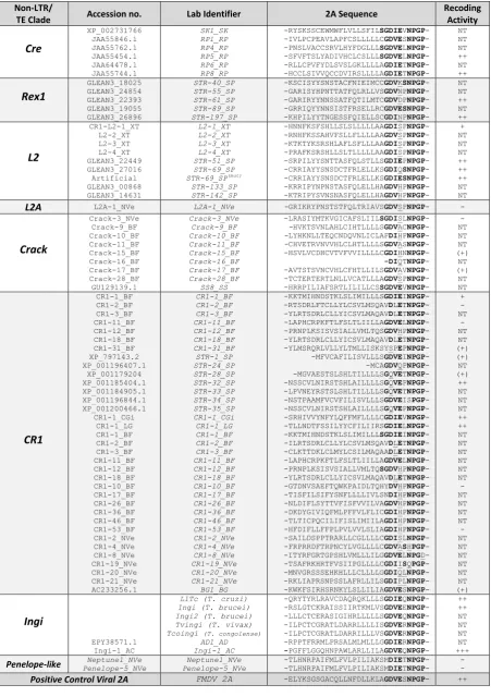
Chapter 3. Non-LTR Associated 2As ... 47
Transposons ... 48
3.1 3.1.1 Non-LTRs ... 50
Non-LTR 2As – Methodology ... 53
3.2 3.2.1 Contributors ... 53
3.2.2 In Silico - Methodology ... 53
3.2.3 In Vitro – Methodology... 54
2As in Non-LTRs – Results ... 57
3.3 3.3.1 Database Probe ... 57
3.3.2 Non-LTR 2As – In Vitro Translational Recoding Analyses ... 61
3.3.3 2As from Putative TE elements – In Vitro Recoding Analyses ... 64
3.3.4 Correlation of a Single ORF and the Presence of 2A ... 65
Non-LTR 2As - Discussion ... 65
3.4 Chapter 4. Ankyrin-Repeat Associated 2As ... 69
Introduction ... 69
4.1 4.1.1 Amphimedonqueenslandica ... 69
4.1.2 Strongylocentrotuspurpuratus ... 70
4.1.3 Ankyrin-Repeat Domains ... 71
Methodology ... 74
4.2 4.2.1 In Silico Searches ... 74
4.2.2 In Vitro – Methodology... 74
Ankyrin 2As – Results ... 76
4.3 4.3.1 Identification of 2As from Ankyrin Proteins ... 76
ix
Table of Contents (continued)
4.3.3 Ankyrin-repeat 2As – Bioinformatic Analyses ... 80
4.3.3.1 Ankyrin 2As - Protein Architecture ... 80
4.3.3.2 Ankyrin 2As - Phylogenetic Relationships ... 82
Ankyrin 2As - Discussion ... 86
4.4 4.4.1 Role of 2A in Ankyrin-Repeat Proteins ... 86
4.4.2 A. queenslandica Ankyrin 2As – Phylogeny ... 87
Chapter 5. Sodium-Dependent Transporter Associated 2As ... 89
Introduction ... 89
5.1 5.1.1 Membrane Embedded Transporter Proteins ... 89
5.1.2 SLC38 Gene Family – SNAT Proteins ... 90
5.1.3 SNAT Signalling ... 92
5.1.4 SNAT Regulation ... 93
Methodology ... 94
5.2 5.2.1 In Silico Searches ... 94
5.2.2 In Vitro – Methodology ... 95
SNAT9 Associated 2As – Results ... 96
5.3 5.3.1 Cataloguing SNAT9 2A-like sequences ... 96
5.3.2 SNAT9 2As - In Vitro Recoding Activity Assays... 99
5.3.3 SNAT9 2As – Bioinformatic Analysis ... 100
5.3.3.1 SNAT9 2A Protein Domain Configuration ... 100
5.3.3.2 SNAT9 2A1 Phylogeny ... 101
x
Table of Contents (continued)
Chapter 6. NLR-Like Protein Associated 2As ... 107
Introduction – NLR-Like Proteins ... 107
6.1 6.1.1 Introducing NLRs ... 107
Methodology ... 110
6.2 6.2.1 Contributions ... 110
6.2.2 In Silico Searches ... 110
6.2.3 In Vitro - Methodology ... 110
NLR 2As - Results ... 111
6.3 6.3.1 Identification of 2A - NLRs ... 111
6.3.2 NLR 2As - Translational Recoding Assays ... 113
6.3.3 NLR 2As - Bioinformatic Analyses ... 114
6.3.3.1 2A-NLR Protein Architecture ... 114
6.3.3.2 NLR 2As - Phylogenetic Relationships ... 114
NLR 2As - Discussion ... 119
6.4 6.4.1 Role of 2A in NLR Proteins ... 119
6.4.2 NLR 2A Phylogeny ... 119
Chapter 7. A Dual Role for 2As as Signal Peptides? ... 121
Introduction ... 121
7.1 7.1.1 Signal Peptides – ER/Transmembrane Trafficking ... 121
7.1.2 Signal Peptides – Intracellular Targeting ... 123
7.1.3 Signal Sequence Prediction ... 124
Methodology - Overview ... 125
7.2 7.2.1 Contributors ... 127
7.2.2 STR6 Mutagenesis Investigations ... 127
7.2.3 STR6 Transfections In Vivo ... 129
7.2.4 AQ27NAGP & SS7NAGP – Signals ... 130
xi
Table of Contents (continued)
Analyses of Dual Function Signal Peptide 2As ... 131
7.3 7.3.1 Identification of the Signal Properties of STR6 and Related Sequences ... 131
7.3.2 STR6 - Protein Localisation ... 134
7.3.2.1 Microscopy ... 134
7.3.2.2 STR6 Immuno-blotting ... 136
7.3.2.3 STR6-Peptides: Mass Spectrometry ... 137
7.3.3 STR6- In Vivo Investigations ... 140
7.3.3.1 STR6 - Tobacco Leaf Infections ... 140
7.3.3.2 STR6 - Echinoderm Transfections ... 140
7.3.4 STR6 Mutants ... 143
7.3.5 AQ27NAGP & SS7NAGP- Mitochondrial Signal Sequences? ... 146
7.3.6 Amino Acid Transporter SNAT9 N-Terminal 2As – Signals? ... 149
Signal 2As – Discussion & Future Experiments ... 152
7.4 7.4.1 Biological Function of Dual Purpose 2As - Signal Peptides? ... 152
7.4.2 Evolutionary Conservation of Extracellular Signals ... 153
7.4.3 SNAT9 Amino Acid Transporter 2As – Signals? ... 153
7.4.3.1 Future Directions ... 154
Chapter 8. 2A Phylogeny & Consensus Sequence Modelling ... 155
Introduction ... 155
8.1 Methodology ... 155
8.2 8.2.1 In Silico Searches and Analyses ... 155
8.2.2 Cloning & In Vitro Translational Recoding Analyses ... 156
Results: 2A Phylogeny & Sequence Composition ... 163
8.3 8.3.1 In Silico Searches ... 163
8.3.2 Phylogenetic Distribution of Eukaryotic 2A Sequences ... 163
8.3.3 Translational Recoding Assay Results ... 165
xii
Table of Contents (continued)
8.3.5 Eukaryotic 2As - Translational Recoding Assays ... 166
8.3.6 Mutagenesis on 2A Sequences – Artificial Intermediates ... 168
8.3.7 2A Sequences – Commonalities in Amino Acid Composition? ... 171
8.3.7.1 Frequency of Each C-Terminal DxxxNPGP Motif ... 171
8.3.7.2 C-Terminal DVTINPGP 2A Sequences... 173
8.3.7.3 C-Terminal DVESNPGP 2A Sequences ... 175
8.3.7.4 C-Terminal DVEENPGP 2A Sequences ... 178
8.3.7.5 C-Terminal DIETNPGP 2A Sequences ... 181
8.3.7.6 C-Terminal DVETNPGP 2A Sequences ... 183
8.3.7.7 C-Terminal DVELNPGP 2A Sequences ... 186
8.3.7.8 C-Terminal DVEVNPGP 2A Sequences ... 188
8.3.7.9 C-Terminal DVERNPGP 2A Sequences ... 190
8.3.7.10 Consensus 2A Sequences – Alignment & Modelling ... 191
Discussion ... 194
8.4 8.4.1 In Vitro Activity Levels– Relationship to Hypothetical Peptide Architecture ... 194
8.4.2 2A Peptide Phylogeny ... 195
Concluding Remarks ... 197
Publications Arising: ... 202
xiii
List of Figures
Figure 1.1 Characteristic life-cycle of a eukaryotic cell infecting virus ... 2
Figure 1.2 Non-canonical mRNA processing by positive sense RNA viruses ... 6
Figure 1.3 Cross-section through a translating eukaryotic ribosome ... 7
Figure 1.4 Eukaryotic ribosome tunnel – potential for nascent chain secondary structure? ... 9
Figure 1.5 Ribosome stalling & 2A skipping peptides contrasted ... 12
Figure 1.6 Picornavirus virion ... 13
Figure 1.7 Picornavirus genome organisation, translation & primary polypeptide processing. ... 14
Figure 1.8 Model of FMDV 2A sequence. ... 17
Figure 1.9 Simple schematic of 2A activity ... 18
Figure 1.10 Phylogenetic analysis of viral RdRp sequences ... 22
Figure 1.11 In vitro reporter assay system & 2A translation products ... 25
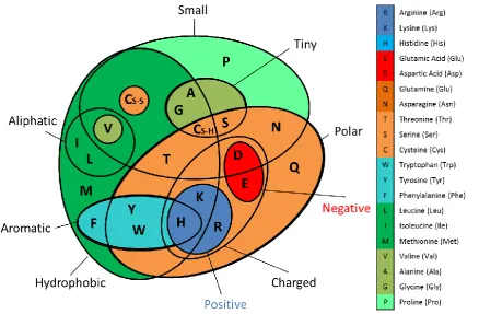
Figure 2.1 Amino acid properties ... 32
Figure 2.2 Calculation of recoding activity analyses ... 38
Figure 3.1 Classification, replication and structure of transposable elements ... 50
Figure 3.2 pSTA1 vector and cloning strategy... 55
Figure 3.3 2A-like sequences within LINEs ... 58
Figure 3.4 Non-LTR 2A sequences – recoding analyses ... 63
Figure 3.5 2As from putative TEs – recoding analyses ... 64
Figure 4.1 Ankyrin-repeat proteins ... 73
Figure 4.2 Recoding activity analyses ... 79
Figure 4.3 Schematic of the protein domain configuration of ankyrin 2A-containing proteins ... 80
Figure 4.4 Structure of A. queenslandica 2A-containing proteins ... 81
Figure 4.5 Cladogram of A. queenslandica ankyrin-repeat proteins ... 83
Figure 4.6 Cladogram of A. queenslandica 2A-ankyrin proteins ... 84
Figure 4.7 Cladogram analysis of ankyrin 2A sequences ... 85
Figure 5.1 SNAT protein topology ... 92
Figure 5.2 SNAT9 2A Recoding activity analyses ... 99
Figure 5.3 SNAT9 protein isoforms ... 100
Figure 5.4 Cladogram of SNAT9 proteins with 2As ... 103
Figure 5.5 SNAT9 2A1 cladogram ... 104
Figure 6.1 NLR protein topology ... 108
Figure 6.2 Recoding activity analyses of NLR-associated 2As ... 113
Figure 6.3 Schematic of the protein domain configuration of NLR 2A-containing proteins ... 114
Figure 6.4 Cladogram of aligned NTPase domains from 2A-NLR proteins ... 116
xiv
List of Figures (continued)
Figure 6.6 Cladogram analysis of 2A sequences from NLR proteins ... 118
Figure 7.1 Signal sequence mediated protein trafficking ... 122
Figure 7.2 Creation of pJN132 ... 128
Figure 7.3 Creation of pSTR6-GFP ... 129
Figure 7.4 STR6wt & STR6NAGPSignal-P analyses ... 132
Figure 7.5 Translation products from pJN132 ... 134
Figure 7.6 Deltavision microscopy of transfected pJN132 constructs ... 135
Figure 7.7 Western blot analysis of STR6 transfections ... 136
Figure 7.8 Supernatant mCherryFP detection ... 137
Figure 7.9 STR6NAGP -mass spectrometry analyses ... 139
Figure 7.10 Tobacco leaves inoculated with STR6 constructs ... 141
Figure 7.11 STR6 Sea-urchin transfection ... 142
Figure 7.12STR6-mutants recoding activity analyses ... 143
Figure 7.13 Deltavision microscopy of transfected STR6-mutants in pJN132 ... 145
Figure 7.14 Plasmids used in SS7NAGP/AQ27NAGP transfections ... 146
Figure 7.15 AQ27NAGP and SS7NAGPtransfections ... 148
Figure 7.16 ORF of mammalian SNAT9 sodium-dependent amino acid transport proteins ... 149
Figure 7.17 SNAT9 2A transfections... 151
Figure 8.1 2A cloning strategy ... 160
Figure 8.2 SS7 mutagenesis cloning strategy ... 161
Figure 8.3 The extant phyla with 2A-like sequences. ... 164
Figure 8.4 Translational recoding analyses of a selection of viral 2As ... 166
Figure 8.5 Eukaryotic 2As - translational recoding assays ... 167
Figure 8.6SK-45 effect of increasing 2A sequence length ... 168
Figure 8.7STR-37 to STR-140 artificial intermediate sequences ... 169
Figure 8.8SS7 to OM-4 artificial intermediate mutants ... 170
Figure 8.9 The most frequently occurring 2A C-terminal DxxxNPG↓P motifs ... 172
Figure 8.10 Determining the viral DVTI consensus 2A sequence ... 174
Figure 8.11 Determining the viral DVESNPGP consensus 2A sequence ... 176
Figure 8.12 Determining the eukaryotic DVESNPGP consensus 2A sequence ... 177
Figure 8.13 Determining the viral DVEENPGP consensus 2A sequence ... 179
Figure 8.14 Determining the eukaryotic DVEENPGP consensus 2A sequence ... 180
Figure 8.15 Determining the viral DIETNPGP consensus 2A sequence ... 182
Figure 8.16 Determining the viral DVETNPGP 2A consensus sequence ... 184
xv
List of Figures (continued)
Figure 8.18 Determining the eukaryotic DVELNPGP consensus 2A sequence ... 187
Figure 8.19 Determining the eukaryotic DVEVNPGP consensus sequence ... 189
Figure 8.20 Determining the eukaryotic DVERNPGP consensus 2A sequence ... 190
Figure 8.21 Consensus 2A sequences aligned ... 191
xvi
List of Tables
Table 1.1 Example ribosome stalling nascent peptides ... 11
Table 1.2 2A sequence types in the Picornaviridae ... 21
Table 1.3 Sample viruses with active ribosome skipping 2As ... 23
Table 2.1 Sequencing primers ... 36
Table 3.1 Non-LTR 2As cloned by means of PCR. ... 56
Table 3.2 2A sequences from putative non-LTRs of unknown clade ... 57
Table 3.3 Non-LTRs containing 2A sequences ... 59
Table 3.4 List of novel 2A sequences associated with putative TEs ... 60
Table 4.1 Gene-block/primer sequences for ankyrin 2A cloning ... 75
Table 4.2 List of ankyrin-associated 2A-containing proteins ... 77
Table 5.1 Properties of human SLC38 products ... 91
Table 5.2 SNAT 2A reverse primers ... 95
Table 5.3 SNAT9 2A-like sequences with canonical DxxxNPGP C-termini ... 97
Table 5.4 SNAT9 2A-like sequences with non-canonical C-termini ... 98
Table 6.1 Gene-block/primer sequences for NLR 2As ... 111
Table 6.2 List of 2A-containing NLR proteins ... 112
Table 6.3 Multi-species comparison of occurrences of NLR-associated 2As ... 115
Table 7.1 PCR primers used in the work reported in Chapter 7. ... 126
Table 7.2 List of high scoring potential exocytic pathway signal NLR-2As ... 133
Table 7.3 Signal peptide analyses of AQ27NAGP and SS7NAGP ... 147
Table 7.4 Signal peptide analyses of SNAT9 amino acid transporter 2As ... 150
Table 8.1 2A sequences incorporated in gene-blocks ... 157
Table 8.2 2A Gene-blocks ... 158
Table 8.3. List of 2A sequences cloned by means of PCR ... 159
xvii
List of Abbreviations
aa – amino acid
ank/ankyrin - ankyrin repeat protein BFA - Brefeldin A
BHK21 - baby hamster kidney cells bp - base pair
DAPI - diamino phenylindole (double-stranded nuclear DNA stain) °C – Degrees Centigrade
DMEM - Dulbecco’s modified eagle medium DMSO - dimethyl sulfoxide
DNA - deoxyribonucleic acid
eEF2 - eukaryotic elongation factor 2 eGFP - enhanced green fluorescent protein eIF4 - eukaryotic initiation factor 4
eRF1 & 3 - eukaryotic release factors 1 and 3 FCS - foetal calf serum
GUS - beta-glucuronidase HGT - horizontal gene transfer
IPTG - isopropyl β-D-1-thiogalactopyranoside kb - kilobase (of DNA or RNA)
LB – Lysogeny broth/agar
mCherryFP - cherry fluorescent protein [35S]-Met - radiolabelled [35S]-methionine MOI - multiplicity of infection
MW - molecular weight
NLR - Natch-like repeat/receptor protein (innate immune protein) Non-LTR - non-long terminal repeat retrotransposon
ORF - open reading frame PBS - phosphate buffered saline PCR - polymerase chain reaction
PEG polyethylene glycol (used here as a transfection reagent) PTC - peptidyltransferase centre
RNA - ribonucleic acid rpm - revolutions per minute
SDS-PAGE - sodium dodecyl sulphate polyacrylamide gel electrophoresis
SNAT - Sodium-dependent amino acid transporter protein (membrane transporter protein) TAE - Tris base, acetic acid and EDTA buffer
TE - transposable element Tm - melting temperature
TnT - cell-free coupled transcription/translation reaction UV - ultraviolet
v/v - volume for volume w/v - weight for volume
1
Chapter 1.
Discovery of 2A-Like Sequences
‘Every thing must have a beginning ... … and that beginning must be linked to something that went before.’
Frankenstein; Or, The Modern Prometheus - Mary Wollstonecraft Shelley, 1823 edition
Viruses
1.1
Viruses are obligate intracellular parasites consisting of infectious particles of genetic material
(RNA or DNA) encapsulated by a protein coat which, in many cases, is surrounded by a lipid
membrane (Figure 1.1). The discovery of viruses as infectious entities was the result of
independent observations by a small number of researchers in the final decade of the 19th century (reviewed in Lustig and Levine, 1992; Bos, 2000; Lecoq, 2001). They concluded that a plant
ailment – namely tobacco mosaic disease, was caused by an agent smaller than any known
bacterium and too small to visualise using light microscopy. This agent could be filter-isolated
from infected plant sap, but it could not replicate outside of its host’s tissue. This agent, now
known as Tobacco Mosaic Virus (TMV) was the first identified virus. The first such filterable agent
from animals, Foot-and-Mouth Disease Virus (FMDV) was identified in 1898, followed shortly, in
1901, of the first isolation of a virus infecting humans, Yellow Fever Virus. With these discoveries
the concept of viruses, infectious filterable particles as disease causing agents began to gain
general acceptance.
Biochemical and electron microscopy analyses in the mid-20th century revealed that viruses were essentially nucleic acid and protein complexes, sized on the order of tens to hundreds of
nanometres in diameter. Deficient in the translational machinery necessary for their replication,
they are obligate parasites of cellular organisms. There have been numerous attempts to classify
viruses based on genome composition, host range, biochemical characteristics or mode of
replication. The Baltimore classification divided viruses into seven categories based upon genome
type and mode of replication (Baltimore, 1971) and has been in widespread use from the 1970s
until the present, but it is now superseded by the International Committee on Taxonomy of Viruses
(ICTV, homepage http://ictvonline.org/) nomenclature system, also based on virus genome type, which further identifies viruses using a Linnaean system where viruses are placed in orders,
2
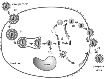
Figure 1.1 Characteristic life-cycle of a eukaryotic cell infecting virus
(a) Adsorption or docking with host cell surface receptor protein, (b) Entry into cell cytoplasm, (c)
biosynthesis of viral components (proteins & nucleic acid), (d) assembly into new viruses, and (e)
budding from the host cell (image modified from Gao et al., 2005).
Positive Sense Single-Strand RNA Viruses – Translational Tricks
1.2
Viruses rely on “hi-jacking” host cell machinery to complete their replication cycle; therefore,
virus-encoded proteins are translated by host cell ribosomes (see Section 1.3) in a similar manner
to cellular proteins. In the case of positive sense RNA viruses their genomic RNA is treated
comparably to host mRNA with cytoplasmic protein translation. All RNA viruses have extremely
compact genomes, the largest being only around 30 kilobases (kb), while most are under 10 kb
(Firth and Brierley, 2012). From these small genomes they must encode all the proteins necessary
for their reproduction. Typically each positive sense RNA viral genome encodes one or more
precursor polyproteins that are both cleaved and modified into mature structural and replicative
viral proteins. However, most viral genomes encode only a single copy of each viral protein
(Palmenberg, 1990), whereas the relative quantities required for a successful infection vary with
infection stage (at the start, replicative proteins and immune system interacting proteins are
required; later, high levels of structural proteins for packaging). Therefore, one of the major
3
translation where typically only a single protein product can be produced from each mRNA strand
(Firth and Brierley, 2012).
Whilst the central dogma of molecular biology stated that a single message or gene (DNA)
encoded a single messenger (RNA), translated into a single product (protein) (Crick, 1970), it is
now known that for some genes, the situation can be considerably more complex, with translational
recoding mechanisms permitting a single gene to encode multiple protein products. Positive sense
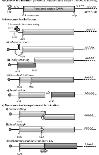
RNA viruses employ a number of translational recoding mechanisms (Figure 1.2) which
successfully subvert the usual ribosome translation of mRNA. In non-canonical initiation the
translation begins at a site other than the canonical AUG codon immediately downstream of a
5’UTR with a methylated 5’ cap protein.
One method of non-canonical initiation makes use of Internal Ribosome Entry Sites (IRES, see
Figure 1.2b i). First discovered in picornaviruses in the late 1980s, these are highly structured RNA
regions that recruit ribosomes to internal positions on mRNAs (Jang et al., 1988; Pelletier and
Sonenberg, 1988). Viral IRESes are often employed to instigate translation initiation while
allowing replication elements and/or packing signals to be accommodated within the 5’UTR. They
can be used to access internal otherwise untranslated ORFs, and can also facilitate viral mRNA
translation when host-cell translation has been inhibited, for example by viral proteases cleaving
the initiation factors required for 5’ cap-dependent translation (reviewed in Firth and Brierley,
2012). Viral IRESes are highly variable, both in the degree to which they depend on host cell
initiation factors and in their precision of initiation site selection. IRESes are presently grouped
into several classes based on their initiation mechanisms (reviewed in in Firth and Brierley, 2012).
In recent years viral IRESes have been successfully used to encode multiple proteins from a single
vector in biotechnological applications (for a recent review see Minskaia et al., 2015): the first
gene being expressed from the 5’ cap and the second from an internal IRES.
IRESes instigate 5’-independent internal translation initiation whereas another form of
non-canonical translation, namely ribosome shunting (Figure 2.1b ii), permits ribosomes to translate
downstream ORFs in a manner that is only partially 5’ dependent. Here alternate translation
products are produced when secondary structure in the 5’ UTR results in translation starting at a
downstream AUG codon (reviewed in Firth and Brierley, 2012).
Leaky scanning is also 5’ cap-dependent, here a significant proportion of the 40 ribosome subunits (see Section 1.3, to follow) that bind near the 5’ cap and scan downstream on the mRNA until they
reach an appropriate AUG codon, will fail to initiate translation at the first AUG codon they
encounter. These ribosomes continue scanning until they reach an alternate downstream initiation
codon where translation commences (reviewed by Kozak, 2002). Leaky scanning results in
4
Non-AUG initiation is similar to leaky scanning in that it results in multiple isoforms of the
translation product, some N-terminally truncated (see Figure 1.2b iv). In eukaryotes, protein
synthesis begins almost exclusively with methionine, encoded by AUG. The tRNA carrier Met–
tRNAi, delivers the initial methionine. Met–tRNAi differs slightly from the standard Met-tRNA
used during elongation. Under certain circumstances Met–tRNAi can be recognised by
near-cognate codons such as CUG or ACG. Initiation at a non-AUG codon is enhanced by particular
flanking nucleotides, and by secondary structure (typically stemloops) forming close to the exterior
of the ribosome. Non-AUG initiation is typically inefficient, occurring only in in 2-30% of cases,
in the remainder of instances leaky scanning mechanisms permit the ribosome to scan downstream
until it encounters the next AUG codon which is treated as a start codon (reviewed in Firth and
Brierley, 2012).
Another non-canonical initiation process used by positive sense RNA viruses is re-initiation
(Figure 1.2b v). This depends on the ribosome 40S subunit (see following, Section 1.3) remaining
associated with the mRNA after protein translation ceases (normally the subunits would
disassociate after translation termination) and resuming scanning to re-initiate translation at a
downstream AUG codon. In cellular genes, re-initiation most commonly occurs after a short (less
than 30 codons in length) upstream ORF, but in some viruses (for example, in the Calicivirus and
Sapovirus genera) re-initiation occurs after a translation of a longer upstream ORF. Re-initiation is
dependent on initiation factors (see following, Section 1.3) remaining attached to the ribosome
during translation (hence why it is more common after short upstream ORFs as there is less time
for the factors to disassociate). After the upstream ORF translation terminates, the 40S subunit is
not immediately competent to reinitiate, but becomes competent after scanning downstream along
the mRNA for some distance. This time spent scanning is thought to permit the reacquisition of the
necessary initiation factors and the eIF2–Met–tRNAi–GTP ternary complex required for
translation initiation. Re-initiation is also thought to rely on complex RNA structure in the region
between ORFs that can delay the scanning and permit initiation factors re-binding, or may be used
to retain the bound ribosome-initiation factor complex between translation of each ORF (reviewed
in Firth and Brierley, 2012; Jackson et al., 2012).
In non-canonical elongation and termination, events during translation, or in the altered reading of
termination signals can result in protein products other than those predicted by an in-frame reading
of the mRNA sequence. Collectively such events are termed ribosome recoding.
Viral-directed ribosome recoding events include frameshifting, whereby a proportion of ribosomes
are directed into an alternate reading frame by mRNA movement back or forwards by one or two
nucleotides (Figure 1.2c i). Frameshifting -1 was discovered to be the mechanism by which the
5
et al., 1988). Many positive-strand RNA viruses, most retroviruses, plus some Totiviridae dsRNA
viruses use -1 frameshifting to express their RdRp or reverse transcriptase domains. Frameshifting
is thought to rely both on a “slippery” mRNA sequence with repeated bases around the point of slippage, and on a strongly structured downstream RNA. It is thought that a delay in “unwrapping”
the downstream RNA and preparing it for reading by the ribosome permits the one or two
nucleotide slippage of the mRNA within the translating ribosome (reviewed in Firth and Brierley,
2012).
Another viral recoding mechanism is readthrough (Figure 1.2c ii), whereby a proportion of
ribosomes fail to terminate at a stop codon but instead insert a standard amino acid (hence the term
readthrough). Termination is generally highly efficient, but the efficiency is known to be
influenced by the particular stop codon (whether UAA, UAG or UGA) and the flanking
nucleotides, especially the immediately adjacent 3’ nucleotide, and in some viruses secondary
structure beginning eight nucleotides downstream in the mRNA sequence. During readthrough the
stop codon is decoded by a near-cognate or suppressor tRNA and termination continues until the
next stop codon is encountered. Readthrough mechanisms are used by some viruses to control
expression of polymerase proteins or to add extensions to a subset of coat proteins (reviewed in
Firth and Brierley, 2012).
Finally, another viral recoding mechanism is stop–carry on, also referred to as ribosome skipping
(Figure 1.2c iii). It is the 2A peptides that instigate ribosome skipping that will form the focus of
this thesis. 2A ribosome skipping will be described in detail (to follow, Chapter 1.9). 2A-mediated
ribosome skipping differs from the other forms of non-canonical mRNA processing as it is
believed to rely solely on the peptide sequence of the nascent protein rather than structural
6
Figure 1.2 Non-canonical mRNA processing by positive sense RNA viruses
7
Ribosomes
1.3
In order to describe the process of ribosome skipping, it is first necessary to introduce briefly
eukaryotic ribosomes. Ribosomes are the protein factories of the cell, they are large complexes of
RNA and proteins that “read” messenger (mRNA) and manufacture new peptide chains. They are
essentially ribozymes, as it is their RNA components that act to catalyse the formation of new
peptide chains, whereas their protein component acts to stabilise the structure. In the last few years,
high resolution cryoEM and crystallography studies (Armache et al., 2010; Demeshkina et al.,
2010; Rabl et al., 2011) have provided detailed ribosomal models (Figure 1.3).
Figure 1.3 Cross-section through a translating eukaryotic ribosome
a) Structure of a translating wheat-germ (Triticum aestivum) ribosome illustrating the relative positions of the ribosomal subunits, the P-site tRNA in the PTC, and the nascent polypeptide (modified from Becker et al., 2009), b) conceptual model which additionally shows the relative position of the translated mRNA and the A and E-site tRNAs (modified from Lafontaine and Tollervey, 2001).
It is now known that both eukaryotic and prokaryotic ribosomes comprise two subunits (termed
large and small), each composed of ribosomal RNA (rRNA) and proteins. Prokaryotic 70S
ribosomes comprise ~4500 nucleotides ribosomal RNA (rRNA) and 54 proteins, their large 50S
subunit contains two RNAs (23S and 5S) and 32 proteins whilst their small subunit possesses a
single 16S RNA and 22 proteins. Eukaryotic ribosomes are substantially larger than prokaryotic,
for example, mammalian ribosomes are approximately 50% larger than those from E. coli bacteria.
Eukaryotic ribosomes comprise ~5500 nucleotides rRNA and 80 ribosomal proteins with their
large 60S subunits containing three RNAs (28S, 5.8S and 5S), and their small 40S subunit one 18S
RNA. The increased size and complexity of eukaryotic ribosomes is due to the presence of
additional rRNA expansion sequences (rRNAs that do not form part of the core ribosome structure)
8
lineages also differ, typically mammalian ribosomes are 12% larger than yeast ribosomes; this
increase is due to additional rRNA expansion sequences (Morgan et al., 2000).
However, the mechanics of protein translation at the central active site of the peptidyltransferase
centre (PTC) is extremely conserved across all domains of life. First, the ribosome binds to the
mRNA and translation initiates at a start (methionine, nucleotides ATG) codon. A supply of the
relevant amino acids are transported to the ribosome 3’ attached to transfer RNAs (tRNAs), a class
of small RNA molecules. Each tRNA is specific for a single amino acid. The ribosome possesses
three tRNA binding sites within the PTC. Firstly, a tRNA with its specific attached amino acid
(aminoacyl-tRNA) encounters the A-site where it is presented to the mRNA being decoded. Slight
changes in ribosome conformation then shunt the tRNA into the peptidyl site (P-site), where it and
its amino acid burden are attached to the C-terminal end of the growing peptide chain as a
peptidyl-tRNA complex. The tRNA-amino acid link is then broken and the deacylated “empty”
tRNA moves into the exit site (E-site) before exiting the PTC, and the ribosome moves forward on
the mRNA to read the next codon to be translated. Translation is an iterative process with one
amino acid residue being added to the chain at each step, at a rate of approximately 15–20 residues
per second in bacteria (reviewed in Lafontaine and Tollervey, 2001).
Unsurprisingly, given the high level of sequence and structure conservation in ribosomal RNA and
proteins between organisms, ribosome synthesis is also very highly conserved. In virtually all
living cells, rRNAs are generated by post-transcriptional processing from a polycistronic precursor
rRNA (pre-rRNA). The rRNAs are then assembled along with the ribosomal proteins to form the
ribosome complex. Events during assembly must follow a strict temporal order in order to permit
the correct folding and modifications of all components. Deviations, either in component
composition, modification, or assembly order will normally result in mis-folding and inhibit
assembly (see Lafontaine and Tollervey, 2001). Ribosome functional impairment is normally lethal
to the cell, hence the extremely conserved structure of the ribosome in all living cells.
Ribosome Exit Tunnel
1.4
Nascent polypeptide chains exit the ribosome through the exit tunnel. This comprises a hollow tube
approximately 80-100 Å in length and 10-20Å in diameter (Figure 1.4), its walls predominately
composed of rRNA (82% rRNA atoms, 18% protein atoms, respectively). Generally the tunnel
walls are negatively charged and hydrophilic (Voss et al., 2006; Bhushan et al., 2010). At one
point the “arms” of two ribosomal proteins, L4 and L17 (L22 in bacteria), form a constriction
reducing the passageway width to 10Å. The tunnel dimensions preclude extensive folding of the
peptide nascent chain. However, it can accommodate 30-40 peptide residues (first experiments
Malkin and Rich, 1967; confirmed by Bernabeu and Lake, 1982; revisited Voss et al., 2006),
9
hypothesis that the nascent chain adopts a partial α-helical confirmation within the tunnel. Recent
fluorescence resonance energy transfer (FRET) (Woolhead et al., 2004) and cryo-EM (Bhushan et
al., 2010) studies confirmed that nascent peptides do indeed form an α-helix within certain tunnel
regions.
Figure 1.4 Eukaryotic ribosome tunnel – potential for nascent chain secondary structure?
Model of the wheat-germ (Triticum aestivum) ribosome tunnel displaying the exit pathway of the polypeptide nascent chain and the regions where some helical folding can occur (modified from Bhushan et al., 2010).
The Ribosome Tunnel & Translational Recoding – Stalling Peptides
1.5
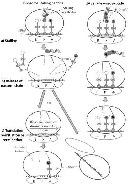
The constricted environs of the ribosome tunnel not only determine the permissible nascent chain
secondary structures which can form within its confines, but in forming into such structures, the
nascent chain can interact with the tunnel walls, and in doing so influence events in the PTC to
alter or halt peptide synthesis. Such events are termed translational recoding (recently reviewed by
Ito et al., 2010; Cruz-Vera et al., 2011; Wilson and Beckmann, 2011; for examples see Table 1.1).
The specific amino acid sequence of each nascent peptide influences its rate and ease of transit
through the ribosome tunnel. Peptide geometry and mass contribute, but the principle governing
factor is electrostatic force, particularly nascent peptide charge and hydropathicity (Lu and
Deutsch, 2008). The tunnel constriction site proteins (Figure 1.4), the vestibule exit protein L39
(bacteria L23), and rRNA residues adjacent to each of these proteins, have also been identified as
essential players. These tunnel wall proteins can interact with specific residues within the nascent
10
causes slight conformational changes in the P- and/or A-sites which block PTC activities including
peptide bond formation and/or tRNA movement. Translation ceases releasing the tRNAs and the
nascent peptide, then, subsequent mRNA rearrangement may permit translation re-initiation from
an ORF downstream to that encoding the stalling peptide.
The majority of these stalling peptides rely on co-effector binding either with the tunnel vestibule
or within the lower reaches of the tunnel to create the particular peptide conformation that causes
stalling (Figure 1.5). Macrolide antibiotics provide the co-effectors for the most bacterial stalling
peptides, and the downstream ORF that is later translated encodes an antibiotic resistance factor,
thus providing the host with antibiotic resistance when antibiotic is present (reviewed in Tenson
and Ehrenberg, 2002; Ito et al., 2010). This provides an evolutionary fitness advantage to the
carriers as they do not have to expend energy producing resistance factors constitutively, instead,
they can divert resources to producing the resistance factors only when and if they are required.
These bacterial antibiotic resistance genes tend to be carried on plasmid DNA and are responsible
for the growing phenomena of drug-resistance (reviewed by Ito et al., 2010). Where the stalling
peptide functions to regulate cellular metabolic pathways, high intracellular levels of a
constitutively expressed molecule can act as the co-effector, such as tryptophan in the case of tnaC,
and arginine for the fungal arginine attenuation peptides, respectively (Fang et al., 2004). Or, more
rarely, the specific amino acid sequence of the stalling peptide can be sufficient to stall the
ribosome without the aid of a co-effector molecule.
A number of the stalling peptides end with proline (see Table 1.1) or possess proline residue(s)
close to their C-terminus. It has been suggested (Jenni and Ban, 2003) that the unique properties of
proline (the only naturally occurring N-alkylamino or “imino” acid in proteins) could contribute to
the ribosome stalling in these instances as there is a greater energy barrier to proline, an imino acid,
forming peptide bonds than with the other naturally occurring 19 amino acids, making proline
slower to bond (Pavlov et al., 2009).
Each stalling peptide occurs in a distinct monophyletic (single ancestor) group of organisms, or in
a single organism, suggesting that while they share a common function, namely ribosome arrest,
stalling peptides are an example of convergent evolution with separate origins for each sequence
(Ito et al., 2010; Cruz-Vera et al., 2011; Wilson and Beckmann, 2011). Additionally, every stalling
peptide discovered to date can only function in either prokaryote or eukaryote ribosomes,
suggesting that subtle differences in tunnel topography and characteristics may influence nascent
11
Table 1.1 Example ribosome stalling nascent peptides
The peptide name, its host organism and the peptide sequence are given in each instance. Where specific residues are known to be essential to function these are been underlined. If the peptide requires a co-effector to function, this is also listed.
Peptide Host organism Active stalling peptide
sequence(s) Co-effector Bacterial cat, cmlA Salmonella spp. Enterobacter spp. Pseudomonas spp. VKTD
KNAD chloramphenicol
ermC,
ermCL Enterococcus spp.
SFVI
MxxxxIFVIs erythromycin
tnaC tnaC Ec tnaC Pv
Escherichia coli
& Proteus vulgaris
KWFNID WxxxDxxIxxxxP*
WxxxDxxLxxxxPK tryptophan
secM secM Ms
Escherichia coli FxxxxWIxxxxGIRxGP
xxxxxxxxxxHAPIRGSP translocation (SecA)membrane
MifM Bs Bacilus subtilis RIxxWIxxxxxMNxxxxxxxxx Fungal/Yeast
CPA1 Saccharomyces
cerevisiae NSQYTCQDYISDHIWKTS arginine
arg Neurospora crassa PSxFTSQDYxSDHLWxAx
Mammalian
AdoMetDC mammals MAGDIS spermidine,
spermine
Β2-AdRec mammals MKLPGVRPRPAAPRRRCTR
No co-effector
RAR- Β2 mammals MIRGWEKDQQPTCQKRGRV
CMV UL4 mammalian
cytomegaloviruses MQPLVLSAKKLSSLLTCKYIPP
Note: underlined residues are essential to function, x=any amino acid. *=unknown residue, (information from Tenson and Ehrenberg, 2002; Ito et al., 2010)
Another class of translational recoding peptide which stalls the ribosome prior to translation
re-initiation was discovered in the Picornaviridae virus family in the early 1990s (Figure 1.2 and
Figure 1.5). Termed 2A ribosome skipping peptides, these are the sequences that form the focus of
this PhD investigation. The mechanism of activity of ribosome skipping 2A will be addressed in
detail in Chapter 1.8, but first, protein production in the Picornaviridae will now be outlined with a
12
Figure 1.5 Ribosome stalling & 2A skipping peptides contrasted
13
The
Picornaviridae
1.6
Under the ICTV system the Picornaviridae virus family is placed in the order Picornavirales. The
Picornaviridae are a large and varied family that contains many socially and economically
significant human and animal pathogens including Polio, Hepatitis A, the Common Cold Virus, and
the cattle disease FMDV. Their classification has recently undergone a major reorganisation due to
the inclusion into the taxonomic system of numerous newly identified Picornavirus and
Picorna-like Viruses, with a current total of 46 known picornavirus species grouped into 26 genera
(Knowles, 2012; Adams et al., 2014).
Figure 1.6 Picornavirus virion
a) Electron microscopy image of the outer view of a FMDV virion, b) schematic diagram detailing the icosahedral arrangement of the protein subunits composing the FMDV virion capsid (images courtesy of Prof. Martin Ryan).
As their name suggests, these viruses possess small (pico = small) RNA genomes. Their
single-stranded positive sense RNA genome is typically between 7500-8000 bases in length and is
organised in a single central open reading frame (ORF), flanked by highly structured 5’ and 3’
untranslated regions (UTRs). Picornavirus replication is thought to be entirely cytoplasmic, with
the viral genomic RNA utilising the translational machinery of infected cells in a similar manner to
host cell mRNAs. They encode their own RNA-dependent RNA-polymerase to enable replication
of their RNA genome. Their single ORF encodes a single precursor polyprotein that is co-and
post-translationally cleaved to form the structural (capsid) and replicative elements of the virus. The
virion particle is composed of a single copy of the RNA genome encapsidated within an
non-enveloped icosahedral capsid (Figure 1.6) composed of 60 protein subunits each consisting of 4
14
Picornavirus Translation
1.7
Picornavirus RNA is capped by an oligopeptide (Vpg or 3B) covalently attached to the 5’ terminus, and the 3’ end terminates in a poly(A) tract. The 5’ UTR folds into a highly structured clover-leaf
configuration which possesses an internal ribosome entry site (IRES). The IRES facilitates
cap-independent translation and allows the virus to effectively halt host-cell protein synthesis and to
henceforth “hijack” the translational apparatus to synthesise their own proteins (reviewed in
Martinez-Salas, 2008; Martinez-Salas et al., 2008). The ORF is translated to form a large precursor
protein of approximately 250kDa, but this protein is rarely observed in cell-culture as it is co- and
post-translationally cleaved to form the active structural and regulatory viral proteins.
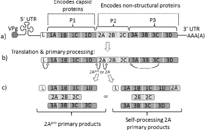
Co-translational cleavages by viral proteases 2A and 3C divide this polyprotein into three regions (P1,
P2 and P3 (Figure 1.7.). P1 contains the four structural proteins (1A-1D) that form the virion
capsid coat; both P2 and P3 contain the non-structural proteins, 2A-2C, and 3A-D, respectively
(Palmenberg, 1990, following the nomenclature proposed by Rueckert and Wimmer, 1984). The
non-structural proteins facilitate viral replication, shut-down of cellular protein synthesis, and the
[image:35.595.79.483.375.629.2]re-arrangement of the cell membranes.
Figure 1.7 Picornavirus genome organisation, translation & primary polypeptide processing. Not to scale. a) RNA genome organisation, note not all picornaviruses possess a Leader (L) region
15
Picornavirus Translational Recoding 2As
1.8
Virtually all picornaviruses encode a 2A protein, but the size, structure and function of 2A differs
widely between genera. In the majority of genera, the 2A region apparently functions in primary
polypeptide processing: for example, in Enteroviruses and Rhinoviruses, the 2A polypeptide is a
thiol proteinase (termed 2Apro) which cleaves the polyprotein at the P1/P2 juncture in cis. Whereas, in the Aphtoviruses and Cardioviruses, the 2A region apparently self-cleaves at its own
C-terminus, meaning that the 2A polypeptide remains as a C-terminal extension of the upstream
polyprotein (P1) until it is removed by secondary proteinase cleavage (Ryan et al., 1991; Ryan and
Drew, 1994). However, in some other picornavirus genera (such as the Parechoviruses, Kobuvirus
and Megrivirus), the 2A region has apparently no protease or protease-like activity, and instead its
apparent function is to alter host cell metabolism as it possesses a high homology to cellular
protein H-rev107 that regulates cell proliferation (H-box 2A) (Hughes and Stanway, 2000). The
processes of recombination and the re-arrangement of genome segments have resulting in some
picornaviruses possessing multiple 2A regions of various types (Table 1.2).
The mechanisms of thiol protease 2As and cell-cycle regulatory 2As were readily apparent.
However, the elucidation of the functional mechanism of the non-protease “cleavage” 2As has
taken over 20 years of careful step-wise investigations, primarily using FMDV 2A as the model
system. Early investigations into the later proteolytic cleavage steps between FMDV 1D and 2A
revealed that the 2A region in FMDV was only 18aa (-LLNFDLLKLAGDVESNPG-) in length
(Belsham, 1993). This was considerably shorter than any than any known protease enzyme, but
there was the possibility that the FMDV 2A-2B cleavage could have resulted from the activities of
exogenous host cell proteases or of another virus encoded protease? Investigative studies
conclusively demonstrated that this was not the case, as neither host cell proteases, nor the FMDV
viral encoded proteases (namely 3Cpro or Lpro)cleaved at the 2A-2B boundary (Ryan et al., 1989; Ryan et al., 1991; Palmenberg et al., 1992). It was also found that it was the amino acid not the
nucleic acid sequence that was instrumental to function as synonymous mutations within the RNA
sequence did not influence function (most recently revisited by Gao et al., 2014).
Sequence comparison of the Enterovirus and Rhinovirus 2Apro thiol proteases with the 2As of Cardioviruses and Aphthoviruses found that although their 2Apro were of a similar length to the Cardiovirus 2A proteins, approximately 150 amino acids (aa) there was no apparent sequence
similarity. However, the C-terminal region of the Cardiovirus 2As were found to be highly similar
to the much shorter (approximately 20-30aa) 2A peptide of the Aphthoviruses (Donnelly et al.,
1997). This difference in 2A region sequence, coupled with dissimilarity to both to the Entero- and
co-16
translational “cleavage” event identified at the end of the Aphthovirus and Cardiovirus 2As,
occurred co-translationally through a novel non-protease mediated system (Palmenberg, 1990).
The next examinable hypothesis was that the FMDV 2A “cleavage” was due to the specific
C-terminus amino acid sequence. The C-C-terminus tri-peptide from FMDV 2A (-NPG-) and the
N-terminal residue of 2B (-P-), together formed a tetrapeptide motif (-NPGP-), which was found very
rarely in natural proteins, making it almost unique to the 2A sequences. Therefore, it was suspected
that this tetrapeptide might be the key to 2A function (Palmenberg, 1990). However, it was shown
that the -NPGP- motif alone was ineffective at instigating 2A “cleavage” (reviewed in Luke et al.,
2010b).
Models of the FMDV 2A nascent peptide (Figure 1.8) suggest that it has the propensity to form
into α-helix along most of its length with a tight reverse turn motif (-ESNPG-) turn at its
C-terminus (Ryan et al., 1999; Donnelly et al., 2001b). Similar to ribosome stalling peptides (Table
1.1 & Figure 1.5), it was proposed that its unusual geometry could assist the FMDV 2A nascent
chain to transiently pause (a pause was observed in puromycin incorporation experiments, see
Donnelly et al., 2001b) in its transverse of the ribosome exit tunnel. Indeed, the current favoured
hypothesis is that 2A activity is a result of the particular geometry of the nascent 2A amino acid
sequence, and its ability to interact with the ribosome exit tunnel (Ryan et al., 1991; Donnelly et
al., 2001b; de Felipe et al., 2003; Atkins et al., 2007; Doronina et al., 2008b). The supposition is
that the N-terminus portion of 2A (the helix) might interact with the tunnel to obtain the specific
stereo-chemical constraints required for the turn motif (-ESNPG-) to literally be in a position to
influence events within the peptidyl transferase centre (PTC) of the ribosome. The nascent chain
pausing in its exit of the ribosome tunnel halts translation and results in minute shifts in the
peptidyl-tRNAGly position within the PTC, leading to a configuration whereby nucleophilic attack on the carbonyl group of the P-site peptidyl-tRNAGly by the A-site aminoacyl-tRNAPro amino group is prevented by the unfavourable energetics for peptide bond formation to the imino acid proline.
The formation of a glycine-proline peptide bond is inhibited whereas the hydrolysis of the
peptidyl-tRNAGly ester bond between glycine and its carrier tRNA is favoured.
It was proposed that release (termination) factors eRF1 and eRF3 contributed to the hydrolysis and
release of the 2A nascent chain even although the proline codon was still occupying the ribosome
A site (Doronina et al., 2008a; Doronina et al., 2008b). Normally, eRF1 & 3 can only form into a
complex with elongating eukaryotic ribosomes whenever a stop codon enters the A-site.
(Zhouravleva et al., 1995), and it is this complex which permits hydrolysis of the ester bond
between the final amino acid and its carrier tRNA. Therefore, in the case of 2A, the stalled
ribosome-2A complex must promote eRF entry without reading the specific mRNA, as the eRFs
17
dissociation must occur to facilitate the entry of prolyl-tRNA to the A site of the ribosome. The
aminoacyl-tRNA is then translocated from the A to the P site to become the initiating N terminus
peptidyl-tRNA of the downstream nascent protein chain (Figure 1.9). However, a newly published
study, using in vitro cell extracts, found that for Encephalomyocarditis Virus (EMCV) 2A-mediated
translational recoding could occur even in the absence of eRFs (Machida et al., 2014). In light of
these recent findings, it will be interesting to determine whether eRFs play any role in the
functioning of other viral 2As, or, if indeed, release factors are redundant for 2A processing. It is
suspected that the concentration of eukaryotic elongation factors may determine whether, after
hydrolysis, the ribosome dissociates or continues to translate the downstream context (Luke and
[image:38.595.212.453.272.496.2]Ryan, 2013).
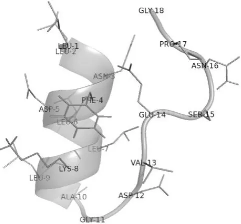
Figure 1.8 Model of FMDV 2A sequence.
The image shows the hypothetical configuration of FMDV 18aa 2A at neutral charge and pH in a non-spatially constrained hydrophilic environment, however the ribosome exit tunnel is known to be spatially constrained with negatively charged walls (model created in PyMOL through alignment of the five best-guess models from PEP-FOLD). Amino acid residues are numbered sequentially from N to termini. Note the N-terminal alpha-helix, and the sharp turn at the C-terminus.
Therefore, there are three possible outcomes when a eukaryotic ribosome translates 2A: either
translational read-through of the 2A sequence; or, translational recoding instigated by the nascent
peptide resulting in non-canonical termination at the final glycine; or, the formation of the
glycine-proline peptide bond can be inhibited. In this third case, two nascent proteins are produced from a
single mRNA transcript due to the “skipping” of the glycine-proline peptide bond (never formed,
as opposed to formed and subsequently broken). This 2A-driven ribosome translational recoding
mechanism has variously been termed “stop-go” (Atkins et al., 2007), “stop carry-on” (Sharma et
18
used in the literature; with no one term emerging as the most popular nomenclature. In the interests
of brevity and clarity this report will refer to the ribosomal recoding peptide as 2A, whereas the
enterovirus-like 2A proteases will be referred to 2Apro.
Figure 1.9 Simple schematic of 2A activity
19
Sequence comparison of FMDV 2A with other Aphthovirus and Cardiovirus 2As found
considerable variation within the N-terminal portion, but revealed the existence of a highly
conserved C-terminus, namely -D[V/I]ExNPG- where x represents any amino acid. This was
always followed by proline (P) as the first Nterminal residue of 2B. This 8 amino acid motif,
-D[V/I]ExNPG↓P-, where ↓=cut in nascent chain, was crucial to function as point mutations within it either severely reduced or ablated activity altogether (Luke et al., 2008).
To function optimally the 2A sequence also required an appropriate upstream sequence (Donnelly
et al., 2001a). Hybrid 2A peptides manufactured by switching the D[V/I]ExNPG↓P motif from
FMDV onto the 22 amino acid upstream sequence from EMCV, and vice versa, showed low or no
activity (Sharma et al., 2012), confirming that the entirety of 2A contributes to function. Sequence
length was also important, because when, in vitro, FMDV 2A was elongated by the addition of
upstream (1D C-terminus) residues, increasing chain length to up to 30 amino acids, its activity
was enhanced (Donnelly et al., 1997; Donnelly et al., 2001b; Minskaia et al., 2013). The same
pattern held for the other picornavirus 2As, with nascent chain lengths of around 30 amino acids
exhibiting the highest activity (Luke et al., 2008). However, it has proven difficult to distinguish
critical residues which interact with the ribosome tunnel from flanking “space-filling” residues.
The majority of substitutions within a 2A sequence tend to reduce its self-cutting activity, which in
wild-type 2As can be as high as 100%. Indeed, it was recently suggested that 2A sequences are:
“fine-tuned to function as a whole” and “each 2A may then represent a specific solution for positioning the
conserved C-terminus within the peptidyl-transferase centre to promote recoding” (Sharma et al., 2012).
Ribosome Skipping 2As - Viral Phylogeny
1.9
Following the discovery of ribosome skipping 2A sequences in the Aphthoviruses and
Cardioviruses, such sequences were also found in a number of other (but not all) Picornavirus
genera (Table 1.2). In all cases the active sequence was short, being fully functional at only 30
amino acids), and ended in the conserved C-terminus motif D[V/I]ExNPG↓P. Interestingly, in a number of picornavirus genera, one or more species possessed ribosome skipping 2As whereas
others within the same genera possessed 2Apro (Table 1.2).
This led to the hypothesis that, for some picornaviruses, due to later recombination events an
ancestral ribosome skipping 2A may have been replaced by 2Apro (Luke et al., 2008). Extending the search for 2A sequences beyond the picornaviruses, further online database probing using the
2A C-terminus motif, revealed the occurrence of ribosome skipping 2A sequences in a number of
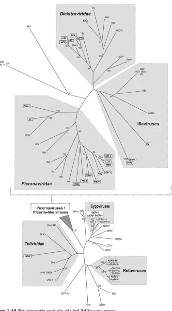
other viruses (Table 1.3, Figure 1.10), namely, in positive-stranded RNA viruses belonging to the
Iflaviridae, Tetraviridae, and Dicistroviridae families (insect-infecting viruses); in the Reoviridae
(mammalian or insect-infecting segmented double-stranded RNA viruses); and the Totiviridae