Original citation:
Salker, Madhuri S., Hosseinzadeh, Zohreh, Alowayed, Nour, Zeng, Ni, Umbach, Anja T.,
Webster, Zoe, Singh, Yogesh, Brosens, Jan J. and Lang, Florian. (2016) LEFTYA activates the
epithelial Na+ Channel (ENaC) in endometrial cells via serum and glucocorticoid inducible
kinase SGK1. Cellular Physiology and Biochemistry, 39 (4). pp. 1295-1306.
Permanent WRAP URL:
http://wrap.warwick.ac.uk/85073
Copyright and reuse:
The Warwick Research Archive Portal (WRAP) makes this work of researchers of the
University of Warwick available open access under the following conditions.
This article is made available under the Attribution-NonCommercial-NoDerivatives 4.0 (CC
BY-NC-ND 4.0) license and may be reused according to the conditions of the license. For
more details see:
http://creativecommons.org/licenses/by-nc-nd/4.0/
A note on versions:
The version presented in WRAP is the published version, or, version of record, and may be
cited as it appears here.
Original Paper
This article is licensed under the Creative Commons Attribution-NonCommercial-NoDerivatives 4.0 Interna-tional License (CC BY-NC-ND) (http://www.karger.com/Services/OpenAccessLicense). Usage and distribution
for commercial purposes as well as any distribution of modified material requires written permission.
© 2016 The Author(s) Published by S. Karger AG, Basel Department of Physiology, University of Tuebingen, Gmelinstr. 5, D-72076 Tuebingen (Germany)
Tel. +49 7071/2972194, Fax +49 7071/295618, E-Mail florian.lang@uni-tuebingen.de
Prof. Dr. Florian Lang
LEFTYA Activates the Epithelial Na
+
Channel (ENaC) in Endometrial Cells via
Serum and Glucocorticoid Inducible Kinase
SGK1
Madhuri S. Salker
aZohreh Hosseinzadeh
a,bNour Alowayed
aNi Zeng
a,c,dAnja T. Umbach
aZoe Webster
eYogesh Singh
aJan J. Brosens
f,gFlorian Lang
a,haDepartment of Cardiology, Vascular Medicine and Physiology I, bExperimental Retinal Prosthetics Group,
Institute for Ophthalmic Research, University of Tuebingen, Tuebigen, Germany; cState Key Laboratory
of Oral Diseases and dDepartment of Cleft Lip and Palate Surgery, West China Hospital of Stomatology,
Sichuan University, Chengdu, China; eES Cell and Transgenics Facility, Medical Research Council Clinical
Sciences Centre, Imperial College London, London, fDivision of Biomedical Sciences, Warwick Medical
School, Clinical Sciences Research Laboratories, University Hospital, Coventry CV2 2DX, gTommy’s
National Centre for Miscarriage Research, University Hospitals Coventry and Warwickshire NHS Trust, Clifford Bridge Rd, Coventry, CV2 2DX, United Kingdom; hDepartment of Molecular Medicine II, Heinrich
Heine University Düsseldorf, Düsseldorf, Germany
Key Words
Na
+channels • Endometrium • Amiloride • Infertility • SGK1
Abstract
Background:
Serum & glucocorticoid inducible kinase (SGK1) regulates several ion channels,
including amiloride sensitive epithelial Na
+channel (ENaC). SGK1 and ENaC in the luminal
endometrium epithelium, are critically involved in embryo implantation, although little
is known about their regulation. The present study explored whether SGK1 and ENaC are
modulated by LEFTYA, a negative regulator of uterine receptivity.
Methods:
Expression levels
were determined by qRT-PCR and Western blotting, ENaC channel activity by whole cell
patch clamp and transepithelial current by Ussing chamber experiments.
Results:
Treatment of
Ishikawa cells, an endometrial adenocarcinoma model cell line of endometrial epithelial cells, with
LEFTYA rapidly up-regulated
SGK1
and
ENaC
transcript and protein levels. Induction of
ENaC
in response to LEFTYA was blunted upon co-treatment with the SGK1 inhibitor
EMD638683.
ENaC
levels also significantly upregulated upon expression of a constitutively active, but not a
kinase dead, SGK1 mutant in Ishikawa cells. LEFTYA increased amiloride sensitive Na
+-currents in
Introduction
A successful pregnancy requires the endometrium to first engage with a competent
embryo, embed the conceptus into the decidua (stroma), and then support invasion of
extra-embryonic trophoblast leading to the formation of the placenta [1, 2]. These carefully
orchestrated events require a specialized uterine microenvironment, commonly referred to
as the window of implantation. Failure of the endometrium to express this phenotype is
thought to be a major cause of subfertility and IVF treatment failure [3, 4].
Unexplained infertility has been associated
with impaired endometrial expression
of
LEFTY2
, encoding LEFTYA (
originally designated as Endometrial Bleeding-Associated
Factor EBAF), a member of the Transforming Growth Factor-β superfamily that antagonizes
Nodal signaling [5].
Induction of LEFTYA in the endometrium is associated with a reciprocal
decrease in NODAL expression during the mid- to late-luteal phase of the menstrual cycle
[6]. Transition of NODAL to LEFTYA dominance appears to be essential for implantation, as
loss of NODAL or overexpression of LEFTYA severely compromises fertility in mice [7].
LEFTYA is highly induced by decidualizing stromal cells and levels rise sharply following
closure of the window of implantation during the late luteal phase of the cycle [8-10]. It
is expressed as a polypeptide that requires processing by proprotein convertase (PC5) for
its activation [11]. In response to falling progesterone levels, LEFTYA is activated [12] and
increases expression of matrix metalloproteinases (MMP)-1, MMP3 and MMP9, which are
enzymes that proteolytically degrade the stratum functionalis prior to menstruation
.
In the
murine endometrium,
LEFTYA
decreases during the peri-implantation period and transient
overexpression during this time results in implantation failure
[13, 14]. Similarly, LEFTYA
secretion is markedly reduced during the implantation window in the endometria and sera
of normal fertile women, whereas elevated LEFTYA levels are associated with infertility
[13]. Taken together, these observations strongly implicate LEFTYA in the regulation of
endometrial receptivity, although its mechanism of action remains unclear.
We previously reported that aberrant levels of endometrial Serum & Glucocorticoid
inducible Kinase (SGK1) was also linked to unexplained infertility in both humans and mice
[15, 16]. SGK1 is a serine/threonine protein kinase with considerable homology to AKT.
Targets of SGK1 include the ubiquitin ligase NEDD4-2, a key hormone-dependent regulator
of sodium (Na
+) transport in mammalian epithelia.
NEDD4-2 binds to and ubiquitinates
amiloride-sensitive epithelial sodium channel
(ENaC) at the cell surface, which targets surface
ENaC for degradation and thus, reduces epithelial Na
+transport [17]. SGK1 regulates Na
+transport by inhibiting the ubiquitin ligase NEDD4-2, thereby enhancing the expression of
ENaC [18-22].
Recently, ion channels in the endometrium have emerged as important players
in regulating endometrial receptivity. Abnormal expression or function of ion channels in
the endometrium may lead to impaired endometrial receptivity and implantation failure
[20, 21]. ENaC is localized at the apical membrane in a wide variety of epithelia, including
endometrial epithelium. ENaC is essential to sodium and water homeostasis in the body [23,
24]. Up-regulation of ENaC leads to absorption of luminal fluid and uterine ‘closure’, which
likely facilitates apposition of the blastocyst to the luminal epithelium [25]. A previous study
demonstrated that deregulated endometrial ENaC expression was associated with failure to
conceive [19, 25]. Furthermore, secreted TGFβ participates in the crosstalk of endometrial
cells and the preimplantation embryo [26].
Our previous study demonstrated that expression of constitutively active SGK1 prevented
expression of key endometrial receptivity genes, perturbed uterine fluid handling
via
ENaC
and abrogated embryo implantation [16]. Little is known about regulation of endometrial
SGK1 expression. The present study explored whether LEFTYA (which antagonizes TGFβ
signalling)
modifies SGK1 expression as well as the expression and function of ENaC. To this
end, qRT-PCR and Western blotting were employed to quantify SGK1 and ENaC expression.
Whole cell patch clamp as well as Ussing chamber experiments were performed to quantify
Materials and Methods
Animal experiments
All animal experiments were conducted in accordance with the Animals (Scientific Procedures) Act under Project Licences granted by the Home Office, United Kingdom and according to the German law for the welfare of animals approved by the respective government authority of the state Baden-Württemberg
(Regierungspräsidium). Prior to the experiments mice had access to food and water ad libitum, and were kept under constant humidity (55 ± 10%), temperature (22 ± 2°C) and 12h light-dark cycle conditions. Experiments were performed using 6-8 week old C57BL/6 wild type female mice (Charles River Ltd, Margate, UK), wild
type (Sgk1+/+) or Sgk1 knockout (Sgk1-/-) female mice [27] at the estrus stage. The mice were sacrificed and the
uterus removed. The excised uterus (ex vivo) was washed and the horns flushed with (100 μl) of either PBS or LEFTYA (500 ng ml−1; in PBS). After 24h the uterus was harvested for further analysis.
Cell culture
Ishikawa cells, a human endometrial adenocarcinoma cell line widely used as a model of luminal endometrial epithelial cells [28, 29], were maintained in Dulbecco’s modified Eagle’s medium/F12-phenol
free media (Invitrogen, Darmstadt, Germany) supplemented with 10% fetal bovine serum (Invitrogen), 2 mM L-Glutamine and 100 U ml−1 penicillin/streptomycin (Invitrogen) and incubated at 37oC in a humid atmosphere maintained at 5% (v/v) CO2, and routinely tested for mycoplasma infection. Where indicated,
the cells were treated with LEFTYA as described previously [30] (R&D Systems, Wiesbaden, Germany) in the absence and presence of SGK1 inhibitor EMD 638683 (50 µM; Tocris, Cologne, Germany) for the indicated
periods and with the indicated concentrations. Ishikawa cells were further transiently transfected with the constitutively active SGK1 mutant S422DSGK1 [hSGK1SD in pIRES-EGFP or in pcDNA 3.1(+)] or the inactive
mutant K127NSGK1 [hSGK1KN in pIRES-EGFP or pcDNA 3.1(+)] [28] using Lipofectamine2000 transfection
reagent (Invitrogen) according to the manufacturer's instructions.
Quantitative Real-time PCR (qRT-PCR).
Total RNA was extracted from Ishikawa cultures or from snap frozen whole uteri using Trizol (Invitrogen) based on a phenol-chloroform extraction protocol. Equal amounts of total RNA (2 µg) were reverse transcribed by using the Superscript III First-Strand synthesis system for RT-PCR (Invitrogen) using an oligo dT primer. The resulting first-strand cDNA was diluted and used as a template in qRT-PCR analysis. Primers were designed with PrimerBlast. L19 and Cyclophilin (Cyclo) represent non-regulated (housekeeping) human and murine genes, respectively, and their expression was used to normalize for variances in input cDNA. Detection of gene expression was performed with KappaFast-SYBR Green (Peqlab, Erlangen Germany) and quantitative RT-PCR (qRT-PCR) was performed on a BioRad iCycler iQTM Real-Time PCR Detection System (Bio-Rad Laboratories, Munich, Germany). The expression levels of the samples were expressed as arbitrary units defined by the ΔΔCt method. All measurements were performed in triplicate. Melting curve analysis and agarose gel electrophoresis confirmed amplification specificity.
Western blotting
Flow cytometry and ELISA
Cyclooxygenase-2 (COX-2) expression was analyzed by flow cytometry. Cultured cells were detached, washed three times with phosphate-buffered saline (PBS) and fixed with 4 % paraformaldehyde for 15 min on ice. Then the cells were incubated for 60 min (37 °C) with anti-COX-2 primary antibody (1:200, #ab23672, Abcam), washed once in PBS, and stained in 1:250 diluted CF™ 488A-labeled anti–rabbit secondary antibody (Sigma) for 30 min (37 °C). Samples were immediately analyzed on a FACS Calibur flow
cytometer (BD Biosciences, Heidelberg, Germany). In parallel cultures, the supernatants were also collected
and soluble 6-keto-PGF1a levels (#ab141709; Abcam) were measured using an Enzyme-linked
immunosorbent assay (ELISA). Soluble levels in the cell culture media were determined according to the manufacturer’s protocol.
Patch clamp
Patch clamp experiments were performed at room temperature in voltage-clamp, fast-whole-cell mode as described previously [31]. Ishikawa cells were continuously superfused through a flow system inserted
into the dish [32]. The bath was grounded via a bridge filled with NaCl Ringer solution. Borosilicate glass pipettes (2-4 MOhm tip resistance; GC 150 TF-10, Clark Medical Instruments, Pangbourne, UK) manufactured by a microprocessor-driven DMZ puller (Zeitz, Augsburg, Germany) were used in combination with a MS314 electrical micromanipulator (MW, Märzhäuser, Wetzlar, Germany). The currents were recorded by an EPC-9 amplifier (Heka, Lambrecht, Germany) using Pulse software (Heka) and an ITC-16 Interface (Instrutech, Port Washington, N.Y., USA). Currents were elicited by 200 ms square wave voltage pulses from -160 to +40 mV in 20 mV steps delivered from a holding potential of -50 mV with an acquisition frequency of 10 and 3 kHz low-pass filtered [33]. The liquid junction potential ΔE between the pipette and the bath solutions and between the salt bridge and the bath solutions was estimated as described previously [34]. Data were corrected for the estimated ΔE values.
Ishikawa cells were superfused with a bath solution containing: 145 mM/l Na-gluconate, 2.7 mM/l KCl, 1.8 mM/l CaCl2, 2 mM/l MgCl2, 5.5 mM/l glucose, 10 mM/l HEPES/NaOH, pH 7.4.The patch clamp pipettes were filled with: 135 mM/l K-gluconate, 6 mM/l NaCl, 2 mM/l MgCl2, 10 mM/l HEPES/KOH, pH 7.2 [19].
Ussing chamber experiments
ENaC activity was estimated from the amiloride-sensitive potential difference and current across the
murine endometrial epithelium. After removing the outer serosal and the muscular layer under a microscope,
tissues were mounted onto a custom-made mini-Ussing chamber with an opening diameter of 0.99 mm and an opening area of 0.00769 cm2. Transepithelial potential difference (V
te) was determined continuously and
transepithelial resistance (Rte) estimated from the voltage deflections (ΔVte) elicited by imposing rectangular
test currents of 1 µA and 1.2 s duration at a rate of 8/min. Rte was calculated according to Ohm’s law [35]. The serosal and luminal perfusate contained (in mM): 145 NaCl, 1 MgCl2, 2.6 Ca-gluconate, 0.4 KH2PO4, 1.6 K2HPO4, 5 glucose. To assess ENaC induced current, 50 µM amiloride (Sigma, in DMSO) was added to the luminal perfusate [36]. Colonic epithelium was used as a positive control.
Statistical analysis
Data are provided as means ± SEM, n represents the number of replicate experiments investigated. Data were tested for significance using ANOVA or Students t-Test as appropriate. P < 0.05 were considered statistically significant.
Results
The present study explored whether LEFTYA modifies endometrial expression of SGK1
and endometrial expression as well as activity of ENaC. In a first series of experiments
qRT-PCR was employed to quantify the effect of LEFTYA on
SGK1
and
ENaC
transcript levels.
As illustrated in Fig. 1A-D, a 5-120 min. treatment of Ishikawa cells with LEFTYA (25 ng/
ml) increased the transcript levels of
SGK1
and of all three ENaC subunits
αENaC
,
βENaC
,
and
γ
ENaC
. As shown by Western blotting for phosphorylated SGK1 and αENaC (Fig.
reflecting activated kinase activity. Conversely levels of phospho-NEDD4-2 decreased. No
change was seen in total NEDD4-2 or SGK1 levels (Fig. 1F&G). To test whether LEFTYA could
regulate COX-2, a key implantation gene, Ishikawa cells were treated with LEFTYA or remained
untreated (Control). As illustrated in Fig. 2, LEFTYA significantly decreased COX-2 expression,
both at transcript and protein level. In keeping with previously published findings, that LEFTYA
can block decidualization-specific genes, we also observed a statistical decrease in Prolactin
(PRL) and Bone morphogenetic protein 2 (BMP2) (Fig. 3). No change was seen in Wingless-type
MMTV integration site family, member 4 (WNT4) (Fig. 3).
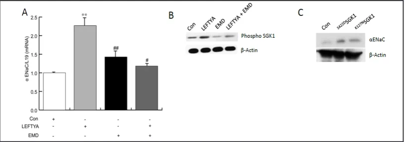
In order to quantify the contribution of SGK1 to the regulation of αENaC by LEFTYA,
Ishikawa cells were treated in the absence and presence of SGK1 inhibitor EMD638683 (50
µM; EMD) with or without LEFTYA. As illustrated in Fig. 4, LEFTYA again increased the
αENaC
levels. The administration of EMD638683 alone slightly but significantly increased
αENaC
transcript levels, an effect presumably unrelated to SGK1 inhibition. In the presence of both
[image:6.595.97.503.89.395.2]EMD638683 and LEFTYA,
αENaC
levels in Ishikawa cells did not change when compared to
Fig. 1. Effect of LEFTYA on SGK1 and ENaC subunit transcript levels as well as SGK1, NEDD4-2, and αENAC protein abundance in Ishikawa cells. A-D. Arithmetic means ± SEM (n = 4) of the (A) SGK1, (B) αENaC, (C) βENaC, (D) γENaC over L19 transcript levels in Ishikawa cells prior to (0) and 5-120 min following treatment with 25 ng/ml LEFTYA. Statistically significant difference was observed when *p<0.05, **p<0.001 using one way ANOVA. E. Original Western blot of phosphorylated SGK1 (p-SGK1), αENAC, phosphorylated NEDD4-2 (pNEDD4-NEDD4-2) and β-actin protein cell lysates from Ishikawa cells prior to (0) and 5-1NEDD4-20 min following treatment with 25 ng/ml LEFTYA. β-actin was used as a loading control. F. Represent the original Western blot of Total-SGK1 and Total-NEDD4-2 from cell lysates prior (0) to and 5-120 min following treatment with 25 ng/ml LEFTYA. β-actin was used as a loading control. G. Arithmetic means ± SEM (n = 5) of
phosho-SGK1/Total-SGK1, αENAC/β-actin and phospho-NEDD4-2/Total-NEDD4-2 protein abundance ratios in cell
Fig. 2. Effect of LEFTYA on COX-2 transcripts and protein levels. Ishikawa cells were treated with 25 ng/ml LEFTYA for 120 min or remained untreated (Control). A. Arithmetic means ± SEM (n = 4) of COX-2 over L19 transcript levels in Ishikawa cells. B. Cells were stained with COX-2 antibody and subjected to FACS. Left, the median fluorescence intensity (MFI) was quantified and Right, original FACS histogram for Control (grey) and LEFTYA (blue). C. In parallel cultures 6-Keto PGF1-α levels were measured using ELISA. Statistically significant difference was observed when **p<0.001 using Student’s t-Test.
Fig. 3. Expression of key uterine implantation genes. Expression genes coding Bone Morphogenetic Protein 2 (Bmp2), Wingless-Type MMTV Integration Site Family, Member 4 (Wnt4), and Prolactin (PRL) was examined by qRT-PCR in Ishikawa cells treated with or without LEFTYA (n=6). Data are presented as arithmetic means
± SEM. * indicates p< 0.05 or ** p< 0.01 (Student’s t-test).
Fig. 4. αENaC transcript levels in Ishikawa cells following LEFTYA treatment in absence and presence of SGK1 inhibitor EMD638683 and following transfection with active or inactive SGK1. A. Arithmetic means ± SEM (n = 3) of the αENaC over L19 transcript levels from untreated (white), LEFTYA treated (grey), EMD638683 treated alone (50 µM; black) or in combination of both LEFTYA and EMD638683 (dark grey). Statistically significant difference was observed between untreated and LEFTYA treated cells (**p<0.01), or EMD638683 treated cells (*p<0.05) as well as between LEFTYA treated cells and LEFTYA+EMD638683 treated cells (#p<0.05) using one way ANOVA. B.Original Western blot of αENAC and β-actin. Protein cell
lysates from untreated Ishikawa cells or cells treated with LEFTYA, EMD638683 treated alone (50 μM; EMD;
[image:7.595.94.503.462.605.2]the control. However, when compared to LEFTYA alone, treatment with both EMD638683
[image:8.595.99.500.85.404.2]and LEFTYA significantly reduced
αENaC transcript levels.
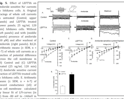
Fig. 5. Effect of LEFTYA on
amiloride sensitive Na+ currents in Ishikawa cells. A. Original tracings of whole cell currents
in untreated (Control; upper
panels) and LEFTYA treated (lower panels; 25 ng/ml, 120 min) Ishikawa cells. Without (left panels) and with (middle
panels) presence of amiloride
(50 µM) and after washout of
amiloride (right panels). B,C,D.
Arithmetic means (± SEM, n = 6-7) of whole cell currents as a
function of potential difference across the cell membrane in
(B) Control and (C) LEFTYA treated (25 ng/ml, 120 min)
(D) Amiloride senstive current
fraction of LEFTYA treated cells in Ishikawa cells. E. Arithmetic means (± SEM, n = 6-7) of inward conductance (nS) of
the cell membrane calculated
by linear fit of I/V-curves (in D) from -80 mV to -140mV in
untreated (Control, white bar) and LEFTYA treated (25 ng/ml, 120 min, black bar) Ishikawa cells. Statistically significant difference from untreated cells compared with LEFTYA (**p<0.01) using Student’s t-Test.
Fig. 6. Effect of LEFTYA on amiloride induced transepithelial current across murine endometrial epithe
-lium. A,B. Representative original tracings showing the effect of amiloride (50 µM) on the transepitheli
-al potenti-al difference across (A) Control and (B) LEFTYA (500 ng/ml) treated murine endometrium. The voltage deflections result from injection of 1 µA current pulses and reflect the transepithelial resistance. Arrow indicates addition of amiloride (50 µM). C. Arithmetic means ± SEM (n = 6) of the amiloride (50 µM) induced equivalent short-circuit current across (µA/cm2) Control (PBS; white bar) and LEFTYA (500 ng/ml;
[image:8.595.93.505.437.591.2]In order to test, whether induction of SGK1 is sufficient for up-regulation of
αENaC
expression, a further series of experiments explored whether
αENaC protein levels in
endometrial cells are modified by SGK1 transfection. As shown in Fig. 4C,
αENaC
levels
were significantly enhanced following transfection of Ishikawa cells with the constitutively
active
S422DSGK1 and significantly down-regulated upon transfection of a kinase dead mutant
(
K127NSGK1). Thus,
αENaC
protein levels in Ishikawa cells are up-regulated by SGK1 even in
the absence of LEFTYA. The negative effect of
K127NSGK1 points to competitive displacement
of endogeneous SGK1 from the target protein by the inactive mutant [37].
Patch clamp experiments were performed to test whether the increase of ENaC protein
[image:9.595.93.504.88.367.2]corresponded to enhanced ENaC activity. To this end, amiloride sensitive Na
+currents in
Ishikawa cells were determined utilizing whole cell patch clamp. As illustrated in Fig. 5A-D,
the amiloride-sensitive whole cell currents were significantly increased by LEFTYA treatment
(25 ng/ml, 120 min). Fig. 5B&C display the whole cell currents as a function of potential
difference across the cell membrane in untreated and LEFTYA treated Ishikawa cells both, in
the absence and presence of amiloride (50 µM). Fig. 5D displays the amiloride sensitive current
fraction as a function of potential difference across the cell membrane. The effect of LEFTYA on
the whole cell current was paralleled by the respective effect on inward conductance calculated
Fig. 7. SGK1 sensitivity of αENaC and key murine implantation genes. A. Arithmetic means ± SEM (n=5) of
from the individual I-V relations by linear regression of inward current between -80 and
-140 mV (Fig. 5E). The reversal potential of the currents under control conditions and after
treatment with LEFTYA were not statistically significant.
Ussing chamber experiments were performed to quantify the amiloride-sensitive
transepithelial current (electrogenic Na
+transport) across murine endometrial epithelium
in situ
. As illustrated in Fig. 6, the amiloride (50 µM) sensitive transepithelial potential
difference and the amiloride-sensitive equivalent short-circuit current across the murine
endometrial epithelium were significantly increased by LEFTYA treatment (500 ng/ml). At
lower concentrations no discernable effects were seen.
A final series of experiments explored whether LEFTYA influenced endometrial
αENaC
transcript levels and whether this effect was modulated by SGK1. To test this conjecture,
uteri from wild type mice (
Sgk1
+/+) and Sgk1-deficient mice (
Sgk1
-/-) were excised and
flushed with LEFTYA (500ng/ml) or with PBS. The uteri were then cultured for 24 hours
and the expression of
αENaC
was examined by qRT-PCR. As illustrated in Fig. 7A&B, LEFTYA
treatment (500 ng/ml) was followed by a significant increase of
αENaC
transcript and
protein levels in endometrium from
Sgk1
+/+mice but not from
Sgk1
-/-mice. Implantation
genes Cox2, Leukemia inhibitory factor (Lif) and Prolactin family 8, subfamily a, member 2
(Prl8a2) tended to be lower in the
Sgk1
-/-mice and the LEFTYA flushed
Sgk1
+/+mice than in
control mice, a difference, however, not reaching statistical significance (Fig. 7C).
Discussion
Gene ablation studies in mice have been pivotal in identifying critical implantation
regulators. Within this network of genes, many encode secreted factors, including growth
factors (e.g.heparin-binding EGF-like growth factor), cytokines (e.g. leukemia inhibitory
factor) and various morphogens (e.g. bone morphogenetic protein 2) [2]. These establish
paracrine gradients that control a distinct temporal-spatial pattern in order to enable the
embryo to breach the luminal epithelium and embed in the decidualizing stroma .
The present study demonstrates that LEFTYA is a major regulator of the expression and
activity of SGK1 and ENaC in the endometrium. Further, we reveal that LEFTYA participates in
the regulation of fluid transport across the endometrial epithelium.
In keeping with previous findings, treatment of Ishikawa cells with LEFTYA (25 ng/
ml) rapdily increased the transcript levels of
SGK1
and
αENaC
,
βENaC
and
γ
ENaC
subunits
within 60 mins [16, 38]. Like TGFβ1 [26], LEFTYA was shown to be a strong stimulator
of SGK1 expression. Further, the effect of LEFTYA on
αENaC
levels was abolished by the
SGK1 inhibitor EMD638683. Transfection of Ishikawa cells with the constitutively active
S422D
SGK1 mutant was sufficient to increase
αENaC
protein levels. By contrast, transfection
with
K127NSGK1, a kinase dead mutant, significantly decreased
αENaC
levels, an observation
pointing to the transdominant inhibitory effect of the mutant. The role of SGK1 in regulating
endometrial
αENaC
expression was further illustrated by comparing wild type mice (
Sgk1
+/+)
and Sgk1-deficient (
Sgk1
-/-) mice. Interestingly, even in the absence of LEFTYA treatment,
αENaC
transcript levels were significantly lower in
Sgk1
knockout mice. Furthermore,
LEFTYA significantly increased
αENaC
transcript levels in wild type but not
Sgk1
-/-mice,
demonstrating that LEFTYA-dependent regulation of ENaC in the endometrium is largely if
not exclusively dependent on SGK1.
Up-regulation of ENaC activity is expected to stimulate endometrial salt and fluid
absorption. Failure to down-regulate LEFTYA secretion during the mid-luteal phase of the
cycle may lead to premature ‘closure’ of the uterine lumen
via
ENaC-mediated fluid absorption,
resulting in implantation failure [20, 39]. The present observations, however, do not provide
insight into the purported inhibitory effects of LEFTYA on decidualization of the endometrial
stroma [40, 41]. A previous study reported that induction of ENaC activity in response to
response to Ca
2+signaling and enhanced prostaglandin E2 (PGE2) production [19]. As PGE2
stimulates differentiation of endometrial stromal cells, up-regulation of ENaC by LEFTYA is
unlikely to account for the negative effect of LEFTYA on decidualization [41]. The possibility
must be considered that LEFTYA upregulates ENaC but by the same token disrupts the link
between ENaC and decidualization. On the other hand, at least in theory, activation of ENaC
prior to implantation may compromize fertility [20, 39], whereas activation of ENaC may be
required during embryo implantation. It must further be kept in mind that, besides SGK1
[42], several other kinases regulate ENaC, including; SGK2&3, Protein Kinase A, Casein
Kinase II, G protein-coupled receptor kinase 2, Inhibitor of Nuclear Factor Kappa-B kinase
subunit beta, Phosphoinositide-Dependent Kinase 1, Protein Kinase C, Extracellular
Signal-Regulated Protein Kinases 1 and 2 and AMP-Activated Protein Kinase [43].
Besides the effect on endometrial fluid transport, ENaC accomplishes Na
+transport
in a wide variety of tissues and contributes to the physiology and pathophysiology of
diverse functions, including renal salt excretion [42], blood pressure [44], cell volume [45],
pulmonary fluid transport [46, 47] and endothelial function [48]. It is tempting to speculate
that LEFTYA may influence ENaC activity in non-uterine tissues. Moreover, SGK1 and ENaC
may also participate in the known effects of LEFTYA on embryonic morphogenesis [49]
.
In conclusion, LEFTYA is a powerful regulator of SGK1 and ENaC in the endometrium
and contributes to the complex regulatory network that controls embryo implantation. Our
findings provide new insight on how deregulation of SGK1, ENaC and LEFTYA contribute to
unexplained infertility and implantation failure in IVF.
Acknowledgements
The authors acknowledge the meticulous preparation of the manuscript by Tanja Loch
and technical support by Elfriede Faber.
This study was supported by the Deutsche Forschungsgemeinschaft
to
F.L
(GRK 1302,
SFB 773 B4/A1, La 315/13-3), the Open Access Publishing Fund of Tuebingen University,
and
to M.S.S the EMBO Long-Term Fellowship (ALTF 20-2013).
Disclosure Statement
All authors declare that there are no conflicts of interest that could be perceived as
prejudicing the impartiality of the research reported.
References
1 Koot YE, Teklenburg G, Salker MS, Brosens JJ, Macklon NS: Molecular aspects of implantation failure. Biochim Biophys Acta 2012;1822:1943-1950.
2 Wang H, Dey SK: Roadmap to embryo implantation: Clues from mouse models. Nat Rev Genet 2006;7:185-199.
3 Evers JL: Female subfertility. Lancet 2002;360:151-159.
4 Teklenburg G, Salker M, Heijnen C, Macklon NS, Brosens JJ: The molecular basis of recurrent pregnancy loss: Impaired natural embryo selection. Mol Hum Reprod 2010;16:886-895.
5 Sakuma R, Ohnishi Yi Y, Meno C, Fujii H, Juan H, Takeuchi J, Ogura T, Li E, Miyazono K, Hamada H: Inhibition of nodal signalling by lefty mediated through interaction with common receptors and efficient diffusion. Genes Cells 2002;7:401-412.
6 Gellersen B, Brosens JJ: Cyclic decidualization of the human endometrium in reproductive health and failure. Endocr Rev 2014;35:851-905.
8 Cornet PB, Galant C, Eeckhout Y, Courtoy PJ, Marbaix E, Henriet P: Regulation of matrix
metalloproteinase-9/gelatinase b expression and activation by ovarian steroids and lefty-a/endometrial bleeding-associated factor in the human endometrium. J Clin Endocrinol Metab 2005;90:1001-1011. 9 Cornet PB, Picquet C, Lemoine P, Osteen KG, Bruner-Tran KL, Tabibzadeh S, Courtoy PJ, Eeckhout Y,
Marbaix E, Henriet P: Regulation and function of lefty-a/ebaf in the human endometrium. Mrna expression during the menstrual cycle, control by progesterone, and effect on matrix metalloprotineases. J Biol Chem 2002;277:42496-42504.
10 Papageorgiou I, Nicholls PK, Wang F, Lackmann M, Makanji Y, Salamonsen LA, Robertson DM, Harrison CA: Expression of nodal signalling components in cycling human endometrium and in endometrial cancer. Reprod Biol Endocrinol 2009;7:122.
11 Ulloa L, Creemers JW, Roy S, Liu S, Mason J, Tabibzadeh S: Lefty proteins exhibit unique processing and activate the mapk pathway. J Biol Chem 2001;276:21387-21396.
12 Druckmann R: Long-term use of progestogens--getting the balance right: Molecular biology and the endometrium. Gynecol Endocrinol 2007;23 Suppl 1:53-61.
13 Tabibzadeh S, Mason JM, Shea W, Cai Y, Murray MJ, Lessey B: Dysregulated expression of ebaf, a novel molecular defect in the endometria of patients with infertility. J Clin Endocrinol Metab 2000;85:2526-2536. 14 Tang M, Mikhailik A, Pauli I, Giudice LC, Fazelabas AT, Tulac S, Carson DD, Kaufman DG, Barbier C, Creemers JW, Tabibzadeh S: Decidual differentiation of stromal cells promotes proprotein convertase 5/6 expression and lefty processing. Endocrinology 2005;146:5313-5320.
15 Feroze-Zaidi F, Fusi L, Takano M, Higham J, Salker MS, Goto T, Edassery S, Klingel K, Boini KM, Palmada M, Kamps R, Groothuis PG, Lam EW, Smith SK, Lang F, Sharkey AM, Brosens JJ: Role and regulation of the
serum- and glucocorticoid-regulated kinase 1 in fertile and infertile human endometrium. Endocrinology
2007;148:5020-5029.
16 Salker MS, Christian M, Steel JH, Nautiyal J, Lavery S, Trew G, Webster Z, Al-Sabbagh M, Puchchakayala G, Foller M, Landles C, Sharkey AM, Quenby S, Aplin JD, Regan L, Lang F, Brosens JJ: Deregulation of the serum-
and glucocorticoid-inducible kinase sgk1 in the endometrium causes reproductive failure. Nature medicine
2011;17:1509-1513.
17 Zhou R, Patel SV, Snyder PM: Nedd4-2 catalyzes ubiquitination and degradation of cell surface enac. J Biol Chem 2007;282:20207-20212.
18 Lang F, Cohen P: Regulation and physiological roles of serum- and glucocorticoid-induced protein kinase isoforms. Sci STKE 2001;2001:re17.
19 Ruan YC, Guo JH, Liu X, Zhang R, Tsang LL, Dong JD, Chen H, Yu MK, Jiang X, Zhang XH, Fok KL, Chung YW, Huang H, Zhou WL, Chan HC: Activation of the epithelial na+ channel triggers prostaglandin e(2) release and production required for embryo implantation. Nature medicine 2012;18:1112-1117.
20 Tsang LL, Chan LN, Chan HC: Altered cyclic expression of epithelial na+ channel subunits and cystic fibrosis transmembrane conductance regulator in mouse endometrium by a low sodium diet. Cell Biol Int 2004;28:549-555.
21 Yang JZ, Ajonuma LC, Tsang LL, Lam SY, Rowlands DK, Ho LS, Zhou CX, Chung YW, Chan HC: Differential
expression and localization of cftr and enac in mouse endometrium during pre-implantation. Cell Biol Int
2004;28:433-439.
22 Zhou M, Fu J, Huang W, Shen L, Xiao L, Song Y, Liu Y: Increased cystic fibrosis transmembrane conductance
regulators expression and decreased epithelial sodium channel alpha subunits expression in early
abortion: Findings from a mouse model and clinical cases of abortion. PLoS One 2014;9:e99521. 23 Ruan YC, Chen H, Chan HC: Ion channels in the endometrium: Regulation of endometrial receptivity and
embryo implantation. Hum Reprod Update 2014;20:517-529.
24 Soundararajan R, Pearce D, Ziera T: The role of the enac-regulatory complex in aldosterone-mediated sodium transport. Mol Cell Endocrinol 2012;350:242-247.
25 Chan LN, Tsang LL, Rowlands DK, Rochelle LG, Boucher RC, Liu CQ, Chan HC: Distribution and regulation of enac subunit and cftr mrna expression in murine female reproductive tract. J Membr Biol 2002;185:165-176.
27 Schmid E, Xuan NT, Zahir N, Russo A, Yang W, Kuhl D, Faggio C, Shumilina E, Lang F: Serum- and
glucocorticoid-inducible kinase 1 sensitive nf-kappab signaling in dendritic cells. Cell Physiol Biochem
2014;34:943-954.
28 Schmidt S, Schneider S, Yang W, Liu G, Schmidt EM, Schmid E, Mia S, Brucker S, Stournaras C, Wallwiener D, Brosens JJ, Lang F: Tgfbeta1 and sgk1-sensitive store-operated ca2+ entry and orai1 expression in endometrial ishikawa cells. Molecular human reproduction 2014;20:139-147.
29 Brosens JJ, Salker MS, Teklenburg G, Nautiyal J, Salter S, Lucas ES, Steel JH, Christian M, Chan YW, Boomsma CM, Moore JD, Hartshorne GM, Sucurovic S, Mulac-Jericevic B, Heijnen CJ, Quenby S, Koerkamp MJ, Holstege FC, Shmygol A, Macklon NS: Uterine selection of human embryos at implantation. Sci Rep 2014;4:3894. 30 Salker MS, Zhou Y, Singh Y, Brosens J, Lang F: Leftya sensitive cytosolic ph regulation and glycolytic flux in
ishikawa human endometrial cancer cells. Biochem Biophys Res Commun 2015;460:845-849. 31 Yan J, Schmid E, Hosseinzadeh Z, Honisch S, Shumilina E, Fuchs J, Lang F: Impact of janus kinase 3 on
cellular ca release, store operated ca(2+) entry and na(+)/ca(2+) exchanger activity in dendritic cells. Cell Physiol Biochem 2015;36:2287-2298.
32 Hosseinzadeh Z, Honisch S, Schmid E, Jilani K, Szteyn K, Bhavsar S, Singh Y, Palmada M, Umbach AT, Shumilina E, Lang F: The role of janus kinase 3 in the regulation of na(+)/k(+) atpase under energy depletion. Cell Physiol Biochem 2015;36:727-740.
33 Pakladok T, Almilaji A, Munoz C, Alesutan I, Lang F: Pikfyve sensitivity of herg channels. Cell Physiol Biochem 2013;31:785-794.
34 Barry PH, Lynch JW: Liquid junction potentials and small cell effects in patch-clamp analysis. J Membr Biol 1991;121:101-117.
35 Rexhepaj R, Dermaku-Sopjani M, Gehring EM, Sopjani M, Kempe DS, Foller M, Lang F: Stimulation of electrogenic glucose transport by glycogen synthase kinase 3. Cell Physiol Biochem 2010;26:641-646. 36 Pasham V, Pathare G, Fajol A, Rexhepaj R, Michael D, Pakladok T, Alesutan I, Rotte A, Foller M, Lang F:
Osr1-sensitive small intestinal na+ transport. Am J Physiol Gastrointest Liver Physiol 2012;303:G1212-1219. 37 Lang F, Klingel K, Wagner CA, Stegen C, Warntges S, Friedrich B, Lanzendorfer M, Melzig J, Moschen I,
Steuer S, Waldegger S, Sauter M, Paulmichl M, Gerke V, Risler T, Gamba G, Capasso G, Kandolf R, Hebert SC, Massry SG, Broer S: Deranged transcriptional regulation of cell-volume-sensitive kinase hsgk in diabetic nephropathy. Proc Natl Acad Sci U S A 2000;97:8157-8162.
38 Waldegger S, Barth P, Raber G, Lang F: Cloning and characterization of a putative human serine/threonine protein kinase transcriptionally modified during anisotonic and isotonic alterations of cell volume. Proc Natl Acad Sci U S A 1997;94:4440-4445.
39 Chan HC, Ruan YC, He Q, Chen MH, Chen H, Xu WM, Chen WY, Xie C, Zhang XH, Zhou Z: The cystic fibrosis transmembrane conductance regulator in reproductive health and disease. J Physiol 2009;587:2187-2195. 40 Li H, Li H, Bai L, Yu H: Lefty inhibits in vitro decidualization by regulating p57 and cyclin d1 expressions.
Cell Biochem Funct 2014;32:657-664.
41 Park CB, Dufort D: Nodal expression in the uterus of the mouse is regulated by the embryo and correlates with implantation. Biol Reprod 2011;84:1103-1110.
42 Soundararajan R, Lu M, Pearce D: Organization of the enac-regulatory machinery. Crit Rev Biochem Mol Biol 2012;47:349-359.
43 Baines D: Kinases as targets for enac regulation. Curr Mol Pharmacol 2013;6:50-64.
44 Rossier BC: Epithelial sodium channel (enac) and the control of blood pressure. Curr Opin Pharmacol 2014;15:33-46.
45 Bondarava M, Li T, Endl E, Wehner F: Alpha-enac is a functional element of the hypertonicity-induced cation channel in hepg2 cells and it mediates proliferation. Pflugers Arch 2009;458:675-687.
46 Althaus M: Enac inhibitors and airway re-hydration in cystic fibrosis: State of the art. Curr Mol Pharmacol 2013;6:3-12.
47 Fronius M: Treatment of pulmonary edema by enac activators/stimulators. Curr Mol Pharmacol 2013;6:13-27.
48 Kusche-Vihrog K, Jeggle P, Oberleithner H: The role of enac in vascular endothelium. Pflugers Arch 2014;466:851-859.