University of Warwick institutional repository:
http://go.warwick.ac.uk/wrap
A Thesis Submitted for the Degree of PhD at the University of Warwick
http://go.warwick.ac.uk/wrap/63780
This thesis is made available online and is protected by original copyright.
Please scroll down to view the document itself.
Understanding the "Rules of Engagement" for
Membrane Protein Folding: Chemical Biology and
Computational Approaches for Determination of
Structure and Dynamics
by
Anthony Nash
Thesis
Submitted to the University of Warwick for the degree of
Doctor of Philosophy
Supervisors:Dr. Ann Dixon and Dr. Rebecca Notman
MOAC Doctoral Training Centre
Contents
1 Introduction 1
1.1 Statement of problem . . . 1
1.2 Transmembrane proteins . . . 3
1.2.1 Membrane-mimetics . . . 6
1.2.1.1 Isotropic solvents . . . 6
1.2.1.2 Detergent micelles . . . 6
1.2.1.3 Lipid bilayers . . . 7
1.2.1.4 Biological membranes . . . 7
1.2.2 TM interaction motifs . . . 8
1.2.2.1 Small-xxx-small TM interaction motifs . . . 9
1.2.2.2 Heptad repeat transmembrane interaction motifs . . . 10
1.2.2.3 Polar transmembrane interaction motifs . . . 12
1.2.3 Protein-lipid mediated oligomerisation . . . 15
1.3 Low complexity sequence scaffolds . . . 16
1.3.1 GxxxG motif low complexity sequences . . . 17
1.3.2 Polar and/or ionic side chain motif low complexity sequences . . . 20
1.3.3 Aromatic motif low complexity sequences . . . 25
1.3.4 Miscellaneous low complexity sequences . . . 26
1.4 De novoTM peptide applications . . . 28
1.5 Aims and objections . . . 30
1.6 Chapter Organisation . . . 31
2.1 Statistical mechanics . . . 33
2.1.1 Ergodicity . . . 36
2.2 Interaction potentials . . . 36
2.2.1 Van der Waals interactions . . . 38
2.2.2 Electrostatic interactions . . . 39
2.2.3 Covalent bonds . . . 39
2.2.3.1 Harmonic bond stretching . . . 40
2.2.3.2 Harmonic angle potential . . . 40
2.2.3.3 Harmonic improper dihedral potential . . . 41
2.2.3.4 Proper dihedral . . . 41
2.3 Coarse grained force field . . . 42
2.4 Potential energy minimisation . . . 43
2.4.1 Steepest descent . . . 43
2.5 Molecular Dynamics . . . 44
2.5.1 Equation of motion . . . 44
2.5.1.1 Time integration algorithms . . . 46
2.5.2 Constant temperature - canonical ensemble . . . 47
2.5.2.1 Berendsen thermostat . . . 48
2.5.2.2 Nosé-Hoover thermostat . . . 48
2.5.3 Constant pressure . . . 49
2.5.3.1 Berendsen barostat . . . 49
2.5.3.2 Parrinello-Rahman barostat . . . 49
2.5.4 Constraints . . . 50
2.5.5 Periodic boundary conditions . . . 50
2.6 Free energy methods . . . 51
2.6.1 Reaction coordinate . . . 52
2.6.2 Umbrella sampling . . . 53
2.6.3 Weighted Histogram Analysis Method . . . 54
3.2 Bacterial strains . . . 57
3.3 Plasmids . . . 58
3.4 Primers . . . 58
3.5 Growth and maintenance ofE. coli . . . 58
3.5.1 Media . . . 59
3.5.2 Maintenance . . . 60
3.5.3 Preparation of competentE. colicells . . . 60
3.5.4 Transformation of competentE. colicells . . . 60
3.6 DNA manipulation and cloning techniques . . . 61
3.6.1 Preparation of plasmid DNA . . . 61
3.6.2 Colony PCR . . . 61
3.6.3 Agarose gel electrophoresis . . . 62
3.6.4 Purification of DNA by gel extraction . . . 62
3.6.5 Restriction endonuclease digestion of DNA . . . 62
3.6.6 Ligation of DNA fragments . . . 62
3.6.7 Sequencing of plasmid DNA . . . 63
3.7 GALLEX methods . . . 63
3.7.1 Chimera expression, insertion and orientation checks . . . 64
3.7.1.1 SDS-PAGE . . . 65
3.7.1.2 Western Blotting . . . 65
3.7.1.3 MalE complementary assay . . . 65
3.7.1.4 Sodium hydroxide extraction assay . . . 66
3.8 Measurement ofβ-galactosidase activity . . . 66
3.8.1 β-galactosidase Assay . . . 66
3.8.2 β-galactosidase Free Energy Assay . . . 67
3.9 Specialist software . . . 68
4 Lipid-Mediated Dimerisation of the Transmembrane Helical Peptide Neu: Insights from Molecular Dynamics Simulations 69 4.1 Introduction . . . 69
4.2.1 Forcefield parameters . . . 71
4.2.2 Simulation parameters . . . 71
4.2.3 Generation of initial configurations of the dimer . . . 71
4.2.4 Generation of Initial Bilayer . . . 72
4.2.5 Insertion of the dimer into the bilayer . . . 73
4.2.6 Equilibration procedure . . . 73
4.2.7 Umbrella sampling and free energy calculations . . . 74
4.3 Results . . . 74
4.3.1 Bulk structural properties of a POPC lipid bilayer . . . 75
4.3.1.1 Bilayer thickness . . . 75
4.3.1.2 Area-per-lipid . . . 76
4.3.2 Free energy profiles as a function of peptide separation . . . 78
4.3.3 Bilayer thickness . . . 82
4.3.4 Lipid tail order parameters . . . 82
4.3.5 Dimer stabilisation throughinterhelical side chain interactions. . . 90
4.4 Discussion . . . 96
4.4.1 The Neu and Neu* dimers are stabilised by side chain interactions. . . 98
4.4.2 Interhelical approach is a lipid-mediated process. . . 100
4.4.3 Both Neu helical orientations have a similar propensity for dimerisation. . . 101
4.4.4 Both Neu* helical orientations have a similar free energy values, yet dimeri-sation of Neu*AGand Neu*IVmay halt along a free energy plateau. . . . 102
4.5 Conclusion . . . 102
5 Self association of low complexity scaffold TM domains in a biological environment using thein vivoGALLEX assay 105 5.1 Introduction . . . 105
5.1.1 Low complexity scaffold design . . . 106
5.1.2 Polyleucine scaffold design . . . 107
5.1.3 Alanine zipper scaffold design . . . 108
5.1.4 Bifunctional alanine zipper . . . 108
5.3 Results . . . 109
5.3.1 Analysis of scaffold insertion and orientation . . . 109
5.3.1.1 Theoretical insertion of scaffold TM domains . . . 109
5.3.1.2 Analysis of protein chimera association with the inner membrane from sodium hydroxide washes . . . 116
5.3.1.3 MalE complementation assay . . . 117
5.3.2 Analysis of low complexity scaffold interactions using GALLEX . . . 117
5.3.2.1 Low complexity scaffolds . . . 118
5.3.2.2 Polyleucine scaffolds . . . 118
5.3.2.3 Alanine zipper scaffolds . . . 118
5.3.2.4 Bifunctional alanine zipper scaffolds . . . 121
5.3.3 Free energy of association in a biological membrane . . . 124
5.4 Discussion . . . 133
5.4.1 Polyleucine scaffolds . . . 133
5.4.2 Alanine zipper scaffolds . . . 136
5.5 Conclusion . . . 138
6 Free energy of association of TM peptides in a POPC bilayer: comparison of coarse-grained and united-atom force fields 139 6.1 Introduction . . . 139
6.2 Simulation methodology . . . 144
6.2.1 Force field parameters . . . 144
6.2.2 Simulation parameters . . . 144
6.2.3 POPC lipid bilayer construction . . . 145
6.2.4 Insertion of dimers into a POPC lipid bilayer . . . 145
6.2.5 Umbrella sampling and free energy calculations . . . 145
6.2.6 Optimisation of the NeuWTCG force field . . . 146
6.3 Results . . . 147
6.3.1 Optimisation of the NeuWTCG force field . . . 147
6.3.3 Bilayer thickness of a CG POPC lipid bilayer . . . 150
6.3.4 Bilayer thickness and area per lipid of mediated through hydrophobic TM domain length . . . 151
6.3.5 Tilt angle mediated through hydrophobic TM domain length . . . 152
6.4 Discussion . . . 156
6.5 Conclusion . . . 158
7 Molecular Dynamics simulations of low complexity TM domains 161 7.1 Introduction . . . 161
7.2 Simulation methods . . . 163
7.2.1 Coarse grained simulation configuration . . . 163
7.2.1.1 Force field parameters . . . 163
7.2.1.2 Simulation parameters . . . 163
7.2.1.3 POPC bilayer construction . . . 163
7.2.1.4 Insertion of dimers into a POPC bilayer . . . 163
7.2.1.5 Umbrella sampling and free energy calculations . . . 164
7.2.1.6 Single-linkage cluster analysis . . . 164
7.2.2 CHI conformational search . . . 164
7.2.3 United atom simulation configuration . . . 165
7.2.3.1 Force field parameters . . . 165
7.2.3.2 Simulation parameters . . . 165
7.2.3.3 POPC bilayer construction . . . 165
7.2.3.4 Generation of initial configurations of the AZ2, AZ2L6S and AZ2L10S dimers . . . 165
7.2.3.5 Insertion of dimers into a POPC bilayer . . . 165
7.2.3.6 Equilibration procedure . . . 165
7.3 Results . . . 167
7.3.1 CG low complexity scaffold free energy of self association . . . 167
7.3.1.1 L17 . . . 167
7.3.1.2 L17(GG4) . . . 171
7.3.1.4 AZ2 . . . 182
7.3.1.5 AZ2(GG4)L . . . 186
7.3.1.6 AZ2(GG4)A . . . 191
7.4 United atom simulations of AZ2, AZ2L6S and AZ2L10S . . . 195
7.4.1 Initial dimer geometry . . . 195
7.4.1.1 AZ2 initial dimer geometry . . . 195
7.4.1.2 AZ2L6S initial dimer geometry . . . 196
7.4.1.3 AZ2L10S initial dimer geometry . . . 196
7.4.2 Bilayer area per lipid . . . 197
7.4.3 interhelical distance calculations and serine hydrogen bonding . . . 198
7.4.4 Helix-helix crossing angle . . . 200
7.5 Discussion . . . 203
7.5.1 L17, L17(GG4) and L17L9Q . . . 204
7.5.2 AZ2, AZ2(GG4)Land AZ2(GG4)A . . . 207
7.5.3 United atom simulations of AZ2, AZ2L6S and AZ2L10S . . . 209
7.6 Conclusion . . . 210
8 Conclusion and Future Work 213 8.1 Conclusions . . . 213
8.2 Limitations . . . 215
8.3 Future work . . . 216
A Helix-helix crossing angle and tilt angle 245 A.1 Minimum distance between helices . . . 245
A.2 Helix-helix crossing angle . . . 247
List of Tables
1.1 Membrane-mimetics: advantages and disadvantages. . . 8
1.2 Leucine zipper-like heptad repeats found in native TM domains. . . 12
1.7 TM domain sequences of low complexity scaffolds. . . 31
2.1 The basic Molecular Dynamics algorithm. . . 45
3.1 BacterialE. colistrains used in this study. . . 57
3.2 List of previously generated plasmids vectors used in this study. . . 58
3.3 Polyleucine DNA primers. . . 58
3.4 Alanine zipper DNA primers. . . 59
3.5 Bifunctional alanine zipper DNA primers. . . 59
4.1 Free energy and structural calculations of Neu and oncogenic Neu dimers. . . 98
5.1 TM domain sequences of low complexity scaffolds. . . 109
5.2 Self-association free energy of low complexity scaffolds in a biological membrane. 126 6.1 Bulk POPC, AZ2 and NeuWTsequences, hydrophobic length, tilt angle, APL and bilayer thickness. . . 160
List of Figures
1.1 Structural types of membrane proteins: (A) Associating membrane protein: prostaglandin
H2 synthase (associates to the membrane of the endoplasmic reticulum), 1PTH. (B)
Integral TM protein: potassium channel KCSA, 1BL8. (C) Polytopic TM protein:
Bacteriorhodopsin, 2AT9. (D)β-barrel Omp G monomeric porin 2JQY. . . 4
1.2 A pictorial representation of the membrane protein two-stage model. . . 5
1.3 Examples of GG4 and polar motifs in TM domains. . . 10
1.4 Heptad repeat motif. . . 11
2.1 The ergodic hypothesis: the time spent in a region of phase space is proportional by the volume occupied by microstates of the same energy. . . 37
2.2 The LJ-Potential. . . 38
3.1 Induction of the GALLEX chimera from IPTG. . . 63
3.2 The GALLEX mechanism. . . 64
4.1 Final frame of a united-atom POPC bilayer. . . 75
4.2 Bilayer thickness of bulk united-atom POPC bilayer. . . 76
4.3 The simulation boxxy-area, parallel to the bilayer. . . 77
4.4 Self-association free energy of NeuIV, NeuAG, Neu*IVand Neu*AG. . . 79
4.5 Frequency histograms from NeuIV, NeuAG, Neu*IVand Neu*AGumbrella sampling simulations. . . 80
4.6 Dimer and individual peptide tilt angles . . . 81
4.7 Bilayer thickness in the presence of two NeuAGand Neu*AGpeptides. . . 83
4.8 Bilayer thickness in the presence of two NeuIVand Neu*IVpeptides. . . 84
4.10 Lipid order parameters in the presence of two Neu*IVpeptides. . . 86
4.11 Lipid order parameters in the presence of two NeuAGpeptides. . . 87
4.12 Lipid order parameters in the presence of two Neu*AGpeptides. . . 88
4.13 Oncogenic Neu and proto-oncogenic Neu hydrophobic lengths. . . 89
4.14 NeuIVand Neu*IVinterhelical atom-atom contact plots. . . 92
4.15 NeuAGand Neu*AGinterhelical atom-atom contact plots. . . 93
4.16 Interhelical distance distributions from NeuIV, NeuAG, Neu∗IV and Neu∗AG free energy umbrella simulation. . . 94
4.17 Final frame of the (A) Neu*IV I659xxxV663 and (B) NeuAG A661xxxG665, in-terhelical motifs. . . 95
4.18 Particle density normal to the bilayer of Neu*AGand Neu*IV. . . 96
4.19 Dimer helical structures of Neu at the free energy minima. . . 97
4.20 Dimer helical structures of oncogenic Neu at the free energy minima. . . 97
5.1 Rationale low sequence complexity building blocks. . . 107
5.2 TM domain visual schematics of complexity scaffolds. . . 112
5.3 Theoretical ∆Gpred of TM domains L17, L17(GG4), L17(GG4)G11I, L17L9Q, AZ2, and AZ2(GG4)L. . . 113
5.4 Theoretical∆Gpredof TM domains AZ2(GG4)LG6I, AZ2(GG4)LG10I, AZ2L6S, AZ2L10S, AZ2(GG4)A, and AZ2(GG4)AG8I. . . 114
5.5 Theoretical∆Gpredof TM domains AZ2(GG4)AG12I, AZ2(GG4)AL6A, and AZ2(GG4)AL10A.115 5.6 Sodium hydroxide washes of polyleucine scaffolds. . . 116
5.7 Semi-quantitative GALLEX assay of low complexity scaffolds. . . 119
5.8 Semi-quantitative GALLEX assay of low complexity L17 scaffolds. . . 120
5.9 Semi-quantitative GALLEX assay of low complexity AZ2 scaffolds. . . 122
5.10 Semi-quantitative GALLEX assay of low complexity AZ2(GG4)Ascaffolds. . . . 123
5.11 GpA titration curve of IPTG concentration to GALLEX signal. . . 126
5.12 G83I titration curve of IPTG concentration to GALLEX signal. . . 127
5.13 L17 titration curve of IPTG concentration to GALLEX signal. . . 128
5.14 L17(GG4) titration curve of IPTG concentration to GALLEX signal. . . 128
5.16 AZ2 titration curve of IPTG concentration to GALLEX signal. . . 130
5.17 AZ2(GG4)L titration curve of IPTG concentration to GALLEX signal. . . 131
5.18 AZ2TF titration curve of IPTG concentration to GALLEX signal. . . 132
5.19 Thermodynamic ‘road map’ of low complexity dimers. . . 135
6.1 Negative hydrophobic mismatch. . . 140
6.2 Positive hydrophobic mismatch. . . 141
6.3 Typical PMF profiles of TM domain association. . . 142
6.4 Coarse-grain solvated bulk POPC lipid bilayer. . . 144
6.5 Comparison of bond angle distributions of CG NeuWT. . . 148
6.6 Umbrella histograms and block averaged PMF profiles of AZ222. . . 149
6.7 Umbrella histograms and block averaged PMF profiles of AZ232. . . 149
6.8 Umbrella histograms and block averaged PMF profiles of NeuWT. . . 150
6.9 PMF profile of AZ2 (22) and AZ2 (32). . . 150
6.10 Average bilayer thickness of bulk POPC bilayer thickness. . . 151
6.11 Average bilayer thickness and final frame snap shots of an alanine zipper dimer in POPC. . . 152
6.12 Bilayer thickness of CG NeuWTdimer. . . 153
6.13 Cross sectional averaged bilayer thickness. . . 153
6.14 AZ2 dimer hydrophobic length over time. . . 154
6.15 NeuWTdimer hydrophobic length. . . 154
6.16 Tilt angle of AZ2 dimers of varying lengths. . . 155
6.17 NeuWTdimer tilt angle. . . 155
6.18 CG AZ2 tilt angle as a function of dimer hydrophobic length. . . 158
6.19 NeuWTCG and united atom PMF profiles. . . 159
7.1 PMF profile of self-association an L17 dimer. . . 168
7.2 L17 Umbrella sampling histograms and converging PMF profiles. . . 168
7.3 CG cluster representative of L17 free energy minimum. . . 168
7.4 Averaged structures from cluster analysis of an L17 conformational search using CHI.169 7.5 Atomistic L17 Interaction energies. . . 170
7.7 L17 Umbrella sampling histograms and converging PMF profiles. . . 172
7.8 L17(GG4) dimers from cluster analysis at the global free energy minimum. . . . 172
7.9 L17(GG4) CHI clusters . . . 173
7.10 Atomistic L17GG4 Interaction energies. . . 174
7.11 PMF profile of self-association an L17L9Q dimer. . . 175
7.12 L17L9Q Umbrella sampling histograms and converging PMF profiles. . . 175
7.13 CG cluster representative of L17L9Q free energy minimum found 0.95 nm along theinterhelical reaction coordinate. . . 176
7.14 CG cluster representative of L17L9Q free energy minimum found 0.5 nm along the interhelical reaction coordinate. . . 177
7.15 Averaged structures from cluster analysis of an L17L9Q conformational search using CHI. . . 179
7.16 Atomistic L17L9Q Interaction energies. . . 180
7.17 Continued... Atomistic L17L9Q Interaction energies. . . 181
7.18 PMF profile of self-association an AZ2 dimer. . . 182
7.19 AZ2 Umbrella sampling histograms and converging PMF profiles. . . 183
7.20 CG cluster representative of AZ2 free energy minimum. . . 183
7.21 Averaged structures from cluster analysis of an AZ2 conformational search using CHI. . . 184
7.22 Atomistic AZ2 Interaction energies. . . 185
7.23 PMF profile of self-association an AZ2(GG4)Ldimer. . . 186
7.24 AZ2(GG4)LUmbrella sampling histograms and converging PMF profiles. . . 187
7.25 CG cluster representative of AZ2(GG4)Lfree energy minimum. . . 187
7.26 AZ2(GG4)Ldimers cluster analysis at a second energy minimum. . . 188
7.27 Averaged structures from cluster analysis of an AZ2(GG4)L conformational search using CHI. . . 189
7.28 Atomistic AZ2(GG4)LInteraction energies. . . 190
7.29 PMF profile of self-association an AZ2(GG4)Adimer. . . 191
7.30 AZ2(GG4)AUmbrella sampling histograms and converging PMF profiles. . . 192
7.32 Averaged structures from cluster analysis of an AZ2(GG4)Aconformational search
using CHI. . . 193
7.33 Atomistic AZ2(GG4)AInteraction energies. . . 194
7.34 Starting configuration of a united atom AZ2L6S dimer as determined using the CHI algorithm. . . 196
7.35 Starting configuration of a united atom AZ2L10S dimer as determined using the CHI algorithm. . . 197
7.36 Area-per-lipid of united-atom AZ2 POPC bilayer. . . 198
7.37 Area-per-lipid of united-atom AZ2L6S POPC bilayer. . . 199
7.38 Area-per-lipid of united-atom AZ2L10S POPC bilayer. . . 199
7.39 Average atom-atom distance of a united atom alanine zipper dimer. . . 200
7.40 Average atom-atom distance calculated during the production run of AZ2L6S. . . . 201
7.41 Average atom-atom distance calculated during the production run of AZ2L10S. . 202
7.42 Crossing angle of alanine zipper and the two serine variants. . . 202
7.43 Thermodynamic pathway of CGde novoTM domain dimers. . . 210
Acknowledgements
I would like to begin by thanking the academics and staffat the MOAC doctoral training centre, without whom this PhD would never have been possible, nor nearly as enjoyable. My gratitude goes
to the Centre for Scientific Computing for their excellent support and high performance computing
facilities, the Chemical Biology department, and EPSRC for funding.
For three years I have been part of two wonderful groups of which I would like to thank: Leo
Bowsher; Sang Young Noh; Jenny Webb; Michael Chow; Dr. Fay Probert, and Dr. Gemma Warren.
Not only did they contribute towards a friendly and intellectually stimulating environment, but also
many hours of fond and happy memories.
Both of my supervisors, Rebecca Notman and Ann Dixon, have contributed so much of their time
and support above and beyond anything I have experienced during my previous jobs in industry. I
cannot thank them enough for their guidance throughout my studies. In particular, I would like to
thank Ann for introducing me to the world of Chemical Biology and experimental science. The
knowledge and experience from the long hours spent in the laboratory has contributed vastly to my
first steps towards my academic career. My other supervisor, Rebecca, I would like to thank for
her time and energy and providing her support and knowledge without which I would not be in an
academic career.
Finally, I would like to thank my wife, Dr. Johanna Nash. Johanna has been by my side throughout
these three years, providing love, support, and what can only be described as an infinite amount of
PhD published papers
• Effects of the oncogenic V664E mutation on membrane insertion, structure, and sequence-dependent interactions of the neu transmembrane domain in Micelles and Model
mem-branes: an integrated biophysical and simulation study. Andrew Beevers, Anthony Nash,
Martha Salazar-Cancino, David Scott, Rebecca Notman and Ann Dixon.Biochemistry. 2012.
Abstract
Approximately one third of genes in the human genome (1) encode transmembrane (TM) proteins
and form more than half of all drug targets (2). However, our understanding of how these proteins
fold into their functional form, as well as how they may misfold into a disease-associated form,
remains a difficult area of study. By observing the effects of single point mutations in the context of a native sequence, in addition to adding and mutating interhelical interaction motifs on a low
complexity sequence background, we aim to elicit ‘rules’ of TM protein domain association.
For the single point mutation in the context of a native sequence, the TM domain of the sequence
Neu, along with its oncogenic substitution V664E form Neu*, were selected. Using molecular
dynamics (MD) a united atom model of each dimer in a model bilayer system was subjected
to umbrella sampling along an interhelical reaction coordinate to yield a free energy profile of
self-association. The lipid order, bilayer thickness, and peptide tilt angle were calculated from
trajectories taken from three points along the reaction coordinate. Helical composition, solvent
accessible surface area, and hydrogen bond analysis (for the V664E substitution) were performed at
the free energy minimum.
Low complexity sequences of polyleucine and polyleucine-alanine heptad repeat sequences, with
and without interaction motifs similar to those present in the Neu model, were ligated into PBLM100
plasmids. TransformedE. colicells were subjected to semi-quantitative homo-interaction analysis
using the GALLEX assay. The same TM sequences were modelled using a coarse grained (CG)
forcefield. Umbrella sampling along an interhelical reaction coordinate was performed to yield a
free energy profile of self-association. Single-linkage cluster analysis of peptides was performed
at the global free energy minimum. A representative structure from each set was compared to an
averaged structure from the clusters of an atomistic conformational search.
The results presented in this study, could contribute to what in theory would be a large database of
motif-driven rules for TM helical domain oligomerisation. This may encourage further investigation
Chapter 1
Introduction
1.1
Statement of problem
TM protein folding, a fundamental process of cellular function and intercellular communication,
remains a persistent nontrivial problem with the number of resolved structures of soluble proteins
far out weighing those of membrane proteins (3). Predicting the protein structure from the primary
sequence is fraught with difficulty. Our understanding of the process behind the spontaneous insertion of a TM protein and its folding is limited, with most information coming from the studies
of native TM protein sequences in model membrane-mimetics. Unraveling the complex relationship
between TM protein sequence and TM protein structure using low complexity sequences has been
described as a ‘divide and conquer’ approach (4). By studying sequences of low complexity, putting
to one side the often complex sequence of native proteins, we are able to improve our understanding
of the interactions betweenα-helical domains. A low complexity sequence is composed from the
smallest variation in amino acid types whilst maintaining a stable TMα-helical structure. The
sequence can be used as a baseline for measuring TM helix-helix interaction and oligomerisation
properties before and after changes to the primary sequence. It is often seen as a ‘bottom-up’
approach to understanding TM helix-helix interactions. Conversely, studying how a membrane
protein folds from the perspective of a complete native TM domain sequence can be considered
as ‘top-down’. A low complexity sequence scaffold is the foundation forde novoTMα-helical peptide designs. Using the current understanding of how TM interactions drive association between
designs forde novomembrane ion channels (5).
Our understanding of how TM proteins fold into their functional form, as well as how they may
misfold into a disease-associated form, remains a difficult area of study. This is due primarily to the hydrophobic nature of TM proteins, which requires the use of membrane mimetics (e.g.
detergent micelles and phospholipid vesicles) for their purification and further study in either the
solution- or crystalline state. Many lipid mimetics may disrupt membrane protein structure and
destabilise the protein (6), yielding poor quality data that is difficult to interpret. Likewise, studying
in silicomembrane model systems incorporating integral TM proteins can be fraught with long
auto correlation times resulting in lengthy simulations.
Despite over three decades of research, the ability to predict membrane protein structure from the
primary sequence is unreliable. Although it is thought that the primary sequence drives protein
folding, the fold is subjected to the constraints imposed on the protein by the membrane bilayer
architecture (7). Despite the experimental challenges, there have been many studies which suggest
that membrane protein folding is dependent upon both protein-protein and protein-lipid interactions;
therefore a greater understanding of both types of molecular interactions is crucial. By way
of predicting the final structure of a membrane protein and in particular, of disease-associated
folds, future therapeutic designs maybe within sight. Buildingde novo membrane proteins for
a novel application is an emerging field, which will perhaps require novel approaches and new
methodologies. As will be discussed in section 1.4, there are a far greater number of purpose built
de novosoluble proteins than those of a membrane protein design.
We have investigated both a ‘bottom-up’ and ‘top-down’ approach using a combination of well
established tools. Our investigation into TM protein folding from a ‘bottom-up’ perspective utilises
in vivoexperiments in cell membranes to investigate the propensity for oligomerisation of low
complexity sequence scaffolds combined withde novoprinciples of helix-helix interactions to modulate the oligomerisation. The results are complemented by calculating the propensity for
dimerisation of the low complexity sequences in model membranes using MD. The low
‘bottom-up’ study of low complexity sequences is accompanied by a ‘top-down’ approach using MD to
investigate the effect of a single point mutation on a native TM domain sequence in a model membrane. PMF calculations are used to yield the free energy of association along a together-apart
studying theinterhelical side chain contacts, the helix-helix crossing angle (see Appendix A for a
formal definition), and analysing macroscopic changes to the bilayer with respect to theinterhelical
distance.
The following review begins with a brief discussion on the importance of TM proteins along with a
short description of their secondary structure (section 1.2). This is complemented with a section on
the characteristics of membrane architecture used in this study, and the studies discussed in this
review (section 1.2.1). We then introduce an in depth review on helix-helix TM interactions motifs
(section 1.2.2), followed by how oligomerisation of TM proteins are affected by the membrane bilayer architecture (section 1.2.3). This is then followed by a review on the limited number of low
complexity TM protein designs in literature (section 1.3). The number of studies on low complexity
sequences reflects the novel value of this study. Finally, the review is concluded with a discussion
on the applications ofde novoTM protein designs (section 1.4).
1.2
Transmembrane proteins
Approximately one third of genes in the human genome (1) encode TM proteins and form more than
half of all drug targets (2). These proteins are able to associate with the cell membrane by either
associating with the surface (monotopic) (Figure 1.1 (A)), or crossing the whole bilayer (integral)
(Figure 1.1 (B)). When an integral TM protein spans the bilayer several times it is known as a
polytopic integral TM protein (Figure 1.1 (C)). To associate with a bilayer, a monotopic TM protein
typically requires a linear arrangement of hydrophobic residues to anchor amongst the hydrophobic
lipid chains in the membrane core (due to the like-for-like arrangement of non-polar side chains
with the non-polar alkyl structure of the lipid chains). Alternatively, the membrane protein can be
anchored to the membrane by electrostatic interactions with the lipid head groups. In the case of an
integral TM protein, the non-polar TM domain spans the non-polar bilayer core, with polar domains
on either side of the membrane. The hydrophobic nature of the TM domain sequence prevents the
protein from disassociating from the bilayer. In the case when a TM domain disassociates from the
bilayer due to a disease-associated cleavage of the TM protein anchor or domain, in a multi-cellular
organism globular aggregates can form in the extra-cellular matrix, potentially disrupting regular
cellular function (8).
ions and small molecules. Extracellular membrane associating proteins on the other hand play a
crucial role in direct cell-to-cell biological processes, such as theα-helical SNAP-25, part of the
plasma membrane bound SNARE (SolubleN-ethylmaleimide sensitive fusion Attachment Protein
Receptor) TM proteins, which drives exocytosis of transport vesicles in neuromediator release (9).
A B C D
Lipid Bilayer
Extramembranous environment
Figure 1.1: Structural types of membrane proteins: (A) Associating membrane protein: prostaglandin H2 synthase (associates to the membrane of the endoplasmic reticulum), 1PTH. (B) Integral TM protein: potassium channel KCSA, 1BL8. (C) Polytopic TM protein: Bacteriorhodopsin, 2AT9. (D)β-barrel Omp G monomeric porin 2JQY.
Theα-helix is the most common secondary structure amongst TM protein domains. By adopting a
right-handed spiral, residues are stacked up to four residues apart, which enable the formation of
a network of hydrogen bonds between the amide hydrogen donor of residueiwith the carbonyl
oxygen acceptor of residuei+4. This shields the polar peptide backbone from the non-polar lipid chains of the membrane. Helices are on average 26.4 nm in length and are tilted with respects to the
bilayer normal by an average 21◦(10). The tilt angle is defined as the angle between the principal
axis of the helix with the bilayer normal. The orientation of the bilayer normal is known to fluctuate
in a liquid crystalline state, however, the bilayer normal can be considered constant for the system
in this study. For a formal definition of a helix tilt angle in a bilayer see Appendix A.
Although far less prevalent thanα-helices, it is thought that 2-5% of the prokaryotic gram-negative
genome encodes for β-barrel porin TM proteins (11) (Figure 1.1 (D)), and many TM proteins
in mitochondrial and chloroplast outer membranes of eukaryotic cells are thought to adopt aβ
-barrel structure (12). The tertiary structure is constructed from an arrangement of anti-parallel β-strands, each consisting of three or more amino acids. Strands form a closed structure through
an arrangement of hydrogen bonds between the peptide backbone amide hydrogen donor and
the structure is lined with non-polar side chains, whilst the inside is composed of polar side chains.
Two decades ago, a simple two-stage model of helical membrane protein spontaneous insertion and
association was proposed (13). The first stage describes the spontaneous insertion of a hydrophobic
protein into the bilayer as an independently stableα-helix, while the second stage describes the
lateral association of helices within the plane of the bilayer (Figure 1.2). Although overly simplistic,
the two-stage model is a reasonable attempt at describing the behaviour of several integral membrane
proteins. A decade later, the two-stage model was extended by an additional step designed to
encapsulate the events of ligand binding, folding of extramembranous loops, insertion of peripheral
domains and the formation of quaternary structures (14). Over the last decade, work by
Jean-Luc Popot has contributed towards designing chemical chaperons for the inserting and folding of
membrane proteins. Hemifluorinated surfactants, constructed from a polar head and a hydrophobic
moiety with a fluorinated central region with a hydrocarbon tip, have been shown to form a good
solvent for membrane proteins whilst not mixing well with alcyl lipid chains (15). These surfactants
promote further thermodynamic quantification of the mechanisms involved in chaperoning and
spontaneously inserting membrane proteins.
Folding and inserting
∆G≈ −50–−70kcal mol−1
Lateral diffusion Association
Figure 1.2: A pictorial representation of the membrane protein two-stage model. During the first stage, a hydrophobic protein spontaneously inserts into the bilayer as an independently stableα-helix. The initial model proposed that a 20 residue long polyalanine sequence is in favour of inserting by∆G≈ −50 –−70 kcal mol−1(13). The second stage
1.2.1 Membrane-mimetics
The following section briefly reviews the characteristics, uses, advantages, and disadvantages of
membrane-mimetic systems discussed in this introduction and used throughout this study. There
are already a significant number of in depth reviews into this topic (16, 17, 18); therefore, the aim
of this subject is simply to introduce the reader to the terms used. The aforementioned points of
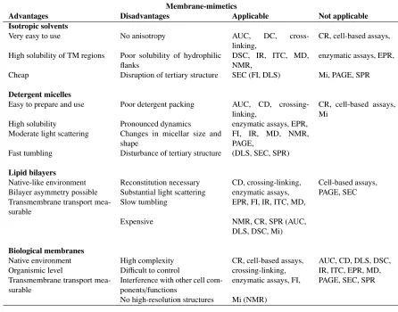
interests are presented in Table 1.1.
1.2.1.1 Isotropic solvents
Some popular isotropic solvents are chloroform, trifluoroethanol (TFE), dimethyl sulfoxide (DMSO),
methanol, ethanol, 1,1,1,3,3,3-hexafluoro-2-propanol (HFIP), and other hydrogenated or
halo-genated alcohols. They are the simplest and crudest hydrophobic membrane mimetic with a very
poor resemblance to biological membranes due to the lack of chemical and structural heterogeneity,
in addition to an absence in anisotropic characteristics. Unlike the more sophisticated membrane
mimetics, such as lipid bilayer vesicles, isotropic solvents have a high solubility for TM peptides
and they are applicable over a broad range of experimental techniques (as listed in Table 1.1).
1.2.1.2 Detergent micelles
With head group regions typically bulkier than their hydrocarbon chains, self-association into
micelle structures shields the hydrophobic chains from the solvent environment by orientating
detergent chains together whilst forming a spherical macroscopic structure. As a membrane mimetic,
they are easy to prepare and use. With a moderate light scattering and fast tumbling, detergent
micelles are ideal for CD and NMR, respectively. By the standard of today’s high performance
computers, micelles are small enough to be studied along with solvent and protein using an all-atom
model (19).
Detergent micelles remain highly soluble for membrane proteins reconstitution. They are also
known to adopt a cross-sectional topology similar to a lipid bilayer. Unfortunately, poor detergent
packing, significant exchange between lipid and solvent, and disruption to protein tertiary structure,
structures and natural bilayers (16).
1.2.1.3 Lipid bilayers
Model bilayer systems, whether as a bilayer slab or as part of a lipid vesicle (often referred to
as a liposome) introduce the anisotropy and structural characteristics similar to those of natural
bilayers. Early planar membrane model systems were composed of a monolayer at the air-water
interface (20). By evaporating organic solvent, lipids are left to orientate their polar heads into the
water leaving the hydrophilic chains orientated into the air. The precise surface pressure, thickness
and phase can be controlled, although high surface tension of water can denature proteins.
The more familiar unilamellar lipid bilayers are constructed from two opposite facing lipid leaflets,
whilst multilamellar lipid bilayers are constructed from stacked bilayer sheets, separated by a
polar solvent. Although the curvature of a vesicle is far less pronounced than a detergent micelle,
more lipids pack in the outer membrane of a vesicle to equalise surface tension between the two
leaflets (21). Mixed lipid bilayers can represent a closer abstraction to natural bilayers and enables
the calculation of changes to lipid aggregation and TM peptide packing by varying the head group
charge, the bilayer thickness, and lateral pressure profiles (mediated through a change in area per
lipid). Despite their obvious advantages and the relative ease in their construction, reconstituting
membrane proteins into phospholipid vesicles requires a lengthy process of dialysing or gel filtering
the detergent so as to solubilise the membrane proteins. In addition, there is little control over the
orientation of the membrane protein across the bilayer (22). Due to the size of the vesicle (the
diameter is typically>50 nm), the system is too large as an all-atom model for even the most sophisticated computing platform.
1.2.1.4 Biological membranes
Biological membranes are complex and often exhibit unpredictable behaviour. They are crowded
with membrane proteins, glycoproteins, and steroids, all of which can interfere with membrane
protein function and structure by contributing to a life-time of non-specific interactions, thus making
free energy calculation of association between membrane proteins of interest an approximation
at best. The variation in lipid composition can also contribute to a change in membrane protein
interactions, such as ToxR (23, 24), TOXCAT (25), GALLEX (26), and POSSYYCAT (27), are
not always reproducible and are prone to experimental fluctuations. Further more, it is possible for
a very small change in the environment, for example: heat shock; starvation; and resistance to or
absence of antibiotics, can to lead to unpredicted cellular responses.
Membrane-mimetics
Advantages Disadvantages Applicable Not applicable
Isotropic solvents
Very easy to use No anisotropy AUC, DC, cross-linking,
CR, cell-based assays,
High solubility of TM regions Poor solubility of hydrophilic flanks
DSC, IR, ITC, MD, NMR,
enzymatic assays, EPR,
Cheap Disruption of tertiary structure SEC (FI, DLS) Mi, PAGE, SPR
Detergent micelles
Easy to prepare and use Poor detergent packing AUC, CD, crossing-linking,
CR, cell-based assays, Mi
High solubility Pronounced dynamics enzymatic assays, EPR, Moderate light scattering Changes in micellar size and
shape
FI, IR, MD, NMR, PAGE,
Fast tumbling Disturbance of tertiary structure (DLS, SEC, SPR)
Lipid bilayers
Native-like environment Reconstitution necessary CD, crossing-linking, Cell-based assays, Bilayer asymmetry possible Substantial light scattering enzymatic assays, PAGE, SEC Transmembrane transport
mea-surable
Slow tumbling EPR, FI, IR, ITC, MD,
Expensive NMR, CR, SPR (AUC, DLS, DSC, Mi)
Biological membranes
Native environment High complexity CR, cell-based assays, AUC, CD, DLS, DSC, Organismic level Difficult to control crossing-linking, IR, ITC, EPR, MD, Transmembrane transport
mea-surable
Interference with other cell com-ponents/functions
enzymatic assays, FI, PAGE, SEC, SPR
[image:29.596.68.514.202.552.2]No high-resolution structures Mi (NMR)
Table 1.1: Recreated from a review on membrane-mimetics by Bordag and Keller (17). CR, conductance recordings; DLS, dynamic light calorimetry; EPR, electron paramagnetic resonance; FI, fluorescence-based methods; Mi, microscopy; SPR, surface plasmon resonance; CD, circular dichroism; MD, molecular dynamics; IR, infra-red-based methods; AUC, analytical ultracentrifuge; ITC, isothermal titration calorimetry; NMR, nuclear magnetic resonance; PAGE, polyacrylimide gel electropharisis methods
1.2.2 TM interaction motifs
TM domain interaction motifs are patterns of amino acids, key to mediating the strong association
of TM domains. Alteration to any part of the motif results in significant disruption to TM domain
association. With an identifiable pattern, motifs have been used as determinants of protein folding to
help predict TM domain interactions and structure. Motifs identified in TM proteins fall into one of
driven by the promiscuous positioning of polar side chains in the lipid chain environment. We look
at each motif in detail below.
1.2.2.1 Small-xxx-small TM interaction motifs
Association of TM helices within a membrane bilayer can be stabilized by interactions between
specific amino acid motifs. Statistical analysis of amino acid patterns of a Swiss-Prot database of
helical TM proteins revealed an over representation of small residues (alanine, glycine and serine)
atiandi+4in association with larger aliphatic residues (28). An interaction motif of this design is
commonly referred to as small-xxx-small. A survey of known membrane protein sequences has
shown that the GG4 motif is over represented in the TM domains of membrane proteins (29, 30).
Based on mutagenesis of the glycophorin A (GpA) TM helix, Lemmonet al.,(31) proposed that
the sequence75L76Ixx79G80Vxx83G84Vxx87T helped establish self-association (see Figure 1.3 (A)). The arrangement of two glycines three residues apart (79Gxxx83G) results in an indentation along the same helical face. A conformational search in a low dielectric environment (32), confirmed
by NMR structural analysis (30), concluded that GpA adopted a right handed crossing angle of
-40◦with closest point of helix-crossing at the79G and83G residues. The tight packing between
interhelical glycines maximise van der Waals interactions, and a high crossing angle ensures
additional enthalpic contributions between bulkierβ-branched amino acids. By systematically
adjusting the number of intermediate residues, three intermediate residues between glycines were
confirmed as the optimum (24). Further analysis determined that the strong helix-helix dimerisation
could be disrupted after substituting a glycine for an isoleucine. The bulkier hydrophobic side chain
disrupts the tight packing arrangement, dramatically destabilising TM domains interactions. This
was confirmed on low complexity scaffolds of polyvaline and polymethionine TM proteins using the TOXR assay (24). Since these early studies, GpA and the G83I mutant have become common
standards for a number ofin vivoandin vitroTM protein association assays (26, 33).
Besides GpA, the GG4 motif has been identified in the TM domain of several other integral
mem-brane proteins. To name a few: the phage M12 coat protein (34); the heparan sulfate proteoglycan
N-syndecan (35); theβ2-adrenergic receptor (36); the amyloid precursor proteinβ-carboxy-terminal
fragment (8); and Japanese encephalitis virus prM protein adopting a heterodimer with E
L76 I75 G79 V80 G83 T87
HIS SER SER
GLN
[image:31.596.93.509.95.299.2]A B C
Figure 1.3: (A) The established minimum residues required to stabilise GpA TM domain. (B) Tar-1 (1VLT) TM domain showing polar residues serine and glutamine packing at the helix-helix interface. (C) BNIP3 (2KA1), small residues
176A,180G and184G provide a close packing of helices which results in a hydrogen bond between serine and histidine.
class B, type I (38); ErbB2 (39); EphA1 (40); BNIP3 (41, 42) (Figure 1.3 (C)), and in the
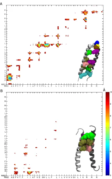
het-erodimers ofαIIb/β3 (43, 44) and ErbB1/ErbB3 (45). Although, neither isoleucine or valine are considered to have a short side chain, the motifs I659xxxV663and A661xxxG665, have been found in
rat Neu receptor tyrosine kinase TM domain (46).
Although the GG4 motif has been shown to play a significant role in TM helix dimerisation, there
are claims that it is in fact the neighbouring aliphatic side chains, which stabilise TM domain
association (47, 48), suggesting that the sequence modulates the strength of dimerisation. This
was emphasised by Unterreitmeieret al(49), who demonstrated that dimerisation of a sequences
containing a GG4 motif could be affected by a phenylalanine, yielding the sequence FxxGxxxG. An extension to the works of sequence context modulation of GG4 demonstrated histidine flanked
with either glycine or serine stabilised GG4 mediated dimersiation (50). However, a minimised
GpA motif (GVxxGVxxT) on a polyleucine peptide in detergent micelles yielded oligomerisation
without the assistance of neighbouring side chains (51), making it apparent that the membrane
mimetic used to solubilise the TM peptide also has a profound influence on oligomerisation.
1.2.2.2 Heptad repeat transmembrane interaction motifs
The heptad repeat motif, composed of a seven residue repeatabcdefg, is prevalent amongst integral
ensures contact between helices every 3.5 amino acids resulting in the seven amino acid repeat.
On rare exceptions, a heptad repeat containing a GG4 motif can form a right handed helix-helix
crossing angle (54), which is often a characteristic of a small-xxx-small mediated TM domain
dimerisation.
The tight packing due to reduced residue turns in a left-handed helix-helix dimer results in a smaller
positive crossing angle, giving the appearance of a ‘coiled coil’ or ’supercoiling’. The heptad
pattern repeats two to three times, with a ‘knobs-into-holes’ arrangement of side chains stabilising
oligomerisation (54). The side chains of one helix protrude into cavities formed by the side chains
of the opposite helix typically using sitesaanddas primary interaction sites (Figure 1.4 (A)). Side
chains at siteseandgcan be charged, resulting ininterhelical salt-bridges. Finally, sitesb,candf
are considered as sites interacting directly with the lipid environment. In the case of a TM trimer the
same interaction sites contribute to oligomeric stability, however interactions are shared between
three helical faces (Figure 1.4 (B)). A
B
C
[image:32.596.134.444.378.721.2]D
Leucine zipper motifs
Swiss-prot ID TM domain sequence
CAD1_XENLA 705ILGGILALLLLLLLLL
CAD3_HUMAN 659VLGAVLALLFLLLVLL
CADB_CHICK 559VLAVLGAVLALLLVLL
CADF_HUMAN 611LASALLLLVLVLLVAL CD72_MOUSE 93LQNFLLGLLLSCLMLG ENV_FRSFB 339LLIILLLLLILLLWTL
EPOR_MOUSE 256LILVLISLLLTVLALL
GPBB_HUMAN 155LALLGLGLLHALLLVL
HEMA_CDVO 38LLFVLLILLVGILALL
LECH_CHICK 27AVYVLLALSFLLLTLL
PVR_MOUSE 35LLVLLLAGGFLALILL
SRPB_MOUSE 35LLSVAVALLAVLLTLV TNRC_MOUSE 223LLAILLSLVLFLLFTT VE5A_BPV1 14AAMQLLLLLFLLLFFL
[image:33.596.171.400.106.345.2]VGLX_HSVBS 395LAIALLVLLFSLVIVL
Table 1.2: Leucine zipper-like heptad repeats found in native TM domains. Reproduced from Gurezka (60).
Early studies in TM domain oligomerisation, found heptad repeatinterhelical ‘knobs-into-holes’
packing in photosynthetic reaction centres (57) (Figure 1.4 (C) and (D)), bacteriorhodopsin (58),
and cytochrome C oxidase (59). Further more, a recent statistical analysis on sequences from a
Swiss-Prot database of TM proteins identified fifteen TM domains (see Table 1.2) which conformed
to a sequence akin to a soluble leucine-zipper (60) (a soluble coiled-coil protein stabilised from the
packing arrangement of stacked leucine side chains).
1.2.2.3 Polar transmembrane interaction motifs
The central region of the helical peptide adjacent to the lipid hydrocarbon region is occupied
predominately by the nonpolar residues alanine, leucine, valine, isoleucine or phenylalanine. Polar
or charged residues are usually found at the flanking region of the TM domain, the lipid-to-water
interface, where residues such as arginine, aspartic acid, asparagine, glutamic acid, glutamine and
lysine, have the potential to form O–H··O or N–H··O hydrogen bonds with lipid head groups regions
of the bilayer or O-H· · ·O hydrogen bonding with water molecules (61). The behaviour described as
the interaction between positively charged side chains and the negatively charged phosphate group
of lipids is known as snorkelling (62). Water molecules are able to permeate the hydrophobic region
of the bilayer by forming a network of hydrogen bonds from the charged side chain through to the
which moderately contribute to helix-helix stability (7).
There is a significant energetic cost of inserting polar groups into the bilayer due to the unfavourable
dipolar mismatch between nonpolar hydrocarbon chains and polar side chains (and polar backbones
for a non-helical structure). Peptide and protein hydrophobicity scales (63, 64, 65, 66) estimate
an energetic cost of 0.3 – 1.0 kcal/mol for the insertion of polar groups into the bilayer, and 1 – 6 kcal/mol for the insertion of peptide bonds or charged side chains. This penalty, however, has been attributed to some interesting mechanisms of TM protein association mediated through polar
residue interactions.
One of the early works on polar residue mediated self association demonstrated the formation
of a TM trimer upon the substitution of valine to asparagine on a low complexity sequence
of a hydrophobic heptad repeat (67). Mutation of asparagine to valine essentially eliminates
oligomerisation. This would suggest that a single asparagine residue buried at the centre of a
very non-polar heptad repeat could provide the thermodynamic drive into membrane protein
self-association. Interestingly, the heptad repeat sequence was derived from the soluble GCN4-P1 two
stranded coiled-coil peptide, part of the yeast transcription factor GCN4 (68). This demonstrates
the viability of retaining the buried hydrophobic residues of a soluble heptad repeat sequence for
membrane protein designs. Gratkowskiet al.,(69) extended this study by substituting the asparagine
for glutamic acid, glutamine, lysine, serine and threonine. They concluded that glutamine, glutamic
acid and histidine were capable of forming trimers due to a hydrogen bond network between polar
side chains after protonation of side chain carboxyl groups. Serine and threonine were thought not
to promote helix-helix association as the short side chain length would favour a hydrogen bond
from the side chain hydroxyl group to the backbone carbonyl oxygen of the residue one helical
turn directly above. Finally, it was concluded that lysine did not form helical bundles as it only has
one polar atom (unlike the two polar groups on the side chain of glutamic acid and asparagine);
therefore it had fewer sites to forminterhelical hydrogen bonds.
A similar study by Zhouet al.,(70) demonstrated that polyleucine self-association could be driven
by the inclusion of a singe glutamic acid, glutamine, histidine, aspartic acid and asparagine, yet
threonine, and serine do not associate more than the polyleucine sequences. Interestingly, the serine
motifs SxxSSxxT and SxxxSSxxT were later found to stabilise TM oligomerisation (71). It is likely
than in the case of a single serine residue, and thus encouraginginterhelical hydrogen bonding. A
further example of the role played by polar residues is the glutamic acid involved in TM protein
oligomerisation of the oncogenic rat Neu and human ErbB-2, which has inspired a significant
number of studies (72, 73, 74, 46), and still remains in contention to this day. A further review of
this TM protein is given in chapter 4.
As it turns out, placing a polar residue into a TM sequence is not always enough to promote
oligomerisation. The propensity for helix-helix association is complicated further by the specific
location of the polar residue with respect to the hydrophobic core of the bilayer and how sequence
context can modulate polar side chaininterhelical association. Learet al.,(75) performed the first
study on the position of asparagine on a heptad repeat template in detergent micelles to elicit free
energy differences using analytical ultracentrifugation. They concluded that an asparagine side chain within the hydrophobic core of the transmembrane dimer significantly stabilised self-association
compared to when placed near the lipid-water interface. This would suggest the ability to shield the
polarity of asparagine by packing beside the lipid-water interface (head group region). Further more,
usingin vitrotranslation of a model protein in the presence of dog pancreas rough microsomes,
Hessa et al., (66) determined such position-specific free energy contributions from all twenty
amino acids. Strongly polar side chains were found to contribute approximately 1.8 kcal mol−1,
+1.8 kcal mol−1, and+1.6 kcal mol−1, per aspartic acid, lysine and asparagine residue, respectively. Statistical analysis over a data set of 170 integral membrane proteins (76), revealed that charged
amino acids predominately favoured the lipid-water interface, a consequence of the energetic penalty
of inserting charged amino acids into membrane bilayers. Further more, the distribution of charged
amino acids was asymmetric, occurring more frequently on the cytoplasmic side of the TM helix.
This asymmetric distribution of charged residues maybe due to the enrichment of sphingolipids
and sterols in the extra-cytoplasmic leaflet (77). As far as the neighbouring sequence is concerned,
Dawson et al.,(78), found that the effectiveness in self-association from interhelical hydrogen bonding between strongly polar residues was greatly influenced by the sequence of a native TM
domain (notably, variants of bacteriophase M13 major coat protein, activated T cell receptor TM
domains, and B cell antigen receptor domains), and Herrmannet al.,(79) concluded that glutamic
acid, asparagine, lysine and arganine were significantly over represented in the presence of GG4
association alone, requiring the additionalinterhelical interactions of neighbouring polar residues,
a further note of contention with regards to the precise dimerisation contribution a GG4 motif can
make by itself.
1.2.3 Protein-lipid mediated oligomerisation
When considering the TM protein folding problem in a membrane environment, there are two
things which should be considered: (i) what affect does the membrane-mimetic have on the intrinsic fold of individual membrane helices; and (ii) how does the membrane-mimetic composition affect helical TM domain aggregation. It is thought that as many as one in four TM domains lack the
predicted hydrophobicity to allow spontaneous insertion into the bilayer (66). This suggests that
the membrane protein folding problem is not simply a case of understanding TM helix-helix
interactions, and that the lipid environment is thought to play a role in TM protein stability (80).
Therefore, the choice in the membrane mimetic must be carefully considered whilst analysing the
dynamic and structure of TM proteins. A recent study by Tulumello and Deber (80) suggested that a
number of detergent micelles were capable of maintaining the helical composition of a TM segment
similar to the helical structure of the intact protein in a native lipid environment. Interestingly,
investigation of GpA in model membranes concluded that strong dimerisation of GpA duringin
vitrostudies may be an artifact of homogeneous model bilayers, given that natural membrane
environments promote destabilising contributions from electrostatic interactions between charged
lipids and charged side chains and nonspecific competition from other membrane proteins (81).
Generally speaking, a model bilayer is typically composed of one to three lipid types and has a
large lipid to protein ratio. On the other hand, almost over half the total mass of natural membrane
systems is contributed to membrane proteins (82). One would expect model membranes to promote
a very different equilibrium for TM domain association.
It is well known that the chemical composition of the bilayer can lead to a condition of hydrophobic
mismatch between TM helix length and bilayer thickness which, in turn, can induce conformational
changes in a protein (83). The idea that the lipid environment may control the dimerisation mode
of TM proteins is not new (84). Unrestrained united atom MD simulations and structural analysis
using solid state NMR confirmed that a change from long chain lipids to short chain lipids increased
however, alter the TM domain secondary structure (86), and cases of hydrophobic mismatch have
been shown to influence association of helical TM domains (87, 88, 89). Alternatively, there are
many examples where the presence of protein has induced changes to the bilayer structure (see
Figure 6.1 and Figure 6.2). For example, TM proteins can adjust to mismatch by stretching or
disordering nearby lipids (90, 91, 83). Also, short helices can cause negative membrane curvature
to the bilayer (92, 93, 94), and there is the suggestion that lipids are arranged according to the
hydrophobic length of the TM domain (95). Polyanskyet al.,demonstrated using computational free
energy calculations, that the interactions between individual TM helices with different membrane-mimetics contributed to either an energy loss or energy gain which would consequently affect TM helical self-association in a family of epidermal growth factor receptors (96). Given the diverse and
complex nature of the lipid bilayer, elucidating relationships between lipid bilayer compositions
with TM protein oligomerisation is difficult and far from complete.
1.3
Low complexity sequence sca
ff
olds
A TM low complexity scaffold is composed from the bare essential amino acids required to assemble anα-helix and to satisfy the required hydrophobicity for spontaneous insertion. These two
requirements are typically fulfilled by using either a polyleucine sequence, a heptad repeat sequence
(composed from two different non-polar amino acids) or some other arrangement of two different non-polar amino acids. What follows is an account in the literature of how low complexity scaffolds have been used to investigate low complexity sequences containing a small-xxx-small motif, a polar
residue, or a duet of polar residues and small-xxx-small motifs. Unlike ‘top-down’ investigations
carried out on native sequences, the available studies are limited. A significant number of low
complexity sequences were constructed for single-site mutagenesis studies derived from the results
of combinatorial plasmid libraries, or iterative reconstruction of a native protein based on heuristics
from structural analysis. The following low complexity sequence review is separated according
to the adjustment made to the low complexity scaffold, notably, GG4 or a small-xxx-small motif, polar or ionic residues, aromatic residues, and all other studies. We are particularly interested in the
arrangement of the low complexity sequence, the experimental procedure performed (in addition to
1.3.1 GxxxG motif low complexity sequences
A polyvaline and polymethionine low complexity scaffold was modified through a process of iterative site-directed mutagenesis to identify the amino acid motif mediating glycophorin A
homod-imerisation (24) (from M13). Neither polymethionine nor polyvaline basic sequences demonstrated
significant dimerisation from ToxR analysis. A single substitution of methionine/valine for glycine had a negligible affect on TM domain association until a second glycine residue was positioned, three residues from the first resulting in a significant increase in homodimerisation. Isoleucine,
leucine, and tyrosine residues were added at the respective position found at the GpA TM
do-main:75L76Ixx79G80Vxx83G84Vxx87T. These additional adjustments to the basic sequence failed to encourage further increase in self-association. These results suggest that in the context of
a polymethionine and polyvaline low complexity scaffold, a GG4 motif stabilises TM domain oligomerisation. Further to this work, a minimised GpA dimerisation motif was systematically
grafted onto a polyleucine low complexity scaffold (51) (from GG). Given the large increase in propensity for dimerisation after the GG4 motif was added and prior to the additional GpA residues,
polyleucine proves to form a stable scaffold to form strong TM domain oligomers. In addition, polyleucine scaffolds were used to investigate the effects of helix-helix association whilst adjusting the length of the hydrophobic helix by adding or removing flanking leucine residues. The ideal TM
domain length according to three different PAGE analysis techniques is 18 to 24 leucine residues. Both studies (24, 51) were performedin vivo. The propensity for specific oligomeric states (dimer
and trimer) was not reported.
The low complexity scaffolds polyleucine and polyalanine (97), were randomised atinterhelical packing sites using a library of plasmid constructs (Randomised A16/A17 library). Sequences with
a high-affinity for dimerisation were isolated using thein vivoTOXCAT system (25). Over 80% of the identified systems contained a GG4 motif. As seen earlier by Herrmannet al.,(79), GG4
isolates were accompanied by flanking residues, notably valine, leucine, isoleucine and threonine
occurring patterns would suggest that these positions and amino acid types are indicative of a
favourable packing motif. Either [large] and [small] residues beside each glycine residue is not
surprising assuming that in either case the side chain would not pack directly at the helix-helix
interface, therefore, the size of the size chain would not affect association.
Using thein vivoPOSSYCATT (27) assay to screen a plasmid library of TM protein sequences for
high helix-helix affinity sequences (49), the over-represented FxxGxxxG sequence was identified and analysed further by grafting the sequence onto a polymethionine low complexity scaffold (from L16). The results from adjusting the position of the phenylalanine yielded a high propensity
for oligomerisation providing the FxxGxxxG motif was preserved. The proximity to the
helix-helix closest point of approach, in addition to the position of the phenylalanine on the helix-helix may
suggest that a GG4 mediated packing motif can be stabilised further by potential π aromatic
interactions. The FxxGxxxG motif was grafted onto two further low complexity scaffolds, AZ2 and polyleucine. Although, both sequences demonstrate a poor propensity for oligomerisation,
after systematically replacing three native residues with an FxxGxxxG motif the AZ2 construct
demonstrated significantly higher levels of oligomerisation when grafted onto the polyleucine low
complexity scaffold. This would suggest that the mode of oligomerisaiton of this motif was highly sequence dependent.
GxxxG and small-xxx-small motifs
Name* Sequence Reference
*Naming convention from literature
M13 MMMMMMMMMMMMM Brosig and Langosch (24).
M12.1 MMMMGMMMMMMMM
M12.2 MMMMMMMMGMMMM
M11 MMMMGMMMGMMMM
M9 MIMMGMMMGMMMT
M8 LIMMGMMMGMMMT
V13 VVVVVVVVVVVVV
V11 VVVVGVVVGVVVV
V9 VIVVGVVVGVVVT
V8.1 LIVVGVVVGVVVT
V7 AAVVGVVVGVVAA
GG LLLLLLGLLLGLLLLLLL Orzáezet al(51).
GVGV LLLLLLGVLLGVLLLLLL
IGVGV LLLILLGVLLGVLLLLLL
GGT LLLLLLGLLLGLLLTLLL
GVGT LLLLLLGVLLGLLLTLLL
GGVT LLLLLLGLLLGVLLTLLL
LLGVT LLLLLLLLLLGVLLTLLL
GVGVT LLLLLLGVLLGVLLTLLL
IGVGVT LLLILLGVLLGVLLTLLL
GGS LLLLLLGLLLGLLLSLLL
GGN LLLLLLGLLLGLLLNLLL
GVGVS LLLLLLGVLLGVLLSLLL
GVGVN LLLLLLGVLLGVLLNLLL
Randomised A16 library ASxxAAxxAAxxAAxAILI Russ and Engelman (97).
Randomised L16 library LSxxLLxxLLxxLLxLILI
L16 LLLLLLLLLLLLLLLL Unterreitmeieret al(49).
L16-L5F/L8G/L12G LLLLFLLGLLLGLLLL
AZ2 LLAALLALLAALLALL
AZ2-L8G/L12G LLAALLAGLAAGLALL
AZ2-L5F/L8G LLAAFLAGLAALLALL
AZ2-L5F/L8G/L12A LLAAFLAGLAAALALL
AZ2-L5F/L8G/L12G LLAAFLAGLAAGLALL
M11 MMMMGMMMGMMMM
MM-M1F FMMMGMMMGMMMM
MM-M2F MFMMGMMMGMMMM
MM-M3F MMFMGMMMGMMMM
MM-M4F MMMFGMMMGMMMM
MM-M5F MMMMFMMMGMMMM
MM-M6F MMMMGFMMGMMMM
MM-M7F MMMMGMFMGMMMM
MM-M8F MMMMGMMFGMMMM
MM-M9F MMMMGMMMFMMMM
MM-M11F MMMMGMMMGMFMM
MM-M12F MMMMGMMMGMMFM
MM-M13F MMMMGMMMGMMMF
MM-M2W MWMMGMMMGMMMM
MM-M2Y MYMMGMMMGMMMM
1.3.2 Polar and/or ionic side chain motif low complexity sequences
A systematic asparagine-scanning mutagenesis of a polyleucine low complexity scaffold was performed and self-association was measured using the ToxR chimeric protein system (98) (from
Leu20). The strongest impact on TM domain self-association was seen when asparagine was
located around the centre of the TM domain sequence, at7L,11L,14L, and18L. These residues map to sitesdand a on a heptad repeat sequence, placing them at the helix-helix interface of a
leucine zipper. In addition, site-mutagenesis on heptad positionsgandeat the centre of the TM domain, were seen to also stabilise oligomerisation suggesting that flanking asparagine side chains
can stabilise a polyleucine low complexity TM domain. Oligomerisation was confirmed using
SDS-PAGE analysis. The polyleucine control was shown to be monomeric, along with N-terminal
asparagine site sequences L1N to L6N, and C-terminal asparagine site sequences L16 to L20. All
other sequences were dimeric.
Position-specific histidine residues, flanked with glycine, serine and/or threonine, in addition to a C-terminal GG4 motif were selected using the TOXCAT assay from a combinatorial library of
plasmid sequences on a polyleucine scaffold (50) (randomised L16 library). Mutational analysis on selected sequences confirmed that histidine side chains would form a network ofinterhelical
hydrogen bonds, mediated via the tight packing of the GG4 motif.
Interhelical ionic interactions between oppositely charged side chains were shown to stabilise TM
domain association depending on sequence context (79) from ToxR and site-directed mutagenesis
analysis (from L16). In the presence of a C-terminal GG4 motif, a single charged aspartic acid
moderately encouraged self-association with respect to a polyleucine sequence and a C-terminal
association is substantially increased. Finally, adding two additional residues, cysteine and tyrosine,
or serine and histidine, increases association further from hydrogen bonding or an ionic-πinteraction
in the case of tyrosine interacting with arganine.
A low complexity scaffold of mixed leucine and alanine (from 9TM+Leu) was used to elicit the minimal dimerisation motif of the bacterial Tar-1 homodimer TM domain (99) (the arrangement
of polar residues in Tar-1 can be seen in Figure 1.3 (B)). The polar residues, glutamine and serine
when placed two residues apart, stabilised self-association. Interestingly, the QxxS motif grafted
onto a polyalanine low complexity scaffold gave no indication of oligomerisation. As it would happen, although SDS-PAGE suggested that the polyalanine chimera proteins were being expressed,
the absence of growth on a maltose minimal media plate suggested that polyalanine would not
spontaneously insert into the bilayer. This may be due to the borderline hydrophilic nature of
alanine in addition to the energetic penalty attributed to the cost of inserting serine and glutamine
into a hydrophobic lipid environment.
A low complexity sequence of polyleucine residues (pVNVV and pVVVV) with an interfacial
sequence of valine-asparagine-valine-valine was used to record free energy values before and after an
asparagine to valine substitution (100). Computational potential of means force (PMF) calculations
enables the decomposition of the free energy from helix-helix and helix-lipid interactions. The
low complexity sequence provided an efficient platform to report significant per-reside energetic contributions, as is the case of a centrally located asparagine residue which suggests favourable
patterns ofinterhelical hydrogen bonding between asparagine on a polyleucine scaffold.
Finally, inspired by a polyleucine heptad repeat with interfacial residues asparagine and valine,
Zhouet al.,(70) positioned a single polar residue in interfacial sites (aandd) on a polyleucine low
complexity sequence (from L23). Polar residues serine, threonine and tyrosine failed to encourage self association compared to the low complexity polyleucine control, possibly due to the propensity
for these residues to form hydrogen bonds with the backbone oxygen. Whereas, asparagine,
aspartic acid, glutamine, glutamic acid and histidine, residues were capable of being simultaneously
hydrogen bond donors and acceptors.
Although, far from being a combinatorial analysis over all naturally occurring amino acids on a