Characteristics of Colorectal Polyps and Cancer;
a Retrospective Review of Colonoscopy Data in Iran
ABSTRACT
BACKGROUND
Early diagnosis and endoscopic resection of adenomatous polyps is the main approach for screening and prevention of colorectal cancer (CRC). We aimed to assess polyp detection rate (PDR) and to characterize demographic, clinical, and pathological features of colorectal polyps in an Iranian population. METHODS
We retrospectively analyzed the data from 5427 colonoscopies performed during 2007-2012 at Masoud Clinic, the main endoscopy center associated with Sasan Alborz Biomedical Research Center, in Tehran, Iran.
RESULTS
Our sample included 2928 (54%) women and 2499 (46%) men, with the mean age of 48.3 years (SD=16.1). The most common reasons for colonoscopy included screening in 25.0%, and gastrointestinal bleeding in 15.2%. Cecal intubation was successful in 86% of patients. The quality of bowel preparation was fair to excellent in 78.1% (n=4235) of colonoscopies. Overall PDR was 42.0% (95% CI: 40.6-43.3). The PDR in men (51.1%, 95% CI: 49.1-53.1) was
significantly higher than women (34.2%, 95% CI: 32.4-35.9, p<0.001). Polyps were more frequently observed in patients after the 6th decade of life (F=3.2; p=0.004). CRC was detected in 2.9% (73/2499) of men and 1.9% (57/2928) of women (p=0.02). The mean age for patients with cancer was significant -ly higher than that for individuals with po-lyps, 60.9 (SD=13.4) year vs. 56.9 (SD=13.7) year, respectively (p=0.001). Almost 82.8% of the lesions were pre-cancerous with tubular type predominance (62.3%) followed by tubulo-villous
(10.3%), villous (6.6%), and serrated (3.6%). Hyperplastic/inflammatory polyps
comprised 17.2% of lesions. CONCLUSION
Distal colon was more prone to develop polyps and cancer than proximal
colon in our series. These findings provide a great infrastructure for next pre -ventive programs and have implications for colorectal cancer screening at population-level.
KEYWORDS
Colon Cancer; Colonoscopy; Colonic Polyps 1. Digestive Oncology Research Center,
Digestive Disease Research Institute, Shariati Hospital, Tehran University of Medical Sciences, Tehran, Iran. 2. Sasan Alborz Biomedical Research
Center, Masoud Gastroenterology and Hepatology Center, Tehran, Iran. 3. Division of Gastroenterology, Feinberg
School of Medicine, Robert H Lurie Comprehensive Cancer Center, North-western University, Chicago, IL 60611, United States.
4. Department of Anatomy, School of Medicine, Tehran University of Medical Sciences, Tehran, Iran.
Alireza Delavari1,2, Fatemeh Mardan1, Hamideh Salimzadeh1*, Faraz Bishehsari3, Pejman Khosravi1,2,
Maryam Khanehzad4, Siavosh Nasseri-Moghaddam1,2, Shahin Merat1,2, Reza Ansari1,2, Homayoon Vahedi1,2,
Bijan Shahbazkhani1,2, Mehdi Saberifiroozi1,2, Masoud Sotoudeh1,2, Reza Malekzadeh1,2
INTRODUCTION
Colorectal cancer (CRC) is the third most prevalent cancer in men * Corresponding Author:
Hamideh Salimzadeh, PhD
Digestive Oncology Research Center, Di-gestive Disease Research Institute, Shari-ati Hospital, Tehran University of Medi-cal Sciences, North Kargar Avenue 14666 Tehran, Iran.
Tel: + 98 21 82415415 Fax:+ 98 21 82415400
Email: hsalimzadeh@sina.tums.ac.ir Received: 10 Apr. 2014
Accepted: 21 Jun. 2014
Please cite this paper as:
Delavari AR, Mardan F, Salimzadeh H, Bishehsari F, Khosravi P, Khanehzad M, Nasseri-Moghaddam S, Merat S, Ansari R, Vahedi H, Shahbazkhani B, Saberifiroozi M, Sotoudeh M, Malekzadeh R. Characteristics of Colorectal Polyps and Cancer; a Retrospective Re-view of Colonoscopy Data in Iran. Middle East J Dig Dis 2014;6:144-150.
and the second in women; accounting for 8% (n=608,700) of all cancer deaths worldwide.1 The
highest increase in the incidence of colon cancer are in the Eastern Europe and Asia.1,2 Recent cancer
statistics indicate a decreasing trend in CRC inci-dence in the Unites States because of the increase in timely detection and removal of precursor lesions through colonoscopy.3
Colorectal cancer is also the third most common cancer in Iranians excluding the skin cancers. It oc-curs at younger ages with an increasing trend simi-lar in the Asia-Pacific countries.1,4 These increasing
rates may result from the young age-structure and low rates of colon cancer in older people of these countries.2,5,6
Colon carcinomas mostly arise from adenoma-tous polyps and the time span for the transition pro-cess is estimated to nearly 10 years on average.7,8
Given the slow progression of colorectal adenomas into invasive adenocarcinoma,9 early detection and
endoscopic resection of these precancerous lesions, have been claimed to be effective in decreasing both the incidence and mortality rate of CRC.10-12
There is a report that colonic precancerous lesions (adenomas) with a high prevalence tend to present at younger ages, therefore undergoing screening among asymptomatic adults aged 50 years for ad-enomas and CRC is strongly recommended.13
There is scant knowledge about the prevalence of colorectal polyps and polyp detection rate (PDR) in Iranian adult population. To the best of our knowl-edge, only few studies are available in the national literature that assessed colorectal polyps,14-17 but
none has explicitly noted the rate of polyp detec-tion and most of them are biased because of their small sample size. Nevertheless, our study provides comprehensive information about clinical and epi-demiological features of colorectal polyps, using a relatively large sample of patients undergoing colo-noscopy.
The mass screening of colorectal cancer is not yet available in Iran, therefore updating the current knowledge in the scope of colorectal polyps and CRC is essential. Hence, identifying the features of colon polyps (e.g., age of onset, changes in
sub-sites distribution, location, and histology type) have great implications for developing national screen-ing guidelines for CRC.18 The aims of the current
study were to measure PDR, and to evaluate the clinical and histological characteristics of colorec-tal polyps in an Iranian population.
MATERIALS AND METHODS
Study design
We conducted a cross-sectional study and retro-spectively assessed the colonoscopy database and pathology reports maintained by Masoud Clinic, a well-known gastrointestinal endoscopy clinic in Tehran, Iran. The Institutional Review Board of Digestive Disease Research Institute, Tehran Uni-versity of Medical Sciences, approved the study protocol.
Patients, procedures and measures
We included all patients aged 15 to 90 years, who underwent their first time colonoscopy from June 2007 to March 2013. The patients with a personal history of colon cancer and polyposis were exclud-ed from the study. Twenty two gastroenterologists certified by the Iranian National Board of Gastroen -terology and Hepatology performed the procedures using two high-quality colonoscopes (OLYMPUS CV-240, and PENTAX EPK-1000) under conscious sedation.
We collected the data on patients’ demographic variables, indications for colonoscopy, quality of bowel preparation, and the rate of successful cecal insertion. For all colorectal lesions, data on clinical and pathological features (i.e., number, size, site, and grade of dysplasia) were obtained.
Pathological features of colorectal lesions were determined using the World Health Organization criteria19 as follows: hyperplastic, precancerous
(serrated, tubular, tubular-villous, and villous), and cancer. The overall polyp detection rate (PDR) was defined as the proportion of procedures in which at least one polyp was detected over the total number of colonoscopies.
the proportion of polyps detected by different co-lonic segments. Proximal colon included transverse colon, hepatic flexure, ascending colon and cecum. Distal colon included rectum, sigmoid, descending colon, and splenic flexure.
Statistical analysis
We reviewed the endoscopic data and pathology records. Patient-level data were used for the esti-mates of PDR, and summary-level data for present-ing pathology features and anatomic site of polyps. Histograms were developed to demonstrate polyp characteristics, i.e., size, counts, and proportion per patient. Categorical data were tested between sub-groups using the Chi-square test or the Fisher exact test, where appropriate. Continuous data were pre-sented as means (SD), and 95% confidence interval (CI). The Student t test was used for comparisons of means. For statistical significance we considered a p value of 0.05 applying 2-tailed statistical tests. All statistical analyses were performed using Stata/ MP software, version 11. Plots were created in R, version 2.15.1.
RESULTS
Demographics and colonoscopy findings
All patients (n=5427) aged 15 to 90 years who underwent their first time colonoscopy from June 2007 to March 2013 were included in this study. Our sample included 2928 (54%) women and 2499 (46%) men, with the mean age of 48.3 years (SD=16.1). The most common reasons for colonos-copy included screening (i.e., asymptomatic adults aged 50 years and older, and first degree relatives of patients with CRC) in 25.0% (n=1356), and gas-trointestinal bleeding in 15.2% (n=824). Other indi-cations for colonoscopy were classified as follows: 7.5% abdominal pain (n=405), 8.5% inflammatory bowel disease (n=462), 6.3% suspected irritable bowel syndrome (n=346). In 37.5% (n=2034) of the patients, indications for colonoscopy were not noted (table 1).
Cecal intubation was successful in 86% (n=4660) patients. The quality of bowel preparation was
ex-cellent to fair in 78.1% (n=4235) of colonosco-pies vs. 9.6% (n=522) with poor to unsatisfactory preparation. However, bowel cleansing was not mentioned for 12.3% (n=670) of examinations. Approximately, 42.0% (n=2277) of patients had at least one polyp, and cancer was detected in 2.4% (n=130) of patients (table 1).
Study outcomes
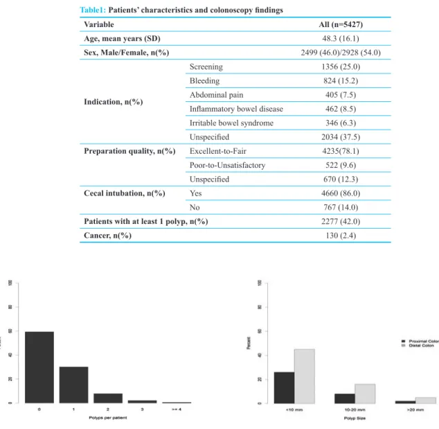
On the basis of colonoscopy reports, the overall PDR was 42.0% (95% CI: 40.6-43.3). Figure 1 de-picts the overall distribution of polyps per patient, where the high proportion of patients with 1 or 2 polyps detected, is visible (figure 1). Almost 56.1% (n=1277/2277) of patients, who had at least one polyp, were men. The PDR in men (51.1%, 95% CI: 49.1-53.1) was significantly higher than that in women (34.2%, 95% CI: 32.4-35.9, p<0.001). The mean age of patients with polyp was 56.9 (SD=13.7) years. Polyps were more frequently observed in pa-tients after the 6th decade of life (F=3.2, p=0.004, table 2).
Colorectal cancer was detected in 2.9% (73/2499) of men and 1.9% (57/2928) of women, suggesting a significantly higher prevalence among men com -pared with women (p=0.02). Age specific preva -lence of CRC is shown in table 2, presenting a peak in the prevalence of CRC after the seventh decade of life (table 2). The mean age of patients with can-cer was significantly higher than individuals with polyps, 60.9 (SD=13.4) year versus 56.9 (SD=13.7) year, respectively (p=0.001).
Characteristics of colonic lesions
A total of 3058 polyps were removed by colo-noscopy. Data about the size of polyps were avail-able for 1838 polyps; of these, 30% (n=549) were more than 10 mm. Additional information on size distribution are highlighted in figure 2, showing a higher proportion of polyps sized 10-20 mm and ≥ 20 mm in distal colon compared with the proximal colon.
Table 3 shows the distribution of cancer and pol-yps, in different colonic segments. Overall, polyps were frequently detected in sigmoid (26.8%),
rec-tum (19.0%), and transverse colon (15.5%). The same colonic distribution was observed for pol-yps’ ≥10 mm in size. Likewise, cancer was more frequently observed in sigmoid (40.0%), rectum (26.2%), and transverse colon (10.0%), (table 2). The prevalence of polyps in the distal colon was higher than that of the proximal colon (64.5% vs.
35.4%, respectively, p<0.001). Accordingly, most of the cancers were located in the distal colon com-pared with the proximal colon (72.5% vs. 26.1, re-spectively, p<0.001).
Colonic distribution of polyps by histology was presented in table 4. Analysis of summary-level data for pathology reports indicated that 82.8% of Table1: Patients’ characteristics and colonoscopy findings
Variable All (n=5427)
Age, mean years (SD) 48.3 (16.1)
Sex, Male/Female, n(%) 2499 (46.0)/2928 (54.0)
Indication, n(%)
Screening 1356 (25.0)
Bleeding 824 (15.2)
Abdominal pain 405 (7.5) Inflammatory bowel disease 462 (8.5) Irritable bowel syndrome 346 (6.3)
Unspecified 2034 (37.5)
Preparation quality, n(%) Excellent-to-Fair 4235(78.1) Poor-to-Unsatisfactory 522 (9.6)
Unspecified 670 (12.3)
Cecal intubation, n(%) Yes 4660 (86.0)
No 767 (14.0)
Patients with at least 1 polyp, n(%) 2277 (42.0)
Cancer, n(%) 130 (2.4)
Fig.1: Overall proportion of colon polyps per patient Fig.2: Size distribution of polyps per colonic segments
Table 2: Polyp detection rates and cancer prevalence by age-group (n=5427) <30 yrs.
(n=916) 30-39 yrs. (n=890) 40-49 yrs. (n=1006) 50-59 yrs. (n=1227) 60-69 yrs. (n=908) >=70 yrs. (n=480) Total(n=5427) Polyp, no (%) 82 (8.9) 179 (20.1) 322 (32.0) 633 (51.6) 648 (71.4) 413 (86.0) 2277 (42.0)
lesions were precancerous with tubular type pre-dominance (62.3%) followed by tubulo-villous (10.3%), villous (6.6%), and serrated (3.6%). Hy-perplastic/inflammatory polyps comprised 17.2% of lesions. Precancerous lesions (i.e., adenomas and serrated polyps) with higher proportion ap-peared in distal colon in comparison with the proxi-mal colon (48.2% vs. 33.6 %, respectively, table 4). High grade of dysplasia was reported among 19.5% (n=445) of resected polyps.
DISCUSSION
We have reported here the features of colorec-tal neoplasia from a referral gastroenterology clinic using a relatively large database of colonoscopy. The overall estimate for PDR in our patients was 42.0%, which would be correspondent to more than 30% rate of adenoma detection.
Older age is the most important predictor for the prevalence of adenomas, and cancer.20 In our study,
the PDR and cancer prevalence reached a peak in the 6th and 8th decades of life, respectively. These
data are consistent with findings reported by Ba -fandeh, Mirzaie, and their colleagues.14-16,21 Studies
from the Middle East and the western countries also mentioned significant increase for the risk of CRC, in particular after the age of 50 years.20,22 Our
pa-tients with cancer were significantly older, 4 years on average, than patients with polyps. This relative-ly small level of difference in mean age is sensible, even though a difference of 10 years that is com-patible with time span required for transformation of a polyp to carcinoma, were explicitly noted by other studies.15,16 Given the increased prevalence of
CRC in the sixth decade of life, the age threshold to start screening for individuals with average risk is 50 years.23,24
The risk of developing polyps and cancer in co-lon is greater in men than in women.25,26 Our study
showed significantly higher rates for both polyps and cancer among men compared with women, which is in line with current evidence that indicates male gender is an important risk factor for polyps and colon cancer.21,25-27 Moreover, other reports
from Iran support gender differences in the preva-lence of colon polyps and cancer.4,14,16,28
The tubular type was the most common histo-logical feature of adenomas in the present study, in accordance with the results of other reports.14,16,29
Distal colon was more prone to develop polyps and cancer than proximal colon in our series, compa-rable with results from the Asian and the Western countries.13,20,23 However, little evidence exists for
the right-ward shift of colonic polyps and can-Table 3: Distribution of polyps (count and size*) and cancer by colonic segments
Rectum Sigmoid Descending colon Splenic flexure Transverse colon Ascending colon Hepatic flexure Cecum Cancer
(n=130) 52(40.0) 34 (26.2) 8 (6.2) 4 (3.1) 13 (10.0) 10 (7.7) 2 (1.5) 7 (5.3)
Polyps*
(n=3023) 573(19.0) 811(26.8) 400 (13.2) 55 (1.8) 470 (15.5) 339 (11.2) (5.4)163 212(7.0)
Polyps**<10
mm (n=1289) 312 (17) 330(18.0) 154(8.4) 24(1.3) 203 (11.0) 119 (6.5) 56 (3) 91 (4.9) Polyps >=10
mm (n=549) 119 (6.5) 185(10.0) 65(3.6) 10(0.5) 63(3.5) 61(3.3) 22(1.2) 24(1.3)
*Location of 35 polyps was not specified; **Size of 1220 polyps was not available. Table 4: Colonic* distribution of polyp count by histologic type, number (%)
Hyperplastic/inflammatory Serrated Tubular Tubulo-villous Villous
Proximal colon (n=1184) 157 (13.3) 26 (2.2) 869 (73.4) 84 (7.1) 48 (4.0)
Distal colon (n=1839) 364 (19.8) 85 (4.6) 1012 (55.0) 228 (12.4) 150 (8.2)
cer17,18,29 across Iranian population. Such an
as-sumption was not further supported by the results of the current study and others.18,28,30 Nonetheless,
because of the significance of adenomas and neo -plasms present in proximal colon,18 complete
colo-noscopy is recommended in screening guidelines for colon cancer.23
Strengths of the current study included use of a relatively large sample of adult patients, and equal number of both genders. The major limitation of our study was the absence of automated interface between our pathology reports and endoscopic database, which prevented us from estimating the detection rate of adenoma, and addressing the pre-dictive factors for them. Finally, our sample in-cluded mostly symptomatic patients, in which the estimates may be different from screening studies with asymptomatic individuals.
In summary, data presented here may provide a good infrastructure for the next preventive pro-grams and have clinical implications for colorec-tal cancer screening through population-level programs. Screening-based studies, however, are required to probe the clinical and epidemiological aspects of colorectal polyps and cancer in Iran.
ACKNOWLEDGEMENTS
The authors thank Mohammad Masoud Male-kzadeh for data processing, and all gastroenterolo-gists and pathologastroenterolo-gists in Masoud Clinic for their contributions in performing colonoscopies, and re-viewing the pathology slides.
CONFLICT OF INTEREST
The authors declare no conflict of interest related to this work.
REFERENCES
1. Jemal A, Center MM, DeSantis C, Ward EM. Global pat-terns of cancer incidence and mortality rates and trends. Cancer Epidemiol Biomarkers Prev 2010;19:1893-907. 2. Yiu HY, Whittemore AS, Shibata A. Increasing colorectal
cancer incidence rates in Japan. Int J Cancer 2004;109: 777-81.
3. Edwards BK, Ward E, Kohler BA, Eheman C, Zauber AG,
Anderson RN, et al. Annual report to the nation on the sta-tus of cancer, 1975-2006, featuring colorectal cancer trends and impact of interventions (risk factors, screening, and treatment) to reduce future rates. Cancer 2010;116:544-73. 4. Somi MH, Mirinezhad K, Farhang S, Jazayeri E, Sani A,
Seif-Farshad M, et al. Gastrointestinal cancer occurrence in
East Azarbaijan: a five year study from North Western Iran. Asian Pac J Cancer Prev 2006;7:309-12.
5. Mousavi SM, Gouya MM, Ramazani R, Davanlou M, Hajsadeghi N, Seddighi Z. Cancer incidence and mortality in Iran. Ann Oncol 2009;20:556-63.
6. Ansari R, Mahdavinia M, Sadjadi A, Nouraie M, Kaman-gar F, Bishehsari F, et al. Incidence and age distribution of colorectal cancer in Iran: results of a population-based cancer registry. Cancer lett 2006;240:143-7.
7. Noffsinger AE. Serrated polyps and colorectal cancer: new pathway to malignancy. Ann Rev Pathol 2009;4:343-64. 8. Levine JS, Ahnen DJ. Adenomatous polyps of the colon. N
Engl J Med 2006;355:2551-7.
9. Huang CS, Farraye FA, Yang S, O’Brien MJ. The
clini-cal significance of serrated polyps. Am J Gastroenterol 2010;106:229-40.
10. Zauber AG, Winawer SJ, O’Brien MJ, Lansdorp-Vogelaar I, van Ballegooijen M, Hankey BF, et al. Colonoscopic pol-ypectomy and long-term prevention of colorectal-cancer deaths. N Engl J Med 2012;366:687-96.
11. Espey DK, Wu XC, Swan J, Wiggins C, Jim MA, Ward E, et al. Annual report to the nation on the status of can-cer, 1975–2004, featuring cancer in American Indians and Alaska Natives. Cancer 2007;110:2119-52.
12. Atkin WS, Edwards R, Kralj-Hans I, Wooldrage K, Hart
AR, Northover J, et al. Once-only flexible sigmoidoscopy
screening in prevention of colorectal cancer: a multicentre randomised controlled trial. Lancet 2010;375:1624-33. 13. Ferlitsch M, Reinhart K, Pramhas S, Wiener C, Gal O,
Bannert C, et al. Sex-specific prevalence of adenomas, ad -vanced adenomas, and colorectal cancer in individuals un-dergoing screening colonoscopy. JAMA 2011;306:1352-8. 14. Mirzaie AZ, Abolhasani M, Moghaddam RM, Kabivar M.
The Frequency of gastrointestinal polyps in Iranian popula-tion. Iranian Journal of Pathologhy 2012;7:183-9. 15. Bafandeh Y, Khoshbaten M, Sadat ATE, Farhang S.
Clini-cal predictors of colorectal polyps and carcinoma in a low prevalence region: results of a colonoscopy based study. World J Gastroenterol 2008;14:1534-8.
16. Bafandeh Y, Daghestani D, Esmaili H, Aharizad S. Distri-bution of cancer and adenomatous polyps in the colorec-tum: study in an Iranian population. Asian Pac J Cancer Prev 2006;7:65-8.
17. Khatibzadeh N, Ziaee S, Rahbar N, Molanie S, Arefian L,
Fanaie S. The indirect role of site distribution in high-grade dysplasia in adenomatous colorectal polyps. J Cancer Res Ther 2005;1:204-7.
18. Eshghi MJ, Fatemi R, Hashemy A, Aldulaimi D, Khoda-doostan M. A retrospective study of patients with colorectal polyps. Gastroenterol Hepatol Bed Bench 2010;4:17-22. 19. Morson BC, Sobin LH. Histological typing of intestinal
tu-mours: World Health Organization Geneva; 1976. 20. Heitman SJ, Ronksley PE, Hilsden RJ, Manns BJ,
Ro-stom A, Hemmelgarn BR. Prevalence of adenomas and colorectal cancer in average risk individuals: a systematic review and meta-analysis. Clin Gastroenterol Hepatol 2009;7:1272-8.
21. Bafandeh YD, Daghestani, Heidar E. Demographic and anatomical survey of colorectal polyps in an Iranian popu-lation. Asian Pac J Cancer Prev 2005;6:537-40.
22. Nam JH, Yang CH. Clinical characteristics and risk factors of colon polyps in Gyeongju and Pohang area. Korean J Gatroenterol 2008;52:142-9.
23. Sung JJ, Lau JY, Young GP, Sano Y, Chiu H, Byeon J, et
al. Asia Pacific consensus recommendations for colorectal
cancer screening. Gut 2008;57:1166-76.
24. Levin B, Lieberman DA, McFarland B, Smith RA, Brooks D, Andrews KS, et al. Screening and Surveillance for the Early Detection of Colorectal Cancer and Adenomatous Polyps, 2008: A Joint Guideline from the American Can-cer Society, the US Multi-Society Task Force on Colorectal
Cancer, and the American College of Radiology. CA Can-cer J Clin 2008;58:130-60.
25. Brenner H, Hoffmeister M, Arndt V, Haug U. Gender dif-ferences in colorectal cancer: implications for age at initia-tion of screening. Br J Cancer 2007;96:828-31.
26. Brenner H, Hoffmeister M, Stegmaier C, Brenner G, Alten-hofen L, Haug U. Risk of progression of advanced adeno-mas to colorectal cancer by age and sex: estimates based on 840 149 screening colonoscopies. Gut 2007;56:1585-9. 27. Nguyen SP, Bent S, Chen Y-H, Terdiman JP. Gender as a
risk factor for advanced neoplasia and colorectal cancer: a systematic review and meta-analysis. Clin Gastroenterol Hepatol 2009;7:676-81.
28. Omranipour R, Doroudian R, Mahmoodzadeh H. Anatomi-cal distribution of colorectal carcinoma in Iran: a retrospec-tive 15-yr study to evaluate rightward shift. Asian Pac J Cancer Prev 2012;13:279-82.
29. Khodadoostan M, Fatemi R, Maserat E. Clinical and patho-logical characteristics of colorectal polyps in Iranian popu-lation. East Afr J Public Health 2010;7:157-9.
30. Hosseini SV, Izadpanah A, Yarmohammadi H.
Epidemio-logical changes in colorectal cancer in Shiraz, Iran: 1980−