ARTIGO DE REVISÃO
Osteoporosis: From Bone Biology to
Individual Treatment Decision
Osteoporose: Da Biologia Óssea à Decisão Terapêutica Individual
1. Rheumatology Research Unit. Instituto de Medicina Molecular. Faculdade de Medicina de Lisboa. Lisboa. Portugal. 2. Serviço de Reumatologia. Hospital de Santa Maria. Centro Hospitalar Lisboa Norte. Lisboa. Portugal.
3. Unidade de Reumatologia. Hospital de Santo Espírito de Angra do Heroísmo, E.P.E.R. Angra do Heroísmo. Açores. Portugal. Recebido: 07 de Fevereiro de 2013 - Aceite: 06 de Julho de 2013 | Copyright © Ordem dos Médicos 2013
Maria João GONÇALVES1,2, Ana Maria RODRIGUES1,3, Helena CANHÃO1,2, João Eurico FONSECA1,2
Acta Med Port 2013 Jul-Aug;26(4):445-455
RESUMO
Introdução: A Osteoporose é uma doença óssea metabólica sistémica de prevalência crescente. Nesta revisão, abordamos os mais recentes estudos epidemiológicos e o seu impacto no tratamento individual dos doentes, assim como os mecanismos moleculares desta doença que levaram à descoberta de novos alvos terapêuticos.
Material e Métodos: Usando os MeSH terms (osteoporose, epidemiologia, Portugal, Europa, patogenia, osteoblastos, osteoclastos,
osteócitos, obesidade, sistema imune, terapia, ensaio randomizado e controlado, eficácia e segurança) como palavras-chave. Foram
revistos artigos originais, revisões e position papers indexados na PubMed.
Resultados: A osteoporose apresenta uma prevalência crescente, mas recentemente foi atingido um plateau na taxa ajustada à idade. Uma nova ferramenta, o FRAX™, foi desenvolvida para a estimativa do risco de fratura, a partir da contribuição de fatores de risco clínicos associados a fraturas de fragilidade. O tratamento da osteoporose é oferecido a uma baixa percentagem de doentes com osteoporose. O tratamento em 40% dos casos inicia-se já em doença estabelecida (na presença de fratura de fragilidade prévia). As questões de segurança associadas a medicamentos para tratamento da Osteoporose, após aprovação para comercialização, têm sido alvo de debate. Por último, os avanços no entendimento da biologia molecular do metabolismo ósseo levaram ao desenvolvimento de novas drogas.
Discussão e Conclusão: Apesar da existência de novas ferramentas diagnósticas e tratamento eficaz, o tratamento para osteopo-rose é oferecido a uma minoria dos doentes, muitas vezes a indivíduos com doença avançada. A mudança deste cenário poderá ser alcançada com novos e mais eficazes tratamentos.
Palavras-chave: Osteoporose/epidemiologia; Osteoporose/tratamento; Ossos; Remodelação Óssea; Osteoblastos; Osteoclastos; Reabsorção Óssea.
ABSTRACT
Introduction: Osteoporosis is a bone metabolic disease with increasing prevalence in ageing societies. Herein we reviewed recent epidemiologic findings and their impact in the individual patient management. In addition we dissected the major disease mechanisms which have uncovered new potential therapeutic strategies.
Material and Methods: Using MeSH terms (osteoporosis, epidemiology, Portugal, Europe, pathogenesis, osteoblasts, osteoclasts, osteocytes, immune system, obesity, therapy, randomized controlled trial, efficacy, safety) as keywords. We have reviewed original studies, reviews and position papers indexed in PubMed.
Results: Osteoporosis is increasingly prevalent, but recently an age-adjusted rate of fracture plateau was reached. A new fracture risk assessment tool was developed, FRAX™, which integrates the contribution of clinical risk factors associated with fragility fractures. It can be used either independently or in combination with bone mineral density. Osteoporosis treatment is offered only to a fraction of the affected individuals. In addition, 40% of the patients receiving Osteoporosis medication had a previous fracture. Relevant safety issues of different drugs used in Osteoporosis have been detected in post-marketing experience. Finally, advances in the understanding of the molecular pathways involved in Osteoporosis led to the development of new drugs
Discussion and Conclusion: Despite the existence of diagnostic tools and several effective treatments, Osteoporosis treatment is still offered only to a fraction of the affected individuals and mainly to a population with advanced disease. New and more effective treat-ments might change this scenario.
Keywords: Osteoporosis/epidemiology; Osteoporosis/therapy; Bone and Bones; Bone Remodeling; Bone Resorption; Osteoblasts; Osteoclasts.
INTRODUCTION
Osteoporosis (OP) is a major public health concern, with a high economic burden in developed and emerging societ-ies. OP is not only a major cause of fractures, it also ranks high among diseases causing disability, dependence and bedridden.1 These may cause life-threatening
complica-tions in elderly people.1 Although OP has been a hot topic
for the last decade in the medical community, OP treatment is still only offered to a minority of the patients.2 Even in
es-tablished OP, in Portugal, only 4.5% to 14.4% of the patients receive anti-OP medication.3
OP is increasingly prevalent, but recently an age-adjust-ed rate of fracture plateau was reportage-adjust-ed.4,5 The OP
estimat-ed prevalence basestimat-ed on bone mineral density (BMD) crite-ria is 11% in women and 2% in men.6 However, osteopenia
ARTIGO DE REVISÃO as high as 40% and fractures most commonly occur in the spine, hip or wrist.7-9 In Portugal, it is estimated a number of
hip fragility fractures around 9500 per year.3 In the year
fol-lowing the fracture, 10-20% of these patients eventually die and 50% lose their baseline functional capacity.3 Moreover,
it is estimated that the annual cost of fragility fractures in Europe is €30 billion.5
OP can be defined as ‘a systemic skeletal disease characterized by low bone mass and microarchitectural deterioration of bone tissue, with a consequent increase in bone fragility’.10 The operational definition of OP is a BMD
that lays -2.5 standard deviations (SD) or more below the average value of young health women (NHANES III), using the femoral neck as the reference site.11 This definition also
stands for male OP, which may lead to different risk of frac-ture for the same BMD.12 For children, the Z-score is used,
which is the SD by which the BMD in an individual differs from the expected mean adjusted for age and gender.11
However half of the subjects who experienced a fragil-ity fracture do not have OP by BMD criteria.13This occurs
because primary OP is more than a mere quantitative issue, as it affects bone qualitatively, influencing both bone tissue material and geometric properties. Failure to account for changes in these parameters limits the accuracy of fracture risk prediction.14
Bone turnover markers (BTM) reflect bone remodeling and are associated with bone fragility and fractures. Pre-sently, available BTM are promising fracture risk predictors but there still exists uncertainty regarding their clinical ap-plication, mainly due to intra and inter variability of the avail-able assays.15
Currently, there are several therapeutic options that ef-fectively decrease fracture risk.16 The clinical challenge that
we are facing today is to accurately select the individuals with high risk of fracture and with indication for treatment, in order to minimize individual and societal costs.
Fracture risk assessment
BMD provides diagnostic criteria and it is usually deter-mined by dual energy X-ray absorptiometry (DXA). Many controlled prospective studies with DXA, particularly in el-derly women, indicate that the risk of fracture doubles for each SD reduction in BMD.17
Still, there is increasing evidence that BMD is a rela-tively weak predictor of fragility fractures.18 One of the
rea-sons for the limitations of BMD in fracture risk assessment is that bone strength is not only influenced by bone density, but also by bone quality, which in turn is influenced by bone turnover, mineralization, microarchitecture, geometry and accumulation of damage.19 Bone quality is still difficult to
be measured in clinical practice, as most techniques (quan-titative computed tomography, magnetic resonance ima-ging, histomorphometry) are still expensive and/or invasive. Several epidemiologic studies showed that some clinical risk factors (CRF) contribute independently from BMD to fracture risk and can help to identify patients at risk of fragi-lity fractures.20 This led to the development of algorithms to
assess fracture risk, without including BMD measurement (which is not accessible to all physicians). The most wide-ly used algorithm is FRAX™. It was developed by WHO (World Health Organization), based on nine prospective population based cohorts (190 000 patient-years), from
Table 1 - Clinical risks factors in FRAX™ Clinical risks factors in FRAX™
Age (Adults between the ages of 40 and 90 years)
Gender
Body mass index (BMI)
History of previous fragility fracture (radiologic vertebral fractures should be considered) History of hip fracture in patient’s mother or father
Alcohol intake (> 3 units/day) Current smoking exposure
Use of oral glucocorticoids >3 months
Diagnosis of Rheumatoid arthritis
Secondary OP – type I diabetes, osteogenesis imperfecta in adults, untreated hyperthyroidism, hypogonadism or premature
menopause (< 45years), inflammatory bowel disease, chronic malnutrition or malabsortion, prolonged immobility (e.g. spinal cord injury, stroke, muscular dystrophy) , chronic liver disease, organ transplantation and chronic obstructive pulmonary disease
Optional – Hip BMD assessed with different equipments
ARTIGO DE REVISÃO Europe, North America, Australia and Japan. FRAX™ is
a multivariate model, country-specific, that calculates the 10-year probability of a major fracture and hip fracture and can be used in untreated subjects over 40 years-old. CRFs used in FRAX™ are described in Table 1 and BMD can be optionally added to the calculation.17 Portuguese FRAX™
was recently validated and a calculator is available online (http://www.shef.ac.uk.uk/frax).21
Within the FRAX™ tool, the risk of falls, the number of fractures and the magnitude of exposure for several CRFs (glucocorticoid use, smoking and alcohol) are not taken into account.20 Other algorithms, such as QFracture
(cons-tructed with UK population data), were also developed. QFracture uses many CRF and incorporate the risk of falls and the dose effect of alcohol and smoking.22 It does not
include BMD or previous fractures. Both algorithms (FRAX and QFracture) seem to be similar in estimating risk, yield-ing high specificity, but low sensitivity.22
These algorithms miss the contribution of several di-seases, which have been studied as secondary causes of OP. Recent evidence shows that an increased susceptibility to fracture is present across the spectrum of chronic kidney disease (CKD) and associated hyperparathyroidism.23,24
Transiliac crest bone biopsy is the gold standard to dia-gnose renal osteodistrophy and OP in patients with signifi-cant kidney dysfunction.24
The effect of obesity on bone is still a topic of discus-sion. Epidemiological evidence suggests that obesity is correlated with increased bone mass and that increased body weight protects against bone loss.25,26 However,
re-cent data points to potential detrimental effects of obesity, especially in disorders involving fat redistribution. Moreo-ver, most studies of the effect of BMI on fracture risk have not addressed exactly the issue of obesity and fat content and distribution. Postmenopausal obesity appears to be a risk factor for fracture at selected sites, such as the tibia and ankle.12 Some studies suggest that the accumulation of
visceral fat leads to increased bone marrow fat and that it would be detrimental to bone health.27
Some common pathways that lead to either osteoblas-togenesis or adipogenesis and the effect of adipokines could also explain the association between obesity and OP,28 as described below.
Bone Biology
Bone is constantly being resorbed and formed in a very dynamic process whose imbalance leads to bone metabolic diseases such as OP. This condition is now considered the result of an imbalance in bone resorption and formation, leading to bone fragility at all hierarchical levels. In fact, with ageing, there is an increase of bone resorption and also bone mineralization is impaired, probably due to osteoblast dysfunction.29,30 However the precise mechanisms are still
unclear.
A cross-talk between osteoblasts and osteoclasts is continuously occurring. Osteoblasts produce type I collagen and the remaining matrix proteins and promote
hydroxyapa-tite crystal deposition. On the other side, osteoclasts acidify the lacunar environment solubilising hydroxyapatite compo-nent and secrete enzymes, like cathepsin K that degrades type I collagen into small peptides. Osteocytes are the re-sult of osteoblasts maturation and they are also actively in-volved in the bone turnover, acting as mechanosensors.31
Wnt/β-catenin signalling has a significant role on os-teogenic differentiation, from mesenchyme stem cells to mature osteoblast.32 Wnt can be downregulated by some
bone morphogenic proteins, through sclerostin (a mediator produced mainly by osteocytes) and DKK-1.33
Osteoblasts secret a major osteoclastogenesis indu-cer, the receptor activator of nuclear factor kappa-B ligand (RANKL), which binds RANK at the surface of monocytes, inducing osteoclast differentiation, proliferation and sur-vival.34,35 On the other hand, osteoblasts also secret
osteo-protegerin, a soluble RANKL receptor that impairs RANKL-RANK binding, contributing for an adequately balanced osteoclast differentiation.35
In addition, osteoblasts produce osteocalcin, which is determinant for bone mineralization.29 Its synthesis is
stimu-lated by 1.25-dihydroxyvitamin D. Inside the osteoblast, os-teocalcin undergoes carboxylation, a process that depends on Vitamin K1. Carboxylated osteocalcin, which has high affinity for hydroxyapatite and other mineral ions, is deter-minant for calcium distribution in bone tissue.36 This vitamin
K dependent mechanism can explain why anticoagulation is a clinical risk factor for OP.
In 2012, a local determinant of bone mass was described - semaphorin 3A (Sema3A). Sema3A exerts an osteoprotective effect by both suppressing osteoclastic bone resorption, through RANKL inhibition and increasing osteoblastic bone formation, through the wnt/β-catenin sig-nalling pathway.37
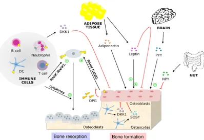
Recently, evidence have mounted suggesting that me-diators from the immune system, the adipose tissue, the gut and even the brain have a major influence on the process of bone remodelling (Fig. 1).
Osteoimmunology is the field that describes the influ-ence of the immune system on bone metabolism.35 Bone
and immune cells share the same progenitors residing in bone marrow and are affected by the same cytokines, which influence hematopoiesis, local immune responses and bone cells as well.38 Immune cells have also a direct influence on
bone cells through RANKL. RANKL is produced by mono-cytes, neutrophils, dendritic cells, B and T lymphocytes. In this way, immune cells have the ability to induce osteoclast differentiation and consequently bone resorption.39
Osteo-blast differentiation blocking can also be mediated by the immune system, as tumour necrosis factor (TNF) induces DKK-1 (a major Wnt inhibitor).40
Inflammatory derangement of normal bone remodelling, leading to high bone turnover, helps to explain the higher prevalence of osteoporosis in inflammatory arthropathies, such as rheumatoid arthritis.41
ARTIGO DE REVISÃO
as leptin or adiponectin), gut-derived appetite-regulatory hormones, namely peptide YY (PYY), glucagon-like pep-tide 1 (GLP-1) and ghrelin and hypothalamic regulators of energy balance, such as neuropeptide Y (NPP Y).25
Leptin is thought to have a direct anabolic effect within the bone, driving the differentiation of osteoblasts and si-multaneously inhibiting the differentiation of osteoclasts.42
Leptin has also been reported to have centrally mediated antiosteogenic actions on trabecular bone.43 Leptin is
ele-vated in obese subjects, but leptin insensitivity is likely to modulate aspects of leptin signalling.25
Adiponectin increases insulin sensitivity, and its circu-lating levels are reduced in obesity and diabetes. Osteo-blasts express both adiponectin and its receptors and show increased differentiation in response to these peptides.44
In contrast to these stimulatory effects on bone, circulating adiponectin has been shown to have a negative effect on bone formation due to stimulation of RANKL and inhibition of osteoprotegerin production by osteoblasts.45
Circulating Peptide YY concentrations are increased in response to acute food intake as well as short-term energy excess. PYY knockout mouse models, with male and fe-male knockouts demonstrated enhanced osteoblast activity and greater trabecular bone mass.46 GLP-1 also increases
in response to food intake, promoting satiety. The effects on bone are not clear.25
Ghrelin is a potent appetite-stimulating hormone,
syn-thesized in the gastric antrum and fundus. The circulating concentrations of ghrelin increase under pre-prandial and fasting conditions. Ghrelin stimulates osteoblast prolifera-tion and differentiaprolifera-tion in vitro, while also promoting osteo-clastogenesis.47
Experimental increases in central NPY expression in mice produce a marked decrease in bone formation and bone mass.48 In light of the antiosteogenic effect of NPY, it
has been postulated that NPY acts as a critical integrator of body weight and bone homeostatic signals.25 Other
neu-ropeptides are thought to have an influence on osteoblasts and osteoclasts. Neurons in the central nervous system seem to integrate clues from the energy homeostasis, gly-caemia or reproductive signals, with the regulation of bone remodelling.49
ARTIGO DE REVISÃO of macro and microstructural bone features by
quantita-tive computed tomography improves our ability to estimate bone strength.50 The development of devices that allow the
assessment of these structural aspects in the appendicular skeleton without the need for a bone biopsy (high resolution quantitative computed tomography (HR-pQCT)) has con-tributed to a better assessment of treatment effects in the context of clinical trials and may become available in clinical practice for selected patients in specialized centres.51
Men appear to have more trabecular thinning than tra-becular drop-out with increasing age, while women have both trabecular thinning and dropout. Furthermore, men have greater bone size than women across age and suf-fer less cortical thinning than women with aging. Overall, age-related changes in trabecular and cortical microstruc-ture in men would thus seem to have less impact on bone strength, thereby, explaining the lower fracture risk in aging men when compared with aging women.52
Bone turnover markers
Bone turnover markers (BTM) at a population level show a very promising potential for clinical applications based on their rapid response to treatment and their value in monito-ring compliance to medications. However, on an individual basis due to the high intra and inter individual variability their use is very limited in the daily clinical practice.53
Serum CTX-1 and P1NP are commonly used BTM. Re-sorption of demineralised organic type I collagen matrix by cathepsin K leads to release of carboxy-terminal collagen cross-linking telopeptides (CTX-1). During the formation of type I collagen, a synthesis marker is released – amino-ter-minal propeptide of type I procollagen (P1NP). Osteocalcin is also a good marker of bone formation, but there is a high biological and circadian variability.53
High levels of BTM (including CTX and bone alkaline phosphatase) are associated with increased risk of OP frac-ture in postmenopausal women, independently of hormone levels and of BMD.54,55 High bone resorption is associated
with an increased risk of OP fracture in elderly men, inde-pendently of BMD.56
OP treatment
In subjects with established OP (history of a previous
fragility fracture), it is now accepted to start treatment with-out the assessment of BMD, especially in countries with lim-ited access to DXA.58 Treatment shall also be started based
on BMD diagnostic criteria (T-score < -2.5) or upon assess-ment of absolute fracture risk (Table 2).58,59
The intervention threshold (in women without previous fractures) was set at the fracture probability equivalent to women with a prior fragility fracture, without knowledge of BMD. Assessment thresholds have been defined at age-specific cut-offs.58,60
In the 2013 clinician’s guide, the National Osteoporosis Foundation (NOF) suggest initiation of treatment in post-menopausal women and men aged 50 and older with low bone mass (T-score between -1.0 and -2.5, osteopenia) at the femoral neck, total hip or lumbar spine by DXA and a 10-year hip fracture probability > 3% or a 10-year major os-teoporosis-related fracture probability > 20% based on the U.S.-adapted WHO absolute fracture risk model.61
OP treatment should be complemented with lifestyle measures58,59,62
In every stage of life, the intake of calcium, vitamin D and protein should be guaranteed according to individual needs. In elderly people, a negative calcium balance leads to parathyroid hormone (PTH) secretion and enhances bone turnover.63
Weight-bearing exercise is essential, as immobilization is a cause of bone loss. The ideal amount of exercise is still controversial. A recent study showed a very strong associa-tion of high activity level and bone mass measures.64
Preventing falls is another important issue and modifia-ble factors should be intervened. Measures include: improv-ing physical condition, correctimprov-ing visual acuity and tapper or suspending drugs that diminish alertness.58,65
Pharmacological treatment
The main characteristics of major anti-OP therapies are described in Table 3. In men, there’s few available data on fracture prevention.12 The effects on BMD are similar
be-tween men and women.
Currently, the majority of approved therapeutic agents are antiresorptive drugs that lower bone turnover but also suppress bone formation.
Table 2 – Clinical indications to initiate OP treatment.
Initiation of osteoporosis treatment
1. Established OP – history of previous fragility fracture58
2. T-score < -2.5 at the femoral neck or lumbar spine by DXA58, 59,61
3. Postmenopausal women and men aged 50 and older wtih BMD osteopenia criteria AND
ARTIGO DE REVISÃO Table 3
- Drugs used in OP
treatment.
Drug
Dosing
Route of
administration
Phase III study
Fractures intervention
Contraindications
Side effects
Biphosphonates
Alendronate
70mg, weekly
Oral
bioavailability
impaired by food
FIT
Extension FLEX
Vertebral (RR 0.53) and
non vertebral fractures,
hip (RR 0.49)
ClCr < 35ml/min, Pregnancy
,
hypersensivity
, hypocalcemia
Flu-like symptoms (IV infusion),
hypocalcemia, Mild GI disturbances,
rarely esophagitis, Esophageal cancer
(?),Atrial fibrillation (possible causal
relation), Osteonecrosis of the jaw
(+cancer patients), Subtrochanteric
fractures (causal relation not
established)
Risendronate
35mg, weekly
Oral
VE
RT
Vertebral (RR 0.59) and
non vertebral fractures,
hip (RR 0.60)
Ibandronate
2.5mg daily OR
150mg monthly
3mg, quartly per
yer
Oral
IV
BONE
DIV
A
Vertebral fractures (RR
0.38 ), Non vertebral
fractures (adhoc analysis)
Zoledronate
5mg, yearly
IV
HORIZON
Vertebral (RR 0.30) and
non vertebral fractures,
hip (RR 0.59)
Strotium ranelate
2g, daily
Oral, 2h after
the last meal
SOTI
TROPOS
Vertebral (RR 0.59) and
non vertebral fractures,
hip (RR 0.85)
ClCr < 30ml/min, history of VTE,
hypersensivity
Nausea and diarrhoea, increased
risk VTE (possible relation), DRESS
syndrome, Increased CDV risk.
Denosumab
60mg, every 6
months
SC
FREEDOM
(against
placebo)
DECIDE
(against
alendronate)
Vertebral (RR 0.32) and
non vertebral fractures,
hip (RR 0.6)
CLCr<30ml/min, Pregnancy
,
hypersensivity
, pre-existing
hypocalc
emia
Rash, musculosketelal pain,
hypocalcemia, osteonecrosis of the
jaw
.
PTH analogs
Teriparatide
(1-34 PTH)
20ug, daily
SC
TOWER
EUROFORS
Vertebral (RR 0.35) and
non vertebral fractures
(RR 0.47)
Hyperparathyroidism, Hypercalcemia,
metabolic bone diseases, skeletal
malignancies or bone methatasis
Nausea, headache, dizziness,
transient ortosthatic hypotension,
Hypercalcemia, Exacerbation of
urolythiasis
1-84 PTH
100ug,daily
SC
TO
P
Vertebral fractures (RR
0.39)
in patients with recent crisis.
SERMS
Raloxifene
60mg, daily
Oral
MORE
Vertebral fractures (RR
0.70)
Pregnancy
, lactation, history of VTE
events
Increased risk of VTE
Hormonal
replacement
therapy
Not approved for OP
therapy
According to WHI: increased risk
coronary heart disease, stroke and
breast cancer
CDV - cardiovascular
, DRESS Drug rash with eosinophilia and systemic symptoms, VTE – venous thromboembolic event, WHI women’
ARTIGO DE REVISÃO Bisphosphonates
Bisphosphonates are antiresorptive drugs, which act through cholesterol biosynthesis pathway enzyme, farnesyl diphosphate synthase. By inhibiting this enzyme, they inter-fere with the attachment of the lipid to regulatory proteins, causing osteoclasts inactivation.66 There is established
evidence of efficacy in reducing fractures rate in postme-nopausal women.67-70 Recently, the duration of treatment
with bisphosphonates has been questioned. The exten-sion of FIT (FLEX) and HORIZON trials showed that bone loss after discontinuation of therapy was only modest as compared with that during continued therapy, suggesting a similarly persistent effect of alendronate and zoledronic acid (after 5 and 3 years of treatment, respectively). The trials extensions were consistent in showing significant re-ductions in the risk of vertebral fracture with continuation of bisphosphonate treatment.71,72 The number of fractures in
other sites was not significantly different. Treatment after 5 years can be considered in patients with high risk of verte-bral fractures, namely patients with low bone mineral den-sity at the femoral neck (T score below −2.5) after treatment and patients with an existing vertebral fracture a T score of less than −2.0.70-72 Recommendations regarding treatment
duration should be limited to alendronate and zoledronic acid, because of insufficient data regarding risendronate and ibandronate.73
Safety questions have also been raised in the last years. The rare occurrence of atypical fractures (namely, subtro-chanteric and diaphyseal femoral fractures) has been re-ported after long term exposure to bisphosphonates.74
Oste-onecrosis of the jaw has been also associated with the use of bisphosphonates, mostly in patients with cancer, treated with high dose EV bisphosphonates. The main known risk factors for osteonecrosis of the jaw are dental procedures, poor dental hygiene, corticosteroid therapy and local radio-therapy.75 There is some conflicting data about esophageal
cancer risk and the use bisphosphonates.76
Strontium ranelate
Strontium ranelate induces opposite effects on osteo-clasts and osteoblasts in vitro via at least three mechanisms involving activation of CaSR and NFATc/Wnt signalling and modulation of OPG/RANKL, an effect that results in im-proved bone architecture and bone strength in osteopenic animal models.75 Studies conducted up to 5 years have
shown anti-fracture efficacy, at spinal and non vertebral sites, in a wide diversity of subjects, independently of the level of fracture risk assessed by FRAX.77-80
Safety questions have been recently addressed. In May 2012, the European Medicine Agency (EMA) has issued cautionary advice to doctors on prescribing strontium rane-late to immobilised patients or patients with higher risk of venous thromboembolism.81 In post-marketing experience,
cases of eosinophilia and systemic symptoms (DRESS), Stevens-Johnson syndrome and toxic epidermal necrolysis were reported. The global incidence of DRESS was cal-culated as one per 47 168 patient-years of treatment.82 In
April 2013, EMA emitted a report advancing an increased cardiovascular risk in patients on OP treatment with stron-tium ranelate. Stronstron-tium is now contraindicated in patients with history of ischaemic heart disease, peripheral arterial disease, or cerebrovascular disease, or in patients with uncontrolled hypertension. The patient’s risk of developing cardiovascular disease should be evaluated before and at regular intervals during treatment. Strontium should only be used for the treatment of severe osteoporosis.82
PTH analogues
Bone quality effects are also noted. PTH analogues re-store the structure of trabecular bone, stimulate periosteal and endosteal bone growth, resulting in increased cortical thickness and cross-sectional area.19 They induce a rapid
increase in bone formation markers.19 PTH analogues are
currently used as a second line treatment in women with postmenopausal osteoporosis with multiple vertebral frac-tures, failing to respond to bisphosphonates. The duration of therapy should be limited to 2 years.58 In normocalcemic
patients, slight and transient elevations of serum calcium concentrations are described.58,83 The use of PTH analogs is
contraindicated in patients with pre-existing hypercalcemia, hyperparathyroidism, Paget’s disease, prior radiotherapy, skeletal malignancies and bone metastasis. Studies in rats have indicated an increased incidence of osteosarcoma, no evidence of this was found in human studies.83
Denosumab
Denosumab is a monoclonal antibody against RANKL. It reduces the formation, activity, and survival of osteoclasts and decreases bone turnover.
FREEDOM, a large placebo randomized controlled trial has demonstrated the antifracture efficacy of denosumab.84
In the STAND study, an effect on BMD after therapy with alendronate was demonstrated, so it is a therapeutic option in OP refractory to oral bisphosphonates.85
At a post hoc analysis of FREEDOM, denosumab showed evi dence of reduction in incident vertebral fractures in patients with estimated glomerular filtration rates (eGFR) between 15-90ml/min, without any adverse renal effects.86
In general, denosumab has a favourable safety profile. In the FREEDOM trial, the only serious adverse events signifi-cantly greater than placebo were skin infections.87
Selective estrogens receptor modulators (SERMS)
SERMs act as agonists or antagonists of the oestrogen receptor depending on the target tissue.58 In bone, its
ef-fects seem to be related to the inhibition of both IL-6 and TNF expression and activity. Osteoclasts differentiation and activity require the presence of factors produced in the bone microenvironment, among which are the proinflammatory cytokines IL-6 and TNF.88,89
devel-ARTIGO DE REVISÃO oped but showed similar results in fractures prevention and similar incidences of vasodilatation, leg cramps and venous thromboembolic events.91 Currently raloxifene is the only
SERM available for prescription.
Hormonal Replacement Therapy (HRT)
The Women’s Health Initiative was designed to test the effects of postmenopausal HRT. Despite confirming HRT as effective for OP, results also indicated that conjugated oestrogen and medroxyprogesterone acetate are associ-ated with a 30% increased risk of coronary heart disease and breast cancer and 40% increase of stroke. HRT is now recommended as a menopausal symptomatic therapy, used as shortly as possible and at the lowest possible dose. It is not recommended as an anti OP treatment.92
Calcitonin
Calcitonin is a polypeptide that binds to high-affinity G protein–coupled receptors on the osteoclast. Fish calcito-nins (eel and salmon) are about 40-fold more potent than mammalian. Calcitonin inhibits extracellular Ca2+ sensing,
a potent antiresorptive signal. Calcitonin withdrawal sensi-tizes an osteoclast to parathyroid hormone–induced (PTH-induced) stimulation.93
EMA considers that there is evidence of a small in-creased risk of cancer with long-term use of these medi-cines and does not approve calcitonin containing drugs to treat osteoporosis.94
Among current treatment options, only indirect compari-sons of RCTs have been used to assess relative efficacy (in the absence of head-to-head trials).
Alendronate was shown cost-effective in the treatment of postmenopausal osteoporosis, in women with a 10-year probability of major fracture above 7.5%.95 Due to his lower
price, it has a lower cost-effectiveness ratio which justifies its common choice as first-line agent.58
The selection of teriparatide versus oral bisphospho-nates as a first-line treatment for severe OP, with prior ver-tebral fractures is supported by some authors.96,97 Strontium
ranelate is also cost effective, but has now a restricted use and is only indicated in established OP.98 Denosumab has a
higher cost-effective ratio than alendronate.99 The increase
of BMD after treatment with alendronate and safety in pa-tients with GFR between 15 to 35ml/min are distinctive fea-tures as mentioned above. Compliance to treatment can be limited in OP, as it is an asymptomatic condition needing
long term treatment. Low treatment adherence is associ-ated with worse outcomes and has a significant impact on cost-effectiveness.100
New Drugs
Odanacatib is a new selective cathepsin K inhibitor that causes a moderate sustained decrease in bone resorption, and a lesser and more transient decrease in bone forma-tion as compared to classic anti resorptive drugs.100 This
agent may uncouple bone formation from resorption.19,101
Odacatinib is undergoing phase III trials in postmenopausal women and older men.
Sclerostin is an antagonist of the Wnt-b catenin pathway and its neutralization leads to an anabolic effect on bone. The anti-sclerostin monoclonal antibody (AMG 785/Romo-sozumab) is currently on phase III trials. Anti-Dkk1 is also being studied as a targeted therapy for the Wnt pathway. Calcium-sensing receptor (CaSR) antagonists stimulate endogenous PTH secretion.102 BA058, a synthetic peptide
analog of human Parathyroid Hormone related Protein (“hPTHrP”) is a bone anabolic compound with the poten-tial to treat severe osteoporosis. Currently, BA058 is being studied as a daily subcutaneous injection (BA058-SC) in a Phase III study.
CONCLUSION
Although an age-adjusted rate of fracture plateau was reached, our ageing society has an increasing prevalence of OP and its associated economic burden. An adequate use of BMD, FRAX™ and BMT could improve fracture pre-diction in postmenopausal women. There are now effective and relatively safe treatment options for OP and additionally the elucidation of several bone biology pathways uncovered new potential future therapeutic strategies.
ACKNOWLEGDEMENT
The authors thank Joana Caetano Lopes for the con-tribution regarding the image that illustrates the interaction between bone, the immune system and adipose tissue.
CONFLICT OF INTERESTS AND FUNDING SOURCES
João Eurico da Fonseca received unrestricted research grants or acted as a speaker for Amgen, MSD, Novartis, Roche and Servier. Helena Canhão acted as a speaker for Amgen, MSD and Servier. Ana Maria Rodrigues acted as speaker for MSD.
REFERENCES
1. Khaltaev N, Pfleger BA, on behalf of WHO, WHO Scientific Group on the Assessment of Osteoporosis at the Primary Health Care Level meet-ing, Summary Meeting Report, Brussels, Belgium, 5-7 May 2004. WHO 2007. [Accessed 2013 Jun 02]. Disponível em: http://www.who.int/chp/ topics/Osteoporosis.pdf.
2. Jennings LA, Auerbach AD, Maselli J, Pekow PS, Lindenauer PK, Lee SJ. Missed opportunities for osteoporosis treatment in patients hospital-ized for hip fracture. J Am Geriatr Soc. 2010;58:650-7.
3. Branco JC, Felicíssimo P, Monteiro J. A epidemiologia e o impacto só-cio-económico das fracturas da extremidade proximal do fémur, uma reflexão sobre o padrão actual de tratamento da osteoporose grave.
Acta Reumatol Port. 2009;34:475-85.
4. Reginster JY,Burlet N. Osteoporosis: a still increasing prevalence. Bone.2006;38:S4-9.
5. Cooper C, Zole CA,Holroyd CR,Earl SC, Harvey NC, Dennison EM, et al. Secular trends in the incidence of hip and other osteoporotic frac-tures. Osteoporos Int.2011;22:1277–88.
6. Cauley JA, Newman AB. Osteoporosis. In: Newman AB, Cauley JA, editors. The Epidemiology of Aging. Amsterdam: Springer; 2012. p. 499-522.
ARTIGO DE REVISÃO
Res. 2008;23:1832-41.
8. Andrew T, Antioniades L, Scurrah KJ, MacGregor AJ, Spector TD. Risk of wrist fracture in women is heritable and is influenced by genes that are largely independent of those influencing BMD. J Bone Miner Res. 2005;20:67-74.
9. Center JR, Center JR,Nguyen TV,Schneider D,Sambrook PN, Eisman JA. Mortality after all major types of osteoporotic fracture in men and women: an observational study. Lancet. 1999;353:878-82.
10. Christiansen C. Consensus development conference (1993): Diagnosis, prophylaxis and treatment of osteoporosis. Am J Med. 1993;94:646-50. 11. Assessment of fracture risk and its application to screening for post-menopausal osteoporosis. Report of a WHO Study Group. World Health Organ Tech Rep Ser.1994;843:1-129.
12. Kanis JA, Bianchi G, Bilezikian JP, Kaufman JM, Khosla S, Orwoll E, et al. Towards a diagnostic and therapeutic consensus in male osteoporo-sis. Osteoporos Int. 2011;22:2789-98.
13. Schuit SC, van der Klift M, Weel AE,de Laet CE,Burger H,Seeman E, et al. Fracture incidence and association with bone mineral density in elderly men and women: the Rotterdam Study. Bone. 2004;34:195-202. 14. Unnanuntana A, Gladnick BP, Donnelly E, Lane JM. The assessment of
fracture risk. J Bone Joint Surg Am. 2010;92:743-53.
15. Vasikaran S, Eastell R, Bruyère O, Foldes AJ, Garnero P, Griesmacher A, et al. Markers of bone turnover for the prediction of fracture risk and monitoring of osteoporosis treatment: a need for international reference standards. Osteoporos Int. 2011;22:391-420.
16. Rachner TD, Khosla S, Hofbauer LC. Osteoporosis: now and the future. Lancet. 2011;377:1276-87.
17. Kanis JA. Diagnosis of osteoporosis and assessment of fracture risk. Lancet. 2002;359:1929-36.
18. Siris E, Miller P, Barrett-Connor E, Abbott T, Sherwood L, Berger M. De-sign of NORA, the National Osteoporosis Risk Assessment Program: a longitudinal US registry of postmenopausal women. Osteoporos Int.1998;8:S62-9.
19. Gallacher SJ, Dixon T. Impact of treatments for postmenopausal os-teoporosis (Bisphophonates, Parathyroid Hormone, Strontium Ranelate and Denosumab) on bone quality: a systematic review. Calcif Tissue Int. 2010;87:469-84.
20. Kanis JA,Johnell O,Oden A, Johansson H,McCloskey E. FRAX and the assessment of fracture probability in men and women from the UK. Osteoporos Int. 2008;19:385-97.
21. Marques A, Mota A, Canhão H, Romeu JC, Machado P, Ruano A, et al. A FRAX model for the estimation of osteoporosis fracture probability in Portugal. Acta Reumatol Port. 2013;38:104-12.
22. Cummins NM, Poku EK, Towler MR, O’Driscoll OM, Ralston SH. Clinical risk factors for osteoporosis in Ireland and the UK: a comparison of Frax and QFractures Scores. Calcif Tissue Int. 2011;89:172-7.
23. Baccheta J,Boutroy S,Vilayphiou N,Juillard L,Guebre-Egziabher F, Rognant N, et al. Early impairment of trabecular microarchitecture as-sessed with HR-pQCT in patients with stage II-IV Chronic Kidney Dis-ease. J Bone Miner Res.2010;25:849-57.
24. Nickolas TL, Leonard MB,Shane E. Chronic Kidney disease and bone fracture: a growing concern. Kidney Int. 2008;74:721-31.
25. Reid IR, Ames R, Evans MC, Sharpe S, Gamble G, France JT,et al. Determinants of total body and regional bone mineral density in normal postmenopausal women- a key role for fat mass. J Clin Endocrinol Me-tab. 1992;75:45-51.
26. Brzozowska MM, Sainsbury A, Eisman JA, Baldock PA, Center JR. Bari-atric surgery, bone loss, obesity and possible mechanisms. Obes Rev. 2013;14:52-67.
27. Gilsanz V,Chalfant J, Mo AO,Lee DC, Dorey FJ, Mittelman SD. Recip-rocal Relations of Subcutaneous and Visceral Fat to Bone Structure and Strength. J Clin Endocrinol Metab.2009;94:3387-93.
28. Zhao LJ, Jiang H, Papasian CJ, Maulik D, Drees B, Hamilton J, et al. Correlation of Obesity and Osteoporosis: Effect of Fat Mass on the De-termination of Osteoporosis. J Bone Miner Res.2008;23:17-29. 29. Rodrigues AM, Caetano-Lopes J, Vale AC, Vidal B, Lopes A, Aleixo I, et
al. Low osteocalcin/collagen type I bone gene expression ratio is associ-ated with hip fragility fractures. Bone. 2012;51:981-9.
30. Rodrigues AM, Caetano-Lopes J, Vale AC, Aleixo I, Pena AS, Faus-tino A, et al. Smoking is a predictor of worse trabecular mechanical performance in hip fragility fracture patients. J Bone Miner Metab. 2012;30:692-9.
31. Datta HK, Ng WF, Walker JA, Tuck SP, Varanasi SS. The cell biology of bone metabolism. J Clin Pathol.2008;61:577-7.
32. Kim JH, Liu X, Wang J, Chen X, Zhang H, Kim SH,et al. Wnt signalling
in bone formation and its therapeutic potential for bone diseases. Ther Adv Musculoskel Dis. 2013;5:13-31.
33. Caetano-Lopes J, Lopes A, Rodrigues A, Fernandes D, Perpetuo IP, Monjardino T,et al. Upregulation of inflammatory genes and downregu-lation of sclerostin gene expression are key elements in the early phase of fragility fracture healing. Plos One. 2011;6:e16947.
34. Manolagas SC. Birth and death of bone cells: basic regulatory, mecha-nisms and implications for the pathogenesis and treatment of osteopo-rosis. Endocr Rev.2000;21:115-37.
35. Kong YY, Yoshida H, Sarosi I, Tan HL, Timms E, Capparelli C, et al. OPGL is a key regulator of osteoclastogenesis, lymphocyte develop-ment and lymph-node organogenesis. Nature. 1999;397:315-22. 36. Amizuka N, Li M,Kobayashi M,Hara K,Akahane S,Takeuchi K, et al.
Vi-tamin K2, a gamma-carboxylating factor of gla-proteins, normalizes the bone crystal nucleation impaired by Mg-insufficiency. Histol Hispathol. 2008;23:1353-66.
37. Hayashi M, Nakashima T, Taniguchi M, Kodama T, Kumanogoh A, Ta-kayanagi H. Osteoprotection by semaphorin 3A. Nature. 2012;485:69-74.
38. Caetano-Lopes J, Canhão H, Fonseca JE. Osteoimmunology – the hid-den imune regulation of bone. Autoimmun Rev. 2009;8:250-5. 39. Zupan J, Matjaz J, Marc J. Osteoimmunology and the influence of
pro-inflamatory cytokines on osteoclast. Biochem Med. 2013;1:43-65. 40. Diarra D,Stolina M, Polzer K,Zwerina J,Ominsky MS, Dwyer D,et
al. Dickkopf-1 is a master regulator of joint remodelling. Nat Med. 2007;13:156-63.
41. Caetano-Lopes J, Nery AM, Canhão H, Duarte J, Cascão R, Rodrigues A,et al. Chronic arthritis leads to disturbances in the bone collagen net-work. Arthritis Res Ther. 2010;12R9.
42. Holloway WR, Collier FM, Aitken CJ, Myers DE, HodgeJM, Malakel-lis M, et al. Leptin inhibits osteoclast generation. J Bone Miner Res. 2002;17:200–9.
43. Jackson MA,Iwaniec UT,Turner RT,Wronski TJ,Kalra SP.Effects of increased hypothalamic leptin gene expression on ovariectomy-induced bone loss in rats. Peptides.2011;32:1575-80.
44. Luo XH, Guo LJ, Yuan LQ, Xie H, Zhou HD, Wu XP, et al. Adiponectin stimulates human osteoblasts proliferation and differentiation via the MAPK signaling pathway. Exp Cell Res. 2005;309:99-109.
45. Luo XH, Guo LJ, Xie H, Yuan LQ, Wu XP, Zhou HD, et al. Adiponectin stimulates RANKL and inhibits OPG expression in human osteoblasts through the MAPK signaling pathway. J Bone Miner Res. 2006;21:1648-56.
46. Wong I, Driessler F, Khor EC, Shi Y, Nguyen AD, Enriquez RF, et al. Peptide YY regulates bone remodeling in mice: a link between gut and skeletal biology. PlosONE. 2012;7:e40038.
47. Costa JL,Naot D,Lin JM,Watson M,Callon KE,Reid IR, et al. Ghrelin
is an osteoblast mitogenandincreases osteoclastic bone resorption in
vitro. Int J Pept.2011;2011:605193.
48. Baldock PA, Sainsbury A, Allison S, Lin EJ,Couzens M,Boey D, et al. Hypothalamic control of bone formation: distinct actions of leptin and Y2 receptor pathways. J Bone Miner Res. 2005;20:1851-7.
49. Florent E. Regulation of bone remodeling by the central and peripheral nervous system. Arch Biochem Biophys. 2008;473:231-6.
50. Brandi ML. Microarchitecture, the key to bone quality. Rheumatology. 2009;48:iv3-8.
51. Burghardt AJ, Link TM, Majumdar S. High-resolution computed tomog-raphy for clinical imaging of bone microarchitecture. Clin Orthop Relat Res.2011;469:2179-93.
52. Amin S, Khosla S. Sex and age-related differences in bone microarchi-tecture in men relative to women assessed by high-resolution peripheral quantitative computed tomography. J Osteoporos.2012;2012:129760. 53. Civitelli R, Armamento-Villareal R, Napoli N. Bone turnover markers:
un-derstanding their value in clinical trials and clinical practice. Osteoporos Int. 2009;20:843-51.
54. Garnero P, Sornay-Rendu E, Claustrat B, Delmas PD. Biochemical markers of bone turnover, endogenous hormones and the risk of frac-tures in postmenopausal women: the OFELY study. J Bone Miner Res. 2000;15:1526-36.
55. Chapurlat RD, Garnero P, Bre´Art G, Meunier PJ, Delmas PD. Serum type I collagen breakdown product (Serum CTX) predicts hip fracture risk in elderly women: the EPIDOS study. Bone. 2000;27:283-6. 56. Meier C, Nguyen TV, Center JR, Seibel MJ, Eisman JA. Bone
resorp-tion and osteoporotic fractures in elderly men: the Dubbo Osteoporosis Epidemiology Study. J Bone Miner Res. 2005;20:579-87.
Abdulgha-ARTIGO DE REVISÃO ni S, et al. Bone histomorphometry revisited. Acta Reumatol Port. 2012;37:294-300.
58. Kanis JA, McCloskey EV, Johanson H, Cooper C, Rizzoli R, Reginster JY. European Guidance for the diagnosis and management of osteopo-rosis in postmenopausal women. Osteoporos Int. 2013;24:23-57. 59. Tavares V, Canhão H, Melo-Gomes JA, Simões E, Romeu JC, Coelho
P, et al. Recomendações para o diagnóstico e terapêutica da osteopo-rose, Sociedade Portuguesa de Reumatologia e Sociedade Portuguesa de Doenças Ósseas Metabólicas. Acta Reumatol Port. 2007;32:49-59. 60. Kanis JA, McCloskey EV, Johansson H, Strom O, Borgstrom F, Oden
A. Case finding for the management of osteoporosis with FRAX®
-assessment and intervention thresholds for the UK. Osteoporos Int. 2008;19:1395-408.
61. Cosman F, Lindsay R, LeBoff MS, de Beur SJ, Tanner B. National Os-teoporosis Foundation, Clinician’s guide to prevention and treatment of osteoporosis 2013. [Accessed 2013 Jun 02]. Disponível em: http://www. nof.org/files/nof/public/content/resource/913/files/580.pdf).
62. Branco JC, Tavares V, Vaz AF, Simões E, Canhão H, George F. Tra-tamento Farmacológico da Osteoporose Pós-menopáusica, Norma da direcção geral de saúde. 027/2011. [Accessed 2013 Jun 02]. Disponível em: http://www.dgs.pt/?cr=21174.
63. Body JJ, Bergmann P, Boonen S, Devogelaer JP, Gielen E, Goemare S, et al. Extraskeletal benefits and risk of calcium, vitamin D and anti-osteoporosis medications. Osteoporos Int. 2012;23:S1-23.
64. Saraví FD,Sayegh F. Bone mineral density and body composition of adult premenopausal women with three levels of physical activity. J Os-teoporos. 2013;2013:953271.
65. Järvinen TN, Sievänen H, Khan KM, Heinonen A, Kannus P. Shift-ing the focus in fracture prevention from osteoporosis to falls. BMJ. 2008;336:124-6.
66. Reszka AA, Rodan GA. Bisphosphonate mechanism of action. Curr Rheumatol Rep. 2003;5:65-74.
67. Liberman UA, Weiss SR, Bröll J, Minne HW, Quan H, Bell NH, et al. Effect of oral alendronate on bone mineral density and the incidence of fractures in postmenopausal osteoporosis. The Alendronate Phase III Osteoporosis Treatment Study Group. N Engl J Med. 1995;333:1437-43.
68. Harris ST, Watts NB, Genant HK, McKeever CD, Hangartner TN, Keller M, et al. Effects of risedronate treatment on vertebral and nonvertebral fractures in women with postmenopausal osteoporosis: A randomized controlled trial. JAMA. 1999;282:1344-52.
69. Black DM, Delmas PD, Eastell R, Reid IR, Boonen S, Cauley JA, et al. Once-yearly zoledronic acid for treatment of postmenopausal osteopo-rosis. N Engl J Med. 2007;356:1809-22.
70. Chesnut III CH,Skag A, Christiansen C,Recker R,Stakkestad JA, Hoi-seth A, et al. Effects of oralibandronate administered daily or intermit-tently on fracture risk in postmenopausal osteoporosis. J Bone Miner Res.2004;19:1241-9.
71. Black DM, Schwartz AV, Ensrud KE, Cauley JA, Levis S, Quandt SA, et al. Effects of continuing or stopping alendronate after 5 years of treat-ment. JAMA. 2006;296:2927-37.
72. Black DM, Reid IR, Boonen S, Bucci-Rechtweg C, Cauley JA, Cosman F, et al. The effects of 3 versus 6 years if zoledronic Acid treatment of osteoporosis: a randomized extension to HORIZON-pivotal fracture trial (PFT). J Bone Miner Res. 2012;27:243-54.
73. Black DM, Bauer DC, Schwartz AV, Cummings SR, Rosen CJ. Continu-ing bisphosphonate treatment for osteoporosis - For whom and for how long? N Engl J Med. 2012;366:2051-3.
74. Black DM, Kelly MP, Genant HK, Palermo L, Eastell R. Bisphosphonates and fractures of the subtrochanteric or diaphyseal femur. N Engl J Med. 2010;362:1761-71.
75. Durie BG, Katz M, Crowley J. Osteonecrosis of the jaw and bisphospho-nates. N Engl J Med. 2005;353:99-102.
76. Vinogradova Y, Coupland C, Hippisley-Cox J. Exposure to bisphospho-nates and risk of gastrointestinal cancers: series of nested case-control studies with QResearch and CPRD data. BMJ. 2013;346:f114. 77. Marie PJ, Felsenberg D, Brandi ML. How strontium ranelate, via
op-posite effects on bone resorption and formation, prevents osteoporosis. Osteoporos Int. 2011;22:1659–67.
78. Reginster JY, Seeman E, de Vernejoul MC, Adami S, Compston J, Phenekos C, et al. Strontium ranelate reduces the risk of non-verte-bral fractures in postmenopausal women with osteoporosis. Treatment of peripheral osteoporosis (TROPOS) study. J Clin Endocrinol Metab. 2005;90:2816-22.
79. Meunier PJ, Roux C, Seeman E, Ortolani S, Badurski JE, Spec-tor TD, et al. The effects of strontium ranelate on the risk of vertebral
fracture in women with postmenopausal osteoporosis. N Engl J Med. 2004;350:459-68.
80. Kanis JA, Johansson H, Oden A, McCloskey EV. A meta-analysis of the effect of strontium ranelate on the risk of vertebral and non-verte-bral fracture in postmenopausal osteoporosis and the interaction with FRAX®. Osteoporos Int. 2011;22:2347-55.
81. European Medicines Agency confirms positive benefit-risk balance of Protelos/Osseor, but recommends new contraindications and revised warnings, EMA/CHMP/185175/2012 (March 2012). [Accessed 2013 Jun 02]. Disponível em: http://www.ema.europa.eu/docs/en_GB/docu-ment_library/Press_release/2012/03/WC500124206.pdf.
82. Pharmacovigilance Risk Assessment Committee (PRAC) recommends restriction in the use of Protelos/Osseor, EMA/220628/2013 (April 2013). [Accessed 2013 Jun 02]. Disponível em: http://www.ema.euro-pa.eu/docs/en_GB/document_library/Medicine_QA/human/000560/ WC500142021.pdf.
83. Bodenner D, Redman D, Riggs A. Teriparatide in the management of osteoporosis. Clin Interv Aging. 2007;2:499-507.
84. Cummings SR, San-Martin J, McClung MR, Siris ES, Eastel R, Reid IR, et al. Denosumab for prevention of fractures in postmenopausal women with osteoporosis. N Engl J Med. 2009;361:756-65.
85. Kendler DL, Roux C, Benhamou CL,Brown JP, Lillestol M,Siddhanti S, et al. Effects of denosumab on bone mineral density and bone turno-ver in postmenopausal women transitioning from alendronate therapy. J Bone Miner Res. 2010;25:72–81.
86. Miller PD. A review of the efficacy and safety of denosumab in post-menopausal women with osteoporosis. Ther Adv Musculoskelet Dis. 2011;3:271-82.
87. Jamal SA, Ljunggren O, Stehman-Breen C, Cummings SR, Mc-Clung MR, Goemaere S,et al. Effects of denosumab on fracture and bone mineral density by level of kidney function. J Bone Miner Res. 2011;26:1829–35.
88. Gianni W, Ricci A, Gazzaniga P, Brama M, Pietropaolo M, Votano S, et al. Raloxifene modulates interleukin-6 and tumor necrosis factor-α synthesis in vivo: results from a pilot clinical study. J Clin Endocrinol Metab. 2004;89:6097-9.
89. Taranta A, Brama M, Teti A, DeLuca V, Scandurra R, Spera G, et al. The selective estrogen receptor modulator raloxifene regulates osteoblast and osteoclast activity in vitro. Bone. 2002;30:368-76.
90. Ettinger B, Black DM, Mitlak BH, Knickerbocker RK, Nickelsen T, Genant HK, et al. Reduction of vertebral fracturerisk in postmenopausal womenwithosteoporosis treated with raloxifene: results from a 3-year randomized clinical trial. Multiple Outcomes of Raloxifene Evaluation (MORE) Investigators. JAMA.1999;282:637-45.
91. Silverman SL, Christiansen C, Genant HK, Vukicevic S, Zanchetta JR, de Villiers TJ. Efficacy of bazedoxifene in reducing new vertebral frac-ture risk in postmenopausal women with osteoporosis: results from a 3-year, randomized, placebo-, and active-controlled clinical trial. J Bone Miner Res. 2008;23:1923-34.
92. Rossouw JE,Anderson GL,Prentice RL,LaCroix AZ,Kooperberg C, Stefanick ML. Risks and benefits of estrogen and progestin in healthy postmenopausal women: Principal results from the Women’s Health Ini-tiative randomized controlled trial. JAMA. 2002;288:321.
93. Zaidi M, Moonga BS, Abe E. Calcitonin and bone formation: a knockout full of surprises. J Clin Invest.2002;110:1769-71.
94. Questions and answers on the review of calcitonin-containing medi-cines, EMA/731082/2012, EMEA/H/A-31/1291 (November 2012). [Accessed 2013 Jun 02]. Disponível em: http://www.emea.europa.eu/ docs/en_GB/document_library/Referrals_document/Calcitonin_31/ WC500134838.pdf.
95. Kanis JA, Adams J, Borgstrom F, Cooper C, Jonsson B, Preedy D, et al. The cost-effectiveness of alendronate in the management of osteoporo-sis. Bone. 2008;42:4-15.
96. Rizzol R, Kraenzli M, Krieg MA, MellinghoffHU, LamyO, Lippuner K. Indications to teriparatide treatment in patients with osteoporosis. Swiss Med Wkly. 2011;141:w13297.
97. Murphy DR,Smolen LJ, Klein TM,Klein RW. The cost effectiveness of teriparatide as a first-line treatmentfor glucocorticoid-induced and postmenopausalosteoporosispatients in Sweden. BMC Musculoskelet Disord.2012;30:213.
98. Hiligsmann M,Vanoverberghe M,Neuprez A, Bruyère O,Reginster JY. Cost-effectivenessof strontium ranelate for the prevention and treat-ment of osteoporosis. Expert Rev Pharmacoecon Outcomes Res. 2010;10:359-66.
postmeno-ARTIGO DE REVISÃO
pausalosteoporosis. Osteoporos Int. 2011;22:967-82.
100. Kanis JA,Cooper C, Hiligsmann M,Rabenda V,Reginster JY,Rizzoli R. Partial adherence: a new perspective on health economic assessment
in osteoporosis.Osteoporos Int. 2011;22:2565-73.
101. Vivien L, Clarke BL. New therapeutics target for osteoporosis: beyond denosumab. Maturitas. 2012;73:269-72.
102. Kimura S, Nakagawa T, Matsuo Y, Ishida Y, Okamoto Y, Hayashi M.
JTT-305, an orally active calcium-sensing receptor antagonist, stimu-lates transient parathyroid hormone release and bone formation in ova-riectomized rats. Eur J Pharmacol. 2011;668:331-6.
103. Roux C, Cooper C, Díez-Pérez A, Martinez L, Ortolani S, Gitlin M. Prevalence of osteoporosis and fractures among women prescribed os-teoporosis medication in five European countries: the POSSIBLE EU®