77
© 2018 by the Serbian Biological Society How to cite this article: Raksha NG, Potalitsyn PY, Yurchenko AV, Halenova TI, Savchuk OM, Ostapchenko LI. Prevention of diet-induced obesity in rats by oral application of collagen fragments. Arch Biol Sci. 2018;70(1):77-86.
Prevention of diet-induced obesity in rats by oral application of collagen fragments
Nataliia G. Raksha*, Pavlo Y. Potalitsyn, Alona V. Yurchenko, Tetyana I. Halenova, Oleksii M. Savchuk and Lydmila I. Ostapchenko
Educational and Scientific Center, Institute of Biology and Medicine, “Taras Shevchenko” National University of Kyiv, 64/13,
Volodymyrska Str., Kyiv, 01601, Ukraine
*Corresponding author: [email protected]
Received: April 1, 2017; Revised: June 1, 2017; Accepted: June 13, 2017; Published online: August 1, 2017
Abstract: The aim of the present study was to determine whether orally applied collagen fragments (CFs) could affect the development of obesity in obese rats. To this end, experimental rats that were exposed to a high-calorie diet (HCD) for four weeks were randomly divided into two groups: HCD and HCD+CFs, with both groups continuing to receive the HCD. However, rats from the HCD+CFs group were also provided with CFs in a 0.05-M citrate buffer (pH 5.0) (1 g·kg-1 of body
weight) by intragastric administration, every other day for the next six weeks. Selected parameters associated with obesity development and insulin resistance, as well as serum markers of oxidative stress and the cytokine profile were assessed at the end of the 10th week. Supplementation with CFs resulted in a decrease in body weight and body mass index when
compared to animals exposed to a HCD. The observed changes were assumed to be caused by a lower food intake and increased water intake by obese rats treated with CFs. Enhanced activity of superoxide dismutase (SOD), catalase (CAT) and decreased malondialdehyde (MDA) concentration were detected in the HCD+CF group of animals when compared to untreated HCD-fed rats. Administration of CFs also lowered the serum concentrations of the proinflammatory cytokines IL-1β and IL-12, whereas the concentration of the anti-inflammatory cytokine IL-10 was significantly increased and the concentration of cytokine IL-4 was near the control value. Decreased concentrations of fasting blood glucose, glycated hemoglobin (GHbA1c) and serum insulin and increased tolerance to glucose in the oral glucose tolerance test (OGTT) were observed in the HCD+CF group of animals when compared to rats in the HCD group. We concluded that CFs mediated a therapeutic effect on obesity development in rats exposed to a HCD by affecting pathways involved in obesity pathogenesis. Key words: collagen fragments; obesity; oxidative stress; inflammation; insulin resistance
INTRODUCTION
Obesity is a medical condition with a multifactorial origin and it is recognized as one of the leading health care problems [1]. The incidence of obesity has sharply increased due to rapid socio-economic development and dramatic lifestyle changes. Obesity is a high-risk factor for the development of many metabolic com-plications, including cardiovascular disease, insulin resistance, type II diabetes, certain forms of cancer and sleep-breathing disorder [2,3]. Despite the many efforts directed at the prevention and treatment of obesity and obesity-related metabolic dysfunction, there is no universally satisfactory treatment. Cur-rently many drugs are available for the treatment of obesity, but most of them become less effective with time or have adverse side effects. Consequently, there
Marine-derived bioactive peptides can be recov-ered from a wide variety of high protein marine by-products, including fish scales. In light of the high yield of collagen from fish, fish scales provide an at-tractive source for collagen extraction [8]. In addition, isolation of collagen peptides from fish-scale waste renders their production both economically and en-vironmentally friendly. Marine collagen peptides have been identified as having a wide variety of activities, including antimicrobial, anti-inflammatory, anti-ulcer, lipid-lowering, wound-healing and anti-skin-aging [9,10]. They have also been observed to affect glucose tolerance and insulin sensitivity in high fat-fed rats and to modulate immune functions [11,12]. Colla-gen peptides can reduce the peroxidation of lipids or fatty acids, scavenge free radicals and chelate transi-tion metal ions, which has led to the hypothesis that these peptides could be suitable supplement agents for several types of illness associated with oxidative stress. It has been incontrovertibly established that obesity per se causes systemic oxidative stress [13] and chronic inflammation [14], and that these disorders play a critical role in the pathogenesis of obesity and obesity-associated metabolic dysfunctions.
We have assumed that treatment with CFs could offer protection for obese individuals by modulating the pathways involved in obesity-related pathogenesis. Therefore, the aim of the present work was to evaluate the effect of CF administration on weight gain and body composition, oxidative stress markers, cytokine profile and some parameters associated with insulin resistance in obese rats.
MATERIALS AND METHODS Preparation of collagen fragments
CFs were prepared from fish scales. Briefly, wild ma-rine fish were caught near the Galindez island (geo-graphical coordinates: 65°15’ south latitude, 64°15’ west longitude) of the Argentine Island archipelago. The material was collected by the XVII (from March 2012 to April 2013) and XVIII (from March 2013 to April 2014) Ukrainian Antarctic expeditions. The scales were washed thoroughly with distilled water and stored until use. Extraction of collagen from fish scales was done in two steps [15]. The fish scales were
washed twice in 10% NaCl solutions (at a ratio of dry scales:solution=1:10) to remove proteins. Deminer-alization was achieved using a 0.4-M HClO4 solution (at a ratio of dry scales:solution=1:15) for 90 min. The demineralized scales were washed three times with distilled water. Collagen from fish scales was then ex-tracted using 0.5 M of acetic acid containing 0.005 M EDTA for 24 h. The extract was centrifuged at 10000 g for 30 min and the residues were re-extracted with the same solution for 24 h with further centrifuga-tion. The supernatant was combined and salted out by adding NaCl to a final concentration of 0.9 M. The precipitated collagen was separated by centrifugation at 10000 g for 30 min, redissolved in 0.5 M of acetic acid and precipitated again with NaCl. The obtained precipitate was dialyzed against distilled water and lyophilized. The procedure of CF preparation was performed as described [16]. The obtained collagen was enzymatically hydrolyzed by pepsin (3000 U g protein-1) at 37oC, pH 2.0 for 8 h. To stop the reac-tion, the mixture was heated to boiling for 5 min and then centrifuged at 4000 g for 30 min. The supernatant was filtered through a ceramic membrane (200 μm) to separate the collagen fragment fraction (26≤kDa). The generated CFs were analyzed by sodium dodecyl sul-fate (SDS) polyacrylamide gel electrophoresis (PAGE) according to Laemmli [17] using a 15% resolving gel.
Animals and experimental design
A total of 30 white male, 8-week-old Wistar rats weighing 170±5g were kept under standard labora-tory conditions with free access to water and food. All animal procedures were in compliance with the European Directive 2010/63/EU (EC, 2010) on the protection of animals used for experimental and other scientific purposes. All manipulations were approved by the Ethical Committee of Educational and Scien-tific Center, Institute of Biology and Medicine, “Taras Shevchenko” National University of Kyiv. The study was conducted after obtaining Ethical Committee clearance (Protocol № 1 from 20th February 2016).
fed with a high-calorie diet prepared in our laboratory. The HCD consisted of a standard meal (60%), pork fat (10%), eggs (10%), sugar (9%), peanuts (5%), dry milk (5%) and sunflower oil (1%) and water ad libitum
[18]. After feeding for 4 weeks, the rats were randomly divided into two groups (with 10 animals per group). The rats from the first group (HCD group) continued to receive the high-calorie diet. The rats from the sec-ond group (CF-treated group) were fed the HCD and were administered intragastrically with CFs (1 g·kg-1 of body weight) in 0.05-M citrate buffer (pH 5.0) every other day for the next 6 weeks. Food and water were available ad libitum. The rats in the control group were treated intragastrically with an equal volume of citrate buffer (pH 5.0) that served as a vehicle for the CFs.
Blood glucose concentration was evaluated us-ing Glucophot-II glucometer (Norma, Ukraine) and the level of glycosylated hemoglobin was measured spectrophotometrically using a commercial assay kit (Pliva-lachema Diagnostika, Czech Republic).
Sample collection
Blood samples were collected in standard biochemical test tubes. Serum for the determination of biochemical parameters was prepared by centrifugation at 1000 g of previously incubated blood samples for 30 min at 37oC. The serum was separated and kept at -20oC until analysis. Protein concentration was determined according to the method of Bradford [19], using crys-talline bovine serum albumin as a standard.
MDA assay
The level of serum MDA was estimated spectrophoto-metrically by the thiobarbituric acid reactive substanc-es (TBARS) assay [20]. A 0.4-mL aliquot of serum was added to 1.6 mL of an aqueous solution of 0.175 M KCl and 0.025 M Tris-HCl (pH 7.4). The total protein frac-tion was separated from the mixture by precipitafrac-tion with 20% trichloroacetic acid and further centrifuga-tion at 5000g for 15 min. After the addicentrifuga-tion of 1 mL of a 0.8% aqueous solution of thiobarbituric acid to 2 mL of the obtained supernatant, the samples were heated for 30 min in a boiling water bath. After cooling, the optical density of the samples was determined with a spectrophotometer (Smart Spec™ Plus, BioRad, USA)
at 532 nm. The concentration of MDA was calculated using the molar extinction coefficient ε532=1.56·10-5 M-1·cm-1 and expressed as nmol·mg-1 of protein.
Antioxidant enzyme activity assays
SOD activity was assayed by the method of Sirota [21] based on the ability of SOD to inhibit the autooxida-tion of adrenaline, as follows: 0.01 mL of serum was added to a cuvette containing 2 mL of 0.2 M carbonate buffer, pH 10.6, followed by the addition of 0.1 mL of 0.1% adrenaline. Specific SOD activity was expressed as U·mg-1·min-1. CAT activity was measured by the method of Korolyuk et al. [22]. A 0.1-mL aliquot of the serum (each sample was previously 10-fold di-luted with 0.05 M Tris-HCl buffer, pH 7.4) was incu-bated in 2 mL of freshly prepared 0.03% H2O2 at room temperature for 5 min. The control sample included 0.1 mL of 0.05 M Tris-HCl buffer (pH 7.4) instead of the biological material. The enzymatic reaction was stopped by the addition of 1 mL of 4% ammonium molybdate; the yellow complex of molybdate and hy-drogen peroxide was measured at 405 nm against the blank (0.1 mL 0.05 M Tris-HCl buffer (pH 7.4), 2 mL distilled water, 1 mL ammonium molybdate). Enzyme activity was calculated from the difference of the H2O2 content in the control and experimental samples, us-ing a calibration curve previously prepared with H2O2, and expressed as mmol H2O2 min-1·mg protein-1.
Cytokine and insulin assays
micro-plate reader (mQuant™, BioTek Instruments, Inc). The cytokine and insulin levels in the control animals was set as 100%, and changes in the cytokine and insulin levels are given as percentages relative to the controls.
OGTT
Glucose was administered per os (p.o.), 2 mg·kg-1in 2 mL. Blood samples were collected from the tail vein just prior to and 30, 60, 120 and 150 min after glucose loading. The blood glucose concentration was assayed using the Glucophort II glucometer (Norma, Ukraine) and a hyperglycemic curve was constructed.
Statistical analysis
Data entry and analysis were performed using StatSoft Statistica ver. 7.0 for Windows. After testing for normality (by Shapiro-Wilk), one-way analysis of variance (ANOVA) was used to compare the means among different groups. Differences were considered to be statistically significant when p<0.05. Data were reported as means±standard deviation (SD).
RESULTS AND DISCUSSION
Changes in body weight, body mass index and selected nutritional parameters
Numerous peptides from various sources have been reported to have anti-obesity effects by modulating the pathways involved in obesity pathogenesis. Anti-obesity activities were found in whey, soy and casein protein hydrolysates [24]. We first examined whether diet-induced obesity was present in rats exposed to a high-calorie diet. Our data showed that by the end of the experiment, the animals that consumed high-calo-rie food suffered from obesity, as evidenced from their higher body weight as compared to the control group. The development of obesity under the applied experi-mental condition was shown in our previous work [25]. The changes in rat body weight over the duration of the study (Fig. 1) showed that the initial weight of the rats on the high-calorie diet was 171±6.8 g. Through-out the monitoring period, we observed a gradual in-crease in body weight of rats in the HCD group, and at the end of the 10th week of the experiment the average
weight of the animals was 463.6±8.7 g. We found that the treatment with CFs prevented the increase in body weight. At the end of experiment, the body weight of the rats in the CF-treated group was 401.8±7.5, which was significantly lower than that of rats in the HCD group (p<0.05). To summarize, in the course of our study we found that the body weight of the control rats increased 1.9-fold, and of rats from the HCD and CF-treated group 2.7- and 2.2-fold, respectively.
To confirm obesity in experimental rats we cal-culated the body mass index (BMI). According to the obtained results (Table 1), the BMI of control rats at the end of the 10th week of the experiment was 0.7±0.05 g·cm-2, which was within the range for rats of corre-sponding age [26]. We observed an increase in the BMI of rats that were on the HCD as compared to the control group, which could indicate the development of diet-induced obesity. The treatment with CFs was also accompanied by an increase in the BMI, but the
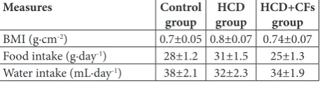
Table 1. Body mass index (BMI) and food and water intake of
control, HCD and HCD+CFs rats.
Measures Control
group groupHCD HCD+CFs group
BMI (g·cm-2) 0.7±0.05 0.8±0.07 0.74±0.07
Food intake (g·day-1) 28±1.2 31±1.5 25±1.3
Water intake (mL·day-1) 38±2.1 32±2.3 34±1.9
All parameters were measured after10 weeks of experiment. Data were ex-pressed as means±SD (n=10). BMI – body mass index; HCD – group of rats fed with a high-calorie diet; HCD+CFs – group of HCD rats that were intra-gastrically administered collagen fragments (CFs) (1 g·kg-1 of body weight).
Fig.1. Changes in body weight of control, HCD and HCD+CFs
value was lower than that of the HCD group and was 0.74±0.07 g·cm-2. To examine the possible mechanisms underlying the action of CFs, we investigated the in-fluence of CFs on food intake by rats that received the HCD. According to our results (Table 1), the animals of the control group consumed an average of 28±1.2 g ofstandard food per day during the monitored pe-riod. Rats in the HCD group consumed approximately 31±1.5 g of food per day, which was about 10% higher than the control rats. Thus, based on the results of the BMI and increase in food consumption, we suggest that the HCD group developed obesity. We established that treatment with CFs suppressed food intake by rats ex-posed to HCD. The animals that were treated with CFs consumed about 25±1.3 g of high-calorie food per day. This value was lower even in comparison with control rats (p<0.05). It is well known that proteins are one of the most satiating macronutrients. A protein-rich diet helps weight loss as well as weight maintenance after dieting. The exact mechanisms by which proteins or peptides affect satiety remains unclear. According to Seale and Lazar (2009) [27], enhanced thermogenesis and increased concentration of glucagon-like peptide-1 (GLP-1) could be involved in the induction of the feel-ing of satiety. Our results could be explained in part as due to the influence of CFs on cholecystokinin, the hor-mone that regulates appetite and gastric emptying and which is associated with satiety [28]. It has been discov-ered that peptides derived from shrimp head protein hydrolysates stimulate the release of cholecystokinin, which acts as an appetite suppressant. Thus, functional foods containing peptides could potentially aid in the control of appetite-related disorders. Our results dem-onstrated the ability of CFs to influence obesity devel-opment in rats exposed to a HCD through reduction of the amount of consumed food, which was accompanied by a decrease in the BMI and body weight when com-pared to animals maintained on a HCD alone.
Changes in serum oxidative stress markers
It is well known that obesity is associated with increased oxidative stress [13]. The level of oxidative stress strong-ly correlates with fat accumulation and plays a critical role in the pathogenesis of obesity-related complica-tions. The increase in the production of reactive oxy-gen species (ROS) during obesity development is more likely due to the activation of mitochondrial and
per-oxisomal oxidation of fatty acids. Another mechanism involves overconsumption of oxygen, which is accom-panied by the generation of free radicals in the mito-chondrial respiratory chain. Antioxidant enzymes, such as SOD (E.C.1.15.1.1.) and CAT (E.C. 1.11.1.6.) are im-portant for protection against oxidative stress. However, when obesity persists for a long time, the antioxidant resources are depleted due to the decreased activity of antioxidant enzymes or/and inhibition of their synthe-sis. It was found that supplementation with antioxidants or ROS inhibitors reduces the risk of complications related to obesity and oxidative stress [29]. Recently, several studies have demonstrated that peptides exert potent antioxidant activity [30]. In some cases, the anti-oxidant effects of peptides were even higher than those observed for the natural antioxidant α-tocopherol, or close to that of synthetic antioxidant butylated hydroxy-toluene (BHT) [31,32]. The exact mechanism through which peptides display antioxidant activity is not fully understood. However, several explanations have been proposed to elucidate their antioxidant properties. The antioxidant activity of bioactive peptides is related to their amino acid composition, structure, and hydro-phobicity. Amino acids, such as histidine, leucine, tyro-sine, methionine and cysteine, are believed to enhance the radical scavenging activity of peptides by donating protons to electron deficient radicals [30]. The presence of hydrophobic amino acids also increases the affinity of peptides to the lipid system, favoring their distribution at the water-lipid interface and enhancing the radical-scavenging activity at the lipid phase [33]. Furthermore, it has been established that peptides could be involved in the induction of the expression of genes for nonen-zymatic antioxidant components such as heme oxy-genase-1 and ferritin [34]. Himaya et al. [35] showed that the 582 Da peptide derived from the skin of the Japanese flounder (Platichthys olivaceus) enhanced the expression of antioxidant enzymes, thus protecting cells from free radical-mediated injury.
CF-treated group was also higher in comparison to the control value (p<0.01), but was 1.3-fold lower when compared to the results for the HCD group (p<0.05). Our result concurred with Himaya et al. [35], who demonstrated that protein hydrolysate reduced the formation of secondary oxidation products, includ-ing MDA. Obesity induced by the HCD provoked a significant reduction in SOD and CAT activities, which were 2- and almost 5-fold lower than in control rats (p<0.01) (Fig. 2B and C), respectively. The decrease in antioxidant enzyme activities during obesity develop-ment is a well-known fact and could be the result of their oxidative modification by ROS and lipid peroxi-dation products, followed by their fragmentation and inactivation [36]. Examination of the activities of anti-oxidant enzymes in the sera of rats treated with CFs revealed increased activities of both enzymes. Thus, the activities of SOD and CAT were 1.4- (p<0.05) and 2.7-fold (p<0.01) higher as compared to the HCD group. The observed antioxidant effect of CFs could be due to their amino acid compositions. Namely, the main amino acid components in the CFs, glycine, proline and alanine, have been confirmed to exhibit significant radical scavenging activity and to exert cytoprotection against free radical-induced injury [37,38]. The anti-oxidant potential of marine collagen peptides was also observed by some research groups [39,40].
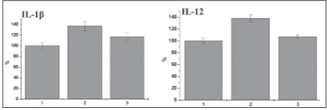
Changes in serum pro- and anti-inflammatory cytokine levels
Evidence has been presented that obesity is linked to a state of chronic low-grade inflammation [41,42]. During the last decades, understanding of the biol-ogy of adipose tissue, especially its secretory func-tions, has dramatically improved. A leading
hypoth-esis in this regard is that adipose tissue is not simply a storage reservoir of fat but also an active endocrine organ that, due to production of a variety of bioac-tive molecules, plays multiple metabolic roles in the regulation of whole-body physiology [43]. Adipocytes and preadipocytes have been identified as sources of pro-inflammatory cytokines, including IL-1ß, IL-6, IL-8, TNF-α, which are involved in the triggering of chronic inflammation in the adipose tissue [44]. In addition, oxidative stress is associated with an irregu-lar production of adipokines that in turn are potent stimulators of the ROS and reactive nitrogen species production by macrophages and monocytes. These changes can amplify the set of metabolic alterations. Chronic inflammation and high levels of associated pro-inflammatory cytokines are closely related with insulin resistance development. Therefore, pharma-ceutical strategies that target the inflammatory mi-lieu could correct obesity-related metabolic disorders. Considering the relationship between oxidative stress and pro-inflammatory cytokine levels, as well as the positive effect of the CF treatment on oxidative stress biomarkers, we tested whether CFs could improve the cytokine balance in obesity rats. As can be
ob-Fig. 2. MDA concentrations (A) and enzymatic activities of SOD(B)and CAT (C) in sera of control, HCD and HCD+CFs rats. 1 – control; 2 – group of rats fed a high-calorie diet (HCD); 3 or HCD+CFs – group of HCD rats that were intragastrically administered collagen fragments (CFs) (1 g·kg-1 of body weight).
served in Fig. 3, the serum level of pro-inflammatory cytokines IL-1β and IL-12 was 1.36- and 1.38-fold higher in rats that were on the HCD than in control animals (p<0.05). IL-1β acts as the instigator of the pro-inflammatory response via induction of other pro-inflammatory cytokines, in particular IL-6 [41]. Therefore, the accumulation of this cytokine in the se-rum of obese rats could indicate a cytokine imbalance. We found that the relative levels of serum IL-1β and IL-12 were significantly lower in the CF-treated rats than in HCD animals (p<0.05). These results are in agreement with the results of other authors [45] who described a lowering effect of marine-derived peptides on IL-1β, IL-6 and TNFα production. The decrease in the relative level of pro-inflammatory cytokines un-der CF treatment could be explained in part by their ability to decrease body weight, which is associated with an improvement in the cytokine profile as well as with a decreased risk of low-grade inflammation. The results of the examination of the anti-inflammatory cytokine levels in the sera of rats exposed to a HCD, and in rats treated with CFs are demonstrated in Fig. 4. Serum anti-inflammatory cytokine IL-4 was at the control level in rats of both investigated group. The level of another anti-inflammatory cytokine, IL-10, was also within the control range in rats of the HCD group. We observed that CF administration resulted in a slight elevation in IL-10 when compared to the control value (p<0.05) and that of the HCD group
(p<0.05). This suggests that while CFs did not exert a significant effect on the anti-inflammatory cytokine level, they influenced the level of pro-inflammatory cytokine.
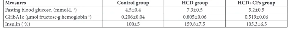
Parameters of insulin resistance
Systemic chronic inflammation and prolonged oxida-tive stress are viewed as important factors in the patho-genesis of obesity-related metabolic dysfunctions, in particular insulin resistance and impaired glucose me-tabolism [14, 46]. It has been shown that the markers of inflammation and oxidative damage are higher in obese individuals and that they are in direct correla-tion with the BMI and percentage of body fat. Insulin resistance, in addition to being caused by obesity, can contribute to the development of obesity-mediated complications [47]. It was reported that fish protein hydrolysates are effective in hyperglycemia manage-ment. It was observed that administration with dietary cod (morrhua) proteins reduced metabolic disorders in individuals suffering from type 2 diabetes mellitus by improving glucose tolerance and insulin sensitiv-ity [48]. Taking into account our results on the abilsensitiv-ity of CFs to improve the oxidative status and cytokine profile, we tested whether the treatment with CFs af-fected the development of insulin resistance in rats with diet-induced obesity. To this end we measured some parameters associated with insulin resistance develop-ment (blood glucose concentration, the level of glycated hemoglobin and insulin concentration). According to our results, diet-induced obesity was accompanied by changes in blood glucose concentration in HCD rats (Table 2). While control rats had a normal concentra-tion of blood glucose after 10 weeks, in HCD animals the blood glucose concentration was 7.3±0.5 mmol·L–1, which was 1.6-fold higher than in the control group (p<0.05). The observed increase in this parameter pointed to initial changes in glucose metabolism in obese rats. The increased concentration of blood glu-cose could be the result of impaired insulin secretion
Fig. 4. Relative changes in anti-inflammatory cytokine (IL-4 and IL-10) levels in sera of control, HCD and HCD+CFs rats. 1 – control; 2 – group of rats fed a high-calorie diet (HCD); 3 or HCD+CFs – group of HCD rats that were intragastrically ad-ministered collagen fragments (CFs) (1 g·kg-1 of body weight).
Table 2. Fasting blood glucose concentration, relative level of insulin, and level of glycated hemoglobin (GHbA1c) in the blood of control, HCD and HCD+CFs rats.
Measures Control group HCD group HCD+CFs group
Fasting blood glucose, (mmol·L–1) 4.5±0.4 7.3±0.5 5.2±0.5
GHbA1c (μmol fructose∙g hemoglobin-1) 0.206±0.04 0.805±0.06 0.519±0.06
due to pancreatic β-cell dysfunction or/and reduced sensitivity of peripheral tissues to the biological effects of insulin. The treatment with CFs did not produce a significant effect on blood glucose concentration when compared to the control. The concentration of blood glucose was 5.2±0.5 mmol·L–1, which was within the normal range (3.5-5.5 mmol·L–1). We therefore exam-ined the influence of CF treatment on the serum level of GHbA1c, which is in direct proportion to the average blood glucose concentration during the period corre-sponding to the lifespan of erythrocytes. We found a significant increase in the concentration of GHbA1c in the HCD group of rats (p<0.01) (Table 2). The serum GHbA1c level in rats treated with CFs decreased from 0.805±0.06 μmol fructose∙g hemoglobin-1 established for the obesity ratsto 0.519±0.06 μmol fructose∙g he-moglobin-1 (p<0.05). To explore potential mechanism that might explain the glucose-regulatory effect of CFs, we estimated the insulin level in HCD rats and HCD rats treated with CFs. Our study revealed that long-term exposure to HCD was accompanied by hyper-insulinemia. We found a 1.58-fold increase in plasma insulin concentration was in HCD rats compared to control animals (p<0.01) (Table 2). This result could be a manifestation of a compensatory reaction that usu-ally takes place at the early stage of insulin resistance development. It is known that high blood glucose con-centration stimulates insulin production by pancreatic β-cells; thus, type 2 diabetic individuals often exhibit excessive insulin production and hyperinsulinemia. The serum insulin concentration in the CF-treated rats was within the control value and significantly lower than in the HCD group (p<0.01). Therefore, we were unable explain the glucose-lowering effect of CFs by increased insulin secretion. There is no doubt that weight loss can improve insulin sensitivity [48]. Normalization of blood glucose concentration and insulin level in the CF-treated HCD rats might be linked with the reduc-tion in body weight that resulted in increased insulin sensitivity. The obtained results are in good agreement with [12], where it was noted that a daily treatment with marine collagen peptides from a fish hydrolysate improved glucose and lipid metabolism in Chinese pa-tients with type 2 diabetes mellitus.
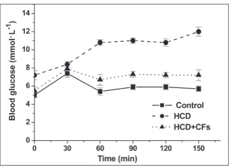
Finally, to confirm the state of insulin resistance in obese rats and to assess whether this state could be improved by CF treatment, we performed OGTT at the end of 10th week. This test estimates the efficiency
of the body to metabolize glucose in the circulation, as indicated by the nature of the glycemic curves fol-lowing glucose administration. Our results show that the development of obesity was accompanied by a de-crease in the sensitivity of peripheral tissues to the hypoglycemic effect of insulin. This conclusion was drawn after a comparison of the OGTT data of the control animals and the HCD animals. Judging from the glycemic curves, the blood glucose concentra-tion in the HCD rats was higher than in the control animals (Fig.5). Glucose loading to normal rats was accompanied by an increase in blood glucose concen-tration from 5.1±0.3 mmol·L-1 to 7.4±0.4 mmol·L-1 at 30 min of monitoring. The glucose concentration returned to normal at 150 min. In contrast, glucose loading of HCD-fed rats produced a gradual increase in serum glucose concentration during the test period, from 7.2±0.5 mmol·L-1 at 0 min, to 12.0±0.5 mmol·L-1 at 150 min. We observed partial normalization in OGTT in HCD rats treated with CFs. In this group, the blood glucose concentrations were lower than in the HCD group at all time points, except at 30 min.
Taken together, the obtained results show an im-provement of glucose metabolism in obese rats treated with CFs. Considering the strong relationship between obesity and insulin resistance, the positive effect of CF administration might be explained by their influence on body weight gain and body fat content (as indicat-ed by the BMI). Weight rindicat-eduction is accompaniindicat-ed by
Fig. 5. Glycemic curves, obtained from the OGTT of rats of
a decrease in intra-abdominal fat and non-esterified fatty acids, which are one of the most critical factors in modulating insulin sensitivity. Another mechanism of CF action could involve the reduction of oxidative stress and inflammatory mediators, as evidenced by the decreased levels in serum pro-inflammatory cy-tokines and MDA alongside the increased activities of SOD, CAT and levels of anti-inflammatory cytokines.
The present study showed that supplementation with CFs exerts a beneficial effect on diet-induced obesity development. Treatment with CFs restored impaired glucose homeostasis in obese rats most likely through an effect on body weight. The decrease in body fat was reflected as a decrease in weight, an improved cytokine profile and increased antioxidant capacity that was impaired with obesity. Therefore, weight loss through consumption of CFs may be the key to reducing the risk of developing pathologies related with obesity. CFs, being safe and multi-func-tional, have a strong potential for long-term use as a supplement agent for different types of illness involv-ing oxidative stress and inflammation. Further inves-tigations should be directed toward the determination of the pathways of CF activity.
Acknowledgments: This research did not receive any specific
grant from funding agencies in the public, commercial or not-for-profit sectors.
Author contributions: SOM conceived the idea. PPY and GTI
carried out of the experiments. RNG managed the analysis and interpretation of data and wrote the first draft of the manuscript. YAV participated in the literature survey and performed the sta-tistical analysis. OLI critically revised the manuscript. All authors read and approved the final manuscript.
Conflict of interest disclosure: The authors declare they have no conflict of interests.
REFERENCES
1. Hossain P, Kawar B, Nahas M. Obesity and diabetes in the developing world - a growing challenge. N Engl J Med. 2007;356(3):213-5.
2. Reaven G.M. Insulin resistance: the link between obesity and cardiovascular disease. Endocrinol Metab Clin North Am. 2008;37:581-601.
3. Mokdad AH, Ford ES, Bowman BA, Dietz WH, Vinicor F, Bales VS, Marks JS. Prevalence of obesity, diabetes, and obe-sity-related health risk factors, 2001. JAMA. 2003;289:76-9.
4. Aneiros A, Garateix A. Bioactive peptides from marine sources: pharmacological properties and isolation proce-dures. J Chromatogr B Analyt Technol Biomed Life Sci. 2004;803(1):41-53.
5. Ngo D-H, Vo T-S, Ngo D-N, Wijesekara I, Kim S-K. Biologi-cal activities and potential health benefits of bioactive pep-tides derived from marine organisms. Int J Biol Macromol. 2012;51:378-83.
6. Harnedy PA, Richard J. FitzGerald RJ. Bioactive peptides from marine processing waste and shellfish: A review. J Func Foods. 2012;4:6-24.
7. Kim S-K, Mendis E. Bioactive compounds from marine pro-cessing byproducts - A review. Food Res Int. 2006;39:383-93. 8. Nagai T, Izumi M, Ishii M. Fish scale collagen.
Prepara-tion and partial characterizaPrepara-tion. Int J Food Sci Tech. 2004;39:239-44.
9. Wang H, Fu Z, Han C. The potential applications of marine bioactives against diabetes and obesity. American J Marine Sci. 2014;2(1):1-8.
10. Zhang H, Dong Y, Qi B, Liu L, Zhou G, Bai X, Yang Ch, Zhao D, Zhao Y. Preventive effects of collagen peptide from deer sinew on bone loss in ovariectomized rats. Evid Based Complement Alternat Med. 2014;2014:627285.
11. Wang B, Wang YM, Chi CF, Luo HY, Deng SG, Ma JY. Isola-tion and characterizaIsola-tion of collagen and antioxidant colla-gen peptides from scales of croceine croaker (Pseudosciaena crocea). Marine Drugs. 2013;11(11):4641-61.
12. Zhu C-F, Li G-Z, Peng H-B, Zhang F, Chen Y, Li Y. Treat-ment with marine collagen peptides modulates glucose and lipid metabolism in Chinese patients with type 2 diabetes mellitus. Appl Physiol Nutr Metab. 2010;35:797-804. 13. Furukawa S, Takuya Fujita T, Shimabukuro M, Iwaki M,
Yamada Y, Nakajima Y, Nakayama O, Makishima M, Mori-hiro M, Shimomura I. Increased oxidative stress in obe-sity and its impact on metabolic syndrome. J Clin Invest. 2004;114:1752-61.
14. Xu H, Barnes GT, Yang Q, Tan G, Yang D, Chou CJ, Sole J, Nichols A, Ross JS, Tartaglia LA, Chen H. Chronic inflamma-tion in fat plays a crucial role in the development of obesity-related insulin resistance. J Clin Investig. 2003;112:1821-30. 15. Zhang F, Wang A, Li Z, He S, Shao L. Preparation and
char-acterisation of collagen from freshwater fish scales. Food Nutr Scie. 2011;2:818-23.
16. Huang Q, Li S, Teng H, Jin Y, Ma M, Song H. Optimizing preparation conditions for angiotensin-I-converting enzyme inhibitory peptides derived from enzymatic hydrolysates of ovalbumin. Food Sci Biotechnol. 2015;24(6):2193-98. 17. Laemmli UK. Cleavage of structural proteins during
the assembly of the head of bacteriophage T4. Nature. 1970;227:680-85.
18. Shen X-H, Tang Q-Y, Huang J, Cai W. Vitamin E regulates adipocytokine expression in a rat model of dietary-induced obesity. Exp Biol Med. 2010;235:47-51.
19. Bradford MM. A rаpid and sensitive method for quantities of utilizing the principle of protein binding. Anal Biochem. 1976;86:193-200.
C60 fullerene nanostructures in liver and brain of rats with streptozotocin-induced diabetes. J Diabetes Metab. 2012;3(8):1-9.
21. Sirota TV. A novel approach to study the reaction of adren-aline autooxidation: A possibility for polarographic deter-mination of superoxide dismutase activity and antioxidant properties of various preparations. Biochem (Moscow) Suppl Series B: Biomed Chem. 2011;5(3):253-59.
22. Korolyuk MA, Ivanov LI, Mayorova IG, Tokarev VE. Method for determining the activity of catalase. Lab Delo.1988;1:16-9. 23. Halenova TI, Vareniuk IM, Roslova NM, Dzerzhynsky ME,
Savchuk OM, Ostapchenko LI, Prylutskyy YuI, Ritter U, Scharff P. Hepatoprotective effect of orally applied water-soluble pristine C60 fullerene against CCl4-induced acute liver injury in rats. RSC Adv. 2016;6:100046-55.
24. Aoyama T, Fukui K, Nakamori T, Hashimoto Y, Yamamoto T, Takamatsu K. Effect of soy and milk whey protein isolates and their hydrolysates on weight reduction in genetically obese mice. Biosci Biotech Biochem. 2000;64:2594-00. 25. Karpovets TP, Konopelnyuk VV, Savchuk OM, Ostapchenko
LI. Food behavior of rats under development of obesity. Res J Pharmaceut Biol Chem Sci. 2014;5(5):253-9.
26. Nоvеllі Е, Dіnіz Y, Gаlhаrdі C. Аnthrоpоmеtrіcаl pаrаmеtеrsаnd mаrkеrsоf оbеsіtyіn rаts. Lаb Аnіmаls. 2007;41:111-19.
27. Sеаlе P, Lаzаr M. Brоwn fаt іn humаns: turnіng up thе hеаt оn obеsіty. Dіаbеtеs. 2009;58(7):1482-4.
28. Moran TH. Cholecystokinin and satiety: current perspec-tives. Nutr. 2000; 16(10):858-65.
29. Higdon J, Frei B. Obesity and oxidative stress: A direct link to CVD? Arterioscler Tromb Vasc. Biol. 2003;23:365-67. 30. Sarmadi BH, Ismail A. Antioxidative peptides from food
proteins: A review. Peptides. 2010;31:1949-56.
31. Qian ZJ, Jung WK, Byun HG, Kim SK. Protective effect of an antioxidative peptide purified from gastrointestinal digests of oyster, Crassostrea gigas against free radical induced DNA damage. Biores Technol. 2008;99:3365-71.
32. Mendis E, Rajapakse N, Byun HG, Kim SK. Investigation of jumbo squid (Dosidicus gigas) skin gelatin peptides for their in vitro antioxidant effects. Life Sci. 2005;77:2166-78. 33. Ranathunga S, Rajapakse N, Kim S-K. Purification and
char-acterization of antioxidative peptide derived from muscle of conger eel (Conger myriaster). Eur Food Res Technol. 2006;222:310-5.
34. Grosser N, Oberle S, Berndt G, Erdmann K, Hemmerle A, Schröder H. Antioxidant action of L-alanine: heme oxygen-ase-1 and ferritin as possible mediators. Biochem Biophys Res Commun. 2004;314(2):351-5.
35. Himaya SW, Ryu B, Ngo DH, Kim SK. Peptide isolated from Japanese flounder skin gelatin protects against cellular oxi-dative damage. J Agric Food Chem. 2012;60:9112-9. 36. Kang JH. Modification and inactivation of
Cu,Zn-superox-ide dismutase by the lipid peroxidation product, acrolein. BMB Rep. 2013;46(11):555-60.
37. Ishizaki-Koizumi S, Sonaka I, Fujitani S, Nishiguchi S. Mechanisms of the protective effect of L-alanine to D-galac-tosamine-induced hepatocellular injury: comparative studies of L-alanine and pyruvate. Biochem Biophys Res Commun. 2002;291:738-43.
38. Lin L, Li B. Radical scavenging properties of protein hydro-lysates from Jumbo flying squid (Dosidicus eschrichitii Steen-strup) skin gelatin. J Sci Food Agric. 2006;86:2290-5. 39. Liu GQ, Zhu CF, Li YF, Peng HB, Lu H, Guo SC. Effect of
marine collagen peptide on the expression of overoxidation stress markers in rats with type 2 diabetes. J Clin Rehabil Tissue Eng Res. 2008;12:4469-72.
40. Lin B, Zhang F, Yu Y, Jiang Q, Zhang Z, Wang J, Li Y. Marine collagen peptides protect against early alcoholic liver injury in rats. British J Nutr. 2012;107:1160-6.
41. Stienstra R, Tack CJ, Kanneganti TD, Joosten LA, Netea MG. The inflammasome puts obesity in the danger zone. Cell Metab. 2012;15:10-8.
42. McArdle MA, Finucane OM, Connaughton RM, McMorrow AM, Roche HM. Mechanisms of obesity-induced inflamma-tion and insulin resistance: insights into the emerging role of nutritional strategies. Front Endocrinol. 2013;4:1-23. 43. Fonseca-Alaniz MH, Takada J, Alonso-Vale MI, Lima FB.
Adipose tissue as an endocrine organ: From theory to prac-tice. J Pediatr. 2007;83:192-203.
44. Rasouli N, Kern PA. Adipocytokines and the meta-bolic complications of obesity. J Clin Endocrinol Metab. 2008;93(1):S64-S73.
45. Ahn CB, Cho YS, Je JY. Purification and anti-inflammatory action of tripeptide from salmon pectoral fin byproduct pro-tein hydrolysate. Food Chem. 2015;168:151-6.
46. Kahn SE, Hull RL, Utzschneider KM. Mechanisms linking obesity to insulin resistance and type 2 diabetes. Nature. 2006;444:840-6.
47. Kahn BB, Flier JS. Obesity and insulin resistance. J Clin Invest. 2000;106(4):473-81.