R E V I E W
Open Access
Autonomic nervous system involvement in
pulmonary arterial hypertension
Mylène Vaillancourt
†, Pamela Chia
†, Shervin Sarji, Jason Nguyen, Nir Hoftman, Gregoire Ruffenach,
Mansoureh Eghbali, Aman Mahajan and Soban Umar
*Abstract:Pulmonary arterial hypertension (PAH) is a chronic pulmonary vascular disease characterized by increased pulmonary vascular resistance (PVR) leading to right ventricular (RV) failure. Autonomic nervous system involvement in the pathogenesis of PAH has been demonstrated several years ago, however the extent of this involvement is not fully understood. PAH is associated with increased sympathetic nervous system (SNS) activation, decreased heart rate variability, and presence of cardiac arrhythmias. There is also evidence for increased renin-angiotensin-aldosterone system (RAAS) activation in PAH patients associated with clinical worsening. Reduction of neurohormonal activation could be an effective therapeutic strategy for PAH. Although therapies targeting adrenergic receptors or RAAS signaling pathways have been shown to reverse cardiac remodeling and improve outcomes in experimental pulmonary hypertension (PH)-models, the effectiveness and safety of such treatments in clinical settings have been uncertain. Recently, novel direct methods such as cervical ganglion block, pulmonary artery denervation (PADN), and renal denervation have been employed to attenuate SNS activation in PAH. In this review, we intend to summarize the multiple aspects of autonomic nervous system involvement in PAH and overview the different pharmacological and invasive strategies used to target autonomic nervous system for the treatment of PAH.
Keywords: Pulmonary arterial hypertension, Autonomic nervous system, Right ventricle, Sympathetic nervous system, Renin angiotensin aldosterone system
Overview of the autonomic regulation of heart and lungs
The autonomic nervous system
The autonomic nervous system is composed of sympa-thetic and parasympasympa-thetic divisions and is often divided by neural and endocrine regulatory components. The sympathetic nervous system (SNS) originates from the thoracolumbar region of the spinal cord (Fig. 1). Short preganglionic fibers from the T1-L2 segments synapse on paravertebral or prevertebral ganglia, enabling long post-ganglionic fibers to innervate target organs such as the heart and lungs. On the other hand, the parasympathetic nervous system originates from cranial nerves III, VII, IX, and X and the sacral nerves S2-S4. In general, parasympa-thetics cause vasodilation of blood vessels including the
pulmonary vasculature, and sympathetics cause vasocon-striction [1] (Table 1).
Autonomic innervation of the pulmonary vasculature The pulmonary vasculature is innervated by sympathetic, parasympathetic, and sensory nerve fibers. Increased vas-cular resistance is mediated by α-adrenoreceptors upon sympathetic nerve stimulation [2]. Noradrenergic fibers are activated by baroreceptors in the pulmonary artery [3] and proximal airway segments [4]. Chemoreceptors re-spond to decreased arterial PO2 levels to increase sympa-thetic nerve stimulation by the sympasympa-thetic chain neurons [2, 5]. Parasympathetic activation via vagal stimulation re-sults in cholinergic-mediated relaxation of pulmonary ar-teries [6]. Many other factors (i.e. non-adrenergic and non-cholinergic mediators, peptides, trophic factors, dif-ferential release of transmitters by high or low frequencies) are implicated in sympathetic and parasympathetic regula-tion of lung vasculature, though their funcregula-tions have not entirely been elucidated [7] (Table 1).
* Correspondence:sumar@mednet.ucla.edu †Equal contributors
Department of Anesthesiology and Perioperative Medicine, Division of Molecular Medicine, David Geffen School of Medicine, University of California Los Angeles (UCLA), Los Angeles, CA BH 520A CHS, USA
Autonomic innervation of the heart
The heart is also innervated by both parasympathetic and sympathetic fibers (Fig. 1). The parasympathetic fibers are responsible for decreasing chronotropy, dromotropy, and inotropy via cholinergic action on cardiac M2 receptors. The SNS acts on β1 adrenergic receptors to increase chronotropy, dromotropy, and inotropy of the heart [8]. Interestingly,β-adrenergic stimulation has been shown to have a significantly greater positive inotropic effect on left ventricular (LV) contractility than on right ventricular (RV) contractility [9]. On the contrary, adrenergic stimula-tion of alpha 1 receptors result in increased inotropy in the LV but decreased inotropy in the RV [10] (Table 1).
Patients with PAH often have normal systemic blood pressures and lung volumes. However, they may suffer
from hypoxia, hypercarbia, acidosis, and, in later stages, RV hypertrophy and failure. Neural pathways controlling the heart and lungs are described in detail within current scientific literature [11].
Autonomic nervous system and RAAS involvement in PAH
Table
1
Adrenergic,
cholinergic,
and
angiotensin
receptors
and
the
effect
of
their
activation
in
heart
and
lungs
(Continued)
Recept
ors
Rece
ptor
Sub-types
Ac
tivators
Hea
rt
Lung
AT2
•
Angi
oten
sin
II
•
Count
eracts
effect
s
of
AT1
rece
ptor
in
cardiac
hypertro
phy
an
d
remo
deling
–
car
diopro
tective
[
88
]
•
Involved
in
cell
growth
,
prolif
eration,
diff
erentiat
ion,
mi
gration,
an
d
apoptos
is
•
Not
clear
if
AT2
agon
ism
ha
s
acut
e
effect
s
on
vascular
ton
e
•
D
ecreases
pulmonary
vasc
ular
res
istance
[
62
]
•
D
ecreased
inflam
mation
•
D
iminish
ed
fibrosis
•
Prom
otes
cell
differ
entiation
an
d
apoptosis
•
Re
duces
cell
prolif
cardiac output and may compensate for that by upregula-tion of neurohormonal systems such as SNS and renin-angiotensin-aldosterone system (RAAS) [12] (Fig. 1). In-flammatory and RAAS molecules are specifically upregu-lated in PAH and are implicated in the development of the disease through their effects on the brain [13].
There is an increasing body of evidence linking auto-nomic nervous system involvement in the pathogenesis of PAH. Here we review the available literature on SNS activation, heart rate variability, baroreflex sensitivity and arrhythmias, and RAAS dysregulation in PAH. We also summarize the therapeutic strategies for modulating the autonomic nervous system and RAAS in PAH.
Sympathetic nervous system activation in PAH Over the last 30 years, accumulating evidence has sup-ported the involvement of the autonomic nervous system in PAH, strengthening the hypothesis for the role of SNS activation in PAH development. Microneurogra-phy was used to compare the muscle sympathetic nerve activity (MSNA) between patients with PAH and healthy controls and showed increased sympathetic nerve traffic in PAH patients [14]. MSNA was also directly correlated with heart rate, presence of pericardial effusion, oxygen saturation, New-York Heart Association class, 6-min walk distance (6-MWD), and pulmonary arterial acceler-ation time and was associated with clinical deterioracceler-ation [14, 15]. Administration of hyperoxia decreased MSNA frequency and burst amplitude, suggesting that periph-eral chemoreceptors contribute in part to increased MSNA [14].
Increased SNS activity in PAH was also assessed by neu-rohormonal activation, despite conflicting results between studies. In a small clinical study comprising 32 patients with PAH, plasma norepinephrine concentration was strongly correlated to PVR and was associated with poor estimated 5-year survival [16]. Some other studies have shown an increase in plasma norepinephrine concentra-tion in PAH patients compared to healthy controls, al-though these norepinephrine levels did not exceed normal values and were not correlated with any hemodynamic parameter [17, 18]. To determine the involvement of cat-echolamine pathway in PAH, many groups have directed their research towards catecholamine receptors.
Bristow and colleagues [19] were the first to describe local changes in β-adrenergic receptors in the failing RV myocardium of PAH patients. Since then, the impact ofβ -adrenergic receptor signaling in PAH development has been extensively studied using different α/β-adrenergic re-ceptor agonists/antagonists. Ishikawa and colleagues [20] showed that administration of arotinolol, a pureα/β -adren-ergic receptor antagonist, prevents monocrotaline (MCT)-induced PH development by keeping cardiopulmonary pressures below pathological threshold and decreasing RV/
body weight ratio in treated rats. Treatment with the non-selective adrenergic receptor antagonist carvedilol was also reported to reverse established RV failure in two different rat models of PH (Sugen/hypoxia and MCT-induced PH) and improved survival in MCT rats [21, 22]. This improve-ment in RV function was associated with reduced RV hypertrophy, dilation, and fibrosis, as well as an improved capillary density of the myocardium [21]. Interestingly, car-vedilol also decreased pro-fibrotic signaling and extracellu-lar matrix remodelling in right and left ventricles of treated rats via transforming growth factor-β1, connective tissue growth factor, SMAD2/3, p38, and metalloproteinases 2 and 3 pathways. Drake and colleagues [23] performed an extensive microarray gene expression analysis on the RV of Sugen/hypoxia PH rats either treated with carvedilol for 4 weeks or untreated. Within the top canonical pathways revealed in this analysis, one was involved in cardiac hyper-trophy by protein synthesis and included regulation of eukaryotic initiation factor 4 and 5 and p70S6k signaling, all of which were downregulated with carvedilol. Ceramide signaling and glucocorticoid receptor signaling pathways, considered injurious for the heart, were also two of the top five canonical pathways and were downregulated in carvedilol-treated rats. Other canonical pathways included the peroxisome proliferator-activator receptor signaling, peroxisome proliferator-activator receptor/retinoid X receptor-activation, and nuclear respiratory factor 2-mediated oxidative stress response. These pathways are in-volved in metabolism, mitochondrial function, and oxida-tive stress response and are critical for adequate heart function. All these findings highlight the role of adrenergic receptors in RV failure and suggest that the use of β -blockers could be beneficial for PAH patients. In the clinical settings, carvedilol showed encouraging results by improv-ing RV ejection fraction in chronic heart failure [24] and did not lead to any notable adverse event or deterioration [25, 26] (Table 2). In a recent 6-month double-blind, ran-domized, controlled trial, patients treated with carvedilol had an improved heart rate recovery after exercising com-pared to those who received placebo [26]. This is important since heart rate recovery after exercise is not only a pre-dictor of increased risk of death but also of clinical worsen-ing in PAH. RV function was also assessed by an improved glycolytic rate related to carvedilol treatment. [26] Clinical trials are still ongoing to confirm the therapeutic benefit and safety of carvedilol in PAH patients (ClinicalTrials.gov Identifier: NCT02120339 and NCT02507011).
Table 2Clinical trials on neurohormonal modulation in pulmonary hypertension
Study Treatment Study design
Follow-up
Subjects Patient description (n patient)
Changes in outcomes Related side effects (n patient)
Pharmacological therapies Grinnan and
colleagues [25]
Carvedilol Single-arm, open-label, pilot
6 months 6 iPAH (3) HPAH (1) APAH (2)
↑RVEF ↑BNP level
Bradycardia (1) Asymptomatic hypotension (1) Mild fatigue (1) Farha and
colleagues [26]
Carvedilol Single-center, double-blind, randomized, controlled trial
6 months 30 iPAH (9) HPAH (12) APAH (5)
PH due to lung disease and/or hypoxemia (2) Chronic
thromboembolic PH (2) ↓HR
↓RVSP at 3 month follow-up ↑RV fractional area change at 3 month follow-up ↑RV glycolytic rate ↑β-adrenergic receptor density
Fatigue (1) Dyspnea (2) Leg swelling (3) Site infection/acute bronchitis (2) Chest pain (1) Blurry vision (1) Cholecystitis (1) Bloating (1) Nausea/vomiting (1) Dizziness (1) van Campen
and colleagues [29]
Bisoprolol Prospective, randomised, placebo-controlled, crossover
1 years 18 iPAH (18) ↓HR
↑PAWP ↓Cardiac index ↓6-MWD
Worsening of fluid retention (1) Hypotension (1) Tiredness (1) Feelings of depression (1) Bandyopadhyay
and colleagues [31]
Atenolol Bisoprolol Carvedilol Metaprolol Nebivelol Propranolol Sotalol
Retrospective cohort
5 years 568 iPAH (260) APAH (308)
None reported Therapy discontinued (60): Hypotension (18) Shortness of breath (14)
Volume overlaod (12)
Fatigue (7) Bradycardia/ Syncope (4) Intolerance (3) Other (2) Moretti and
colleagues [32]
Atenolol Bisoprolol Metaprolol Nadolol Propranolol
Prospective cohort
2 years 94 iPAH (14) APAH (27) Post pulmonary embolism (13) Out of proportion (8) Mixed (5)
Unknown (27)
↑TAPSE ↓RV diameter ↓HR
↓Systolic blood pressure
None reported
So and colleagues [33]
Acebutolol Atenolol Bisoprolol Metaprolol Nadolol Propranolol
Prospective cohort
2 years 94 iPAH (53)
Drug/toxin-iduced PAH (2)
APAH (39)
↑Cardiac index None reported
Thenappan and colleagues [34]
Atenolol Carvedilol Labetalol Metaprolol Nadolol Propranolol
Retrospective cohort study, propensity score analysis
5 years 564 iPAH (250) HPAH (17) Drug/Toxin-induced (21)
APAH (273) Other (3)
None reported None reported
Bozbas and colleagues [ 60]
Losartan Nifedipine
Prospective, randomized
2 months 63 PH due to left heart disease (40)
PH due to lung disease and/or hypoxemia (23)
↓mean PAP ↑RVEF ↑6-MWD ↓VE ↓VE/VCO2
↑PETCO2
↑CPET test duration
None reported
fibrosis and myocardial inflammation. Bisoprolol restored
β-adrenergic receptor signaling assessed by increased phosphorylation of its downstream targets, myosin binding protein C and troponin I. In contrast, decreased phosphor-ylation of these proteins in PAH cardiomyocytes leads to an increase in sarcomere Ca2+ sensitivity, thus impairing RV relaxation and contributing to RV stiffness [28]. How-ever, these beneficial effects were not confirmed in an ex-plorative study involving 18 PAH patients (Table 2) [29]. In this randomized, placebo-controlled, crossover study, bisoprolol did not improve patients’conditions. Despite a trend to increase RV ejection fraction, patients had a sig-nificant decrease in cardiac index and a near sigsig-nificant drop in 6-MWD, demonstrating no real benefit of bisopro-lol in PAH (Table 2).
Finally, Perros and colleagues [30] compared the effects of nebivolol, a third generation β-adrenergic receptor blocker, to the first generation β1-adrenergic receptor
blocker metoprolol in MCT-induced PH rats. Nebivolol is a β1-adrenergic receptor antagonist and β2,3-adrenergic receptor agonist and has vasodilator properties in addition to its adrenergic-modulating characteristics. Daily adminis-tration of nebivolol for one week in established PH im-proved cardiopulmonary hemodynamics and partially reversed RV hypertrophy and pulmonary vascular remodel-ing with a greater effect than metropolol. In vitro nebivolol, but not metropolol, significantly decreased human pul-monary endothelial cell proliferation as well as the produc-tion of pro-inflammatory cytokines such as interleukin-6 and monocyte chemoattractant protein-1, epidermal and fibroblast growth factors, and the vasoconstrictor endothelin-1. Furthermore, human smooth muscle cell proliferation was decreased when cells were cultured in the endothelial cell + nebivolol-conditioned media [30]. A clin-ical trial is currently recruiting patients to assess the thera-peutic potential and safety of nebivolol in clinical Table 2Clinical trials on neurohormonal modulation in pulmonary hypertension(Continued)
Study Treatment Study design
Follow-up
Subjects Patient description (n patient)
Changes in outcomes Related side effects (n patient)
Maron and colleagues [69]
Spironolactone ±Ambrisentan
Retrospective analysis of randomized, placebo-controlled trials ARIES-1 and 2
Nonprimary PAH (73) ↓BNP level ↓WHO class
Pulmonary hypertension (21) Edema or prevention of edema (11) RV failure (7) Electrolyte imbalance (1) Invasive strategies
Chen and colleague [74]
PADN Prospective
cohort
3 months 21 iPAH (21) ↓mean PAP
↓PVR ↓RVSP ↓TPG
↓Pericardial effusion ↑PA compliance ↑6-MWD ↓WHO class ↓NT-proBNP level ↓Rehospitalization
Chest pain (10)
Chen and colleagues [75]
PADN Prospective
cohort, single-arm
1 year 66 iPAH (20)
APAH (19) PH due to left heart disease (18) Chronic
thromboembolic PH (9)
↓mean PAP ↓PVR ↓RVSP ↑Cardiac output ↓Right atrial diameter ↓RV diameter ↓pericardial effusion ↑6-MWD
↓NT-proBNP level
Chest pain (47) Sinus bradycardia (1) Intolerance to dyspnea (3)
managment of the PAH patients (ClinicalTrials.gov Identi-fier: NCT02053246).
Despite encouraging results of adrenoreceptor block-ade in experimental PH, the use ofβ-blockers in clinical PAH is still largely debated due to the poor beneficial re-sults as well as safety concerns revealed in clinical trials [31–36] (Table 2). Although β-blockers partially reverse RV structural and molecular remodeling [23, 24] and seem beneficial and well-tolerated at low or escalating doses [25, 26, 29, 31–34], their negative inotropic and chronotropic effects may actually impair RV function in severe heart failure. Thus, the choice based on the speci-ficity of theβ-blocker used to target one or the otherβ -receptor sub-type may be of importance. The divergent results found in clinical studies may also be the conse-quence of several limiting factors, such as study design, cohort size, diversity in pulmonary hypertension etiology within cohorts, and variability in pre-existing comorbidi-ties and treatments taken by patients.
Heart rate variability and baroreflex sensitivity in PAH
Heart failure is associated with abnormalities in autonomic nervous system control, including decreased heart rate vari-ability and blunted baroreflex sensitivity [37], which were predictors of cardiovascular mortality among post-myocardial infarction patients [38, 39]. Considering the evi-dence of an altered autonomic control in PAH, these two parameters were investigated in different PAH cohorts. In a small cohort of 9 PAH patients, heart rate variability was assessed by a time-domain method and was found to be significantly decreased compared to the control group (n= 20) [40]. In a larger cohort, Wensel and colleagues [41] used a frequency-domain method to show a reduction of total power of heart rate variability in PAH patients (n= 42) compared to healthy subjects (n= 41) but without any significant difference between low/high frequency power ratio. Baroreflex sensitivity was also reduced in PAH pa-tients, determined by the controlled breath method and the
α-index of low frequency. Both heart rate variability and baroreflex sensitivity parameters were correlated with de-creased peak oxygen uptake during exercise, demonstrating the impact of cardiac autonomous modulation in exercise capacity [41]. In a cohort of 47 children with severe PAH, heart rate variability was correlated with 6-MWD and was predictive of transplantation and/or mortality [42]. On bi-variate analysis, standard deviation of mean values for the beat-to-beat interval over 5 min predicted outcome inde-pendently of functional status, syncope, RV function, and haemodynamic parameters [42]. More recently, Yi and col-leagues [43] also confirmed that time-domain heart rate variability parameters, as well as frequency-domain indices, were significantly decreased in PAH patients (n= 26) com-pared to the controls (n = 51). Furthermore, heart rate
variability parameters correlated to mean pulmonary arter-ial pressure (PAP) in patients [44, 45]. Further studies in larger patient cohorts are needed to confirm if the heart rate variability and baroreflex sensitivity changes seen in PAH are indeed associated with a decrease in the vagal tone and whether they carry prognostic significance.
Cardiac arrhythmias in PAH
In experimental PH, our group and others have described spontaneous ventricular fibrillation events and have exten-sively investigated their mechanisms [46–48]. Benoist and colleagues [46] showed in isolated hearts that the mono-phasic action potential duration at 25, 50 and 90% repolari-zation was significantly prolonged in hearts from MCT-induced PH rats compared to controls for both RV and LV. Futhermore, RV monophasic action potential duration at 90% repolarization strongly correlated with cardiac hyper-trophy. These changes in the action potential duration were associated with decreased K+and L-type Ca2+channel ex-pressions as well as an increase in T-type Ca2+channels in the RV of MCT rats [46]. In a subsequent study, the same group monitored MCT rats using electrocardiograms and showed an increased QT interval in RV failure from day 15 post-MCT injection consistent with the prolonged action potential duration [47]. Alternans of both T-wave amplitude and QT interval were also observed in animals with heart failure. In isolated hearts, alternans occurred in four out of six failing hearts and were always discordant. Action poten-tial duration alternans have been associated with dysfunc-tional Ca2+homeostasis, resulting in increased intracellular Ca2+concentration and contractile alternans. Ca2+transient alternans were confirmed in isolated myocytes from failing RVs and were shown to be provoked by dysregulation of sarcoplasmic Ca2+ uptake, load, and release. Consistent with this, failing RVs of MCT rats showed a reduced SER-CA2a activity, an increased sarcoplasmic reticulum Ca2+ re-lease fraction, and an increased Ca2+spark leak. [47].
death period, suggesting that mortality during this critical period may be caused by early afterdepolarizations-mediated triggered activity in the RV [48].
In clinical PAH, cardiac arrhythmias are common in patients and are associated with worsening prognosis. Cardiac arrhythmias include supraventricular arrhyth-mias (SVA), which are the most common with a re-ported incidence between 11 and 30% [49–53], and the ventricular arrhythmias, which were reported to be relatively rare in PAH [40]. The most common SVAs seen in PAH patients are atrial fibrillation, flut-ter, and tachycardia [49, 51, 53]. In a 6-year retro-spective single-center analysis including 231 PAH patients, 31 SVA episodes were detected in 27 pa-tients (cumulative incidence 11.7%, annual risk 2.8% per patient) [49]. In most episodes (n = 26), onset of supraventricular tachycardia resulted in clinical deteri-oration and/or RV failure. Furthermore, mortality was significantly higher in patients with persistent atrial fibrillation compared with the patients in whom sinus rhythm was restored. In a 5-year prospective study including 239 patients, at least one episode of atrial flutter or atrial fibrillation was detected in 20% of the patients [50]. Compared to patients who did not de-velop SVA, patients who dede-veloped atrial flutter and fibrillation had significantly higher baseline values for right atrial pressure, mean PAP, PVR, lower baseline
values for cardiac output, and mixed venous oxygen saturation. Futhermore, the estimated 1, 2, 3, and 5 year survival rates after diagnosis of PH in patients with permanent atrial fibrillation were 80, 66, 22, and 22%, respectively [50]. Presence of SVA in PAH pa-tients is also associated with deterioration of the RV function, assessed by an increase in B-type natriuretic peptide, atrial and ventricular diameter, mean atrial pressure, and PVR, as well as a reduction of the car-diac index [51]. The estimated survival rate is lower in patients who develope SVA, and is significantly worse in patients with permanent SVA compared to transient or without SVA [51, 52]. A good under-standing of cardiac arrhythmias and other autonomic nervous system dysregulations is important not only because they are determinants of the outcome for the patients, but they also determine the clinical manage-ment of PAH in these patients [49, 53].
Renin-angiotensin-aldosterone-system dysregulation in PAH
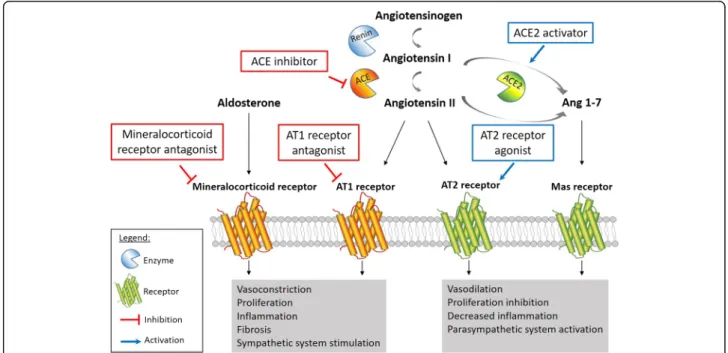
Chronic activation of the RAAS in PAH patients is well de-scribed. As extensively reviewed by Maron and Leopold, RAAS activation promotes pulmonary vasoconstriction, cell proliferation, migration, extracellular matrix remodeling and fibrosis resulting in pulmonary vascular remodeling in experimental PH (Fig. 2) [54].
Renin, angiotensin I, and angiotensin II
In PAH patients, circulating renin activity as well as plasma angiotensin I and II are significantly up-regulated and associated with disease worsening, suggesting a role for systemic RAAS activation in PAH progression [12, 55]. Futhermore, plasma renin and angiotensin II were strongly associated with an increased risk of death or lung transplant, making them potentially reliable biomarkers for clinical prognosis [12]. To determine whether local RAAS activation was also involved in PAH, de Man et al. [12] investigated angiotensin II signaling pathway in hu-man PAH lung tissues. They found a two-fold increase of angiotensin II receptor AT1, but not AT2, in the pulmon-ary vasculature of PAH patients along with an increase in activity of its downstream targets Src and ERK. This is an important finding given that AT1 signaling is involved in vasoconstriction, oxidative stress, inflammation, and pro-liferation, while AT2 signaling leads to vasodilation and is vasculoprotective (Fig. 2) [54]. Exposing pulmonary endo-thelial cells to angiotensin I revealed a significatly in-creased angiotensin II production by cells isolated from PAH patients compared to control, which was abolished with the angiotensin-converting enzyme (ACE) inhibitor enalapril [12]. This high angiotensin II production by endothelial cells induces smooth muscle cell proliferation, resulting in pulmonary vascular medial hypertrophy and obliteration. In vivo, pharmacological AT1receptor antag-onism using losartan significantly delayed disease progres-sion in MCT-induced PH rats by reducing RV afterload, restoring ventricular–arterial coupling, and improving RV diastolic function. Furthermore, losartan significantly re-duced pulmonary vascular remodeling in treated-rats but without any change in RV hypertrophy [12]. However, these results are inconsistent with previous studies in MCT rats using the same AT1 antagonist, in which re-searchers did not find any prophylactic effect of losartan against the development of PAH [56, 57]. Two independ-ent groups also tested losartan in a pressure-overload right heart failure model. Borgdorff and colleagues [58] treated pulmonary artery banded (PAB)-rats with combined losar-tan + eplerenone treatment until RV failure criteria were met or for a maximum of 11 weeks. Combined losartan + eplerenone treatment did not prevent adverse RV remod-eling or clinical RV failure. Interestingly, treated PAB rats did have a significant decrease in LV peak and aortic pres-sures, highlighting inherent differences between right and left ventricles and their significance in research for therap-ies in the context of PAH [58]. PAB model was also used to compare the preventive effect of losartan and bisoprolol on development of RV hypertrophy and dysfunction [59]. After 6 weeks of treatment with either losartan or bisopro-lol, rats did not show any signs of improvement in RV hypertrophy, dysfunction, fibrosis or capillary density. Fur-thermore, neither losartan nor bisoprolol reversed gene
expression levels of cardiac hypertrophy and dysfunction biomarkers.This study is additional evidence of how the RV differs substantially from the LV when responding to inhibition of the increased neurohormonal activation oc-curring in heart failure.
In the clinical setting, 33 PH patients from different etiologies were followed after 8 week- treatment of losar-tan (Table 2) [60]. A modest but significant decrease in mean PAP and increase in RV ejection fraction were ob-served in subjects taking losartan compared to their baseline. They also found an improvement in 6-MWD and in several cardiopulmonary exercise testing parame-ters. However, the duration of treatment was relatively brief, and the study was lacking a control group. Fur-thermore, since the cohort was comprised of subjects from different PH classification groups, further studies are needed to assess the real potential of losartan in PAH patients.
ACE2/Angiotensin-(1–7)/Mas axis
by the reduction of LC3B-II protein level. Li and col-leagues [64] demonstrated the prevention of PH using the pharmacological compound resorcinolnaphthalein, an ACE2 activator, in a severe PH model. Briefly, they injected a single dose of MCT (40 mg/kg) 1 week after performing pneumonectomy in rats. At the time of MCT injection, they implanted osmotic minipumps containing either resorcinolnaphthalein or the vehicle for 21 days of infusion. ACE2 activation decreased mean PAP and subse-quent RV hypertrophy without any effect on systemic pressure. This change in mean PAP was at least in part due to a partial restoration of acetylcholine-induced pul-monary vasorelaxation. Resorcinolnaphthalein also de-creased the neointimal formation in small pulmonary arteries, from 95% in MCT + pneumonectomy group to 29% in treated rats. In accordance with the findings of Fer-reira and colleagues [61], beneficial effects of ACE2 activa-tion in PAH seem to be at least partly mediated through angiotensin-(1–7)/Mas axis, since improvements of mean PAP, RV hypertrophy, pulmonary vasorelaxation, and neo-intimal formation were all abolished by the Mas antagon-ist A779 [64]. Finally, the role of ACE2/angiotensin (1–7)/ mas axis in autonomic nervous system modulation was demonstrated using diminazene aceturate, a putative angiotensin 1–7 converting enzyme activator [65]. Dimin-azene treatment resulted in significant improvement in power spectrum parameters, such as normalized high and low frequency components in treated rats compared to MCT alone, thus reversing the imbalance in the auto-nomic nervous system modulation seen in PH. Clinical trials are now in progress to assess mechanism, safety, and efficacy of ACE-2 treatment for PAH patients (Clinical-Trials.gov Identifier: NCT01884051 and NCT03177603).
Aldosterone
Another key player of RAAS is the steroid hormone al-dosterone. Maron and colleagues [66] found an increase of aldosterone levels in plasma and lung tissues of MCT- and Sugen/hypoxia-induced PH animals. Elevated aldosterone levels were mediated by endothelin-1 via
peroxisome proliferator-activated receptor gamma
coactivator-1α/steroidogenesis factor-1, and increased oxidative stress in pulmonary artery endothelial cells, leading to an inhibition of nitric oxide production. Both in preventive and rescue protocols, spironolactone, a mineralocorticoid receptor antagonist, improved RV hypertrophy, PAP, PVR, and pulmonary artery remodel-ing along with decreased reactive oxygen species gener-ation and restorgener-ation of nitric oxide production [66]. The therapeutic effect of spironolactone was further confirmed in a chronic hypoxia-induced PH mouse model [67]. Furthermore, the latest study demonstrated that mineralocorticoid receptor, once activated by aldos-terone, induces transcriptional activity and proliferation
in pulmonary arterial smooth muscle cells, which was prevented by spironolactone [67].
In a clinical study, Maron and colleagues [68] confirmed hyperaldosteronism in a small cohort of PAH patients.. Among controls (n= 5) and treatment-naïve PAH patients (n = 6), aldosterone levels also positively correlated with PVR and transpulmonary gradient and inversely corre-lated with cardiac output. Maron and colleagues [69] also analyzed the data from patients in whom spironolactone use was reported during ARIES-1 and -2 studies, which were randomized, double-blind, placebo-controlled trials assessing the effect of the endothelin receptor antagonist ambrisentan for 12 weeks on clinical outcomes in PAH (Table 2). Compared to patients treated with ambrisentan alone (n = 57), patients in whom spironolactone therapy was reported during the trial (n= 10) had a trend toward further improvement in 6-MWD, a 1.7 fold increase in B-type natriuretic peptide plasma levels, as well as im-provement in World Health Organization functional clas-sification. However, a recent and larger study showed no association between B-type natriuretic peptide levels, 6-MWD, Borg dyspnea score, RV systolic pressure, cardiac output, or cardiac index [70]. Aldosterone levels were also not associated with mortality. This discrepancy highlights the need for further investigations on larger cohorts to de-fine the usefulness of aldosterone antagonism in PAH. Clinical trials are actually ongoing to define the effect, safety and tolerability of the use of aldosterone antagonists in PAH (ClinicalTrials.gov Identifier: NCT01468571, NCT01712620, NCT02253394).
Invasive strategies for modulating the autonomic nervous system in PAH
Sympathetic ganglion block for the attenuation of experimental PH
levels in the lung tissues. These findings support SGB as a potential novel therapeutic approach to treat PAH.
Pulmonary artery denervation for the treatment of PAH Baroreceptors and sympathetic nerve fibers are localized in or near the bifurcation area of the main pulmonary tery. Chen and colleagues [72] first tested pulmonary ar-tery denervation (PADN) in baloon-occlusion-induced PAH by occluding the left pulmonary interlobar artery in 10 Mongolian dogs. Five minutes after the occlusion, the mean absolute hemodynamic changes reached their peak
with mean PAP Δ16.6 mmHg, RVSP Δ14.1 mmHg, and
PVR Δ1.144 dynes/s/cm5 compared to baseline. These changes at five minutes were completely abolished with the PADN treatment compared to baseline. Recently, the nerve distribution around the pulmonary artery has been investigated in a swine model to determine the effect of radiofrequency PADN on acute PH induced by vasocon-striction using thromboxane A2agonist [73]. Mean PAP was significantly decreased following thromboxane A2 agonist injection in swine treated with PADN (n= 4) com-pared to the sham group (n = 4). This change correlated with the number of histological denervation lesions in pulmonary arteries. Furthermore, they demonstrated that the depth of histological changes induced by radiofre-quency energy delivery varied with anatomic location and wall thickness, indicating that the location is critical to successful PADN.
Chen and colleagues [74] tested for the first time the safety and efficacy of PADN intervention in patients with PAH (PADN-1 study). At 3 months follow-up, patients who underwent PADN procedure (n= 13) showed signifi-cant reduction of mean PAP and PVR and signifisignifi-cant im-provement of 6-MWD, World Health Organization class, and N-terminal brain natriuretic peptide level compared to control group (n= 8). A few years later, a phase II of this study was performed in a cohort of 66 patients with mixed PH ethiologies who all underwent PADN treatment [75]. Compared to baseline, the 6 months follow-up con-firmed an improvement of parameters cited above. None of the parameters changed between the 6 months and 1 year follow-up, confirming the maintenance of benefical effects (Table 2). These clinical studies are the demonstra-tion of a promising new strategy for treatment of PAH. However, these results should be interpreted carefully since either there was no control group, or whenever there was one, that group did not undergo the catheter insertion (sham) procedure. Furthermore, PADN procedure should be tested in combination with current pharmacological therapies used in PAH. In summary, further clinical trials testing PADN in addition to current therapies, including a control (sham) group, are needed to assess the effective-ness and safety of PADN strategy in the treatment of PAH. Clinical trials are ongoing to assess the efficacy of
this technique in PAH (ClinicalTrials.gov Identifier: NCT02516722, NCT02220335 and NCT02525926).
Catheter-based renal denervation as a treatment of PAH Catheter based renal denervation is an intervention that reduces activation of the SNS and the RAAS (Fig. 1) by destroying sympathetic nerve fibers of the renal periar-terial nerve plexus via small bursts of radiofrequency en-ergy along the lengh of the nerve. Qingyan and colleagues [76] reported the first preclinical results of this strategy in a proof-of-concept study that evaluated the efficacy of catheter renal denervation as a treatment for PAH in a canine MCT experimental model. Renal denervation improved cardiopulmonary hemodynamics, attenuated pulmonary vascular remodeling, and de-creased myocardial fibrosis in experimental PH. At the molecular level, renal denervation reduced angiotensin II type-1, but not type 2, receptor expression in the pul-monary arterial tissue [76]. However, these beneficial ef-fects should be analyzed cautiously, since this study lack of control-stimulation (sham) group. This is particularly important since a large, prospective, single-blind, ran-domized, sham-controlled trial reported a significant de-crease of the systolic blood pressure in the sham group (n = 171) as well as in systemic hypertensive patients (n= 364) [77]. Very recently, a group responded to this important issue by performing renal denervation 24 h and 2 weeks after MCT injection in rats, with control groups undergoing a sham renal denervation surgery [78]. After a 35 days follow-up, they confirmed the bene-ficial effects of this procedure on the lung and cardiac histopathology. Interestingly, the sooner the surgery was performed during the development of the disease, the better were the results. These exciting results support a potential benefit of catheter-based renal denervation for the treatment of PAH patients.
Conclusions
Although PAH is generally associated with increased sym-pathetic nervous system activation, the precise role of autonomic nervous system involvement in the pathogen-esis of PAH is still not fully understood. Pharmacologic adrenergic blockade, reduction of neurohormonal activa-tion, and novel direct methods to attenuate sympathetic nervous system activation such as renal denervation and pulmonary artery denervation may be beneficial in PAH. Further studies are needed to determine the extent of in-volvement of the autonomic nervous system in PAH and to assess the effectiveness and safety of targeting the auto-nomic nervous system and RAAS for the treatment of PAH patients.
Abbreviations
activity; PAB: Pulmonary artery banding; PADN: Pulmonary artery denervation; PAH: Pulmonary arterial hypertension; PAP: Pulmonary arterial pressure; PH: Pulmonary hypertension; PVR: Pulmonary vascular resistance; RAAS: Renin-angiotensin-aldosterone system; RVs: Right ventricle; SGB: Sympathetic ganglion block; SNS: Sympathetic nervous system; SVA: Supraventricular arrythmias
Acknowledgments
None.
Funding
Foundation for Anesthesia Education and Research (FAER) Mentored Research Training Grant (MRTG) (SU).
Availability of data and materials
Not applicable (review article).
Authors’contributions
MV: Conception, design, writing and revising the manuscript, preparation of the figure, preparation of the table. PC: Conception, design, writing and revising the manuscript. SS: Writing and revising the manuscript, preparation of the figure. JN: Writing and revising the manuscript, preparation of the table. NH: Conception, design, writing and revising the manuscript. GR: Preparation of the figure, writing and revising the manuscript. ME: Conception, design, writing and revising the manuscript. AM: Conception, design, writing and revising the manuscript. SU: participated in the conception and design of the study, drafting of the manuscript, modifying the figure, overall supervision of the project and revising it critically for important intellectual content. All authors read and approved the final manuscript.
Ethics approval and consent to participate
Not applicable.
Consent for publication
Not applicable.
Competing interests
The authors declare that they have no competing interests.
Publisher’s Note
Springer Nature remains neutral with regard to jurisdictional claims in published maps and institutional affiliations.
Received: 3 April 2017 Accepted: 13 November 2017
References
1. Maron BA, Leopold JA. Emerging concepts in the molecular basis of pulmonary arterial hypertension. Circulation. 2015;131:2079–91. 2. Guidotti TL. The lung: scientific foundations. JAMA. 1997;278:2117. 3. Juratsch CE, Jengo JA, Castagna J, Laks MM. Experimental pulmonary
hypertension produced by surgical and chemical denervation of the pulmonary vasculature. Chest. 1980;77:525–30.
4. Barthélémy P, Sabeur G, Jammes Y. Assessment of an airway-to-pulmonary circulation reflex in cats. Neurosci Lett. 1996;211:89–92.
5. Szidon JP, Flint JF. Significance of sympathetic innervation of pulmonary vessels in response to acute hypoxia. J Appl Physiol. 1977;43:65–71. 6. McMahon TJ, Hood JS, Kadowitz PJ. Pulmonary vasodilator response to
vagal stimulation is blocked by N omega-nitro-L-arginine methyl ester in the cat. Circ Res. 1992;70:364–9.
7. Kummer W. Pulmonary vascular innervation and its role in responses to hypoxia: size matters! Proc Am Thorac Soc. 2011;8:471–6.
8. Glick D. Autonomic nervous system. In: Miller RD, Pardo Jr MC, editors. Basics of anesthesia. 6th ed. Philadelphia: Elsevier Saunders; 2011. 9. Jerzewski A, Pattynama PM, Steendijk P, Leeuwenburgh BP, de Roos A, Baan
J. Differential response of the right and left ventricle to beta-adrenergic stimulation: an echo planar MR study in intact animals. J Comput Assist Tomogr. 1998;22:569–76.
10. Wang G-Y, McCloskey DT, Turcato S, Swigart PM, Simpson PC, Baker AJ. Contrasting inotropic responses to alpha1-adrenergic receptor stimulation
in left versus right ventricular myocardium. Am J Physiol Heart Circ Physiol. 2006;291:H2013–7.
11. Squire L, Berg D, Bloom F, du Lac S, Ghosh A, Spitzer N. Fundamental Neuroscience. 3rd ed. London: Academic Press; 2008.
12. de Man FS, Tu L, Handoko ML, Rain S, Ruiter G, François C, et al. Dysregulated Renin–Angiotensin–Aldosterone system contributes to pulmonary arterial hypertension. Am J Respir Crit Care Med. 2012;186:780–9. 13. Hilzendeger AM, Shenoy V, Raizada MK, Katovich MJ. Neuroinflammation in
pulmonary hypertension: concept, facts, and relevance. Curr Hypertens Rep. 2014;16:469.
14. Velez-Roa S, Ciarka A, Najem B, Vachiery J-L, Naeije R, van de Borne P. Increased sympathetic nerve activity in pulmonary artery hypertension. Circulation. 2004;110:1308–12.
15. Ciarka A, Doan V, Velez-Roa S, Naeije R, van de Borne P. Prognostic significance of sympathetic nervous system activation in pulmonary arterial hypertension. Am J Respir Crit Care Med. 2010;181:1269–75.
16. Nootens M, Kaufmann E, Rector T, Toher C, Judd D, Francis GS, et al. Neurohormonal activation in patients with right ventricular failure from pulmonary hypertension: relation to hemodynamic variables and endothelin levels. J Am Coll Cardiol. 1995;26:1581–5.
17. Nagaya N, Nishikimi T, Uematsu M, Satoh T, Kyotani S, Sakamaki F, et al. Plasma brain natriuretic peptide as a prognostic indicator in patients with primary pulmonary hypertension. Circulation. 2000;102:865–70. 18. Mak S, Witte KK, Al-Hesayen A, Granton JJ, Parker JD. Cardiac sympathetic
activation in patients with pulmonary arterial hypertension. Am J Physiol Regul Integr Comp Physiol. 2012;302:R1153–7.
19. Bristow MR, Ginsburg R, Umans V, Fowler M, Minobe W, Rasmussen R, et al. Beta 1- and beta 2-adrenergic-receptor subpopulations in nonfailing and failing human ventricular myocardium: coupling of both receptor subtypes to muscle contraction and selective beta 1-receptor down-regulation in heart failure. Circ Res. 1986;59:297–309.
20. Ishikawa M, Sato N, Asai K, Takano T, Mizuno K. Effects of a pure alpha/beta-adrenergic receptor blocker on monocrotaline-induced pulmonary arterial hypertension with right ventricular hypertrophy in rats. Circ J Off J Jpn Circ Soc. 2009;73:2337–41.
21. Bogaard HJ, Natarajan R, Mizuno S, Abbate A, Chang PJ, Chau VQ, et al. Adrenergic receptor blockade reverses right heart remodeling and dysfunction in pulmonary hypertensive rats. Am J Respir Crit Care Med. 2010;182:652–60. 22. Okumura K, Kato H, Honjo O, Breitling S, Kuebler WM, Sun M, et al.
Carvedilol improves biventricular fibrosis and function in experimental pulmonary hypertension. J Mol Med. 2015;93:663–74.
23. Drake JI, Gomez-Arroyo J, Dumur CI, Kraskauskas D, Natarajan R, Bogaard HJ, et al. Chronic carvedilol treatment partially reverses the right ventricular failure transcriptional profile in experimental pulmonary hypertension. Physiol Genomics. 2013;45:449–61.
24. Quaife RA, Christian PE, Gilbert EM, Datz FL, Volkman K, Bristow MR. Effects of Carvedilol on right ventricular function in chronic heart failure. Am J Cardiol. 1998;81:247–50.
25. Grinnan D, Bogaard H-J, Grizzard J, Van Tassell B, Abbate A, DeWilde C, et al. Treatm\ent of group I pulmonary arterial hypertension with Carvedilol is safe. Am J Respir Crit Care Med. 2014;189:1562–4.
26. Farha S, Saygin D, Park MM, Cheong HI, Asosingh K, Comhair SA, et al. Pulmonary arterial hypertension treatment with carvedilol for heart failure: a randomized controlled trial. JCI Insight. 2017;2(16). doi:10.1172/jci.insight.95240
27. de Man FS, Handoko ML, van Ballegoij JJM, Schalij I, Bogaards SJP, Postmus PE, et al. Bisoprolol delays progression towards right heart failure in experimental pulmonary hypertension. Circ Heart Fail. 2012;5:97–105. 28. Rain S, Bos Dda G, Handoko ML, Westerhof N, Stienen G, Ottenheijm C, et
al. Protein changes contributing to right ventricular Cardiomyocyte diastolic dysfunction in pulmonary arterial hypertension. J Am Heart Assoc. 2014;3(3): e000716.
29. van Campen JSJA, de Boer K, van de Veerdonk MC, van der Bruggen CEE, Allaart CP, Raijmakers PG, et al. Bisoprolol in idiopathic pulmonary arterial hypertension: an explorative study. Eur Respir J. 2016;48:787–96. 30. Perros F, Ranchoux B, Izikki M, Bentebbal S, Happé C, Antigny F, et al.
Nebivolol for improving endothelial dysfunction, pulmonary vascular remodeling, and right heart function in pulmonary hypertension. J Am Coll Cardiol. 2015;65:668–80.
32. Moretti C, Grosso Marra W, D’Ascenzo F, Omedè P, Cannillo M, Libertucci D, et al. Beta blocker for patients with pulmonary arterial hypertension: a single center experience. Int J Cardiol. 2015;184:528–32.
33. So PP-S, Davies RA, Chandy G, Stewart D, Beanlands RSB, Haddad H, et al. Usefulness of beta-blocker therapy and outcomes in patients with pulmonary arterial hypertension. Am J Cardiol. 2012;109:1504–9. 34. Thenappan T, Roy SS, Duval S, Glassner-Kolmin C, Gomberg-Maitland M.β
-blocker therapy is not associated with adverse outcomes in patients with pulmonary arterial hypertension: a propensity score analysis. Circ Heart Fail. 2014;7:903–10.
35. Bristow MR, Quaife RA. The adrenergic system in pulmonary arterial hypertension: bench to bedside (2013 Grover conference series). Pulm Circ. 2015;5:415–23.
36. Peacock A, Ross K. Pulmonary hypertension: a contraindication to the use of
β-adrenoceptor blocking agents. Thorax. 2010;65:454–5.
37. Eckberg DL, Drabinsky M, Braunwald E. Defective cardiac parasympathetic control in patients with heart disease. N Engl J Med. 1971;285:877–83. 38. Kleiger RE, Miller JP, Bigger JT, Moss AJ. Decreased heart rate variability and
its association with increased mortality after acute myocardial infarction. Am J Cardiol. 1987;59:256–62.
39. De Ferrari GM, Sanzo A, Bertoletti A, Specchia G, Vanoli E, Schwartz PJ. Baroreflex sensitivity predicts long-term cardiovascular mortality after myocardial infarction even in patients with preserved left ventricular function. J Am Coll Cardiol. 2007;50:2285–90.
40. Folino AF, Bobbo F, Schiraldi C, Tona F, Romano S, Buja G, et al. Ventricular arrhythmias and autonomic profile in patients with primary pulmonary hypertension. Lung. 2003;181:321–8.
41. Wensel R, Jilek C, Dörr M, Francis DP, Stadler H, Lange T, et al. Impaired cardiac autonomic control relates to disease severity in pulmonary hypertension. Eur Respir J. 2009;34:895–901.
42. Lammers AE, Munnery E, Hislop AA, Haworth SG. Heart rate variability predicts outcome in children with pulmonary arterial hypertension. Int J Cardiol. 2010;142:159–65.
43. Yi H-T, Hsieh Y-C, Wu T-J, Huang J-L, Lin W-W, Liang K-W, et al. Heart rate variability parameters and ventricular arrhythmia correlate with pulmonary arterial pressure in adult patients with idiopathic pulmonary arterial hypertension. Heart Lung J Acute Crit Care. 2014;43:534–40.
44. Naeije R, van de Borne P. Clinical relevance of autonomic nervous system disturbances in pulmonary arterial hypertension. Eur Respir J. 2009;34:792–4. 45. Raffestin B, Leroy M. Clinical relevance of autonomic nervous system
disturbances in pulmonary arterial hypertension. Eur Respir J. 2010;35:704–5. 46. Benoist D, Stones R, Drinkhill M, Bernus O, White E. Arrhythmogenic substrate in hearts of rats with monocrotaline-induced pulmonary hypertension and right ventricular hypertrophy. Am J Physiol Heart Circ Physiol. 2011;300:H2230–7. 47. Benoist D, Stones R, Drinkhill MJ, Benson AP, Yang Z, Cassan C, et al. Cardiac
arrhythmia mechanisms in rats with heart failure induced by pulmonary hypertension. Am J Physiol Heart Circ Physiol. 2012;302:H2381–95. 48. Umar S, Lee J-H, de Lange E, Iorga A, Partow-Navid R, Bapat A, et al.
Spontaneous ventricular fibrillation in right ventricular failure secondary to chronic pulmonary hypertension. Circ Arrhythm Electrophysiol. 2012;5:181–90. 49. Tongers J, Schwerdtfeger B, Klein G, Kempf T, Schaefer A, Knapp J-M, et al.
Incidence and clinical relevance of supraventricular tachyarrhythmias in pulmonary hypertension. Am Heart J. 2007;153:127–32.
50. Olsson KM, Nickel NP, Tongers J, Hoeper MM. Atrial flutter and fibrillation in patients with pulmonary hypertension. Int J Cardiol. 2013;167:2300–5. 51. Wen L, Sun M-L, An P, Jiang X, Sun K, Zheng L, et al. Frequency of
supraventricular arrhythmias in patients with idiopathic pulmonary arterial hypertension. Am J Cardiol. 2014;114:1420–5.
52. Cannillo M, Grosso Marra W, Gili S, D’Ascenzo F, Morello M, Mercante L, et al. Supraventricular arrhythmias in patients with pulmonary arterial hypertension. Am J Cardiol. 2015;116:1883–9.
53. Małaczyńska-Rajpold K, Komosa A, Błaszyk K, Araszkiewicz A, Janus M, Olasińska-Wiśniewska A, et al. The Management of Supraventricular Tachyarrhythmias in patients with pulmonary arterial hypertension. Heart Lung Circ. 2016;25:442–50.
54. Maron BA, Leopold JA. The role of the renin-angiotensin-aldosterone system in the pathobiology of pulmonary arterial hypertension (2013 Grover conference series). Pulm Circ. 2014;4:200–10.
55. Martyniuk TV, Chazova IE, Masenko VP, Volkov VN, Belenkov IN. Activity of renin-angiotensin-aldosterone system (RAAS) and vasopressin level in patients with primary pulmonary hypertension. Ter Arkh. 1998;70:33–6.
56. Cassis LA, Rippetoe PE, Soltis EE, Painter DJ, Fitz R, Gillespie MN. Angiotensin II and monocrotaline-induced pulmonary hypertension: effect of losartan (DuP 753), a nonpeptide angiotensin type 1 receptor antagonist. J Pharmacol Exp Ther. 1992;262:1168–72.
57. Kreutz R, Fernandez-Alfonso MS, Ganten D, Paul M. Effect of losartan on right ventricular hypertrophy and cardiac angiotensin I-converting enzyme activity in pulmonary hypertensive rats. Clin Exp Hypertens. 1996;18:101–11. 58. Borgdorff MA, Bartelds B, Dickinson MG, Steendijk P, Berger RMF. A
cornerstone of heart failure treatment is not effective in experimental right ventricular failure. Int J Cardiol. 2013;169:183–9.
59. Andersen S, Schultz JG, Andersen A, Ringgaard S, Nielsen JM, Holmboe S, et al. Effects of Bisoprolol and Losartan treatment in the hypertrophic and failing right heart. J Card Fail. 2014;20:864–73.
60. BozbaşSS, BozbaşH, Atar A, Ulubay G, Oner Eyüboğlu F. Comparative effects of losartan and nifedipine therapy on exercise capacity, Doppler echocardiographic parameters and endothelin levels in patients with secondary pulmonary hypertension. Anadolu Kardiyol Derg. 2010;10:43–9. 61. Ferreira AJ, Shenoy V, Yamazato Y, Sriramula S, Francis J, Yuan L, et al.
Evidence for Angiotensin-converting enzyme 2 as a therapeutic target for the prevention of pulmonary hypertension. Am J Respir Crit Care Med. 2009;179: 1048–54.
62. Bruce E, Shenoy V, Rathinasabapathy A, Espejo A, Horowitz A, Oswalt A, et al. Selective activation of angiotensin AT2 receptors attenuates progression of pulmonary hypertension and inhibits cardiopulmonary fibrosis. Br J Pharmacol. 2015;172:2219–31.
63. Shenoy V, Kwon K-C, Rathinasabapathy A, Lin S, Jin G, Song C, et al. Oral delivery of Angiotensin-converting enzyme 2 and Angiotensin-(1-7) bioencapsulated in plant cells attenuates pulmonary hypertension. Hypertension. 2014;64:1248–59.
64. Li G, Liu Y, Zhu Y, Liu A, Xu Y, Li X, et al. ACE2 activation confers endothelial protection and attenuates neointimal lesions in prevention of severe pulmonary arterial hypertension in rats. Lung. 2013;191:327–36. 65. Rigatto K, Casali KR, Shenoy V, Katovich MJ, Raizada MK. Diminazene
aceturate improves autonomic modulation in pulmonary hypertension. Eur J Pharmacol. 2013;713:89–93.
66. Maron BA, Zhang Y-Y, White K, Chan SY, Handy DE, Mahoney CE, et al. Aldosterone inactivates the Endothelin-B receptor via a Cysteinyl Thiol Redox switch to decrease pulmonary endothelial nitric oxide levels and modulate pulmonary arterial HypertensionClinical perspective. Circulation. 2012;126:963–74.
67. Preston IR, Sagliani KD, Warburton RR, Hill NS, Fanburg BL, Jaffe IZ. Mineralocorticoid receptor antagonism attenuates experimental pulmonary hypertension. Am J Physiol Lung Cell Mol Physiol. 2013;304:L678–88. 68. Maron BA, Opotowsky AR, Landzberg MJ, Loscalzo J, Waxman AB, Leopold
JA. Plasma aldosterone levels are elevated in patients with pulmonary arterial hypertension in the absence of left ventricular heart failure: a pilot study. Eur J Heart Fail. 2013;15:277–83.
69. Maron BA, Waxman AB, Opotowsky AR, Gillies H, Blair C,
Aghamohammadzadeh R, et al. Effectiveness of Spironolactone plus Ambrisentan for treatment of pulmonary arterial hypertension (from the [ARIES] study 1 and 2 trials). Am J Cardiol. 2013;112:720–5.
70. Safdar Z, Thakur A, Singh S, Ji Y, Guffey D, Minard CG, et al. Circulating Aldosterone Levels and Disease Severity in Pulmonary Arterial Hypertension. J Pulm Respir Med. 2015;5(5). Epub ahead of print
71. Na S, Kim OS, Ryoo S, Kweon TD, Choi YS, Shim HS, et al. Cervical ganglion block attenuates the progression of pulmonary hypertension via nitric oxide and Arginase PathwaysNovelty and significance. Hypertension. 2014;63:309–15. 72. Chen S-L, Zhang Y-J, Zhou L, Xie D-J, Zhang F-F, Jia H-B, et al. Percutaneous
pulmonary artery denervation completely abolishes experimental pulmonary arterial hypertension in vivo. EuroIntervention. 2013;9:269–76. 73. Rothman AMK, Arnold ND, Chang W, Watson O, Swift AJ, Condliffe R, et al.
Pulmonary artery Denervation reduces pulmonary artery pressure and induces histological changes in an acute porcine model of pulmonary hypertension. Circ Cardiovasc Interv. 2015;8:e002569.
74. Chen S-L, Zhang F-F, Xu J, Xie D-J, Zhou L, Nguyen T, et al. Pulmonary artery Denervation to treat pulmonary arterial hypertension: the single-center, prospective, first-in-man PADN-1 study (first-in-man pulmonary artery Denervation for treatment of pulmonary artery hypertension). J Am Coll Cardiol. 2013;62:1092–100.
Denervation in patients with pulmonary arterial hypertension of different causes. Circ Cardiovasc Interv. 2015;8:e002837.
76. Qingyan Z, Xuejun J, Yanhong T, Zixuan D, Xiaozhan W, Xule W, et al. Beneficial effects of renal Denervation on pulmonary vascular remodeling in experimental pulmonary artery hypertension. Rev Esp Cardiol. 2015;68:562–70. 77. Bhatt DL, Kandzari DE, O’Neill WW, D’Agostino R, Flack JM, Katzen BT, et al.
A controlled trial of renal Denervation for resistant hypertension. N Engl J Med. 2014;370:1393–401.
78. Liu Q, Song J, Lu D, Geng J, Jiang Z, Wang K, et al. Effects of renal denervation on monocrotaline induced pulmonary remodeling. Oncotarget. 2017 Jul 18;8(29):46846–55.
79. Chapter 14. Adrenergic Agonists & Antagonists. In: Butterworth JF, Mackey DC, Wasnick JD. Morgan Mikhails Clinical Anesthesiology. 5th ed. New York: The McGraw-Hill Companies; 2013. https://accessmedicine.mhmedical.com/ content.aspx?aid=57231933
80. Else T, Hammer GD. Disorders of the adrenal medulla. In: Hammer GD, McPhee SJ, editors. Pathophysiology of disease: an introduction to clinical medicine. 7th ed. New York: McGraw-Hill; 2013. http://accessmedicine. mhmedical.com/content.aspx?bookid=961§ionid=53555693. 81. Douglas IS. Acute right heart syndromes. In: Hall JB, Schmidt GA, Kress JP,
editors. Principles of critical care. 4th ed. New York: McGraw-Hill Education; 2015. http://accessmedicine.mhmedical.com/content.aspx?sectionid= 80031364&bookid=1340&Resultclick=2.
82. Leblais V, Delannoy E, Fresquet F, Bégueret H, Bellance N, Banquet S, et al.
β-adrenergic relaxation in pulmonary arteries: preservation of the endothelial nitric oxide-dependentβ2 component in pulmonary hypertension. Cardiovasc Res. 2008;77:202–10.
83. Pérez-Schindler J, Philp A, Hernandez-Cascales J. Pathophysiological relevance of the cardiacβ2-adrenergic receptor and its potential as a therapeutic target to improve cardiac function. Eur J Pharmacol. 2013;698:39–47. 84. Belmonte KE. Cholinergic pathways in the lungs and anticholinergic therapy for chronic obstructive pulmonary disease. Proc Am Thorac Soc. 2005;2:297–304. 85. Chapter 60. The Autonomic Nervous System and the Adrenal Medulla. In:
Guyton, Arthur C. and John E. Hall. Textbook of medical physiology. 11th ed. Philadelphia: Elsevier Inc.2006. https://www.elsevier.com/books/ textbook-of-medical-physiology/hall/978-0-7216-0240-0
86. Li D-L, Liu B-H, Sun L, Zhao M, He X, Yu X-J, et al. Alterations of muscarinic acetylcholine receptors-2, 4 andα7-nicotinic acetylcholine receptor expression after ischaemia / reperfusion in the rat isolated heart. Clin Exp Pharmacol Physiol. 2010;37:1114–9.
87. Niu X-M, Lu S. Acetylcholine receptor pathway in lung cancer: new twists to an old story. World J Clin Oncol. 2014;5:667–76.
88. Zhu Y-C, Zhu Y-Z, Lu N, Wang M-J, Wang Y-X, Yao T. Role of angiotensin AT1 and AT2 receptors in cardiac hypertrophy and cardiac remodelling. Clin Exp Pharmacol Physiol. 2003;30:911–8.
• We accept pre-submission inquiries
• Our selector tool helps you to find the most relevant journal
• We provide round the clock customer support
• Convenient online submission
• Thorough peer review
• Inclusion in PubMed and all major indexing services
• Maximum visibility for your research
Submit your manuscript at www.biomedcentral.com/submit