Original Research Article
Role of image guided fine needle aspiration cytology in the diagnosis of
intra-abdominal and intra-thoracic lesions
Mehnaz Chowdhary, Rajat Gupta*, Kuldeep Singh
INTRODUCTION
Image guided FNAC is widely accepted as an accurate and safe technique for obtaining the tissue for cytological examination of the lesions located in virtually any region of the body.1,2 With the increased sophistication of
radiological imaging techniques the sensitivity of detecting non palpable, deep seated lesions has greatly improved.3 FNAC under image guidance is a very well
established method for diagnosing inaccessible, deep seated intra thoracic and intra-abdominal lesions as well.
The technique is relatively painless, reliable, less time consuming and non-expensive. The techniques of image guided FNAC not only permits precise anatomic imaging and targeting of the lesions, but also allows the planning of a safe access route with constant visualization of the needle tip during insertion; thereby reducing the risk of complications.4 It is also a suitable procedure when
patients are debilitated or have multiple lesions.5 The
technique of image guided FNAC provides a diagnosis of both neoplastic and inflammatory conditions of both thorax and abdomen. It also helps to localize a solid Department ofPathology, Government Medical College Jammu, Jammu and Kashmir, India
Received: 21 January 2019
Accepted: 08 March 2019
*Correspondence:
Dr. Rajat Gupta,
E-mail: rajatpatho@gmail.com
Copyright: © the author(s), publisher and licensee Medip Academy. This is an open-access article distributed under the terms of the Creative Commons Attribution Non-Commercial License, which permits unrestricted non-commercial use, distribution, and reproduction in any medium, provided the original work is properly cited.
ABSTRACT
Background: Intrathoracic and intra-abdominal tumors at inaccessible sites pose difficulty in diagnosis. Ultrasonography and computed tomography guided fine needle aspiration cytology has an important role in the diagnosis and distinguishing them as benign and malignant lesions. Image guided FNA has proved to be safe, quick, reliable and cost-effective method for obtaining tissue for cytopathological examination. The objective was to describe the pattern of intra-abdominal and intra thoracic masses on FNAC.
Methods: This cross-sectional study was done in the postgraduate Department of pathology Government, Medical college Jammu i.e. 1st September 2017 to 30th September,2018 for a period of one year under image guided FNAC.
Air dried and wet fixed smears were stained with may Grunwald Giemsa (MGG) and Papinacolau (PAP) stains respectively. Acid fast bacilli stain was done on additional smears in case of suspected tubercular lesions.
Results: A total of 60 patients were subjected to ultrasonography and CT guided intra-abdominal and intra thoracic FNACs in a period of one year. FNAC was performed from various anatomical sites of which intra-abdominal lesions were 40 (liver:21 cases, gallbladder:8 cases, ovary: 3 cases, lymph nodes 3 cases, pancreas: 2 cases, omentum 2 cases, GIT 1 case). Intrathoracic lesions were twenty (20); out of which lung cases were eighteen (18) and two (2) were mediastinal aspirations.
Conclusions: Percutaneous fine needle aspiration cytology under image guidance well described the pattern of deep-seated lesions.
Keywords: Deep seated lesions, Fine needle aspiration cytology, Intra-abdominal lesions, Intra thoracic lesions
focus within a cystic lesion. Even in a solid tumour, image helps to determine the necrotic and hemorrhagic areas. Thus, it avoids unnecessary sampling of such areas. It also helps in guiding small swelling which is present adjacent to a vessel. The procedure’s low complication rate is an additional advantage which allows FNAC to be performed as an outpatient procedure. It is also suitable in debilitated patients or in the patients having multiple lesions.
The objective of this study was to describe the pattern of deep-seated intra-abdominal and intra thoracic lesions on FNAC.
METHODS
The present descriptive study was cross sectional in nature conducted in the department of pathology for a period of one year (1st September 2017 to 30th September
2018) with prior permission from Institutional Ethics Committee GMC Jammu, Jammu and Kashmir, India (IECGMCJ).
A total of 60 USG and CT guided aspirations were performed in patients who were clinically and radiologically diagnosed with intra-abdominal and intra thoracic lesions in collaboration with the Department of Radiology after obtaining informed consent from patients. Complete clinical history, examination and details of relevant investigations of the patients were recorded. The laboratory investigation i.e. prothrombin time index (PTI) was checked for every patient. The aspirates were obtained from various anatomical sites such as liver, lungs, lymph nodes, gall bladder, ovary, pancreas, omentum, pelvis and mediastinum. The mass to be aspirated was re-localized by US and/or CT scanning and the aspiration was carried out by trained pathology resident with the help of a cytology technician. The site of puncture was marked on the skin and the area was cleaned with antiseptic solution. The lumbar puncture needle of 20gauge was inserted under image guidance into the lesion by rotatory movement and moved in 0.5-1 cm increments back and forth in vertical planes several times before suction was released. The aspirate was spread a glass slides, air dried and fixed in 90% alcohol; followed by staining with May Grunwald Giemsa (MGG) and Papanicolaou stain respectively and Zehl-Nelson stain for acid fast bacilli (AFB) in case of suspected tubercular lesions.
RESULTS
Out of 60 cases who underwent ultrasound and CT guided FNAC for intra-thoracic and intra-abdominal lesions.
Intrathoracic lesions
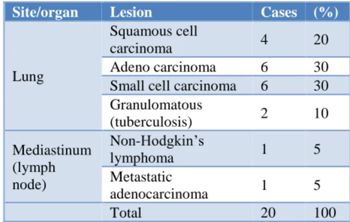
This group comprised of 20 cases; out of which 18 were from lung and 2 cases from mediastinum (Table 1).
Table 1: Cytological diagnosis of intra thoracic lesions.
Site/organ Lesion Cases (%)
Lung
Squamous cell
carcinoma 4 20 Adeno carcinoma 6 30 Small cell carcinoma 6 30 Granulomatous
(tuberculosis) 2 10
Mediastinum (lymph node)
Non-Hodgkin’s
lymphoma 1 5 Metastatic
adenocarcinoma 1 5 Total 20 100
The samples were taken by CT guided (12 cases) and rest were USG guided (8 cases) aspirations. Maximum cases 6(30%) were adenocarcinoma and small cell carcinoma 6 (30%). Authors also reported one case each of non-Hodgkin lymphoma and metastatic adenocarcinoma from mediastinum (Table 2).
Table 2: Cytological diagnosis of intra-abdominal lesions.
Site/ organ Lesion Cases (%)
Liver (n=21)
Hepatocellular
carcinoma 4 10 Adenocarcinoma 12 30 Squamous cell
carcinoma 3 7.5 Small cell carcinoma 1 2.5 Hydatid cyst 1 2.5 Gall bladder
(n=8) Adenocarcinoma 8 20 GIT(n=1) Gastrointestinal
Stromal tumor 1 2.5
Ovary (n=3)
Endodermal sinus tumor 1 2.5 Papillary
adenocarcinoma 1 2.5 Serous
cystadenocarcinoma 1 2.5 Lymph
node (n=3)
Metastatic
Adenocarcinoma 1 2.5 Hodgkins lymphoma 1 2.5 Tuberculosis 1 2.5 Omentum
(n=2)
Metastatic
adenocarcinoma 2 5 Pancreas
(n=2)
Pancreatic cyst 1 2.5 Adenocarcinoma 1 2.5 Total 40 100
Intra-abdominal lesions
lymph nodes 3 cases, pancreas: 2 cases, omentum 2 cases, GIT 1 case). The samples were taken by USG guided (32 aspirations) and CT guided (8 aspirations).
Figure 1: Distribution of cases.
Regarding age distribution of cases, maximum cases 24 (60%) belonged to age group of 45-60 years of age followed by 61-75 years (28.3%). Males were more in number 33(55%) as compared to females i.e. 27 (45%).
DISCUSSION
FNAC is safe, accurate, minimally invasive and cost-effective procedure. Image guided FNAC has facilitated easy collection of cellular material with greater accuracy.6 It provides immediate management of the
patient with intrabdominal, intrathoracic and retroperitoneal lesions at inaccessible and deep-seated sites. With this technique, probable diagnosis can be made before surgery and without subjecting the patient to open biopsy and thus two staged surgical procedures can be avoided. In order to increase the accuracy of sampling; CT/US room should have a microscope so that the
pathologist can look for the adequacy of the aspirated sample with the help of fast stain (toluedene blue) and thus cytological specimen can be interpreted immediately.7
In the present study no major complication was observed, except for the mild pain and discomfort at the puncture site for a short time only. In present study, liver and lungs were the common sites of FNAC; as shown in (Tables 1 and 2) respectively; which is comparable to the studies done by Adhikari RC et al.8 Liver was also the common
site of aspiration performed in the abdomen in a study done by Lokhande R et al.9
The age range of the patients in present study was 10-83 years given in (Table 3).
In the study by Tan KB et al, the age range of the patients was 11 to 82 years.10 The similar age ranges were
observed in the study conducted by Parahjuli S et al.11
The male to female ratio in present study was 1.23:1 comparable with study done by Lokhande R et al, with male:female ratio of 1.69.9
In present study, the most common sites for FNAC were liver and lungs as shown in (Table 1 and 2) which is comparable to studies done by other authors as well.1,8,9
The most common malignancy encountered in the abdomen was metastatic adenocarcinoma of the liver followed by carcinoma gall bladder which corroborated with other author as well.8,9,11
In present study, there were 2 cases of metastatic adenocarcinoma in omentum which is comparable to study of Parahjuli S et al, who also found 2 cases of metastatic adenocarcinoma omentum.11 Maximum
number of malignant lesions were encountered in 45-75 years which were almost similar to studies by Mukherjee S et al.12
Table 3: Age wise distribution of intra thoracic and intra-abdominal lesions.
Lesion/Age group 0-15 16-30 31-45 46-60 61-75 Above-75 Total
Adenocarcinoma - - 2 15 9 6 32
Squamous cell carcinoma - - - 3 2 2 7
Small cell carcinoma - 2 1 2 2 - 7
Non-Hodgkins lymphoma - 1 - - - - 1
Hepatocellular carcinoma - - - 1 3 - 4
Tuberculosis - - 1 2 - - 3
Hydatid cyst 1 - - - 1
Pancreatic cyst - 1 - - - - 1
GIST - - 1 - - - 1
Endodermal sinus tumor 1 - - - 1 Serous cystadeno carcinoma - - - 1 - - 1
Hodgkins ymphoma - - - - 1 - 1
Total 2 4 5 24 17 8 60
35%
30% 13%
5% 5% 4%
3% 3% 2%
Liver Lung Gall Bladder
ovary lymphnode pancrease
Table 4: Gender wise distribution of intra thoracic and intra-abdominal lesions.
Lesions Male Female Total Adenocarcinoma 12 20 32 Squamous cell carcinoma 5 2 7 Small cell carcinoma 6 1 7 Non-Hodgkin lymphoma 1 - 1 Hepatocellular carcinoma 4 - 4 Tuberculosis 2 1 3 Hydatid cyst 1 - 1 Pancreatic cyst 1 - 1
GIST 1 - 1
Endodermal Sinus tumor - 1 1 Serous cystadenocarcinoma - 1 1 Hodgkin lymphoma - 1 1
Total 33 27 60
Among the lung lesions, non-small cell carcinomas (10 cases) were the most common lesions, similar to other studies as well.9,11,12 In this study, FNAC is helpful in
diagnosing benign and malignant neoplasms as well as non-neoplastic lesions like tuberculosis, hydatid cyst and pancreatic cyst. Barrios S et al, recommended that image guided FNAC should be used as a routine procedure in the study of intra-abdominal and pulmonary lesions for retroperitoneal masses.13 CT guidance is very useful
especially in localization of small lesions.13-17
CONCLUSION
Our experience indicates that fine needle aspiration cytology of intrathoracic and intraabdominal lesions under image guidance is a simple and cost-effective outpatient procedure for diagnosis of deep seated, inaccessible lesions with minimal complications. It obviates the need for surgical procedure for reaching at a diagnosis.
Funding: No funding sources Conflict of interest: None declared
Ethical approval: The study was approved by the Institutional Ethics Committee
REFERENCES
1. Sheikh M, Sawhney S, Dey P, Ai‐Saeed O, Behbehani A. Deep‐seated thoracic and abdominal masses: Usefulness of ultrasound and computed tomography guidance in fine needle aspiration cytology diagnosis. Aust Radiol. 2000;44(2):155-60.
2. Das DK, Pant CS. Fine needle aspiration cytologic diagnosis of gastrointestinal tract lesions. A study of 78 cases. Acta Cytol. 1994;38(5):723-9.
3. Reddy VB, Gattuso P, Abraham KP, Moncada R, Castelli MJ. Computed tomography-guided fine
needle aspiration biopsy of deep-seated lesions. A four-year experience. Acta cytol. 1991;35(6):753-6. 4. Sobha Rani G. Md K Faheem N, Sai Prasad BV,
Sudhakar Reddy E. Efficiency of ultrasound guided aspiration cytology in deep seated lesions-a diagnostic evaluation. Int J Med Health Sci. 2012;1:1-2.
5. Pitman MB. Fine needle aspiration biopsy of the liver: principal diagnostic challenges. Clinic Lab Med. 1998;18(3):483-506.
6. Nobrega J, dos Santos G. Aspiration cytology with fine needle in the abdomen, retroperitonium and pelvic cavity: a seven-year experience of the Portuguese institute of oncology. Eur J Surg Oncol. 1994;20:37-42.
7. Lillie RD, HJ Cohn’s biological stains. Baltimore: Williams and Wilkins; 1977:420-429.
8. Adhikari RC, Tuladhar A, Shrestha S, Sharma SK, Sharma SK. Deep-seated thoracic and abdominal lesions: usefulness of ultrasound guided fine needle aspiration cytology, a 3-year experience. Nepal Med Coll J. 2010;12(1):20-5.
9. Lokhande R, Patni A, Sajith SL, Shankara R. Study of image guided fine needle aspiration cytology (FNAC) in the intra-abdominal and intra thoracic masses. Asian Pac. J. Health Sci.2017;4(1):19-26. 10. Tan KB, Thamboo TP, Wang SC, Nilsson B,
Rajwanshi A, Salto-Tellez M. Audit of transthoracic fine needle aspiration of the lung: cytological subclassification of bronchogenic carcinomas and diagnosis of tuberculosis. Singapore Med J. 2002;43(11):570-5.
11. Parahjuli S, Tuladhar A, Basnet RB. Ultrasound and computed tomography guided fine needle aspiration cytology in diagnosing abdominal and intra-thoracic lesions. J Pathol Nepal. 2011;1(1):17-21. 12. Mukherjee S, Bandyopadhyay G, Bhattacharya A,
Ghosh R, Barui G, Karmakar R. Computed tomography-guided fine needle aspiration cytology of solitary pulmonary nodules suspected to be bronchogenic carcinoma: experience of a general hospital. J Cytol/Indian Acad Cytol. 2010;27(1):8. 13. Barrios S, Hamana N, Quiros E. Cytology and
biopsy by fine needle aspiration with ultrasound guidance in abdominal tumors. GEN. 1989;43(3):155-60.
14. Stewart CJ, Coldewey J, Stewart IS. Comparison of fine needle aspiration cytology and needle core biopsy in the diagnosis of radiologically detected abdominal lesions. J Clinic Pathol. 2002;55(2):93-7. 15. Livraghi T, Sangalli G, Giordano F, Vettori C. Fine aspiration versus fine cutting needle, and comparison between smear cytology, inclusion cytology and microhistology in abdominal lesions. Tumori J. 1988;74(3):361-4.
17. Sauthier PG, Bélanger R, Provencher DM, Gauthier P, Drouin P. Clinical value of image-guided fine needle aspiration of retroperitoneal masses and lymph nodes in Gynaecologic oncology. Gynecol Oncol. 2006;103(1):75-80.
Cite this article as: Chowdhary M, Gupta R, Singh K.Role of image guided fine needle aspiration cytology in the diagnosis of intra-abdominal and intra-thoracic lesions. Int J Res Med Sci