Original Research Article
A study of pulmonary function abnormalities in obese individuals
Bhumika T. Vaishnav, Tushar V. Tonde*
INTRODUCTION
Obesity is defined as abnormal or excessive fat accumulation that presents a risk to health.1 Obesity is a
public health crisis of mammoth proportions in the developed world.2 The prevalence is also increasing
rapidly in numerous developing nations worldwide. In 2008, 1.5 billion adults were overweight (BMI >25 kg/m2). Of these over 200 million men and nearly 300
million women were obese (BMI >30 kg/m2).1 The
overall prevalence rate of obesity was 27.8% in urban Indian population. Obesity was found to be more common in female subjects.3 Hypertension, coronary
heart disease, diabetes mellitus, Hypercholesterolemia and hypertriglyceridemia were found to be associated with obesity. Obesity may be associated with a number of pulmonary abnormalities namely decrease effort tolerance, physical deconditioning, and obstructive sleep apnea.
These include reduced chest wall compliance, increased work of breathing, increased minute ventilation due to increased metabolic rate, and decreased total lung capacity and functional residual capacity.4 The
characteristic abnormality seen in obesity is a decreased expiratory reserve volume, caused a markedly decreased Department of Medicine, Dr. D. Y. Patil Medical College, Hospital and Research Center, Pimpri, Pune, Maharashtra, India
Received: 25 December 2019
Accepted: 20 January 2020
*Correspondence:
Dr. Tushar V. Tonde, E-mail: [email protected]
Copyright: © the author(s), publisher and licensee Medip Academy. This is an open-access article distributed under the terms of the Creative Commons Attribution Non-Commercial License, which permits unrestricted non-commercial use, distribution, and reproduction in any medium, provided the original work is properly cited.
ABSTRACT
Background: Previous studies suggest that obese individuals are prone to pulmonary function abnormalities. The aim of this study was to evaluate pulmonary function tests in obese individuals and to relate pulmonary abnormalities if any found to lipid abnormalities and to the extent and duration of obesity.
Methods: This prospective study was done on 40 obese patients attending to Dr. D. Y. Patil Hospital, Mumbai with complaints of pulmonary functions during the period from January to December 2012. Pulmonary function test was done with the help of Jaegers pneumoscreen. The percentage of body fat was determined by using triceps skin fold thickness technique by using Vernier callipers. Fasting serum samples was collected to analyses cholesterol and triglycerides.
Results: Female preponderance was seen in the study (57.5%). Forced expiratory volume, forced vital capacity, maximum mid expiratory flow rate was significantly reduced and the ratio of forced expiratory volume in one second to forced vital capacity was significantly increased in individuals who had abnormal pulmonary function. Decrease in pulmonary function was noted with increased levels of cholesterol and triglyceride but the correlation was not significant.
Conclusions: Obese individuals although asymptomatic have significant lung function abnormality in the form of restrictive as well as obstructive pattern. Hence, reduction in the body weight may help in reversal of the pulmonary function indices.
Keywords: Abnormalities, Obesity, Pulmonary function indices
functional residual capacity with a relatively well preserved residual volume. The major change in pulmonary mechanics in obesity is due to overall decrease in compliance, due to increase in weight, an increase in airway resistance and decreased respiratory muscle functions.5
Many studies were done to correlate the obesity with diabetes mellitus, insulin resistance, dyslipidemia and other cardiac abnormalities, but there are few Indian studies revealing the relationship between obesity and pulmonary functions.6-8 Hence, the current study was
undertaken to study pulmonary function tests in obese individuals and to relate pulmonary abnormalities if any found to lipid abnormalities and to the extent and duration of obesity.
METHODS
This was a prospective study done on 40 obese patients during the period from January to December 2012 attending to the Dr. D. Y. Patil Hospital, Mumbai. The study was reviewed and approved by institutional ethics committee. Informed consent was taken from all the patients. Obese individuals (male or female) above 18 years were included in the study. Patients with diabetes mellitus, hypertension, ischemic heart disease, cerebrovascular diseases, history of gastroesophageal reflux disease, peptic ulcer, hiatus hernia, bronchial asthma, COPD, occupational lung diseases, history of smoking, any other addiction, other major coexisting illness and individuals with anaemia, hematological disorders were excluded.
Patient evaluation included, body fat determination, spirometry, lung volumes and fasting serum samples were collected to analyses cholesterol and triglycerides.
Baseline pulmonary function test was done with the help of Jaegers pneumoscreen (COSMED, model- Quark PFT). Physical parameters such as height and weight of the individual were entered in the machine, which then generated the predicted normal values for that particular individual. FVC (forced vital capacity) in liters, FEV1 (forced expiratory volume in first second) in liters, FEV1/FVC%, PEF (peak expiratory flow) rate in liters and PEF (25-75%) (forced expiratory flow), MMEFR (maximum mid expiratory flow rate) were the pulmonary function parameters tested.
In patients with obstructive pattern, 2 puffs of salbutamol each of 100 ug was administered and pulmonary function tested again after half hour for reversibility. The pneumoscreen calibrated for temperature, humidity and ambient environment. Body surface area was calculated as per height and weight.
The percentage of body fat was determined by using triceps skin fold thickness technique by using Vernier
calipers. Body fat calculation indices included BMI (based on weight and height kg/m2) and waist hip ratio.
Statistical analysis was performed using SPSS software (SPSS, Chicago). Pearson correlation matrix analysis was used to assess relationships between the indices of obesity and the variables of pulmonary function. p value <0.05 was considered statistically significant.
RESULTS
Demographic characteristics of the patients was shown in Table 1. Mean age of the patients was 13.320 years. Out of 40 patients, 23(57.5%) were females and 16(40.3%) were males. All the study subjects underwent pulmonary function testing. Abnormality was noted in 25 cases. Of them, obstructive lung abnormality was seen in 9 patients, restrictive abnormality in 7 patients and both abnormalities were noted in 9 patients. The incidence of lung disease was found to be statistically significant in obese patients (p<0.05) (Table 2) and females are more prone to pulmonary function abnormality.
Table 1: Demographic characteristics of study participants (n=40).
Characteristics Findings
Age (in years) (mean±sd) 43.32±10.73 Sex (female:male) 1.5:1 Height (in cm) (mean±sd) 160.7±18.43 Weight (in kg) (mean±sd) 82±9.68 BMI (kg/m2) (mean±sd) 31.7±2.38
Skin fold fat (in mm) (mean±sd) 22.42±3.33 Waist hip ratio (mean±sd) 1.04±0.072 Grading of cholesterolemia
Desirable n (%) 15(37.5) Borderline n (%) 15(37.5) Undesirable n (%) 10(25) Grading of triglyceridemia Desirable n (%) 14(35) Borderline n (%) 9(22.5) Undesirable n (%) 15(37.5) Highly undesirable n (%) 2(5)
Table 2: Result of pulmonary function test in study population (n=40).
Number
of subjects Normal Obstructive Restrictive Both
40 15 9 7 9
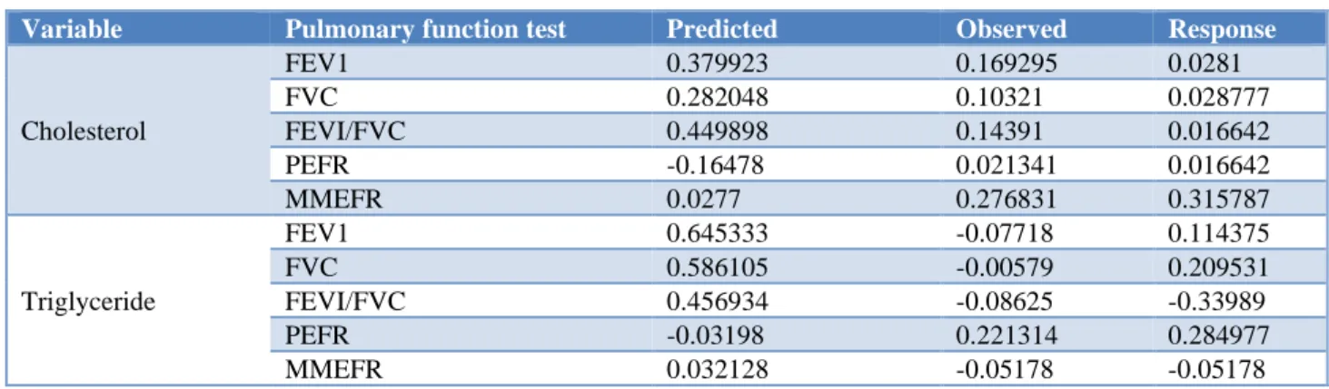
No significant correlation was observed between the cholesterol and triglyceride levels in the patients with obstructive and restrictive pulmonary functional indices (Table 4 and 5). Cholesterol showed negative correlation with the predictive value of PEFR (r=-0.16478) while triglyceride levels showed negative correlation with the observed values of FEV1 0.07718), EVC 0.00579), FEV1/FVC 0.08625) and MMEFR (r=-0.05178) in obstructive cases. In restrictive pulmonary
abnormality cases, the level of cholesterol showed negative correlation with FVC (r=-0.17962) and FEV1/FVC (r=-0.3905) in the observed values and with PEFR (r=-0.2479) and MMEFR (r=-0.1456) in the predictive values. Triglycerides showed negative correlation with FEVI/FVC (r=-0.4631) and MMEFR 0.2444) in observed values and with PEFR (r=-0.0714) in predictive values.
Table 3: Pulmonary function test abnormalities in the obese individuals and their bronchodilator response (n=25).
Pulmonary function test Predicted Observed p value Response p value
FEV1 2.31±0.62 1.787±10. 0.011* 1.9±0.54 0.861 FVC 2.9±0.64 2.32±0.80 0.006** 2.24±0.67 0.756 FEV1/FVC 82.62±3.75 87.33±10.69 0.047* 86.65±7.74 0.828
PEFR 345±49.8 312.6±95.78 0.061 332±105.74 0.991 MMEFR 3.49±1.03 2.27±0.94 0.064 2.27±0.98 0.548 *p<0.05, **p<0.001.
Table 4: Correlation of cholesterol and triglyceride levels with obstructive pulmonary abnormality function.
Variable Pulmonary function test Predicted Observed Response
Cholesterol
FEV1 0.379923 0.169295 0.0281
FVC 0.282048 0.10321 0.028777
FEVI/FVC 0.449898 0.14391 0.016642 PEFR -0.16478 0.021341 0.016642 MMEFR 0.0277 0.276831 0.315787
Triglyceride
FEV1 0.645333 -0.07718 0.114375 FVC 0.586105 -0.00579 0.209531 FEVI/FVC 0.456934 -0.08625 -0.33989 PEFR -0.03198 0.221314 0.284977 MMEFR 0.032128 -0.05178 -0.05178
Table 5: Correlation of cholesterol and triglyceride levels with restrictive pulmonary abnormality function.
Variable Pulmonary function test Predicted Observed Response
Cholesterol
FEV1 0.36511 0.478 0.52909
FVC 0.27405 -0.17962 0.6271 FEVI/FVC 0.6943 -0.3905 -0.31322 PEFR -0.2479 0.1295 0.21769 MMEFR -0.1456 0.0356 -0.03661
Triglyceride
FEV1 0.4822 0.0496 0.3583
FVC 0.45344 0.0321 0.7275
FEVI/FVC 0.477 -0.4631 -0.6827 PEFR -0.0714 0.34809 0.564 MMEFR 0.0978 -0.2444 0.125
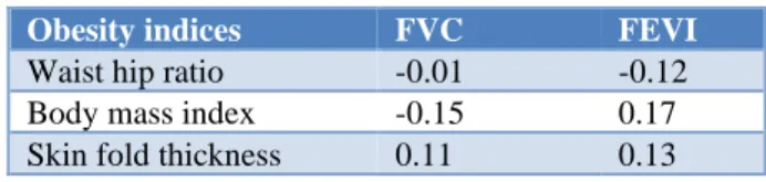
Effect of obesity indices in pulmonary function was evaluated but no significant correlation was seen between them (p>0.05). From the Table 6, it was evident that a negative correlation was identified between FVC (r=-0.01) and FEV1 (r=-0.12) with waist hip ratio. BMI was found to have negative correlation with FVC (r=-0.15).
However, Skin fold thickness showed positive correlation with both FVC (r=0.11) and FEV1 (r=0.13).
Table 6: Correlation of pulmonary function test parameters and indices of obesity in study population.
Obesity indices FVC FEVI
Waist hip ratio -0.01 -0.12 Body mass index -0.15 0.17 Skin fold thickness 0.11 0.13
Table 7: Correlation of pulmonary function parameters and duration of obesity.
Normal PFT
(N=15) Abnormal PFT (N=25)
Duration of obesity
0-2 yrs
2-4 yrs
4-6 yrs
>6 yrs Number of
subjects 15 6 6 5 8 Percentage
of total 37 24 24 20 32
The response to bronchodilator was noted in 8 cases. Of them minimal improvement in pulmonary abnormalities was noted in 7 cases and good in case.
DISCUSSION
Obesity is acknowledged as a global phenomenon that increases morbidity and mortality. It is considered to be a major risk factor for cardio and cerebrovascular diseases and diabetes.9 Obesity shows a profound effect on the
physiology of breathing.10 Obesity is found to be
involved in decreasing the respiratory compliance due to involvement of mechanical factors such as increased weight on thoracic cage and abdomen. Severe obesity may lead to obesity hypoventilation syndrome and sleep apnea.
The most common pulmonary function abnormality in obese individuals is FERV. This happens because mass loading effect of obesity decreases FRC which in turn decreases ERV.11 As given in literature, in mild obesity,
spirometry values are normal. In obese individuals decrease in expiratory flow, FVC and FEV1 and increase in FEV1/FVC ratio was observed.12,13 Similar effects was
also noted in this study.
In the present study, the PEFR and MMEFR values are significantly decreased in individuals who had abnormal pulmonary function test. This was in accordance with the studies of Srinivas et al, and Ofuya et al.7,14
Previous studies conducted on relationship between metabolic abnormality and pulmonary function, elevated serum triglycerides and cholesterol levels were considered as independent predictors of pulmonary abnormalities.15,16 In this study, positive correlation was
observed between the cholesterol and triglyceride levels with predictive values of FVC, FEV1 and FEV1/FVC in the patients with obstructive and restrictive pulmonary
functional indices but the association was not significant. The exact mechanism of the relationship is not known yet. It was predicted that the composition of pulmonary surfactant might be a possible linkage between increased HDL cholesterol level and decreased pulmonary function.17
Body fat distribution also had an impact on the effect of obesity on lung function. Study by Collins et al, showed a significant inverse relationship between adiposity and spirometry values.18 The best measure of fat distribution
was skinfold thickness. Another study by Lazarus et al, suggested that central abdominal obesity has a great impact on spirometric measures, but the relationship diminishes with age.19 In this study, no significant
correlation between pulmonary function parameters and duration of obesity was observed.
The Pearson co-relation coefficient in Collin et al, study for FVC and FEV1 in relation to waist hip ratio, body mass index, triceps fold thickness was -0.18, -0.18, -0.52 and -0.11, 0.15, 0.13 respectively.18 Similar observations were also noted in this study (waist hip ratio, body mass index, and triceps fold thickness were -0.01, -0.15, 0.11 and -0.12, 0.17, 0.13 respectively) and found no significant correlation between them.
Limitations of this study was number of asymptomatic patients included in study was 40, and large number of patients with sizable subgroups, namely; symptomatic and asymptomatic, smokers and nonsmokers, would be required to express firm opinions regarding PFT abnormalities in obese individuals.
CONCLUSION
Based on the observations, authors can conclude that obesity causes change in respiratory functions. Obese individuals although asymptomatic, high BMI, body fat, cholesterol and triglyceride levels may be associated with significant lung function abnormality in the form of restrictive as well as obstructive pattern. Hence, it is not only advisable to control obesity for its well-established complications like type 2 diabetes, hypertension, IHD, osteoarthritis but also, to avoid its adverse impact on pulmonary function and complications arising thereof.
Funding: No funding sources Conflict of interest: None declared
Ethical approval: The study was approved by the Institutional Ethics Committee
REFERENCES
1. Obesity. Available at: https://www.nhp.gov.in/disease/non-communicable-disease/obesity. Accessed on 15 October 2018. 2. Kuczmarski RJ, Flegal KM, Campbell SM, Johnson
Examination Surveys, 1960 to 1991. JAMA. 1994;272(3):205-11.
3. Gopinath N1, Chadha SL, Jain P, Shekhawat S, Tandon R. An epidemiological study of obesity in adults in the urban population of Delhi. J Assoc Phys Ind. 1994;42(3):212-5.
4. Ray CS, Sue DY, Bray G, Hansen JE, Wasserman K. Effects of obesity on respiratory function. Am Rev Respir Dis. 1983;128(3):501-6.
5. Parameswaran K, Todd DC, Soth M. Altered respiratory physiology in obesity. Can Respir J. 2006;13:203-10.
6. Kyrou I, Randeva HS, Tsigos C. Clinical Problems Caused by Obesity. In: Feingold KR, Anawalt B, Boyce A, et al., eds. Endotext. South Dartmouth (MA): MDText.com, Inc.; 2000. Available at: https://www.ncbi.nlm.nih.gov/books/NBK278973/ 7. Srinivas CH, Madhavi Latha M, Surya Kumari N,
Surendranath Y. Comparative study of dynamic lung function tests in obese and non-obese individuals. J Evol Med Dental Sci. 2013;2(35):6736-42.
8. Paralikar SJ, Kathrotia RG, Jani MB. Assessment of pulmonary functions in obese adolescent boys. Lung Ind. 2012;29(3):236-40.
9. National Task Force on the Prevention and Treatment of Obesity. Overweight, obesity and health risk. Arch Intern Med. 2000;160:898-904. 10. Luce JM. Respiratory complications of obesity.
Chest. 1980;78:626-30.
11. Ray CS, Sue DY, Bray G, Hansen JE, Wasserman K. Effects of obesity on respiratory function. Am Rev Respir Dis. 1983;128:501-6.
12. Steele RM, Finucane MF, Griffin SJ, Wareham NJ, Ekelund U. Obesity is associated with altered lung function independently of physical activity and fitness. Obesity. 2009;17:578-84.
13. Shah B, Selot B, Patel SV, Patel NJ. Pulmonary function test in obese and non-obese individuals. IRPMS. 2017;2(5):43-50.
14. Ofuya ZM, Georgewill AA, Agu GO. A study of cardiovascular and respiratory parameters in obese and non-obese subjects resident in Port Harcourt. Afr J of Appl Zool Environ Biol. 2005;7:11-3. 15. Naveed B, Weiden MD, Kwon S, Gracely EJ,
Comfort AL, Ferrier N, et al. Metabolic syndrome biomarkers predict lung function impairment: a nested case-control study. Am J Respir Crit Care Med. 2012;185:392-9.
16. Leone N, Courbon D, Thomas F, Bean K, Jego B, Leynaert B, et al. Lung function impairment and metabolic syndrome: the critical role of abdominal obesity. Am J Respir Crit Care Med. 2009;179:509-16.
17. Park JH, Mun DS, Choi DP, Lee JY, Kim HC. Association between high-density lipoprotein cholesterol level and pulmonary function in healthy Korean adolescents: the JS high school study. BMC Pulm Med. 2017;17:190.
18. Collins LC, Hoberty PD, Walker JF, Fletcher EC, Peiris AN. The effect of body fat distribution on pulmonary function tests. Chest. 1995;107:1298-302.
19. Lazarus R, Sparrow D, Weiss ST. Effects of obesity and fat distribution on ventilatory function. Chest. 1997;111:891-8.