TECHNICAL UNIVERSITY OF CLUJ-NAPOCA
ACTA TECHNICA NAPOCENSIS
Series: Applied Mathematics, Mechanics, and Engineering Vol. 62, Issue IV, November, 2019
DESIGN OF AN INNOVATIVE MEDICAL ROBOTIC INSTRUMENT FOR
MINIMALLY INVASIVE TREATMENT OF LIVER TUMORS
Bogdan GHERMAN, Nadim AL HAJJAR, Alin BURZ, Iosif BIRLESCU, Paul TUCAN, Florin GRAUR, Doina PISLA
Abstract: The paper presents the design of a robotic needle insertion instrument the cancer treatment of the hepatocellular carcinoma. The instrument is able to perform multiple needle insertion on a linear trajectory using specially designed guides. It has only 2 degrees of freedom and is able to insert up to 6 needles successively, featuring preoperative designed needles storages that fit the final needles placement in the liver. Thus it is less dependent of the robot motions during the needles insertion assuring a higher overall accuracy.
Key words: medical instrument, brachytherapy, multiple needles.
1. INTRODUCTION
Liver cancer is one of the most spread types of cancer originating in the liver and due to its aggressiveness many times it leads to death. Hepatocellular carcinoma (HCC) is associated with cirrhosis and for the treatment planning the doctor should take into account the tumor stage and the patient general condition [1].
HCC is the fifth most and the third deadliest is the 3rd because of its aggressiveness and high
vascularity of the liver feeding the tumor. Liver cancer has a lethality index of 0.93 [2]. Although treatment possibilities have advanced over the years, it still remains one of the most difficult cancers to treat. In the case of an early detected HCC, surgery, transplant and local therapy might be a solution, but even so, the high reoccurrence incidence remains a big problem. A curative treatment solution is the tumor resection but this is applicable approximate to 20% of the patients [3]. A thing which must be mentioned is that the only 10-20% of the total number of resections can be performed in a minimally invasive way and the survival rate at 5 years ranges between 25-50%.
Some alternative treatments were proposed like percutaneous local ablation, transarterial chemoembolization or radioembolization but this kind of treatments have low accuracy in
reaching the targeted areas where as the treatment of tumors which are located near important vascular structures of the liver can’t be treated efficiently [4].
Another alternative for HCC treatment is the interstitial brachytherapy. It consists in placing radioactive seeds into the tumor in predetermined locations (using medical imagery). The seeds are placed by using a certain number of needles which are inserted (preferably) on a linear trajectory in the form of a matrix.. The catheters of these needles are connected to a source of radiation which is able to precisely position radioactive seeds (i.e. iodine 125) into the tumor. After each treatment, the cables are disconnected [5].
In this paper the authors present a medical robotic instrument for needle insertion in minimally invasive treatment of inoperable liver tumors using brachytherapy. A 5-DOF parallel robotic system should be used to guide the instrument and position it above the insertion are.
2. MEDICAL PROTOCOL
brachytherapy is the selected one, the best solution is to perform a minimally invasive approach in which the catheters are inserted from the outside of the body on a straight line trajectory towards the liver, in the tumor, guided by the intraoperatory ultrasound probe. Following the precise needle placement, the
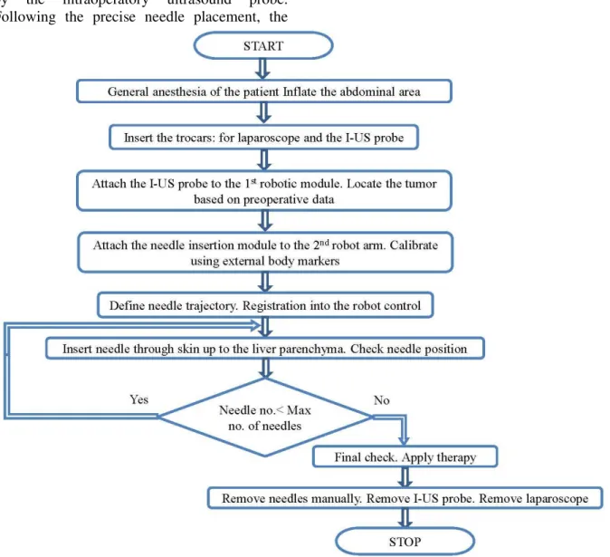
treatment is applied using the specialized equipment for radioactive seeds placement into the tumor. Fig. 1 presents the main steps in achieving the HCC minimally invasive treatment using the proposed procedure.
Fig. 1. Schematic representation of the medical protocol for the HCC treatment using brachytherapy
The patient undergoes general anesthesia and the abdominal area is inflated with carbon dioxide up to a pressure of 12 mmHg creating the necessary cavity for the medical instruments. The two medical instruments used in this procedure are: the intraoperatory ultrasound (I-US) probe and the brachytherapy robotic instrument for needle insertion (BNI). Each of the two instruments have been designed and
Fig. 2. Virtual operating room and possible operating table motion [6]
The needles trajectory is assured by using a set of guides (having the aspect of a sieve) [7]. The first needle guide (Fig. 2) is manually sewed onto the patient’s abdominal cavity, while the other, having a modular structure, is inserted into the patient’s abdominal cavity, assembled there and placed on the liver just above the tumor (Fig. 2). The brachytherapy needles are inserted into the abdominal cavity through this pair of guides up to the tumor.
The second robotic arm holds the BNI and is calibrated with respect to the patient using several external body markers defined in the preoperative stage and registered also with respect to the first arm.
A set of points for the needle are defined based on the preoperative data (I-insertion and T-Target). As the two robotic arms are registered one with respect to the other, the I-US probe is realigned to the defined needle trajectory in such a way that the active probe head and the needle trajectory are in the same plane. This will enable the real-time visualization of the needle as it is inserted in the liver tumor. Using the robotic arm, the needle is positioned with the tip in the insertion point and the final orientation defined by the I-T pair of points [8, 9].
Once all the needles are inserted, the I-US probe is used to verify and validate the final needle positions and the specific therapy is applied using specific medical devices.
When the therapy is finished the needles are extracted manually and using the laparoscopic camera the surgical field is inspected for any signs of bleeding. If necessary, haemostasis is applied. The I-US probe is retracted, using the robotic arm, followed by the manual extraction of the laparoscopic camera and the procedure is finished.
3. ROBOTIC INSTRUMENT DESIGN
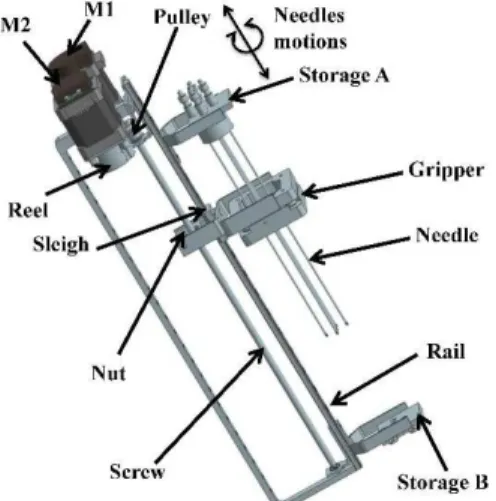
The robotic instrument for needle(s) insertion, [10] presented in figure 4 has two main mechanical subsystems designed to load one needle at a time from storage (Storage A) and then insert them on a linear path until the established depth based on the preoperative data. The position of the needle in the storage A is also established on the preoperative data and can be exchanged from a procedure to other. A possible arrangement of the needles is presented in the figure 4.
Fig. 4. Parallel Insertion Surgical System (BNI)
Fig. 5. Possible brachytherapy needle arrangement (detail of storage B)
The first subsystem is the needle storage mechanism, the capacity of needle storage is set based on the preoperative data and the second subsystem is the loading/insertion mechanism with the main component being the needle gripper.
The two subsystems are actuated by two stepper motors (the closed loop is achieved using rotary encoders for an accurate positioning) which work together with synchronized motion as follows:
1. With the action of motor 1 (M1), both needle storage A and the needle gripper (Fig. 6) will
move on a linear trajectory along the rail. The needle is guided (by the storage A) through a circular channel and at the end of the channel is grabbed by the gripper. The motor 2 (M2) rotate the gripper using a flexible cable placed on reel one from top of the reel and another one from bottom of the reel. The gripper is configured different from each procedure for each configuration of the needle(s).
Fig. 6. The BNI gripper for the current needles arrangement
2. After the loading stage, the needle is fixed in the gripper and the pre-insertion stage begins (actuated by motor M1 through a trapezoidal screw-nut transmission). The needle moved on a linear path until enters form the needle tip guiding element (storage B) followed by the release of the needle.
3. With the needle inserted the loading mechanism is actuated back (motor M1) to the needle loading position.
4. After all the needles are inserted the storage B will be removed manually from the robotic instrument and it is sewn on abdomen. During the treatment procedures, BNI is attached to the PROHEP-LCT robotic module, specially designed module to guide such an instrument. The robotic module has five degrees of freedom (translations along the three axes and two rotations) used for an accurate positioning and orientation of the BNI instrument.
4. MEDICAL ENVIRONMENT
area of the liver where the tumor(s) is located; as mentioned, three instruments are needed to safely perform the procedure: a manually guided laparoscopic camera used to visually supervise the procedure inside the patient, the intra-operatory ultrasound (I-US) probe guided by one of the parallel robotic modules of PROHEP-LCT robotic system [11] and the robotic instrument (BNI) used for treatment and guided by the second robotic module. The robotic module will position the instrument at the insertion area and BNI will insert successively the brachytherapy needles.
Fig. 7. Parallel Insertion Surgical System (BNI)
Fig. 8. Real operating room (Courtesy of Prof. Dr. Octavian Fodor Regional Institute of Gastroenterology
and Hepatology Cluj-Napoca) 5. CONCLUSION
The paper presents the design of an innovative robotic instrument for multiple needle insertion in brachytherapy cancer treatment of HCC. The instrument has a simple and lightweight structure, very often needed to avoid an overload at the tip (end-effector) of the robot. Once the instrument is positioned above
the insertion area (using a 5-DOF parallel robotic system) it will solely insert all the required needles without further involvement from the robot. The instrument has a very important feature: it uses only two motors to insert the needles and to achieve a superior accuracy a set of linear guides are used. Since only two motors are used, a preoperative decision concerning the needle placement is required in order to custom build the storages of the instrument needles. Further work concerns the achievement of the demonstrator and its validation in relevant conditions.
6. ACKNOWLEDGEMENT
The results presented in this paper were obtained in the framework of the GNaC 2018 ARUT grant “Innovative robotized instruments for treatment in surgical abdominal procedures”, research Contract no. 3216/06.02.2019, with the financial support of the Technical University of Cluj-Napoca and the grant of the Romanian Ministry of Research and Innovation, PCCCDI – UEFISCDI, project number PN-III-P1-1.2-PCCDI-2017-0221 / 59 PCCDI/2018 (IMPROVE), within PNCDI III
7. REFERENCES
[1] Balogh, J. et al: Hepatocellular carcinoma: a review, Journal of Hepatocellular Carcinoma, vol. 3, pp. 41-53, (2016)
[2] Vaida,C., Al Hajjar, N., Lazar, V., Graur, F., Burz, A., Elisei, R., Mois, E., Pisla, D.:
Robotics in minimally invasive procedures:
History, current trends and future challenges.
In: the 6th Int. Conf. on Advancements of Medicine and Health Care through Technology – MediTech2018, 17-20 October (2018
[3] Khan, A., et al: Assessment and optimization of liver volume before major hepatic resection: Current guidelines and a narrative
review, International Journal of Surgery vol.
52, pp. 74–81, (2018)
[4] Yang, Z.-W., et al: The efficacy and safety of long- versus short-interval transarterial
hepatocellular carcinoma, Journal of Cancer, vol. 9(21), pp. 4000-4008, (2018)
[5] High-Dose Rate (HDR) Brachytherapy with
Interstitial Implants for the Treatment of
Gynecological Cancers,
https://www.mskcc.org/cancer-care/patient- education/high-dose-rate-brachytherapy-interstitial-implants
[6] Vaida C., Plitea N., Al Hajjar N., Burz A., Graur F., Pisla D.: A new robotic assisted approach in minimally invasive treatment of
liver tumours, Proceedings of the Romainan
Academy, Series A: Mathematics, Physics, Technical Sciences, Information Science, in Press
[7] Graur, F., Al Hajjar, N., Vaida, C., Mois, E., Pisla, D., Furcea, L., Popa, C., Elisei, R.:
Dual matrix system for the guidance of minimally invasive assisted brachytherapy for hepatic tumors using a modular plate
assembled inside the patient, Patent pending
no. A01142/27.12.2018
[8] Pisla D., Tucan P., Gherman B., Crisan N., Andras I., Vaida C., Plitea N. Development of a parallel robotic system for transperineal
biopsy of the prostate, Mech Sci vol.8, pp.
195-213, 2017
[9] Birlescu I., Craciun F., Vaida C., Gherman B., Pisla D. An innovative automated
instrument for robotically assisted
brachytherapy used in cancer treatment,
Acta Technica Napocensis, Series: Applied Mathematics, Mechanics, and Engineering,
vol. 60(4), pp. 633-638, 2017
[10] Gherman B., Birlescu I., Tucan P., Pisla D.:
Medical robotic instrument for liver tumors
treatment using brachytherapy, Patent
pending 2019
[11] Plitea, N., Pisla, D., Vaida, C., Gherman, B., Tucan, P. PRoHep-LCT- Parallel robot for the minimally invasive treatment of
hepatic carcinoma, Patent pending no.
A1017/03.12.2018
DESIGNUL UNUI INSTRUMENT ROBOTIZAT MEDICAL INOVATIV PENTRU TRATAMENTUL MINIM INVAZIV AL TUMORILOR DE FICAT
Lucrarea prezintă designul unui instrument medical robotizat destinat inserţiei acelor de brahiterapie în tratamentul carcinomului hepatocelular. Instrumentul este capabil să efectueze inserarea mai multor ace pe o traiectorie liniară folosind un set de ghidaje special concepute în acest sens. Instrumentul a fost proiectat astfel încât să asigure o independenţă sporită în timpul inserţiei successive a acelor de robotul care îl ghidează în scopul unei precizii de poziţionare ridicate a acelor în tumoare.
Bogdan GHERMAN, Conf. Dr. Ing., CESTER - Research Center for Industrial Robots Simulation and Testing, Technical University of Cluj-Napoca, [email protected], Office Phone: +40 -264-401684, 103-105 Muncii blv., Cluj-Napoca, Romania
Nadim AL HAJJAR, Prof. MD PhD., “Iuliu Haţieganu University of Medicine and Pharmacy”, [email protected], Phone: +40 -264-334871, Croitorilor 19-21, Cluj-Napoca, Romania
Alin BURZ, Ing., CESTER - Research Center for Industrial Robots Simulation and Testing, Technical University of Cluj-Napoca, [email protected], Office Phone: +40 -264-401684, 103-105 Muncii blv., Cluj-Napoca, Romania
Iosif BIRLESCU, Ing., CESTER - Research Center for Industrial Robots Simulation and Testing, Technical University of Cluj-Napoca, [email protected], Office Phone: +40 -264-401684, 103-105 Muncii blv., Cluj-Napoca, Romania
Paul TUCAN, As. Dr. Ing., CESTER - Research Center for Industrial Robots Simulation and Testing, Technical University of Cluj-Napoca, [email protected], Office Phone: +40 -264-401684, 103-105 Muncii blv., Cluj-Napoca, Romania
Florin GRAUR, S.l. MD PhD., “Iuliu Haţieganu University of Medicine and Pharmacy”, [email protected], Phone: +40 -744-807242, Croitorilor 19-21, Cluj-Napoca, Romania
![Fig. 2. Virtual operating room and possible operating table motion [6] The needles trajectory is assured by using a](https://thumb-us.123doks.com/thumbv2/123dok_us/7994446.2119950/3.918.171.797.125.505/virtual-operating-possible-operating-motion-needles-trajectory-assured.webp)