Prospective, Comparative Assessment of Peri-Implant Mucosal
Architecture at Different Implant Abutment Interfaces.
A 1 year Evaluation.
Praephun Limpiphipatanakorn, DDS
A thesis submitted to faculty of the University of North Carolina at Chapel Hill in partial fulfillment of the requirements for the degree of Master of Science in the
school of Dentistry, (Prosthodontics).
Chapel Hill 2013
Approved by:
Dr. Lyndon F. Cooper, DDS, PhD
Dr. John Moriarty, DDS, MS
ii ©2013
iii ABSTRACT
PRAEPHUN LIMPIPHIPATANAKORN: Prospective, Comparative Assessment of Peri-Implant Mucosal Architecture at Different Implant Abutment Interfaces.
A 1 year Evaluation.
(Under the direction of Lyndon F Cooper)
The purpose of this study is to compare the buccal soft tissue changes occurring
around single-tooth replacements in the maxilla using three different
implant-abutment interface designs over a one year period. The study was an open,
prospective, randomized multicenter study in 141 subjects. Subjects were
randomized to Group A (Conical interface, n=48), B (Flat-to-flat interface, n=49) or C
(Platform switch, n=44). This study evaluated the soft tissue changes longitudinally
through collection of standardized oral photographs using Canfield apparatus.
Comparisons between restorative platform types and between time points were
evaluated statistically. There was not statistically significant change of buccal soft
tissue level (mean -0.1 ± 0.7 mm) at 12 month followed implant placement, with no
statistically significant difference between three implant abutment designs. Overall
papilla height showed slight increase (mesial papilla 0.3±0.5 mm, distal papilla
iv
ACKNOWLEDGEMENTS
To my mentor and program director, Dr. Lyndon F. Cooper for providing me with the
opportunity to be one part of UNC graduated prosthodontics residents and for
sharing experience, knowledge and skill with me.
To my committee member: Dr. Ingeborg De Kok and Dr. John Moriarty for all help,
patient and teaching during the research project.
To my two best sisters Sandra and Ghadeer, thank you for all of your help, love and
care. You are always being there for me.
To UNC graduated prosthodontics residents for friendship and all great memories.
To my family and Golf, thank you for all of your love, support, and motivation. I am
v
Table of Contents
LIST OF FIGURES ... vii
LIST OF TABLES ... viii
1. Introduction ... 1
1.1 The evolution of single tooth implant replacement. ... 1
1.2 Survival of single tooth implants ... 9
1.3 Complications for single tooth Implants ... 10
1.4 The issues with evolving single tooth implant therapy ... 13
1.5 Implant esthetics ... 17
1.6 Patient satisfaction... 24
1.7 Implant esthetics assessment ... 26
1.8 Factors Affecting Peri Implant Buccal Tissues ... 28
2. Materials and methods ... 37
2.1 Patient selection ... 38
2.2 Clinical protocol and procedures ... 39
2.3 Data Collection ... 43
2.4 Evaluation Methods ... 44
vi
3. Results ... 48
3.1 Buccal soft tissue (Gingival zenith) response ... 50
3.2 Mesial papilla response ... 52
3.3 Distal papilla response ... 53
4. Discussion ... 55
vii
LIST OF FIGURES
Figure 1. Implant abutment interfaces: (A) Conical, (B) Flat-to-flat, and (C) Flat
platform switch ... 37
Figure 2. Flowchart of clinical protocol and procedures over a 1 year period ... 40
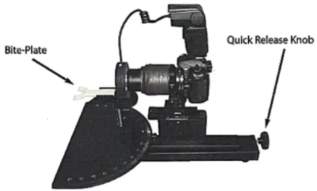
Figure 3. Canfield apparatus ... 43
Figure 4. Standardized clinical photograph of each site in each visit, (A) one with periodontal probe and (B) without periodontal probe ... 44
Figure 5. Assessment method for buccal soft tissue change ... 45
Figure 6. Assessment method for papilla height change ... 46
Figure 7. Study population flow chart ... 49
Figure 8. Comparison of gingival zenith change between implant types from placement of permanent crown to 1 year follow up. ... 51
viii
LIST OF TABLES
Table 1. Factors implicating tooth preservation or replacement ... 7
Table 2. Estimate survival rate of single tooth replacement in 5 and 10 year follow-up
... 10
Table 3. Description of indices for assessment of single implant esthetics* ... 28
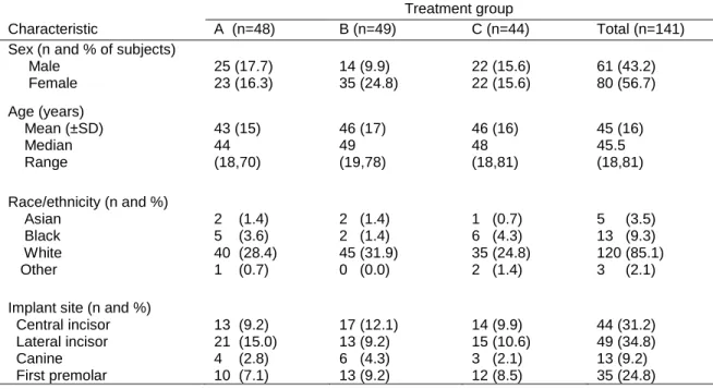
Table 4. Demographic and implant characteristics of the study sample ... 48
Table 5. Comparison of mean gingival zenith changes (±SD) between groups over
time ... 50
Table 6. Comparison of mean mesial papilla changes (±SD) between groups over
time ... 52
Table 7. Comparison of mean distal papilla changes (±SD) between groups over
1. Introduction
1.1 The evolution of single tooth implant replacement.
Ever since dental implant therapy evolved as a treatment option for replacement of
missing teeth, the success of this treatment modality was defined as
Osseointegration1. Presently this would not be sufficient to describe successful
implant based restorations unless osseointegration is accompanied by a pleasing
esthetic appearance and patient satisfaction both functionally and esthetically.
Therefore, there have been continuous efforts for optimizing all factors related to
producing a satisfactory esthetic result especially when dealing with restorations in
the esthetic zone, and among the most challenging restorations in this regard are
the single implant tooth replacements. In this regard, there exists several treatment
options for single tooth replacement including resin-bond prostheses, fixed partial
denture, removable partial denture and implant supported single crown. Decision
making not only depends upon clinical examination and radiographic assessment,
but a long term survival and complication rate of each of treatment modality should
be considered. There exists continued controversy regarding whether to preserve
and restore a problematic tooth or to extract and replace missing teeth with a single
2
The fixed partial denture has been used in dentistry for replacing missing teeth for
decades. Several studies2,3 evaluated the average of survival rate of fixed partial
denture in a least 5 year follow up. Survival of 93.8% and 92.3% for conventional
fixed partial denture and cantilever designed fixed partial dentures were calculated.
A more recent systematic review4 compared the survival rate of tooth supported
fixed prostheses and implant supported single crowns. The authors indicated that
the 5 year single implant crown survival (94.5%) was not statistically different than
the 5 year survival rate of conventional (93.8%) and cantilever fixed partial
dentures (91.4%). At the 10 year evaluation point, however, conventional fixed
partial denture and implant supported single crown have equal estimate survival rate
of 89.4% and cantilever fixed partial dentures demonstrated a reduced survival rate
of 80.3%. If outcomes could be measured purely from a survival estimate alone,
implant and tooth restorations represent comparable solutions for tooth replacement.
One critical consideration is the condition of adjacent teeth. Conservation of tooth
structure adjacent to the missing area seems to be one of the superior advantages
compare to other treatment options.
With good survival rates, fixed partial dentures seem to be a reasonable treatment
option for replacing missing tooth when adjacent teeth needs restoration or
retreatment. Conversely, removing good tooth structure of adjacent teeth is
aggressive and might cause more complication for adjacent natural dentition in the
future.
In addition, there have been a number of complications reported for fixed partial
3
supported fixed partial dentures and implant supported single crown showed that the
5 year observation period found complication in conventional fixed partial denture of
15.7% and in cantilever fixed partial restorations of 20.6%.The most frequency
complication were biological complications; whereas, technical complication were
more commonly reported in implant restorations. Another review that evaluated the
clinical complications for fixed partial dentures over the average of 8 year follow up5
indicated a high complication incidence in conventional fixed partial denture (27%).
The most common complications were caries; follow by need of endodontic
treatment, loss of retention, esthetics, periodontal disease, tooth and prosthetic
fracture. While resin boned prostheses (26%) also present similar rate of
complication and prosthetic debonding was the most common complication. Little or
no mention of patient satisfaction or esthetics was discussed in this or other reports
of fixed dental prosthesis outcomes.
Resin bonded fixed prostheses are minimally invasive alternative solution to
replacing missing single tooth. However, systematic reviews4,6 reported that resin
bonded fixed prostheses (87.7%) had lower survival rated compared to conventional
fixed prosthesis (93.8%) and implant supported single crown (94.5%) in 5 year follow
up. In addition, a more recent study7 also supported that resin bonded bridges are
technique sensitive and the longevity of the restoration is still limited. New materials
had been introduced to improve outcome of resin bonded prostheses included
fiber-reinforced resin-bonded bridges and alumina ceramic. Van Heumen et al, reported
the estimate of the overall survival rate of fiber-reinforced resin bonded bridges was
4
fracture of the restoration and delamination of veneer composite8. Whereas, Kern
and Sasse presented the 10-year survival rate of glass-infiltrated alumina ceramic
resin-boned restoration was 73.9%9. However, there is no other evidence supporting
that new materials will decrease complication rates of resin bonded prostheses.
Location of the edentulous area is an important factor that should be considered
prior to decision making. Cantilever fixed restoration has been used to replace single
missing tooth especially in unbound edentulous space. A systematic review by
Pjetterson et al, reported that cantilever showed inferior survival rate with higher
complication especially posterior cantilever restorations 4. In addition, survival and
complication might depend on abutment teeth condition.
The condition of tooth-supported fixed partial dentures is one of the main concerns
for making treatment decision. A clinical study by De Baker et al presented the
survival rate of 3-unit FPDs decreased in root canal treated abutment follow by post
and core (60.5%) compare to vital abutment tooth (83.2%) at 20 year follow up. The
study concluded that a post and core abutment significantly increase failure rate
especially when used as abutment supported several unit fixed restorations 10. This
finding also confirmed by another prospective study11 that non-vital abutment tooth
supported fixed restoration decreased the survival rate compared to vital abutment
tooth. Therefore, single implant restoration might be a proper treatment option, in
case fixed partial denture abutment teeth have questionable prognosis due to root
5
Another factor that also influences complication and success rate of the fixed
restoration is restorative material. Metal-ceramic fixed partial restorations has high
survival, with a significantly greater 5-year survival rate than all-ceramic fixed partial
restorations 5. Recently, All-ceramic material has been the primary focus for
clinicians due to esthetic and economic advantages. Differences in complications
were unknown, but evidence indicated that the complication incidence of
metal-ceramic FDPs was lower than that of all-metal-ceramic FDPs. A systematic review by
Schley et al, reported that estimate 5 year survival rate of zirconia based fixed partial
restoration was 94.29% (range from 70.54-100%) which is comparable to the
survival rate of metal ceramic material (93.8%)4. However, zirconia based fixed
restoration present several complications include porcelain chipping (79.4%
complication free), marginal inaccuracy, loss of retention, and biological
complications such as caries, loss of tooth vitality and abutment teeth fracture12.
Another study by Christensen and Ploeger, compared clinical performance between
metal, zirconia, and alumina 3 unit posterior fixed partial restoration frameworks
reported that metal framework provided the highest survival rate of 95%, follow by
zirconia framework of 85%, and alumina framework had an unacceptable survival
rate of 64%. The veneer chipping had higher incidences in zirconia framework (56%)
than metal framework (28%) posterior 3 unit fixed restoration. 13. These results were
also confirmed by a recent study14 where zirconia all ceramic restoration had higher
complication rate compared to metal ceramic fixed partial restoration. Therefore,
metal ceramic material is still a standard material of choice and veneer ceramics for
6
Furthermore, the cost of treatment is usually a concern for the patient. According to
Torabinejad et al, reported 3-unit fixed partial denture (2,300-3,000 USD) had
comparable initial cost as implant supported single crown (2,850 USD include
extraction, implant, abutment and crown). However, the author calculated only initial
fee of each treatment and did not include clinical and radiographic examination fee,
and other additional treatment fee such as provisional restoration, foundation (core
built up) and soft and hard tissue graft15. Another recent study by Buchard et al
utilized cost effectiveness model compared between single implant restorations and
3 unit fixed partial prostheses. The study reported fixed partial prostheses
(6286+/-3774 euros/success) presented higher mean cost-effectiveness than implant
restorations (3819+/-1454 euros/success), whereas; single implant restorations
(92%) presented higher success rate in 20 year compare to fixed prostheses (62%).
The author concluded that implant therapy is the first-line treatment is less costly
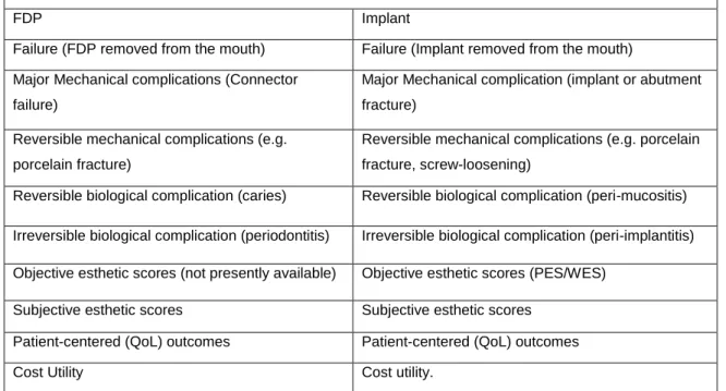
and more efficient over time than bridge first-line therapy16.Table 1 presents
implicating factors for decision making between fixed partial denture and single
7
Table 1. Factors implicating tooth preservation or replacement
Potentially Relevant Outcomes in Evaluation Tooth Replacement
FDP Implant
Failure (FDP removed from the mouth) Failure (Implant removed from the mouth)
Major Mechanical complications (Connector
failure)
Major Mechanical complication (implant or abutment
fracture)
Reversible mechanical complications (e.g.
porcelain fracture)
Reversible mechanical complications (e.g. porcelain
fracture, screw-loosening)
Reversible biological complication (caries) Reversible biological complication (peri-mucositis)
Irreversible biological complication (periodontitis) Irreversible biological complication (peri-implantitis)
Objective esthetic scores (not presently available) Objective esthetic scores (PES/WES)
Subjective esthetic scores Subjective esthetic scores
Patient-centered (QoL) outcomes Patient-centered (QoL) outcomes
Cost Utility Cost utility.
In addition to all the factors involved in decision making that consider options for
single tooth replacement, it’s noteworthy that the decision to restore pulpal and/or
apical pathological condition dentition with root canal therapy or extract the tooth and
replace with single implant restoration has been an issue of controversy for many
years. A systematic review by Torabinejad et al15 , presented that in patient with
periodontally sound teeth, root canal therapy (97%) present survival rate equally to
single tooth implant (97%) therapy but higher than fixed partial prostheses (82%) at
6 year follow up. The study also reported that the successful rate of single implant
therapy was higher than root canal (84%) and fixed partial denture treatment (80%).
The author concluded that implant supported restoration has similar survival rate as
root canal treated tooth; however the implant restoration showed longer time to
8
between root canal treated tooth and single tooth implant also concluded that there
was no significant difference in survival rate between two treatment modalities.
Since the survival rate between implant and root canal therapy are not different, the
decision must be based on other factors such as final restoration after root canal
treatment and its survival and complication rate, remaining sound tooth structure,
patient perception and preference and economic outcome19.
A recent review by Jiloski et al, reported that the presence of a 1.5- to 2-mm ferrule
has a positive effect on fracture resistance of endodontically treated teeth20.
However, several evidences support that root canal treated tooth followed by post
and core increased the complication rate, according to Goodacre et al reported that
post and core had at least 10% complication incidences5. In addition,
Holm-Pederson reported that existing periapical pathology dramatically decreased the
survival of non-vital teeth to less than 80% after 5 years21. Moreover, the high risk for
tooth loss appears to be the presence of perforation during retreatment decreasing
the 5 year survival rate to as low as 42%22. Therefore, condition of tooth should be
taken in consideration before decision making. In addition, overall oral rehabilitation
plan will play a part in the definitive decision for keeping or removing the tooth in
question.
Patient perception and preference also have influence on treatment decision. Gatten
et al evaluated patient’s perception of quality of life compare between endodontic
treatment and implant restoration using the Oral Health Impact Profile (OHIP-14) as
9
present similar overall OHIP score with high satisfaction rate. The major concerns
for patient included overall health status, treatment fee and insurance coverage,
patient perception, treatment outcome, treatment duration and number of visits23.
Cost of treatment is also another factor influencing patient decisions. Review
studies18,24 reported that endodontically treated teeth followed by definitive
restoration show less expensive and less clinical visits than implant supported
restoration. However, Derhalli et al recommended that a treatment cost analysis
should be considered in all possible treatment required. For example, crown
lengthening and foundation restorations should be included in root canal treatment
plan, on the other hand; hard and soft tissue graft and 3-dimention x-ray must also
be concerned in implant restoration therapy25. A recent study by Pennington et al
reported that root canal treatment is highly cost effective as a first line intervention.
Re-treatment is also cost-effective, but surgical re-treatment is not. Therefore,
extraction and replacement with single implant should be considered when
endodontic re-treatment is not effective26.
1.2 Survival of single tooth implants
Implant supported single crowns are growing as a first choice treatment modality for replacing missing single tooth164
. Long term studies have reported excellent implant survival rate in single tooth
implant replacement.A systematic review by Jung et al reported that the average
survival of single implant crown was 96.8% after 5 year and 94.5% after 5 year of
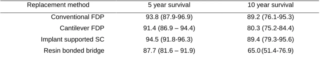
function6. A systematic review by Pjetursson and Lang in 2008 compared the 5 and
10
(Table 2) showed that in 5 year, single implant restoration (94.5%) presented similar
high survival rate as conventional fixed restoration (93.8%) and cantilever restoration
(91.4%). Resin bond prosthesis showed the acceptable survival rate of 87.7%. After
10 year of service, single implant restoration and conventional fixed restorations
showed reasonable survival rate around 89% and cantilever showed inferior survival
rate of 80.3%. Whereas, resin boned prosthesis show unacceptable survival rate of
65%27.
Table 2. Estimate survival rate of single tooth replacement in 5 and 10 year follow-up
5- and 10- year survival estimates for single tooth replacement *
Replacement method 5 year survival 10 year survival
Conventional FDP 93.8 (87.9-96.9) 89.2 (76.1-95.3)
Cantilever FDP 91.4 (86.9 – 94.4) 80.3 (75.2-84.4)
Implant supported SC 94.5 (91.8-96.3) 89.4 (79.3-95.6)
Resin bonded bridge 87.7 (81.6 – 91.9) 65.0 (51.4-76.9)
*Adapted from Pjetursson and Lang, 2008
This implies that the design of fixed partial prosthesis has a high influence to survival
of the restoration in long term. Conventional fixed prostheses or implant supported
single crown represent first treatment options. A more recent systematic review by
Jung et al(2012), also reported the 5 year survival rate of implant supported single
crown was 96.3% and 89.4% after 10 year28. This fact reinforces the previous
conclusions that single implant restorations are a predictable treatment modality with
a longer term evidence base of support.
1.3 Complications for single tooth Implants
According to published evidence, single implant restorations present high survival
11
by how long the restoration remains in oral cavity but should reflect complications of
the implant supported restoration that affect its appearance, its biologic influence on
local and systemic tissue, and patient acceptance.
Berglundh et al29 reported implant loss was the most commonly reported
complication in literature(96-100% described in the study) while other biological
complication (40-60%) and technical complications (60-80%) were underestimated.
The author also reported that the incidence of implant loss in treatment of single
implant crown over at least five year showed 0.76% of implant loss prior prosthetic
placement and an incident of 2.06-2.50% loss during 5 years of function. This review
also indicated that incident of overall implant loss prior to function (2.5%) and during
function (2-3%) was three time higher than incident of single tooth implant loss
(0.76%)29. A recent review by Jung et al28, reported in systematic review that the 5
year survival rate of implants supported single crowns was 97.2% whereas the
survival rate of single crowns supported by implant showed was 95.2%. After 10
year of function, the survival rate of implants supported single crowns decreased to
96.3% whereas the survival rate of single crown supported by implant showed was
89.4% This implies that complications that occur in restoration that supported by
implant might increase the failure rate of overall single implant restoration therapy.
Complications found in single implant restoration can be divided to technical,
biological and esthetic complications. Systematic reviews4,6 reported the annual
technical complication rate of complication in single implant crown was 0.92. The
common technical complications in implant supported restoration were abutment
12
and implant fracture (0.14%). More recent review28 also reported similar result that
the most common technical complication was screw loosening of 8.8%, followed by
loss of retention (4.1%), fracture of veneer (3.5%) and implant fracture (0.18%) and
screw fracture (0.18%). In addition, Salinas and Eckert mentioned that all-ceramic
restorations presented more biomechanical and technical complication incidences
that with metal-ceramic restorations30.
Biological complications include peri-implant hard and soft tissue changes, infection
and inflammation. Overall soft tissue complication is limited in presenting with
0.1-0.3 per person incident rate and among dental implant complications29. Systematic
reviews4,6 reported the annual complication rates of implant supported single crown
were 2.03 in 5 year follow up. Biological complications were presented with
peri-implant soft tissue complication (9.7%) and bone loss more than 2 mm (6.3%). More
recent review in 201230 reported that the 5 year soft tissue complications, including
peri-implantitis, were observed in 9.7%, whereas bone loss exceeding 2 mm was
reported on 6.3%.
Esthetic complications can be determined by clinician or patient interpretations of the
appearance of the restoration itself and/or appearance of soft tissues around the
implant restoration. Biological complications have high influence on the esthetic
outcome of single implant restorations. A systematic review of dental implant
complication by Goodacre et al31 showed that peri-implant soft tissue complication
were a concern for the single tooth implant. Peri implant soft tissue complication
includes dehiscence, fistula and gingival inflammation. Soft tissue dehiscence in
13
esthetic outcome and affect patient satisfaction. Causes of soft tissue complications
include poor oral hygiene, abutment implant misfit or micro-gap at abutment implant
junction, which can lead to bacterial migration and changes of soft tissue level
around implants-abutments which affects the esthetic result adversely30.
A recent review by Jung et al reported the esthetic complication due to
dis-satisfaction of esthetic appearance was 7.1%28 and 9% of esthetic complication rate
was also reported by Salinas et al30 who also mentioned that minimizing esthetic
complications is challenging and related to both technical and biological
complication.
1.4 The issues with evolving single tooth implant therapy
A conventional concept of dental implant therapy recommend placement of dental
implant in healed extraction sites with two stage surgery and a 3-6 months unloading
period32. Due to long treatment duration, number of surgeries and complicated
prosthetic procedures, new concepts include one stage surgery, immediate
placement, immediate loading, and early loading have been introduced.
Evidence33,34 shows that there was no difference in survival rate of implant
restoration between 1-stage and 2-stage surgical protocol. However, a 2-stage
protocol could be indicated when an implant has not obtained an optimal primary
stability. In addition, A systematic review by Den Hartog et al35 compared treatment
outcome of immediate, early and conventional single-tooth implants in the esthetic
zone, also reported that there is no statistically significant difference of survival rate
14
95.5% after 1 year. A recent systematic review by Strub et al also supported that
immediate loading had high survival rate ranged from 96.4-100%36. On the other
hand, a review by Atieh et al, 2009 reported that immediate loading in single implant
crown increased risk of implant failure (relative risk 5.07) compared to conventional
method37.Therefore, immediate loading protocol is considered a successful
procedure in selective cases.
In addition, these contemporary approaches were reported to help minimizing
peri-implant bone loss and provide better soft tissue healing by which possibly improving
the esthetic result especially in anterior esthetic areas. A clinical study by Cooper et
al38, of single-tooth implants reported that early/ immediate loading of single implant
restoration within 3 weeks after implant placement using 1-stage surgical protocol
provided a good survival rate of 94% and acceptable mean marginal bone changes
of 0.4 mm per year. In addition, the authors also reported that 1-stage surgical
protocol and early loading with proper abutment and provisional restoration improve
peri implant soft tissue outcome (A mean gain in papilla length was 0.61 mm at 1
year and 0.74 mm at 3 year and a gain in buccal gingiva was 0.34 mm.at one year
and 0.51mm at 3 year). More evidence39-41 supported that shortening of loading
period and establishing proper provisional crown allow soft tissue adaptation and
permit papilla formation. While, 2-stage surgical protocol might limit soft tissue
healing and inhibit formation of soft tissues that follow anatomical contour. A recent
clinical study42 showed that there is an improvement of papilla level after 1 year
follow up with immediate placement and provisionalization procedure. This has been
15
implant placement protocol provided advantages to hard and soft tissue
preservation. In this review, the author also reported that early placement presented
higher level of patient satisfaction compared to conventional procedure 43.
These more aggressive approaches such as immediate placement and immediate
loading should be implemented with caution and should be preceded by careful
patient selection and treatment planning. In the esthetic zone, tooth loss or removal
is associated with reduced or insufficient facial wall bone which may be a major
factor affecting soft tissue recession when placing an implant immediately in the
socket 44. This can lead to failure of esthetic results such as loss of harmonious
gingival margin, exposure of metal collar, and improper contour of definitive
restoration. A clinical study45 suggested that immediate placement did not alter the
fact that after extraction, there is a decrease of bone dimension which affects soft
tissue changes. De Rouck et al, 2008 also reported that immediate implant
placement in anterior maxilla provide a good survival rate (range 78.6-100%) and
predictable papilla level (-0.39-0.53 mm in 1 year), but undesirable mid facial soft
tissue outcome (-0.55-0.75 mm in 1 year) with average of buccal bone loss range
0.22-1.05 46.This implied that managing soft tissues around implant restorations is
unpredictable and it might need additional steps or procedures to establish
satisfactory outcome. Placement of implant immediately in extraction socket in
patients with insufficient bone might increase the risk of hard tissue defect which
affect long term esthetic outcome. A prospective, randomized-controlled clinical
study by Sanz et al, reported that the immediate placement of an implant resulted in
16
vertical dimension) in 16 weeks after implant placement47. In addition, a recent study
evaluated the thickness of buccal bone at the maxillary anterior region using cone
beam computed tomography (CBCT). The study reported limitation of buccal bone
thickness in majority of patients (the median thickness at the midroot was 1.03 mm
in the premolar area and 0.70 mm for the other anterior maxillary teeth)48.
Therefore, several surgical techniques have been used to correct the bony defect
prior to implant placement such as ridge perseveration following extraction, onlay
grafting, guide bone regeneration (GBR) with barrier membrane, and a combination
of block bone grafts and barrier membrane. These bone augmentation procedures
have been well documented. The ridge preservation approach has demonstrated
success in preserving ridge dimension49. In addition, clinical studies50 demonstrated
that horizontal bone augmentation can be predictably obtained with GBR technique,
whereas vertical bone augmentation seems to be more difficult to achieve satisfy
outcome. Bone grafting procedures improve tissue contour and allow clinicians to
place implant in favorable position which affects the esthetic result. A more recent
study by Hof et al, 2011 reported that bone augmentation at anterior maxilla prior
implant placement allow achieving favorable esthetic results51. This result was also
supported by Buser et al, 2013 that the follow-up of 5 to 9 years presented low risk
of soft tissue recession with early implant placement and bone augmentation with
guided bone regeneration (GBR) maintained a facial bone wall in 95% of patients
which improve satisfactory esthetic outcomes52.
However, implant placement in augmented site might leave a question to the
17
Jensen and Terheyden 53 provided strong evidence that implants placed in
augmented bone have comparable survival rates as implants placed in good quality
bone, however; the available data did not allow identifying one surgical procedure
offered better esthetic outcomes than another. Moreover, success rate of implant
therapy with or without bone augmentation also depend on various factors including
patient health status and life style, size and site of bony defect and surgical
procedures54.
1.5 Implant esthetics
Providing reproducible, highly esthetic outcomes is a challenge of dental implant
therapy. The general esthetic goal of dentistry is to establish the harmonious
appearance the restoration with adjacent teeth and tissue. For implant restorations,
both the peri-implant soft tissue to the surrounding mucosa around the adjacent
teethand the final restoration have distinctly different supports (abutment and
implant) compared to the natural roots and tissue of the adjacent natural dentition.
Soft tissue changes around implants affect the esthetic outcome of the implant
restoration such as absence of interdental papilla, gingival recession and might lead
to esthetic failure of final restoration. Meanwhile, underlying hard tissue structure
plays a key role in the establishment of esthetic soft tissue especially in anterior
maxilla.
To achieve satisfactory outcome, good planning prior implant placement is
bio-18
physiological response of soft and hard tissue surrounding dental implant is
necessary.
Maintenance of the interproximal papillae height is one main esthetic goal in single
tooth implant restoration. Published evidence showed that the level of interproximal
bone crests affects the presence of interproximal papilla. According to a clinical
study 55, showed that if the distance between the contact point of the restoration to
crest of the bone was greater than 5 mm then this would be a critical point at which
the papilla no longer predictably filled the interdental space. This observation has
been confirmed with implant-supported restorations56. In addition, not only crestal
bone height has influence on preserving interproximal soft tissues but the distance
between implants is also associated to papilla response. A study by Tarnow et al,
demonstrated that the mesio-distal distance between implants more than 3 mm
(0.45 mm bone loss) present less interproximal bone loss than distance less than 3
mm (1.04 mm bone loss) 57. The same author also reported that the mean height of
papillary tissue between two adjacent implants was 3.4 mm (range of 1 mm to 7
mm). This implied that esthetic outcome related interproximal papilla might be limited
when placing implant adjacent to each other 58. Zetu et al introduced aesthetic
triangle as a reference of management of interproximal papilla explained that in
order to achieve optimal esthetic outcome, management of interproximal papilla
should be planned prior to tooth extraction. Having good bone foundation for support
soft tissue will lead to higher esthetic satisfaction; however, in case that hard and
soft tissue esthetic cannot achieve by surgical procedure, restorative procedure
19
outcome59. A prospective clinical study in 200660 evaluated a mean marginal bone
resorption at the facial and lingual aspect of the implant was 0.7 and 1.3mm at the
time of abutment connection, while mean proximal bone loss was only 0.1mm. This
small interproximal bone change might lead to positive papilla response around the
implant crown. The study reported the interproximal papilla fill of more than 50% was
shown in 32% at crown placement visit and 86% at 1 year. The result showed the
improvement of papilla height, while facial soft tissue presented negative response.
Gingival biotype is also widely considered to influence the esthetic outcomes for
dental implants. Kois et al described that a thick gingival biotype is more resistant to
recession but more prone to create periodontal pocket at teeth. On the other hand,
thin gingival biotype usually has less osseous supported and high risk to recess after
the surgery 61.A clinical study by Kan et al evaluated that gingival biotype has
influence on final position of implant platform. A thin biotype will require implant
placement more palatal to hide metal color show-through. However, placing implant
too far palatal will limit establishment of ideal emergence profile62. Therefore, patient
who has thin gingival biotype with high smile line should be offered less esthetic
expectations. In addition, recent studies63,64 evaluated relationship of thickness of
facial plate and gingival biotype using CBCT as an assessment, found that there is a
relationship between thickness of facial plate and gingival biotype. Another study by
Kan et al indicated that gingival biotype associated with midfacial soft tissue
changes but have no strong influence on interproximal papilla level42. In contrast, a
study by Si et al reported that thickness of gingival mucosa prior to implant
20
appears that a complex situation involving more than gingival biotype affects peri
implant tissue. Various factors including implant position, the implant abutment
interface, implant/abutment materials, and bone dimension may influence tissue
responses and esthetics.
Evidence supported that the concept of a physiological response responsible for the
biological seal around natural dentition is known as the biological width66. This
organization of connective tissue and epithelium also occurs around dental implant.
This biological seal which forms along the implant abutment consisted of junctional
epithelium and avascular connective tissue attachment with an average of 3 mm in
height. This process of the soft tissue attachment around non-submerged dental
implant that was reported to be properly established after several weeks following
surgery 67.According to Linkevicius et al68,biologic width is a stable biological
response that acts as a soft tissue protector around dental implants, and the
interference of biologic width might lead to hard and soft tissue changes. A more
recent study also reported that biological width around one-piece implants occurred
with similar manner comparing between immediate, early and conventional loading
procedures69 and one-piece implant present similar biologic respond to natural
dentition in comparison to 2-piece implant70. The aggregate information concerning
biologic width at dental implants suggests this anatomic organization of connective
tissue and epithelium is consistently observed at all implants.
A series of animal studies by Hermann et al indicated that a chronic inflammatory
response influenced the crestal bone loss as a result of biofilm accumulation and the
21
biology. Biologic response of crestal bone starts around a 2-piece implant after
abutment installation and crestal bone remodels to a level approximately 2.0 mm
apical to the implant abutment interface71-74. A study by Collan et al, used DNA probe
analysis to identify location of bacteria colonization reported that the bacteria
colonization found at internal surfaces and healing abutment screw-threads within 25
days after second stage surgery and placement of healing abutment. This implies
that a microgap at implant abutment interface allow bacterial infiltration creates
inflammation that might lead to peri-implant hard tissue changes75.
A longitudinal radiographic study by King et al, evaluated the influence of microgap
on crestal bone level showed that 2-piece implant presented greater crestal bone
loss compared with 1-piece implants. The stability of the interface has an influence
on the early wound healing stage around implant but not the size of interface 76.
Moreover, several studies73,77,78 also demonstrated that the location of implant
abutment junction has an influence on vertical bone loss around implants. The
vertical bone remodeling will be increased (average 1.3-1.8 from animal studies) if
the implant abutment junction is located deeper into the bone. Therefore, placement
of implant too far sub-crestal (apical position of implant abutment interface) might
lead to unnecessary bone loss around implant. Bone loss that occurred due to
implant abutment microgap not only affected interproximal crestal bone but also
affects the facial bone resorption which might lead to facial soft tissue deficiency.
Thus, good examination of facial bone dimension prior tooth extraction will lead to
22
Facial bone is an important anatomical architectural feature that affects the final
esthetic outcome of single implant restoration. Deficiency of height and thickness of
facial bone affected the stability and harmony of facial soft tissue around implant
restoration and adjacent teeth79. Clinical and radiographic examination will help
determine amount of facial bone which leads to proper management. A prospective
multicenter study evaluated the thickness of buccal and palatal bone walls at
extraction site prior implant placement using a caliper instrument. The study reported
the reduction of buccal bone height (-0.1 mm) was more significant than palatal bone
loss (-0.5mm) at extraction site 47. A study by Lau et al analyzed the thickness of the
buccal bone at their mid-root and apical level using 300 cone beam radiographs
reported that the mean thickness of buccal bone at the mid root level was 0.9+/-0.4
mm and at the apical level was 2.04+/-1.01 mm. This information showed that there
is a limitation of patient facial bone width and a traditional protocol with site
preparation prior to implant placement might provide more pleasing esthetic
outcome80. Kan et al also reported facial bone influenced the clinical outcome of
recession facial gingival tissue stability following immediate placement and
provisionalization of maxillary anterior single implant. The study showed that
absence of grafting the gap between the implant might affect the facial tissue
recession42. Due to limitation of anatomic structure, tissue augmentation might be an
answer to create proper adequate bone volume that lead to esthetic success.
According to Buser et al, to achieve esthetic outcome, placement of implant in a
correct three dimensional position is crucial 44. The three dimensional concept has
23
that in mesio-buccal view, a minimum of 1.5-2 mm of distance between adjacent
tooth and implant was recommended to provide space for prosthetic restoration and
allow establishing proper physiological response of peri-implant tissue. The
thickness of facial bone and emergence profile of the restoration are used to
determine the bucco-lingual position. Limitation of facial bone that leads to place
implant more palatal might result in an unsatisfactory emergence profile. The
apico-coronal position, placing implant too shallow will affect esthetic outcome but placing
the implant too deep might result in an undesirable hard and soft tissue loss around
implant81. A more recent article by Cooper in 2008 explained esthetic objective
criteria for single implant in anterior areas. The author recommended using gingival
zenith level which is the most apical point of definitive crown as a reference level for
implant placement in esthetic zone. The author explained that it is suggested to
place implant 3 mm apical and 2 mm palatal to the planned gingival zenith in order
to provide proper implant position in esthetic areas, however, if facial bone is
insufficient to follow this guideline, bone augmentation prior to implant placement
must be performed82. Several guidelines and recommendations have been
introduced attempting to achieve esthetic satisfaction. However, to obtain high
patient satisfaction, careful examination and diagnostic procedure seems to be a
leading key to proper management in each particular patient. In addition, clinicians
should understand patient’s perspective and expectation to minimize failure of
24 1.6 Patient satisfaction
Esthetics is subjective; therefore, satisfaction of single implant restoration must
depend on each individual perceptions, preference and experience. To assess the
satisfaction of esthetic outcome of implant supported single crown is challenging.
Visual analog scale (VAS) and questionnaires have been used to evaluate single
implant crown satisfaction. A study83 compared patient and dentist satisfaction of
esthetic outcome of implant single crown in maxillary anterior areas showed that
patient give higher values of outcome satisfaction compared to dental professional.
Most variables in the patients' assessments revealed mean values above 90%.
Factors considered by dental professionals to be of significance for the esthetic
result such as surrounding soft tissue appearance and form of the crown may not be
of decisive importance for the patient's satisfaction. Another study84 that used VAS to
evaluate patient’s satisfaction of implant single crown in esthetic zone compared to
the contralateral natural tooth, reported similar result with high degree of patient
satisfaction (mean value of 96%). A more recent study by Meijndert et al85 reported
that the score Implant Crown Aesthetic Index showed that only 66% of the cases
had acceptable esthetic outcome from dental professionals, while the score from
satisfaction questionnaire by patient showed 100% acceptable result. However, both
patient and dental professional rated less satisfactory of peri-implant mucosa than
the implant-supported crown. This result implies that peri-implant mucosal changes
have high influence to dental professional perception but might be of minor concern
to patient. Another retrospective study86 evaluated outcome of early placed maxillary
25
objective esthetic assessment showed that there is no statistically significant
correlations between total of PES/WES and patient satisfaction using VAS. Some
patients provide high outcome satisfaction whereas PES/WES scores from dental
professional did not correlate with VAS score. This supported previous studies that
the patient’s perception of dental restoration from esthetic point of view differs from
dental professionals. On the other hand, A recent study by Cho et al used PES/WES
to evaluate maxillary single implant restoration in esthetic zone and found that there
was a statistically significant correlation between patients’ esthetic perception and
dentists’ perception except in premolar region. In this study also reported that soft
tissue augmentation has been performed in some patients who have high smile line
and thin gingival biotype which helped to increase level of patient satisfaction87.
From all these evidences, it may be implied that the level of patient satisfaction is the
overall appearance when smiles. Surgical procedures such as soft tissue grafting
26 1.7 Implant esthetics assessment
Several assessment measures (Table 3) for single implant restoration have been
developed to help determine success of the esthetic outcome. In 1997, Jemt88
developed a Papilla Filled Index (PFI), an assessment of interproximal gingival
papilla size around single tooth implant; however, this index does not assess other
esthetic influences of the soft tissue around implant such as facial soft tissue level,
color, and texture. Another esthetic evaluation instruments is Implant Crown
Aesthetic Index (ICAI) developed by Meijer et al in 2005. The ICAI evaluated both
restoration itself and surrounding soft tissue (color, anatomic contour and surface
texture of crown and soft tissue)89. In the same year, Furhauser et al90 introduced
Pink Esthetic Scores (PES), an objective esthetic assessment for single implant
crown in the esthetic zone using clinical photographs for evaluation. PES evaluates
only soft tissue and seven soft tissue characteristics were utilized compare to a
reference tooth including mesial and distal papillae, soft tissue level and contour,
alveolar process deficiency, and the color and texture of the facial marginal
peri-implant mucosa. Among all variable, level of soft tissue margin (facial recession) and
color presented lowest scores, while papilla present has the best score. Evidence
supports PES as a reproducible esthetic assessment of single implant crowns in
both the short and long term51,91-94.
In 2009, Belser et al86 evaluated esthetic outcome in early placed maxillary anterior
single tooth implants using a new objective esthetic assessment called Pink Esthetic
Score/White Esthetic Score (PES/WES). PES/WES is an index that is modified from
27
including: mesial and distal papillae, curvature of facial mucosa, level of facial
mucosa, and root convexity/soft tissue color and texture, then adding 5 implant
restoration variables consist of tooth form, tooth outline/volume, tooth color and
surface texture, and translucency. This index evaluates both soft tissue and implant
restoration using both clinical photographs and diagnostic models. Belser also
reported that PES/WES index is a suitable assessment for evaluation of esthetic
outcome of single implant restoration.
In a recent study by Weinlander et al96, another esthetic evaluation method of the
peri implant mucogingival complex by collection of standardized photographs and
computer-assisted measurement of reproducible data was used. Six soft tissue
parameters have been used which include mesial and distal papillae areas and
heights, soft tissue-crown perimeter, and gingival recession. The study found that a
standardized oral photograph is considered an accurate and reproducible method for
the evaluation and measurement of soft tissue changes that can affect the esthetic
outcome.
According to esthetic assessment indexes, facial soft tissue around implant is one of
the main variables that use to determine esthetic outcome in almost of implant
esthetic. A study by Furhauser el al, reported that level of soft tissue margin
presented lowest scores while gingival papilla has high esthetic score90. A study by
Lai et al evaluated soft tissue around single tooth implant also reported that soft
tissue level present significant changes compare of other variable91. In addition a
28
while papilla height showed small change 42.Therefore, soft tissue margin level
should be given special attention by the clinician.
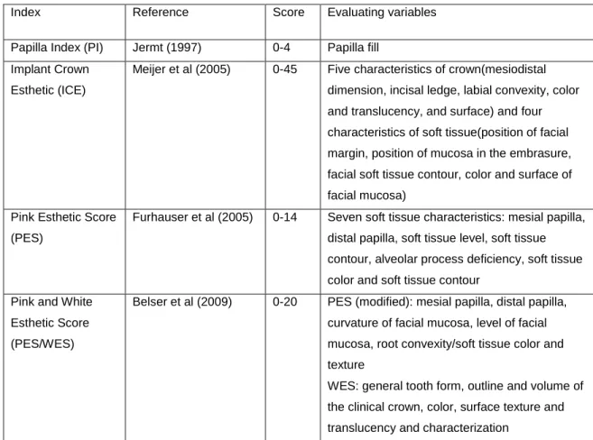
Table 3. Description of indices for assessment of single implant esthetics*
Index Reference Score Evaluating variables
Papilla Index (PI) Jermt (1997) 0-4 Papilla fill
Implant Crown
Esthetic (ICE)
Meijer et al (2005) 0-45 Five characteristics of crown(mesiodistal
dimension, incisal ledge, labial convexity, color
and translucency, and surface) and four
characteristics of soft tissue(position of facial
margin, position of mucosa in the embrasure,
facial soft tissue contour, color and surface of
facial mucosa)
Pink Esthetic Score
(PES)
Furhauser et al (2005) 0-14 Seven soft tissue characteristics: mesial papilla,
distal papilla, soft tissue level, soft tissue
contour, alveolar process deficiency, soft tissue
color and soft tissue contour
Pink and White
Esthetic Score
(PES/WES)
Belser et al (2009) 0-20 PES (modified): mesial papilla, distal papilla,
curvature of facial mucosa, level of facial
mucosa, root convexity/soft tissue color and
texture
WES: general tooth form, outline and volume of
the clinical crown, color, surface texture and
translucency and characterization
*Modified from Benic et al, 201297
1.8 Factors Affecting Peri Implant Buccal Tissues
Buccal soft tissue changes affect esthetic outcome of single implant restoration.
Various factors influence facial soft tissue responses and range from surgical
protocol, augmentation procedure, gingival biotypes, implant design and implant
29
Different implant surgical techniques might have influence to facial soft tissue
responses differently. A review study by De Rouck et al, reported that immediate
placement and provisionalization showed unpredictable buccal soft tissue recession
and the changes of facial soft tissue occurred since tooth extraction46. Van
Keresteren et al compared immediate protocol and delayed implants placement after
extraction and ridge preservation utilizing Straumann Tissue level implants with a 1.8
mm transgingival collar. The study reported that the overall mid facial recession is
small (0.17 +/- 0.47 mm) without significant difference between the immediate and
delayed treatment groups. The authors also reported that immediate placement
group showed a greater ridge resorption compared to ridge preservation and
delayed placement group98. Another study by Raes et al compared between
immediate and conventional implant placement in anterior areas found that
immediate placement presented fairly stable midfacial soft tissue levels with only 7%
of cases showing advanced recession. In addition, the investigators also reported
that less midfacial recession in flapless surgery compared to flap surgery99.
Moreover, in a prospective study of 35 immediate implants with an overall follow up
time of 4 years, the tissue changes at Sterioss replace implants was measured. The
midfacial mucosal changes at 1 year (0.55mm +/- 0.55 mm) were extended to 1.13
mm (+/- 0.87 mm) at the terminal evaluation period42. The authors also reported that
11% of patients complained about esthetic issues which might be the result for
gingival tissue changes. The relationship of surgical protocol and facial tissue
recession has been controversial. Facial hard and soft tissue might also depend on
30
Augmentation procedure is recommended in situations where there is inadequate
bone volume to place implant in proper position. Therefore, augmentation procedure
is also another factor that could affect facial soft tissue.
Due to the limitation of facial bone volume, several augmentation techniques have
been recommended to improve hard and soft tissue contour. Several studies were
interested in the changes of facial soft tissue around single implant restoration with
and without augmentation procedure. A study by Cosyn et al evaluated the esthetic
outcome of crown and soft tissue around single tooth implant with early placement
following extraction and GBR. The study reported that clinical crown and facial soft
tissue presented small changes (0.3 mm) in 21 months and that patient satisfaction
might be related to adequate bone volume at implant placement due to bone
augmentation100. Verdugo et al reported the onlay graft technique would predictably
reconstruct function and esthetics. The study also reported stable bone volume
around implants at an average of 3.5 years. 101. Moreover, Kan et al reported in the
study of immediate placement of single tooth implant that the absence of grafting the
gap between the implant and facial bone could have influenced the clinical outcome
of recession42. On the other hand, Jemt and Lekholmperformed a 6-year
prospective clinical study evaluated the stability of facial contour after buccal block
bone graft from patient bone. The author reported that block graft procedure
provided sufficient bone volume for implant placement after 6 months, however,
bone remodeling patterns in each individual might lead to unpredictable results for
long-term prognosis. The author also reported that proper abutment and crown
31
prior to implant placement to establish adequate bone volume represents one
management approach to minimize undesirable facial soft tissue deficiency.
However, there is not a gold standard protocol to recommend what type of bone
graft procedure can maintain buccal contour of implant restoration better than
others.
In addition, gingival biotype is another factor that might be related to facial soft tissue
around the implant crown. A study by Verdugo et al, reported grafted site phenotype
did not seem to be influenced by the adjacent teeth biotype101. Kan et al, 2010
reported in an immediate implant placement study that a thin biotype was statistically
associated with greater midfacial recession at implants and not relate to
interproximal tissue levels42. Van Keresteren et alcompared immediate protocol and
delayed implants placement reported that no effect of biotype on the changes of soft
tissues98. Raes et al99 similarly concluded that biotype did not affect facial soft tissue
alterations. A recent review by Lee et al, presented the relationship of gingival
biotype and peri-implant soft tissue found that thin gingival biotype present higher
risk of hard and soft tissue changes after implant surgery. Furthermore, modified
implant abutment interfaces such as platform switched design also failed to maintain
peri-implant soft tissue in thin gingival biotype. The relationship between
immediate/delayed placement, gingival biotype and soft tissue recession is still
controversial. According to this review, several investigators presented that
immediate placement in patient who have thin biotype may increase risk of gingival
recession more than with thick biotype, while some studies reported no significant
32
also mentioned that in thin gingival biotype abutment material color might affect color
of marginal soft tissue. Zirconia abutment is recommended that can be used for
esthetic purposes in all gingival biotype103. On the other hand, Bressan et al reported
that the thickness of the peri-implant soft tissue did not appear to be a crucial factor
in the abutment impact on the soft tissue color. The peri-implant soft tissue color
differs from the soft tissue color around natural teeth, no matter which type of
restorative material is selected and the grey colored and titanium abutments invoked
significantly higher color differences than gold or zirconia abutments.
Implant design is another factor that might have influence on facial hard and soft
tissue contour. A contemporary implant design of rough neck surface reported that
preserve marginal bone level compares to smooth neck surface implant. Reduction
of smooth surface minimized marginal bone resorption around implant. Micro-rough
and nano-rough surface extending to the implant neck and a fine thread in the
cervical region has shown that the crestal bone level was stabilized by with
transmitting loading force to the adjacent bony structures 104. A study of immediate
loading/provisionalization of single implant in esthetic zone showed that micro
threaded, TiO2 grit-blasted implants maintains crestal bone level and improved soft
tissue dimension105. Moreover, Den Hartog et al compared marginal bone level
changes in different neck designs for single anterior tooth replacement. Three
different implant neck designs included Steri Oss replace (smooth group), Replace
Groovy (rough group) and Nobel Perfect implant (scalloped group) systems. They
observed greater marginal bone loss over one year at the scalloped neck design,
33
measured changes in interproximal tissues, they failed to measure changes from
baseline to 6 months for the midfacial tissues106.
Recent studies pay more attention to implant abutment interface and micro motion
that influence hard and soft tissue changes. Micro gap formation during function may
induce bacteria invasion into the connection between implant and abutment and the
continued micromotion of transmucosal abutment may also create mechanical
irritation of peri implant soft tissue, causing chronic inflammation and subsequent
vertical bone resorption107. In addition, a study by Todescan et al demonstrated the
closer the implant abutment junction to the crest of the bone, the more bone
resorption occurred77. Therefore, implant designs that allow microbial leakage and/or
micromotion of the implant-abutment connection can lead to chronic inflammation
and bone loss. Ryser et al demonstrated that implants with connections that possess
micromotion (flat-to-flat) are associated with reactionary crestal bone loss108. A
current strategy to reduce the related inflammatory impact of flat-to-flat interface
designs is the lateralization of the interface from the implant bone connection or
“platform switching” 109. Canullo et al, reported that in immediate single implant
restorations in anterior area, the use of platform switching help preserve peri-implant
alveolar bone-level. The study showed that the average of bone reduction level of
0.30 mm (SD = 0.16 mm) in platform switching group, while the average reduction in
the control group is 1.19 mm (SD = 0.35 mm)110. A systematic review about platform
switching by Atieh et al,reviewed 10 studies with 1,239 implants showed that the
marginal bone loss around platform-switched implant was significantly less than
34
proved platform-switching design preserve peri-implant facial soft tissue better than
platform-match design111. A recent study by Pieri et al compared peri implant soft
tissue level between two different implant abutment interfaces (one platform
switched, the other flush), the authors measured bone and soft tissue changes one
year following implant placement into premolar extraction sockets. The control
implants (flat-to-flat) revealed 0.73 +/- 0.52mm midfacial recession while the test
(platform switched) implant revealed 0.61mm +/-0.54 mm midfacial recession (NS).
Most of the changes were recorded during the first 4 months of evaluation. The
radiographic measures for control and test groups at 12 months revealed the
average mesial and distal bone level changes were 0.49 +/-0.25mm and 0.19+/-
0.17 mm change for control and test groups respectively. The authors observed that
the soft tissue changes did not reflect the bone changes as suggested previously by
many investigators (greater bone loss in control group than test group but no
significant change for facial soft tissues) 112.
Rather than platform-switching or flat-to-flat interfaces, several studies have
revealed the relative absence of reactive bone loss at implants with conical implant
abutment connections that lack micromotion and lead to decrease marginal bone
loss. A clinical trial on single-tooth replacements with the Astra Tech implant system
demonstrated minimum marginal bone loss (0.06 mm at first year and mean total
bone loss 0.14 mm in 5 years)113. Another retrospective study of immediate
placement and provisionalization using micro-threaded Astra conical
implant-abutment interface implants. The study reported that conical implant-implant-abutment
35
bone loss 0.33+/-0.40 and distal bone loss 0.28+/-0.37) and maintain soft tissue
around implants105. A more recent prospective multicenter clinical study evaluated
marginal bone level of immediate loading of single implants between placed in
healed ridges and placed in extraction sockets. A 1 year result showed that there is
increasing of mean marginal bone level of 1.30 mm in extraction socket group
whereas in healed ridged group present a reasonable 0.40 mm of mean marginal
bone loss. In addition, the investigators also observed with conical implant abutment
interface, the mucosal zenith was stable or gain following definitive crown placement
in both groups114. The evidence supports that conical interfaces help preserve
marginal bone due to elimination of inflammatory zone at implant abutment interface
and might lead to maintaining facial soft tissue level. A systematic review comparing
long term marginal bone responses of different implant systems illustrates the
potential influence of implant design on outcomes 115.
The implant abutment interface design is another factor affecting peri-implant tissue
responses. The different facial soft tissue responses to different implant abutment
interfaces are of current interest. Unfortunately, no systematic, prospective
comparison of peri-implant tissue responses at implants of varying implant/abutment
interface designs has been undertaken. Therefore, the aim of this study is to
compare the buccal soft tissue changes occurring around single-tooth replacement
in the maxilla using three different implant-abutment interface designs. The
null-hypothesis of this prospective clinical study is there is no statistically significant of
36
three different implant-abutment interface designs, namely, conical interface,
2. Materials and methods
The study was an open, prospective, randomized multicenter study. The study
population consisted of individuals requiring one or more single tooth replacement in
the maxilla within region 5 to 12. 141 Subjects distributed among four centers were
treated and followed for one year duration. The treatment included implant and
abutment installation in a one-stage procedure with immediate provisionalization
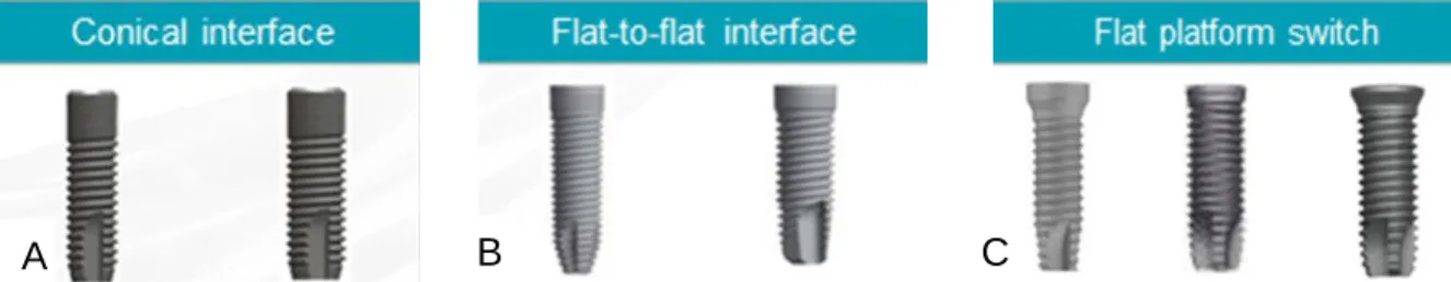
followed by final restoration. Eight main clinic visits were involved. Subjects were
randomized into three groups: group A (Conical interface design-OsseoSpeed),
group B (Flat-to-flat interface design-NobelSpeedy Replace) or group C (Flat
platform switch design-NanoTite Certain Prevail) (Figure.1).
Figure 1. Implant abutment interfaces: (A) Conical, (B) Flat-to-flat, and (C) Flat platform switch
The change in peri-implant tissue from baseline (provisional restoration) to one year
was compared. Measurements of peri-implant mucosal changes were made from
standardized photographs at visit 4 (4 weeks after implant placement and
provisionalization), visit 6 (provisional restoration, more than 8 weeks after implant