THE HEMIDESMOSOMAL PROTEIN BP180 (COLLAGEN XVII) IN SKIN CANCER AND INFLAMMATION
Bin-Jin Hwang
A dissertation submitted to the faculty at University of North Carolina at Chapel Hill in partial fulfillment of the requirements for the degree of Doctor of Philosophy in the
Department of Microbiology and Immunology
Chapel Hill 2018
Approved by: Zhi Liu
iii ABSTRACT
Bin-Jin Hwang: The hemidesmosomal protein BP180 (collagen XVII) in skin cancer and inflammation
(Under the direction of Zhi Liu)
BP180, also known as collagen XVII, is a transmembrane glycoprotein located in the hemidesmosome of basal keratinocytes, and functions as a key cell-matrix adhesion molecule. Loss of BP180 function in human disease leads to subepidermal blistering, and can occur by either autoantibody production (bullous pemphigoid) or inherited mutations in the BP180 gene COL17A1 (junctional epidermolysis bullosa). However, its other biological functions and involvement in different pathological conditions are
unknown. To uncover new functions of BP180, we generated a novel BP180
dysfunctional mouse strain lacking the NC16A domain of BP180 (termed ∆NC16A). We found that ∆NC16A mice developed a proinflammatory microenvironment in the skin accompanied by an influx of immune cells, including mast cells and MDSCs. ΔNC16A mice show spontaneous skin inflammation accompanied by TSLP-dependent itch. When tested in the B16 mouse melanoma models, ∆NC16A mice showed significantly increased melanoma progression. NC16A deletion in the skin or epidermis was
iv
microenvironment and increased tumor progression. Mast cell-deficient ∆NC16A mice had drastically reduced MDSCs in the skin and developed significantly reduced
melanoma. Mast cell reconstitution restored the skin infiltration of MDSCs and
increased melanoma progression in mast cell-deficient ∆NC16A mice. More importantly, MDSC depletion significantly reduced the tumor progression in mast cell-sufficient ∆NC16A mice. These findings provide the first evidence suggesting that BP180 in basal
iii
ACKNOWLEDGEMENT
I would like to thank Dr. Zhi Liu for his mentorship and support during the past six and a half years. It is my greatest honor to work in this lab. I would like to thank every member in my committee for their guidance over the past years. Without Dr. Maureen Su’s expertise in tumor immunology, my melanoma project probably would not have progressed as smoothly as it did. Dr. Scott Williams provided me the valuable training and materials to generate stabilized primary keratinocyte cultures, which proved to be critical in my projects. I would like to thank Dr. Jenny Ting and Dr. Yisong Wan for their comments and suggestions on my project.
I would also like to give my gratitude to Dr. Robert Bourret for his tremendous support during my years in the Department of Microbiology and Immunology.
iv
like to thank to Dr. Donna Culton, Dr. Ye Qian, our current chair Dr. Nancy Thomas and the former chair Dr. Luis A Diaz. They are wonderful to work with and my work could have not been done without the full support of the UNC Department of Dermatology.
v
vi
TABLE OF CONTENTS
LIST OF FIGURES………....VIII LIST OF TABLES………...XIII
LIST OF ABBREVIATIONS………...XIV
CHAPTER 1 BACKGROUND………...….1
1.1 BP180 and the hemidesmosome………..1
1.2 Skin inflammatory diseases and BP180……….…….3
1.2.1 Autoimmune Bullous Pemphigoid………...3
1.2.2 Junctional Epidermolysis Bullosa (JEB) ………..4
1.2.3 The possible correlation between BP180 dysfunction and skin inflammation.…….……….……….6
1.3 Inflammation, myeloid-derived suppressor cells (MDSCs), mast cells and cancer / melanoma progression.………7
1.3.1 Inflammation and Cancer………,,,………7
1.3.2 Myeloid-Derived Suppressor Cells (MDSCs) ………...11
1.3.3 Mast Cells.……….…….14
1.4 Known correlation between altered expression of BP180 and skin cancers.………..15
1.5 Mouse model for BP180 physiological functions.………..……..16
1.6 Working hypothesis for my dissertation………..18
vi
CHAPTER 2 THE DYSFUNCTION OF BP180/COLLAGEN XVII IN KERATINOCYTES LEADS TO MAST CELL-DEPENDENT SKIN INFILTRATION OF MYELOID DERIVED SUPPRESSOR
CELLS AND ACCELERATED MELANOMA PROGRESSION………..…..27
2.1 Overview and Significance….…,,,……….………...28
2.2 Introduction………...29
2.3 Results….………...………...32
2.4 Discussion……...………..………...40
2.5 References….………...71
CHAPTER 3 BP180 DYSFUNCTION TRIGGERS SPONTANEOUS SKIN INFLAMMATION IN MICE……….76
3.1 Overview and Significance…...………...76
3.2 Introduction….……….…...78
3.3 Results….………...80
3.4 Discussion….………...88
3.5 Materials and Methods….………...93
3.6 References….………...118
CHAPTER 4 DISCUSSION AND FUTURE DIRECTIONS………123
4.1 Summary and Findings….………...123
vii
4.3 New Function of BP180 in melanoma progression
through skin basal keratinocytes………..………128
4.4 Possible roles of BP180 in modulating the proinflammatory signaling pathway in keratinocytes, which leads to skin
inflammation and melanoma progression. ………..………132
4.5 Proposed future experiments………..………137
viii
LIST OF FIGURES
1.1 The structure of BP180 protein……….2 1.2 Epidermolysis bullosa (EB)..……….5
1.3 Types of Inflammation in cancer progression. ………..9 2.1. BP180 dysfunction ΔNC16A mice develop spontaneous
skin inflammation……….………...50
2.2. ΔNC16A mice show significantly increased melanoma
progression in both flank and ear injection models………..……….52
2.3. Skin- and basal keratinocyte-specific BP180 dysfunction
are sufficient to promote B16 melanoma progression. ……….………...54
2.4. Mast cells are crucial for melanoma progression in
∆NC16A mice………..56
2.5. Whole body-, skin- and basal keratinocyte-specific ΔNC16A mice have increased infiltration of MDSCs
in the skin………...……..58
2.6. Mast cells and MDSCs are crucial for MDSC infiltration
in ∆NC16A mice………,,,……….….60
2.7. BP180 dysfunction in skin basal keratinocytes triggers MC infiltration into skin, which leads to MDSC promoted
ix
S2.1. Generation of skin-specific ΔNC16A mice………...63 S2.2. Skin-specific ΔNC16A is sufficient to promote skin
inflammation………..……...64
S2.3. Myeloperoxidase (MPO) results indicate that ΔNC16A
promotes the influx of myeloid cells into skin………..………65
S2.4. ΔNC16A leads to the upregulation of proinflammatory
cytokines in skin……….………..…66
S2.5. Generation of basal keratinocyte conditional
K14Cre/ΔNC16A mice……….………..….67
S2.6. Anti-Gr1 antibody mediated myeloid cell depletion significantly reduces Gr1 positive immune cells in the
skin of ΔNC16A mice………..……....68
3.1. ΔNC16A mice exhibit skin inflammation with itch………..100 3.2. Skin-specific ΔNC16A (SkinΔNC16A) mice develop skin
inflammation with increased immune cell infiltration,
increased epidermal thickness, itch, serum IgE and TSLP…………..………….…102
3.3. Basal keratinocyte-specific ΔNC16A (K14Cre/ΔNC16A)
mice exhibit skin inflammation………..……….104
3.4. ΔNC16A mice deficient in both T and B cells
x
as whole body ΔNC16A mice……….……105
3.5. Itch in ΔNC16A mice is independent of IgE and histamine, but dependent on TSLP in keratinocytes………..………...106
S3.1. ΔNC16A mice exhibit skin inflammation with itch……….108
S3.2. ΔNC16A mice exhibit increased immune cell infiltration………109
S3.3. Quantification of cytokines at mRNA levels………..…110
S3.4. Generation of skin-specific ΔNC16A (skinΔNC16A) mice. ………...111
S3.5. Skin ΔNC16A mice exhibit increased immune cell infiltration and a defective skin barrier……….….………...112
S3.6. Basal keratinocyte-specific ΔNC16A (K14Cre/ΔNC16A) mice exhibit skin inflammation………..……..………...113
S3.7. ΔNC16A mice deficient in both T and B cells (Rag1-/-ΔNC16A) develop similar skin inflammation as whole body ΔNC16A mice...………….………...114
S3.8. B cell-deficient ΔNC16A mice develop skin inflammation………...115
4.1. Summary and conclusion of my past research covered at Chapter 2 and 3………125
xi
4.3 Mast cells play a significant role in the concentration of
CCL2 and CXCL1 in ∆NC16A mice………..……….………....133
4.4. Keratinocytes lacking NC16A promote the production of
CXCL1 through NF-κB……….…135
4.5. ∆NC16A significantly upregulates the phosphorylation
of FAK in vitro………...………..…….….136
4.6. FAK and Src inhibition significantly reduce the increased
CXCL1 production in ∆NC16A keratinocytes……….………..136
4.7. Two future experiments I proposed……….…………..………138
4.8. NF-κB inhibitor ammonium pyrrolidinedithiocarbamate (PDTC) significantly reduces the level of stem cell factor
xiii
LIST OF TABLES Table
2.1. The NC16A KO mice showed an increased expression of proinflammatory cytokines and chemokines which are
associated with the proinflammatory tumor microenvironment………..……...69
3.1. Increased expression of proinflammatory cytokines in
ΔNC16A mice. Skin RNA was isolated from three age and sex matched NC16A (WT) and ΔNC16A (KO) mice (8 weeks old), and was used for whole transcriptome microarray analysis. increased expression of proinflammatory cytokines and
xiv
LIST OF ABBREVIATIONS AD: atopic dermatitis
BCC: basal cell carcinoma
BMZ: basement membrane zone BP: Bullous Pemphigoid
BSA: bovine serum albumin FITC: fluorescein isothiocyanate
H1R: histamine 1 receptor H4R: histamine 4 receptor IB: immunoblotting
IF: immunofluorescence IFNγ: interferon γ
IL: interleukin
JEB: junctional epidermolysis bullosa MC: mast cells.
MBP: major basic protein
MDSC: Myeloid-derived suppressor cells.
xv MPO: Myeloperoxidase
PMN-MDSC: Polymorphonuclear-Myeloid-derived suppressor cells.
qPCR: quantitative polymerase chain reaction
SCC: squamous cell carcinoma TNFα: tumor necrosis factor α
1
CHAPTER 1 BACKGROUND 1.1 BP180 and the hemidesmosome
BP180, also known as collagen XVII, is a structural protein which plays a critical role in maintaining the linkage between the intracellular and the extracellular structural
elements involved in epidermal adhesion. The gene encoding BP180 (Col17A) was first isolated from human keratinocytes in 1990 and identified as a hemidesmosomal protein by the group led by Dr. Luis A. Diaz (Diaz et al. 1990). The gene encoding BP180 is located in the long arm of chromosome 10, locus 10q24.3 (Diaz et al. 1990). BP180 plays a key role in maintaining skin integrity through the hemidesmosome (Nahidiazar et al. 2015). In order to anchor epidermal keratinocytes into the underlying basement membrane, BP180 interacts with other proteins to form the hemidesmosome, which include BP230, plectin, keratins 5/14, integrin α6β4 and type VII collagen (Nahidiazar et al. 2015).
BP180 is a type II transmembrane glycoprotein: the extracellular
2
Figure 1.1 The structure of BP180 protein. BP180 (Collagen XVII) is a
transmembrane glycoprotein expressed by skin basal keratinocytes. It has a mostly non-collagen intracellular domain, a transmembrane domain and an extracellular domain with 15 collagen domains (black bars). The non-collagen domain NC16A contains the major epitope for autoantibodies in the patients of bullous pemphigoid (BP). In order to generate a human BP animal model, we generated humanized NC16A mice by replacing mouse NC14A with the human NC16A domain.
3
2011)(Matsumura et al. 2016). In addition, BP180 also plays a role in keratinocyte migration through β4 integrin signaling (Löffek et al. 2014). Therefore, BP180 is not only a cell-ECM adhesion molecule, its diverse physiological functions need broader
investigation.
1.2 Skin inflammatory diseases and BP180
BP180 dysfunction leads to skin blister diseases with inflammatory features, which include bullous pemphigoid (BP) and junctional epidermolysis bullosa (JEB). BP is caused by autoreactive antibodies targeting BP180 in patients (Nousari and Anhalt 1999)(Leighty et al. 2007). JEB is caused by genetic mutations of BP180 gene (Kiritsi et al. 2011)(Fine 2010a)(Jonkman et al. 1982).
1.2.1 Autoimmune bullous pemphigoid
BP is an autoimmune skin blistering disease which occurs most commonly in the elderly population (Hammers and Stanley 2016). The skin lesions on BP patients
4
from severe itchy sensations (Nousari and Anhalt 1999)(Leighty et al. 2007)(Hammers and Stanley 2016).
Since the 1970s, scientists started to develop an animal model for human BP. However, due to the fact that amino acid sequence of the autoantigen, NC16A, is poorly conserved in murine BP180 NC14A, passive transfer of BP patient serum failed to demonstrate BP in an animal setting (Liu et al. 1993). To tackle this issue, our lab generated a humanized mouse model replacing the mouse NC14A domain with human NC16A (termed hNC16A mice, or WT mice). Therefore, we can passively transfer patient antiserum into hNC16A mice to replicate human BP disease in vivo (Liu et al. 2008)(Leighty et al. 2007).
1.2.2 Junctional epidermolysis bullosa (JEB)
Mutations in COL17A1, the gene encoding BP180, lead to junctional
epidermolysis bullosa (Junctional EB, JEB) (Jonkman et al. 1995). EB refers to a group of heterogeneous heritable disorders characterized by skin fragility, the formation of blisters at sites of minor friction or trauma, and impaired wound healing (Fine
5
Mutations in 14 different genes, which are related to the cutaneous basement membrane zone, lead to EP diseases (Uitto et al. 2016). In addition, mutations on the genes encoding laminin 322, α6β4 integrin and BP180, can lead to JEB (Fine 2010a).
Junctional EB (JEB) is characterized into two major subtypes: Herlitz JEB and
non-Herlitz JEB (Sawamura et al. 2010). non-Herlitz JEB is the more severe form of the
condition. From birth or early infancy, affected individuals have blistering over large
regions of the body. Because the signs and symptoms of Herlitz JEB are so severe,
infants with this condition usually do not survive beyond the first year of life. The milder
form of JEB is called non-Herlitz JEB (JEB-nH). Its blistering may be limited to the
hands, feet, knees, and elbows, and it often improves after the newborn period. Most
affected individuals do not have extensive scarring or granulation tissue formation, so
breathing difficulties and other severe complications are rare. JEB-nH is typically
associated with a normal lifespan (Sawamura et al. 2010)(Lin and Carter 1993)(Uitto et
6
Figure 1.2 Epidermolysis bullosa (EB). Based on the level of tissue separation and the genes mutated which are the cause of the disease, there are three types of EB: EB simplex; junctional EB and dystrophic EB. (Adapted from Daisuke Sawamura, J.
Dermatol. 2010;37(3):214–219.)
Herlitz JEB results from severe mutations within any of the three genes which
encode for the three chained adhesion protein laminin-332 (previously called laminin-5).
The majority of patients with JEB-nH have less severe mutations within the same
targeted genes, although a minority have mutations instead within the gene
COL17A1(Fine 2010a). Mutations in the COL17A1 gene prevent the normal function of
BP180. As a result, the skin is less resistant to friction and minor trauma and forms
blisters easily. There are around 60 reported cases of BP180 mutations (Kiritsi et al.
2011) and most COL17A1 gene mutations cause a form of non-Herlitz JEB, called
generalized atrophic benign epidermolysis bullosa (GABEB) (Jonkman et al. 1982). The clinical features of GABEB are continuous blistering since birth, cigarette paper-like atrophic depigmented skin at the sites of recurrent blistering, normal growth, lack of anemia, moderate improvement during aging, dystrophic nails and minor/partial hair loss. Although there is no effective treatment for any of the EB diseases yet,
7
1.2.3 The possible correlation between BP180 dysfunction and skin inflammation. Previous reports have shown that itch is one of the most bothersome symptoms in BP, EB and JEB (Nousari and Anhalt 1999)(Hammers and Stanley 2016)(Snauwaert et al. 2014)(Mabuchi et al. 2007). Atopic dermatitis (AD) is one of the most common inflammatory skin disease in children and its symptoms include losing skin barrier function, increased serum IgE, infiltrating inflammatory immune cells, epidermal hyperplasia and itch (Bieber 2008). Even though JEB is a very rare disease and there are very limited cases of JEB with BP180 mutation (Kiritsi et al. 2011), we found two reported cases indicating that JEB patients with COL17A1 mutations show significantly increased itch and skin inflammation, accompanied with a significant increase in
eosinophil skin infiltration (Mabuchi et al. 2007)(Cifuentes et al. 2013). In addition, AD can co-occur with EB (Lapinski et al. 1998)(Sibaud et al. 2002)(Snauwaert et al. 2014). A reported clinical case found that children can have a combination of EB and AD
(Snauwaert et al. 2014). There are also two cases reported of patients diagnosed as EB with concomitant AD that had an exacerbated blistering condition (35, 36). Based on the clinical findings above, we hypothesized that BP180 dysfunction may promote skin inflammation/dermatitis even without autoimmunity (such as BP). In addition, this hypothesis later developed into another hypothesis that BP180 dysfunction may trigger a skin inflammation which promotes melanoma progression (discussed in Chapter 3).
8 1.3.1 Inflammation and cancer
Cancers are known to be caused by genetic mutations; however, more recent findings have shown that the immune system, especially inflammation, also plays a crucial role in cancer development. The first documented proposition of an association between inflammation and cancer has been attributed to the German pathologist Rudolf Virchow in the mid-19th century (Grivennikov et al. 2010). In recent decades, the scientific field has gradually recognized that tumor-associated inflammation is a key hallmark of cancer (Hanahan et al. 2011)(Grivennikov et al. 2010)(Colotta et al. 2009). The reason is that the vast majority of cancers (90%) are linked to somatic mutations and environmental factors, and most of the environmental causes of cancer and risk factors are associated with some form of chronic inflammation (Grivennikov et al. 2010).
There are four major types of inflammation which promote tumorigenesis and cancer progression (Figure 1.3): 1) Inflammation caused by environmental and dietary exposure e.g., smoking. 2) Therapy-induced inflammation during cancer treatments. 3) Tumor-associated inflammation, e.g., inflammatory cytokines secreted by tumor cells. 4) Chronic inflammation caused by infection or other causes. My research project is
9
Figure 1.3 Types of Inflammation in cancer progression. The inflammatory responses which promote tumor progression and tumorigenesis are marked in purple, for example: promoting angiogenesis, immunosuppression and metastasis. However, in some cases, therapy-induced inflammation can also enhance antigen presentation, leading to immune-mediated tumor eradication. (Adapted from Grivennikov et al., Cell. 2010;140:883–899.)
Inflammation promotes cancer progression mainly through two mechanisms: 1) by increasing the genetic mutation rate or promoting genomic instability, which
10
tumor cells to proliferate, survive and metastasize independently from acquiring new genetic mutations. The inflammatory microenvironment can stimulate angiogenesis, cause localized immunosuppression, and promote the formation of a hospitable microenvironment in which malignant cells can survive, expand and eventually
metastasize (Grivennikov et al. 2010). It is this second mechanism that I will address in Chapter 2 of my thesis.
The pro-inflammatory tumor microenvironment is a very complicated system composed of various players, which includes tumor cells, untransformed host cells and cytokines/chemokines. Untransformed host cells, which include immune cells and stroma cells, play dual roles in both supporting and inhibiting tumor progression,
especially immune cells. Beginning in the 1970s, immune cells were considered to play a significant role in anti-tumor immunological surveillance (Burnet 1970). Therefore, for the transformed cells to establish tumor growth, they have to use various methods to bypass immunosurveillance, which includes immunoediting (constant changing of tumor surface markers), MHC downregulation and induction of immunogenic tolerance
(Shalapour and Karin 2015). Tumors have been considered “the wound that never heals”, and tumor cells promote a chronic inflammatory microenvironment (Trinchieri 2012)(Grivennikov et al. 2010)(Coussens and Werb 2002). Chronic inflammation, especially triggered by tumor cells, can shape both local and systemic immunity to promote formation of an immunosuppressive tumor microenvironment through
11
1.3.2 Myeloid-derived suppressor cells (MDSCs)
Myeloid-derived suppressor cells (MDSCs) are defined as a group of
heterogeneous immature myeloid cells with immunosuppressive functions, which play a key role in linking chronic inflammation and tumor progression in vivo
(Ostrand-Rosenberg and Sinha 2009a)(Condamine et al. 2015). MDSCs inhibit anti-tumor immune surveillance and promote tumor progression. MDSCs express the surface markers CD11b+Gr1+ in mice (Gr1 comprised of two surface markers, Ly6G and Ly6C), and are comprised of two groups of cells, CD11b+Ly6G+Ly6C- polymorphonuclear-MDSCs (PMN-polymorphonuclear-MDSCs) and CD11b+Ly6G-Ly6C+ monocytic-MDSCs (M-MDSCs)(Youn et al. 2008b)(Bronte et al. 2016). PMN-MDSCs share the same surface markers and morphology with neutrophils (PMNs), while M-MDSCs share similar surface markers with monocytes. However, unlike the neutrophils and monocytes in healthy individuals, MDSCs exhibit the ability to suppress the immune response against tumor (Ostrand-Rosenberg and Sinha 2009a)(Condamine et al. 2015)(Youn et al. 2008a)(Bronte et al. 2016).
MDSCs result from the expansion of myeloid precursor cells in bone marrow in response to chronic inflammation, infection, trauma and cancer (Talmadge and
12
normal physiological conditions, IMCs are part of the normal process of myelopoiesis, which is controlled by a complex network of cytokines produced by bone marrow, which includes GM-CSF, M-CSF, SCF, G-CSF, IL3, FLT3 and other soluble factors. IMCs migrate to different peripheral organs and differentiate into macrophages, dendritic cells and neutrophils under normal conditions. However, the factors described above are also produced during acute/chronic inflammation and in tumor microenvironments at these sites. As a result, IMCs gradually developed into MDSCs, accumulate in circulation, spleen and the site of inflammation/tumor (Gabrilovich et al. 2012)(Kumar et al. 2016)(Gabrilovich and Nagaraj 2009). PMN-MDSCs may eventually differentiate into tumor associated neutrophils (TANs) and M-MDSCs may eventually differentiate into tumor associated macrophages (TAMs), which may still retain immunosuppressive functions and promote tumor progression (Bronte et al. 2016).
13
al. 2012)(Ostrand-Rosenberg and Sinha 2009b), including B16 melanoma tumor volume (Jablonska et al. 2010).
MDSCs inhibit immune responses through the expression of immunosuppressive molecules. To inhibit the antigen-specific cytotoxic T cell response against tumor cells, MDSCs use two different immunosuppressive enzymes: inducible nitric oxide synthase (iNOs) or arginase-1. M-MDSCs, which have monocyte-like morphology, preferentially express iNOs, while PMN-MDSCs, which have neutrophil like morphology, express a high levels of arginase-1 (Movahedi et al. 2008). Both types of MDSCs inhibit the T cell-mediated anti-tumor response. MDSCs expressing arginase-1 reduce the availability of L-arginine, and as a result, T cells lose the expression of CD3ζ, which impairs their anti-tumor function. The production of NO through iNOs can also impair T cell function through multiple pathways that are independent of arginase-1(Talmadge and Gabrilovich 2013)(Gabrilovich and Nagaraj 2009)(Nagaraj et al. 2014). Besides the production of arginase-1 and NO, MDSCs can also inhibit anti-tumor immunity through the production of reactive oxygen species (ROS), and immunoregulatory cytokines and chemokines, such as IL10 and TGFβ. To date, reports suggest that M-MDSCs are more potent than PMN-MDSCs in promoting tumor progression in vivo (Youn and Gabrilovich 2010).
14
addition to evasion of immunosurveillance, MDSCs may also promote tumor
progression through increased angiogenesis (Murdoch et al. 2008). MDSCs inhibit T cell-mediated anti-tumor immunity while T cell activation eliminates MDSCs through Fas-mediated apoptosis [35]. In summary, I hypothesized that BP180 dysfunction triggers a skin inflammatory microenvironment, which leads to the accumulation of MDSC in skin local microenvironment and eventually leads to increased tumor progression.
1.3.3 Mast cells
Mast cells are granulated tissue-resident cells and function as regulators of immunity, especially inflammation (St John and Abraham 2013). Mast cells accumulate in various types of skin cancer, including melanoma (Ch’ng et al. 2006). The infiltration of mast cells positively correlates with the severity of melanoma (Duncan et al. 1998). Upon activation, mast cells release inflammatory mediators from their cytoplasmic granules. The granules of mast cells are packed with immunoregulatory and
proinflammatory mediators, including heparin, protease, TNFα, and histamine. Mast cell promoted B16 melanoma progression, and B16 melanoma progression is significantly reduced in mast cell-deficient mice (Starkey et al. 1988)(Oldford et al. 2014). However, the exact role that mast cells play in tumor progression is relatively less known and more research is needed to develop anti-melanoma therapy by targeting mast cells.
15
cutaneous carcinogenesis (Hart et al. 2001)(Townley et al. 2002). However, the mechanism by which mast cells may promote immunosuppression is still unclear. Recently, it was reported that mast cells are required for B16 melanoma metastasis promoted by MDSCs (Saleem et al. 2012). Also, mast cells were reported to promote hepatocarcinoma progression through increasing the trafficking of MDSCs (Yang et al. 2010). These findings suggest that mast cells play a role in melanoma progression through their interaction with MDSCs (Chen et al. 2015). In my research, we link mast cells and MDSC with basal keratinocytes through the skin inflammation triggered by BP180 dysfunction.
1.4 Known correlation between altered expression of BP180 and skin cancers
Since the 1980s, multiple reports have shown that altered BP180 expression is positively associated with various types of skin cancers, including squamous cell carcinoma (SCC) (Parikka et al. 2003)(Parikka et al. 2006)(Stelkovics et al. 2008)(Karppinen et al. 2016), basal cell carcinoma (BCC) (Parikka 2001), and melanoma (Krenacs et al. 2012). BP180 is also reported to be associated with the invasiveness of SCC (Herold-Mende et al. 2001)(Moilanen et al. 2015) and colorectal cancer (Moilanen et al. 2015). Untransformed melanocytes do not express BP180; when transformed, however, melanoma cells acquire the expression of BP180 as it increases its invasiveness (Krenacs et al. 2012). However, it is still unclear whether that the BP180 expressed by skin basal keratinocytes plays a positive or negative in
16
Recent findings indicate that skin keratinocytes can affect the metastasis of nearby melanoma cells (Golan et al. 2015). When melanoma cells are at the early stage, they are surrounded by basal keratinocytes and invasiveness is suppressed (Golan et al. 2015). However, as the melanoma cells progress and directly contact the differentiated epidermal cells, Notch signaling triggered invasion is activated (Golan et al. 2015). This finding shows that keratinocytes may play an important role in the
progression of melanoma cells. To our knowledge, there is no report showing BP180 in skin basal keratinocytes may play a role in affecting melanoma progression. However, It is reported that BP180 null human keratinocytes secrete significantly increased CXCL1 in response to LPS or TNFα stimulation compared to BP180 sufficient human
keratinocytes in vitro (Van den Bergh et al. 2012). CXCL1 is one of the major
chemoattractants for MDSCs, especially for PMN-MDSCs (Sawanobori et al. 2008). Therefore, this finding indicates that BP180 may also play a role in tumor progression through modulating chemokine secretion from keratinocytes.
1.5 Mouse model for BP180 physiological functions
17
Nishimura et al in Japan (Nishie et al. 2007). With the mouse model, Dr. Nishimura’s group found BP180 plays a crucial role in the aging of hair follicle stem cells (HFSC), and maintains a functional niche for melanocyte stem cells through the aging process of HFSC (Tanimura et al. 2011)(Matsumura et al. 2016). However, considering that more than 80% of all Col17m- mice died before two weeks after birth, this imposed a
significant limitation on using BP180 null mice to investigate the function of BP180 in the context of disease progression. Dr. Kaisa Tasanen’s group in Finland also developed a BP180 null mouse called Col17a1-/- (Hurskainen et al. 2012). The Col17a1-/- was
developed with a similar strategy compared to Dr. Nishimura’s group, and also showed a similar mortality rate; 90% of them died within two week of birth. Therefore, the high mortality rate of BP180 null mice severely restricts its application in investigating BP180 function in vivo.
18
To develop the BP passive transfer mouse model, our lab generated a humanized mouse with the mouse NC14A domain being replaced with the human NC16A domain (termed hNC16A or WT mice). Our lab purposely flanked the NC16A-encoding
sequence with loxP sites (Liu et al. 2008). As we crossed the humanized NC16A mice with mice carrying different promoter-driven Cre genes, we generated systemic or conditional NC16A deficient mice (termed ∆NC16A). With conditional ∆NC16A mice, we can investigate the role of BP180 in local skin and basal keratinocytes in vivo.
1.6 Working hypothesis for my dissertation
19
1.7 REFERENCES
Van den Bergh F, Eliason SL, Burmeister BT, Giudice GJ. Collagen XVII (BP180) modulates keratinocyte expression of the proinflammatory chemokine, IL-8. Exp. Dermatol.
2012;21(8):605–11
Van Den Bergh F, Eliason SL, Giudice GJ. Type XVII collagen (BP180) can function as a cell-matrix adhesion molecule via binding to laminin 332. Matrix Biol. J. Int. Soc. Matrix Biol. Elsevier B.V.; 2011;30(2):100–8 Available from: http://www.ncbi.nlm.nih.gov/pubmed/21034821
Bergh F Van Den, Eliason SL, Giudice GJ. Type XVII collagen ( BP180 ) can function as a cell − matrix adhesion molecule via binding to laminin 332. Matrix Biol. Elsevier B.V.; 2011;30(2):100– 8
Bieber T. Atopic Dermatitis. New Engl. J. Medcine. 2008;358:1483–94
Borradori L, Chavanas S, Schaapveld RQ, Gagnoux-Palacios L, Calafat J, Meneguzzi G, et al. Role of the bullous pemphigoid antigen 180 (BP180) in the assembly of hemidesmosomes and cell adhesion--reexpression of BP180 in generalized atrophic benign epidermolysis bullosa keratinocytes. Exp Cell Res. 1998;239(2):463–76
Bronte V, Brandau S, Chen S-H, Colombo MP, Frey AB, Greten TF, et al. Recommendations for myeloid-derived suppressor cell nomenclature and characterization standards. Nat. Commun. Nature Publishing Group; 2016;7:12150
Burnet F. The concept of immunological surveillance. Prog Exp Tumor Res. 1970;13:1–27
Ch’ng S, Wallis RA, Yuan L, Davis PF, Tan ST. Mast cells and cutaneous malignancies. Mod. Pathol. 2006;Jan;19(1):149–59
Chen X, Churchill MJ, Nagar KK, Tailor YH, Chu T, Rush BS, et al. IL-17 producing mast cells promote the expansion of myeloid-derived suppressor cells in a mouse allergy model of colorectal cancer. 2015;6(32)
Cifuentes L, Kiritsi D, Chen W, Pennino J, Ring J, Weidinger S, et al. A case of junctional
20
Colotta F, Allavena P, Sica A, Garlanda C, Mantovani A. Cancer-related inflammation, the seventh hallmark of cancer: Links to genetic instability. Carcinogenesis. 2009;30(7):1073–81
Condamine T, Ramachandran I, Youn J-I, Gabrilovich DI. Regulation of tumor metastasis by myeloid-derived suppressor cells. Annu. Rev. Med. 2015;66(2):97–110 Available from: http://europepmc.org/articles/PMC4324727/?report=abstract
Coussens LM, Werb Z. Inflammation and cancer. Nature. 2002;420(6917):860–7
Diaz LA, Ratrie H, Saunders WS, Futamura S, Squiquera HL, Anhalt GJ, et al. Isolation of a Human Epidermal cDNA Corresponding to the 180-kD Autoantigen Recognized by Bullous Pemphigoid and Herpes Gestationis Sera. J Clini. Invest. 1990;86(October):1088–94
Duncan LM, Richards L a, Mihm MC. Increased mast cell density in invasive melanoma. J. Cutan. Pathol. 1998;25(1):11–5
Fine J-D. Inherited epidermolysis bullosa. Orphanet J. Rare Dis. 2010a;5(1):12 Available from: http://ojrd.biomedcentral.com/articles/10.1186/1750-1172-5-12
Fine JD. Inherited epidermolysis bullosa: Past, present, and future. Ann. N. Y. Acad. Sci. 2010b. p. 213–22
Fine J-D. Inherited epidermolysis bullosa: recent basic and clinical advances. Curr. Opin. Pediatr. 2010c. p. 453–8
Gabrilovich DI, Nagaraj S. Myeloid-derived suppressor cells as regulators of the immune system. Nat. Rev. Immunol. 2009;9(3):162–74 Available from:
http://www.ncbi.nlm.nih.gov/pubmed/19197294
Gabrilovich DI, Ostrand-Rosenberg S, Bronte V. Coordinated regulation of myeloid cells by tumours. Nat. Rev. Immunol. Nature Publishing Group; 2012;12(4):253–68 Available from: http://dx.doi.org/10.1038/nri3175
Golan T, Messer AR, Amitai-Lange A, Melamed Z, Ohana R, Bell RE, et al. Interactions of Melanoma Cells with Distal Keratinocytes Trigger Metastasis via Notch Signaling Inhibition of MITF. Mol. Cell. Elsevier Inc.; 2015;59(4):664–76 Available from:
21
Grivennikov SI, Greten FR, Karin M. Immunity, inflammation, and cancer. Cell. 2010. p. 883–99
Hammers CM, Stanley JR. Mechanisms of Disease: Pemphigus and Bullous Pemphigoid. Annu. Rev. Pathol. Mech. Dis. 2016;11(1):175–97 Available from:
http://www.annualreviews.org/doi/abs/10.1146/annurev-pathol-012615-044313
Hanahan D, Weinberg RA, Pan KH, Shay JW, Cohen SN, Taylor MB, et al. Hallmarks of Cancer: The Next Generation. Cell. 2011. p. 646–74
Hart PH, Grimbaldeston MA, Finlay-jones JJ. BRIEF REVIEW SUNLIGHT ,
IMMUNOSUPPRESSION AND SKIN CANCER : ROLE OF HISTAMINE AND MAST CELLS. 2001;(March 2000):1–8
Herold-Mende C, Kartenbeck J, Tomakidi P, Bosch FX. Metastatic growth of squamous cell carcinomas is correlated with upregulation and redistribution of hemidesmosomal components. Cell Tissue Res. 2001;306(3):399–408 Available from:
http://www.ncbi.nlm.nih.gov/pubmed/11735040
Hurskainen T, Kokkonen N, Sormunen R, Jackow J, Löffek S, Soininen R, et al. Deletion of the Major Bullous Pemphigoid Epitope Region of Collagen XVII Induces Blistering,
Autoimmunization, and Itching in Mice. J. Invest. Dermatol. 2014;(C):1–8 Available from: http://www.ncbi.nlm.nih.gov/pubmed/25310407
Hurskainen T, Moilanen J, Sormunen R, Franzke C-W, Soininen R, Loeffek S, et al.
Transmembrane collagen XVII is a novel component of the glomerular filtration barrier. Cell Tissue Res. 2012;348(3):579–88 Available from: http://www.ncbi.nlm.nih.gov/pubmed/22457199
Jablonska J, Leschner S, Westphal K, Lienenklaus S, Weiss S. Neutrophils responsive to endogenous IFN-beta regulate tumor angiogenesis and growth in a mouse tumor model. J Clin Invest. 2010;120(20237412):1151–64
Jonkman MF, de Jong MC, Heeres K, Pas HH, van der Meer JB, Owaribe K, et al. 180-kD bullous pemphigoid antigen (BP180) is deficient in generalized atrophic benign epidermolysis bullosa. J. Clin. Invest. 1995;95(3):1345–52
Jonkman MF, Jong MCJM De, Heeres K, Pas HH, Meer JB Van Der, Owaribe K, et al. 180-kD Bullous Pemphigoid Antigen ( BP180 ) Is Deficient in Generalized Atrophic Benign
22
Karppinen SM, Honkanen HK, Heljasvaara R, Riihilä P, Autio-Harmainen H, Sormunen R, et al. Collagens XV and XVIII show different expression and localisation in cutaneous squamous cell carcinoma: Type XV appears in tumor stroma, while XVIII becomes upregulated in tumor cells and lost from microvessels. Exp. Dermatol. 2016. p. 348–54
Kiritsi D, Kern JS, Schumann H, Kohlhase J, Has C, Bruckner-Tuderman L. Molecular mechanisms of phenotypic variability in junctional epidermolysis bullosa. J. Med. Genet. 2011;48(7):450–7
Ko JS, Bukowski RM, Fincke JH. Myeloid-derived suppressor cells: A novel therapeutic target. Curr. Oncol. Rep. 2009;11(2):87–93
Krenacs T, Kiszner G, Stelkovics E, Balla P, Teleki I, Nemeth I, et al. Collagen XVII is expressed in malignant but not in benign melanocytic tumors and it can mediate antibody induced melanoma apoptosis. Histochem. Cell Biol. 2012;138(4):653–67 Available from: http://www.ncbi.nlm.nih.gov/pubmed/22688676
Kumar V, Patel S, Tcyganov E, Gabrilovich DI. The Nature of Myeloid-Derived Suppressor Cells in the Tumor Microenvironment. Trends Immunol. Elsevier Ltd; 2016;37(3):208–20 Available from: http://dx.doi.org/10.1016/j.it.2016.01.004
Lapinski P, Lapiere JC, Traczyk T, Chan LS. Sporadic dystrophic epidermolysis bullosa with concomitant atopic dermatitis. Br. J. Dermatol. 1998;138(2):315–20
Leighty L, Li N, Diaz LA, Liu Z. Experimental models for the autoimmune and inflammatory blistering disease, Bullous pemphigoid. Arch. Dermatol. Res. 2007;299(9):417–22
Lin AN, Carter DM. Epidermolysis Bullosa. Annu. Rev. Med. 1993;44:189–99
Liu Z, Diaz L a., Troy JL, Taylor AF, Emery DJ, Fairley J a., et al. A passive transfer model of the organ-specific autoimmune disease, bullous pemphigoid, using antibodies generated against the hemidesmosomal antigen, BP180. J. Clin. Invest. 1993;92(5):2480–8
Liu Z, Sui W, Zhao M, Li Z, Li N, Thresher R, et al. Subepidermal blistering induced by human autoantibodies to BP180 requires innate immune players in a humanized bullous pemphigoid mouse model. J. Autoimmun. Elsevier Ltd; 2008;31(4):331–8 Available from:
23
Löffek S, Hurskainen T, Jackow J, Sigloch FC, Schilling O, Tasanen K, et al. Transmembrane Collagen XVII Modulates Integrin Dependent Keratinocyte Migration via PI3K/Rac1 Signaling. PLoS One. 2014;9(2):e87263 Available from: http://dx.plos.org/10.1371/journal.pone.0087263
Mabuchi E, Umegaki N, Murota H, Nakamura T, Tamai K, Katayama I. Oral steroid improves bullous pemphigoid-like clinical manifestations in non-Herlitz junctional epidermolysis bullosa with COL17A1 mutation. Br. J. Dermatol. 2007;157(3):596–8
Matsumura H, Mohri Y, Binh NT, Morinaga H, Fukuda M, Ito M, et al. Hair follicle aging is driven by transepidermal elimination of stem cells via COL17A1 proteolysis. Science.
2016;351(6273):1–14
Moilanen JM, Kokkonen N, Löffek S, Väyrynen JP, Syväniemi E, Hurskainen T, et al. Collagen XVII expression correlates with the invasion and metastasis of colorectal cancer. Hum. Pathol. Elsevier Inc.; 2015;46(3):434–42 Available from:
http://linkinghub.elsevier.com/retrieve/pii/S0046817714004936%5Cnhttp://www.ncbi.nlm.nih.gov /pubmed/25623077
Movahedi K, Guilliams M, Bossche J Van Den, Bergh R Van Den, Gysemans C, Beschin A, et al. Identification of discrete tumor-induced myeloid-derived suppressor cell subpopulations with distinct T cell–suppressive activity. 2008;111(8):4233–44
Murdoch C, Muthana M, Coffelt SB, Lewis CE. The role of myeloid cells in the promotion of tumour angiogenesis. Nat. Rev. Cancer. 2008;8(8):618–31 Available from:
http://www.ncbi.nlm.nih.gov/pubmed/18633355%5Cnhttp://www.nature.com/doifinder/10.1038/n rc2444
Nagaraj S, Schrum AG, Cho H, Gabrilovich DI. Mechanism of T Cell Tolerance Induced by Myeloid-Derived Suppressor Cells. 2014;
Nahidiazar L, Kreft M, van den Broek B, Secades P, Manders EMM, Sonnenberg A, et al. The molecular architecture of hemidesmosomes, as revealed with super-resolution microscopy. J. Cell Sci. 2015;128(20):3714–9
Nishie W, Sawamura D, Goto M, Ito K, Shibaki A, McMillan JR, et al. Humanization of autoantigen. Nat. Med. 2007;13(3):378–83 Available from:
24
Nousari HC, Anhalt GJ. Pemphigus and bullous pemphigoid. Lancet. 1999;354:667–72
Oldford SA, Haidl ID, Howatt MA, Leiva CA, Johnston B, Marshall JS. A Critical Role for Mast Cells and Mast Cell-Derived IL-6 in TLR2-Mediated Inhibition of Tumor Growth. 2014;
Ostrand-Rosenberg S, Sinha P. Myeloid-derived suppressor cells: linking inflammation and cancer. J. Immunol. 2009a;182(8):4499–506 Available from:
http://www.pubmedcentral.nih.gov/articlerender.fcgi?artid=2810498&tool=pmcentrez&rendertyp e=abstract
Ostrand-Rosenberg S, Sinha P. MDSCs: Linking Inflammation and Cancer. J. Immunol. 2009b;182(8):4499–506
Parikka M. Altered expression of collagen XVII in ameloblastomas and basal cell carcinomas. J. Oral Pathol. 2001;589–95
Parikka M, Kainulainen T, Tasanen K, Vaananen a., Bruckner-Tuderman L, Salo T. Alterations of Collagen XVII Expression During Transformation of Oral Epithelium to Dysplasia and
Carcinoma. J. Histochem. Cytochem. 2003;51(7):921–9 Available from: http://jhc.sagepub.com/lookup/doi/10.1177/002215540305100707
Parikka M, Nissinen L, Kainulainen T, Bruckner-Tuderman L, Salo T, Heino J, et al. Collagen XVII promotes integrin-mediated squamous cell carcinoma transmigration-A novel role for αIIb integrin and tirofiban. Exp. Cell Res. 2006;312(8):1431–8
Saleem SJ, Martin RK, Morales JK, Sturgill JL, Gibb DR, Graham L, et al. Cutting Edge: Mast Cells Critically Augment Myeloid-Derived Suppressor Cell Activity. 2012;
Sawamura D, Nakano H, Matsuzaki Y. Overview of epidermolysis bullosa. J. Dermatol. 2010;37(3):214–9
Sawanobori Y, Ueha S, Kurachi M, Shimaoka T, Talmadge JE, Abe J, et al. Chemokine-mediated rapid turnover of myeloid-derived suppressor cells in tumor-bearing mice. Blood. 2008;111(12):5457–66
25
Sibaud V, Roul S, Leaute-Labreze C, Memeguzi G, Taieb A. Atopic dermatitis: therapeutic challenge in an infant with dystrophic epidermolysis bullosa. Br. J. Dermatol. 2002;147(2):350–2
Snauwaert JJL, Yuen WY, Jonkman MF, Moons P, Naulaers G, Morren MA. Burden of itch in epidermolysis bullosa. Br. J. Dermatol. 2014;171(1):73–8
St John AL, Abraham SN. Innate immunity and its regulation by mast cells. J. Immunol. 2013;190(9):4458–63
Starkey JR, Crowle PK, Taubenberger S. Mast-cell-deficient W/Wv mice exhibit a decreased rate of tumor angiogenesis. Int. J. Cancer. 1988;42(1):48–52
Stelkovics E, Korom I, Marczinovits I, Molnar J, Rasky K, Raso E, et al. Collagen XVII/BP180 protein expression in squamous cell carcinoma of the skin detected with novel monoclonal antibodies in archived tissues using tissue microarrays and digital microscopy. Appl. Immunohistochem. Mol. Morphol. 2008;16(5):433–41 Available from:
http://www.ncbi.nlm.nih.gov/pubmed/18633319
Talmadge JE, Gabrilovich DI. History of myeloid-derived suppressor cells. Nat. Rev. Cancer. Nature Publishing Group; 2013;13(10):739–52 Available from:
http://www.nature.com/doifinder/10.1038/nrc3581
Tanimura S, Tadokoro Y, Inomata K, Binh NT, Nishie W, Yamazaki S, et al. Hair Follicle Stem Cells Provide a Functional Niche for Melanocyte Stem Cells. Cell Stem Cell. Elsevier Inc.; 2011;8(2):177–87 Available from:
http://linkinghub.elsevier.com/retrieve/pii/S193459091000651X
Townley SL, Grimbaldeston M a, Ferguson I, Rush R a, Zhang S-H, Zhou X-F, et al. Nerve growth factor, neuropeptides, and mast cells in ultraviolet-B-induced systemic suppression of contact hypersensitivity responses in mice. J. Invest. Dermatol. 2002;118(3):396–401
Trinchieri G. Cancer and inflammation: an old intuition with rapidly evolving new concepts. Annu. Rev. Immunol. 2012;30:677–706
26
Uitto J, Bruckner-tuderman L, Christiano AM, Mcgrath JA, Has C, South AP, et al. Progress toward Treatment and Cure of Epidermolysis Bullosa : Summary of the DEBRA International Research Symposium EB2015. J. Invest. Dermatol. The Authors; 2016;136(2):352–8
Yang Z, Zhang B, Li D, Lv M, Huang C, Shen GX, et al. Mast cells mobilize myeloid-derived suppressor cells and Treg cells in tumor microenvironment via IL-17 pathway in murine hepatocarcinoma model. PLoS One. 2010;5(1):1–9
Youn J-I, Gabrilovich DI. The biology of myeloid-derived suppressor cells: the blessing and the curse of morphological and functional heterogeneity. Eur. J. Immunol. 2010;40(11):2969–75
Youn J-I, Nagaraj S, Collazo M, Gabrilovich DI. Subsets of myeloid-derived suppressor cells in tumor-bearing mice. J. Immunol. 2008a;181(8):5791–802 Available from:
http://www.pubmedcentral.nih.gov/articlerender.fcgi?artid=2575748&tool=pmcentrez&rendertyp e=abstract
CHAPTER 2
The dysfunction of BP180/collagen XVII in keratinocytes leads to mast cell-dependent skin infiltration of myeloid derived suppressor cells and accelerated melanoma progression1
2.1 OVERVIEW AND SIGNIFICANCE Overview
BP180, also termed collagen XVII, is a hemidesmosomal transmembrane glycoprotein expressed in basal keratinocytes, and functions as a cell-matrix adhesion molecule in the dermal-epidermal junction of the skin. While altered expression of BP180 has been shown in melanoma, whether BP180 is directly involved in melanoma progression remains unknown. We have previously generated a mouse strain with BP180 dysfunction (termed ∆NC16A), which develops spontaneous skin inflammation accompanied by an influx of mast cells (MCs) and myeloid derived suppressor cells
1My contributions: Bin-Jin Hwang designed the concept of this project, developed the experimental methodology, performed the experiments, acquired most data, analyzed the data and wrote the manuscript. This chapter has been prepared to the following manuscript and is under reviewing in the Journal of “Oncogene”:
Hwang BJ, Zhang Y, Brozowski J, Liu Z, Burrette SW, Lough K, Li N, Williams SE, Su M, Thomas N, Diaz L, and Liu Z.
(MDSCs). Here, we utilize the B16 mouse melanoma model to demonstrate that BP180 dysfunction in either skin or basal keratinocytes promotes skin inflammation and tumor progression. MC-deficient ∆NC16A mice had drastically reduced MDSCs in the skin and show reduced melanoma progression, restored by local reconstitution of MCs. MDSC depletion also reduced tumor progression in MC-sufficient ∆NC16A mice, demonstrating a critical role for the MC-mediated MDSC inflammatory response in melanoma progression.
SIGNIFICANCE:
28 2.2 INTRODUCTION
BP180, also known as collagen XVII, is a transmembrane glycoprotein of the hemidesmosome (Hammers and Stanley 2016). The intracellular region of BP180 is linked to the intermediate filament network, and its extracellular portion is anchored into the basement membrane zone (BMZ) through interacting with extracellular matrix proteins (Koster et al. 2003)(Hopkinson and Jones 2000)(Margadant et al. 2008). In the skin, BP180 serves as a critical cell-cell matrix adhesion molecule to maintain the integrity in dermal-epidermal junction. Loss of BP180 function either by mutations in the COL17A1 gene in the genetic disease termed junctional epidermolysis bullosa (JEB) or by autoantibodies against BP180 in the skin autoimmune disease bullous pemphigoid (BP) leads to subepidermal blistering in human and mice (Leighty et al. 2007)(McGrath et al. 1995). BP autoantibodies mainly target the extracellular non-collagenous 16A (NC16A) domain of BP180 (Van den Bergh et al. 2012)(Giudice et al. 1992). Antibodies against the human NC16A and the mouse corresponding NC14A of BP180 are
pathogenic in antibody passive transfer models of BP (Nishie et al. 2007)(Liu et al. 2008).
29
metastasis of nearby melanoma cells (Golan et al. 2015); however, whether BP180 in basal keratinocytes is involved in skin cancer progression remains to be determined.
It is well established that inflammation is crucial for cancer development (Hanahan et al. 2011). Mast cells (MCs) are the key regulator of tissue inflammation and various immune responses (Sayed et al. 2008). Tumor microenvironment has increased MC influx (Duncan et al. 1998)(Ch’ng et al. 2006)(Oldford et al. 2014).
Recently, several reports showed that MCs play an important role in migration of MDSC to the tumor site (Yang et al. 2010) and melanoma progression promoted by MDSCs (Saleem et al. 2012).
30
To identify new biological functions of BP180, we generated a novel mouse strain carrying a deletion of the NC16A domain of BP180 (termed ΔNC16A mice). ΔNC16A mice developed spontaneous skin inflammation, and showed accelerated tumor
31 2.3 RESULTS
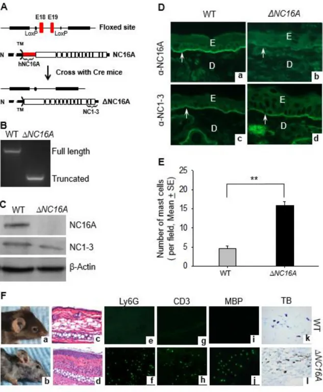
ΔNC16A Mice Develop Skin Inflammation With An Influx Of Mast Cells And MDSCs
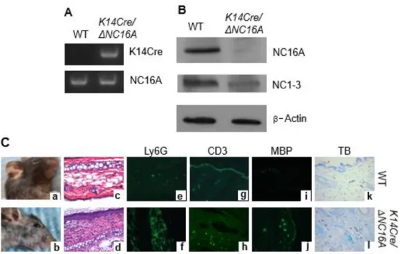
We previously generated a humanized NC16A mice (termed WT mice) by replacing the mouse NC14A domain with the human NC16A counterpart to study the disease mechanisms of BP (Liu et al. 2008). The NC16A domain is encoded by exons 18 and 19 of BP180 gene, which were flanked by lox-P sites (Fig. 2.1A). When crossed with germline Cre mice, Cre recombination removes the loxP-flanked exons 18 and 19 and maintains the remaining reading frame, resulting in mice expressing NC16A domain truncated BP180 (termed ΔNC16A). Genotyping of mouse tail DNA confirmed the lack of exons 18 and 19 in ΔNC16A mice (Fig. 2.1B). Lack of NC16A domain in ΔNC16A mice was confirmed by immunoblotting. Anti-NC16A antibody recognized full-length BP180 in the skin protein extract of WT mice and not ΔNC16A mice, while anti-NC1-3 antibody interacted with both full-length BP180 in WT mice and NC16A truncated BP180 in ΔNC16A mice (Fig. 2.1C). Similarly, anti-NC16A antibody stained the skin of WT mice and not ΔNC16A mice, while anti-NC1-3 antibody stained the skin of both WT and ΔNC16A mice (Fig. 2.1D).
32
the age of 8 weeks (Fig. 2.1F, panel d). In adult ΔNC16A mice, skin lesions were also accompanied by an infiltrate of immune cells (Fig. 2.1F, panel d), including neutrophils (Fig. 2.1E, panel f), T cells (Fig. 2.1E, panel j), and eosinophils (Fig. 2.1E, panel l) identified by immune staining, and significantly increased mast cells as identified by toluidine blue histochemical staining (Fig. 2.1F, panel l and Fig. 2.1E).
BP180 was originally identified and extensively studied in epidermal
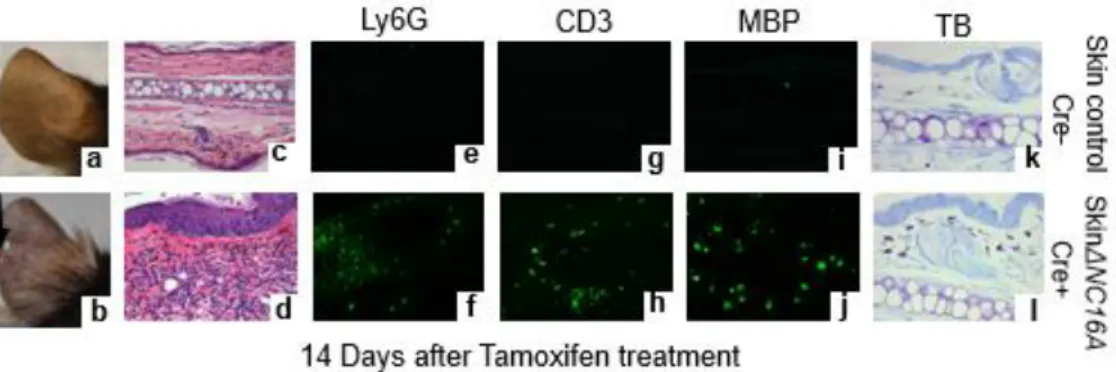
keratinocytes of the skin (1; 4); but, it is also expressed in many other tissues/organs (23). To determine whether the skin inflammation seen in ∆NC16A was caused by deletion of NC16A in the skin or in non-skin tissues/organs, we generated tamoxifen inducible Cre-NC16A mice (termed TamCre-NC16A mice) by crossing NC16A mice with tamoxifen inducible Cre mice. When treated with tamoxifen topically, TamCre-NC16A mice became skin-specific ΔNC16A (termed SkinΔNC16A mice). Efficient deletion of NC16A deletion in skinΔNC16A mice was confirmed by PCR, RT-PCR, immunoblotting and immunofluorescence (Fig. S2.1). Like ΔNC16A mice with the whole body deletion of the NC16A domain, skin-specific ΔNC16A developed skin inflammation with
33
is sufficient to promote the influx of inflammatory neutrophils (Fig. S2.3). Results from cytokine array analysis also showed that there is a significant increase in the skin level of multiple proinflammatory cytokines of skinΔNC16A (Fig. S2.4).
To determine whether ΔNC16A directly promotes skin inflammation through basal keratinocytes, we crossed NC16A mice with K14Cre mice to generate basal keratinocyte-specific ΔNC16A (termed K14Cre/ΔNC16A) mice (Fig. S2.5). Like
ΔNC16A and skinΔNC16A mice, K14Cre/ΔNC16A also showed skin inflammation with
significantly increased influx of MCs, neutrophils, T cells and eosinophils.
Mice Lacking NC16A Exhibit Accelerated B16 Melanoma Progression
Inflammation is one of hallmarks of cancer (Hanahan et al. 2011). Altered BP180 expression was reported to be associated with the increased invasiveness of melanoma cells (Krenacs et al. 2012). We, therefore, hypothesized that BP180 in basal
keratinocytes plays a role in melanoma progression through affecting the skin
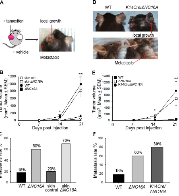
microenvironment. To test this hypothesis, WT and ΔNC16A mice were injected with B16 melanoma cells (1x10⁵ cells, s.c) into the flank, and monitored for tumor
34
Mouse melanocytes are mainly distributed in hair follicles, while human melanocytes are located at the basal keratinocyte layer (Bleehen 1998). The only mouse sites that are comparable to human skin in terms of melanocyte distribution are on the ears and tail (Silvers 1979)(Bleehen 1998). Injection of melanoma cells into the ear has been considered a more clinically relevant model for human melanoma
progression and metastasis (Rozenberg et al. 2010)(Bobek et al. 2010). Therefore, we injected B16 melanoma cells (1 x 10⁶ cells) into the ears of WT and ΔNC16A mice and monitored the tumor growth locally on the ears and metastasis on the neck for 3 weeks (Fig. 2.2D). Similar to the flank injection model, tumor volumes were significantly
increased in ΔNC16A mice compared to WT mice starting day 14 and day 21 after injection (Fig. 2.2E). 60% of ΔNC16A mice developed lymphatic metastases compare to none of the WT at 21 days post injection (Fig. 2.2F). These results demonstrated that NC16A deletion leads to drastically increased melanoma growth, metastasis and mortality, suggesting that BP180 plays a role in melanoma progression.
35
melanoma progression. To elucidate whether local skin BP180 dysfunction is sufficient enough to promote melanoma progression, we expanded our experiments to compare the tumor progression between WT mice treated with Tamoxifen (Control) and Tam-CreNC16A (SkinΔNC16A) mice. SkinΔNC16A developed a significant increased tumor
volume (Fig. 2.3B) and more lymphatic metastasis compare to the control mouse ears at 21 days post melanoma cell injection (77% of SkinΔNC16A vs. 20% of Control ears, Fig. 2.3C). More importantly, melanoma progression in skin-specific ΔNC16A mice is similar to whole body ΔNC16A mice (Fig. 2.3B and 3C). These results suggest that whole body deletion of NC16A domain does not alter systemic tumor
immune-surveillance, and the increased melanoma progression in ΔNC16A mice is caused by local deletion of NC16A in the skin. Therefore, BP180 local dysfunction is sufficient to promote B16 melanoma progression in vivo.
BP180 is mainly expressed by basal keratinocytes in local skin (Leighty et al. 2007). To test whether BP180 dysfunction in basal keratinocytes is sufficient to promote melanoma progression, K14Cre/ΔNC16A mice were injected at the ear with B16
melanoma cells. Both tumor volumes (Fig. 2.3D and 3E) and metastasis (Fig. 2.3F) in K14Cre/ΔNC16A mice were comparable to whole body ΔNC16A mice, but significantly
increased compared to WT mice starting on day 14 after injection. These results demonstrated that BP180 dysfunction in basal keratinocytes leads to increased melanoma progression through changing the skin microenvironment
36
MCs are critical in melanoma progression (Oldford et al. 2014)(Saleem et al. 2012), and there is a significant increase in the skin influx of MCs in both ΔNC16A (Fig. 2.1E, Fl) and skinΔNC16A mice (Fig. S2.2E ). To determine whether MCs play a role in increased melanoma progression in ∆NC16A mice, TamCre-NC16A mice were crossed with MC-deficient c-kiW-sh/W-sh (Wsh) mice (Grimbaldeston et al. 2005) to generate MC-deficient TamCre-NC16A mice (termed TamCre-NC16AWsh). Upon topical tamoxifen treatment, TamCre-NC16AWsh mice became MC-deficient skin-specific ∆NC16A mice (skin∆NC16AWsh). Skin∆NC16AWsh mice exhibited significantly reduced skin
inflammation compared to MC-sufficient skin∆NC16A mice as evidenced by histological examination (Fig. 2.4A) and by measuring skin extract enzyme activity of MPO
(neutrophil cell marker) (Fig. 2.4B). When tested in the B16 melanoma model,
skin∆NC16AWsh mice showed significant reduction in tumor progression as compared to MC-sufficient skin∆NC16A mice (Fig. 2.4C and D).
To rule out the possibility that reduced tumor progression in MC-deficient skin∆NC16A mice is due to c-kit mutation instead of MC deficiency,
37
Mast Cell-Dependent MDSC Influx Is Required for Increased Melanoma Progression in ΔNC16A Mice
MCs play an important role in cutaneous cancers; however, their exact roles in skin cancers are relatively unclear (Ch’ng et al. 2006). One mechanism for
MC-promoting melanoma is that MCs help mobilize MDSCs to tumor site for MDSC pro-tumor activity in melanoma (Yang et al. 2010)(Saleem et al. 2012). We, therefore, hypothesized that MCs promote melanoma progression through upregulating MDSC influx into the skin of ΔNC16A mice.
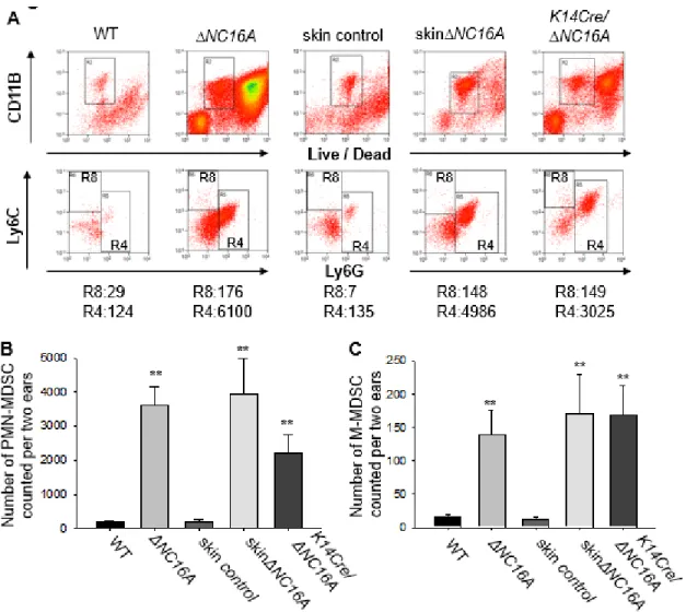
Flow cytometry analysis was performed to test our hypothesis, and we found that there was a significantly increased MDSCs influx in the skin of ΔNC16A, skinΔNC16A and K14Cre/ΔNC16A mice (Fig. 2.5A), with PMN-MDSCs being predominant (Fig. 2.5B) and a smaller population of M-MDSCs (Fig. 2.5C). Taken together, these results
38
skin∆NC16A mice exhibited a significant reduction of both PMN-MDSCs and M-MDSCs compared to MC- sufficient skin∆NC16A mice (Fig. 2.6A-C). Furthermore, MC
reconstitution restored the MDSC infiltration in MC-deficient skin∆NC16A mice (Fig. 2.6A-C). These results demonstrated that MDSC skin infiltration is dependent on MCs. More significantly, skin∆NC16A mice treated with anti-Gr1 antibody to deplete Gr1 positive MDSCs (Gr1+ cells include both Ly6G+ and 6C+ populations) showed
significantly reduced Gr1 positive immune cells in the skin (Fig.S2.6), melanoma tumor volume (Fig. 2.6D) and the rate of developing metastasis (Fig. 2.6E). Taken together, these results demonstrated that MC-dependent MDSC infiltration is crucial for
accelerated melanoma progression caused by BP180 dysfunction.
In conclusion, BP180 dysfunction in basal keratinocytes promotes the
39 2.4 DISCUSSION
BP180 is well established as a key cell-matrix adhesion molecule in the skin (Leighty et al. 2007). In this study, we used the ∆NC16A mouse strain and found that mice with dysfunctional BP180 develop a spontaneous skin inflammationand exhibit accelerated melanoma progression when challenged with B16 mouse melanoma cells. Our findings provide the first direct evidence linking BP180 and melanoma progression. Our in vivo and in vitro data also identified critical cellular events involved in this newly developed model of melanoma: 1) Basal keratinocytes with deletion of BP180’s NC16A region recruit MCs into the skin; 2) Increased infiltrating MCs recruit MDSCs to create a proinflammatory microenvironment in local skin; and 3) Increased infiltrating MDSCs promote melanoma progression (Fig. 2.7). These findings show that MCs are a critical cellular link between dysfunction of BP180 in basal keratinocytes and MDSCs during melanoma progression in our model setting. However, how BP180 dysfunction leads to increased MC infiltration in the skin remains to be determined. Our future studies will aim to identify mediators critical for MC recruitment.
JEB is a rare autosomal recessive disorder caused by defects in genes encoding hemidesmosomal proteins and hemidesmosome-associated proteins including α6β4 integrin, BP230, BP180, laminin 332 and collagen VII (Fine 2010a)(Bubier et al. 2010). BP180 mutations lead to the partial or complete loss of BP180 function, which causes JEB, most commonly non-Herlitz JEB (Fine 2010b)(Kiritsi et al. 2011). The symptoms of non-Herlitz JEB caused by BP180 mutation includes itch and the infiltration of
40
similarity with our ∆NC16A mice and the reported NC14A deficient mice (Hurskainen et al. 2014). Not only sharing the symptoms of BP180 mutated patients, ∆NC16A mice also exhibit minor dermal-epidermal separations starting at around eight weeks after birth, which further indicate that lacking the NC16A domain of BP180 significantly jeopardizes the normal cell/ECM adhesive function of BP180. Our ∆NC16A mouse model is a dysfunctional BP180 animal model, which reflects some pathophysiological features of JEB, including skin inflammation and subepidermal blistering (Fine
2010a)(Kiritsi et al. 2011)(Mabuchi et al. 2007). There is a case report showing a patient with BP180 mutation developed JEB, which shared phenotypical similarity with ∆NC16A mice which includes itch and inflammatory immune cell influx (Mabuchi et al. 2007). This case report supports the clinical relevance of our ∆NC16A mouse model as the physiological consequence of BP180 dysfunction in patients (Mabuchi et al. 2007).
41
Due to the rarity of all EB disease combined, in which the prevalence of all EB disease is less than eight per one million population, and melanoma only accounting for less than 5% of all skin cancers, only limited case reports indicate an increased risk of melanoma associated with any types of EB diseases (Fine et al. 2009)(Mallipeddi 2002). But, JEB is linked to significantly increased susceptibility to SCC (Fine 2012)(Mallipeddi 2002). A research based on clinical data collected between
1986~2002 in the US also found children with recessive dystrophic EB have increased susceptibility to melanoma (Fine et al. 2009). Nevertheless, our animal model findings do provide the reverse-genetic evidence, implicating that the melanoma in patients with BP180 deficiency, such as JEB, may progress more severely, compared to patients with functional BP180. Future clinical studies with more cases of JEB associated with
melanoma may provide more definite evidence to support our conclusion.
Skin cancers, including SCC (Parikka et al. 2003)(Parikka et al. 2006)(Stelkovics et al. 2008), BCC (Parikka 2001), and melanoma (Krenacs et al. 2012) are associated with altered BP180 expression, often having increased levels of the protein. BP180 binding partners, including α6β4 integrin and laminin 332, also play a pro-cancer role in cancer cell adhesion and motility (Stewart and O’Connor 2015)(Van Den Bergh et al. 2011). To our surprise, BP180 plays a negative role in melanoma progression in ∆NC16A mouse model: dysfunction of BP180 in basal keratinocytes surrounding
42
development of melanoma and other skin cancers. Future studies would need to directly address this scenario.
MCs are crucial in tissue inflammation and MDSC recruitment to the tumor
location, helping MDSCs promote melanoma progression (Sayed et al. 2008)(Duncan et al. 1998)(Ch’ng et al. 2006)(Oldford et al. 2014)(Yang et al. 2010)(Saleem et al. 2012). Increased expression of key MC and MDSC tissue homing chemokines both at mRNA and protein levels and infiltrating MC and MDSC are present in the skin of ∆NC16A mice (Fig. S2.4 and Table 2.1). More importantly, MDSC recruitment depends on MCs, and both MCs and MDSCs are required for accelerated melanoma progression,
providing a critical cellular link between BP180 dysfunction, pro-tumor microenvironment and tumor growth/metastasis.
Our study not only highlights the possible anti-melanoma activity of BP180, but also points out the important role of basal keratinocytes in changing the tumor
43
functions as a key regulator of skin inflammation and inflammation-driven melanoma progression through MCs and MDSCs. Basal keratinocytes, when losing NC16A
domain, are the cell source initiating pro-tumor microenvironment. How BP180 performs its anti-inflammation activity by directly and/or indirectly modulating necessary signaling pathways for production of MCs and MDSCs-recruiting mediators remains to be
44 2.5 MATERIALS AND METHODS
Generation of Mice
To study the immunopathogenesis of BP using autoantibodies from BP patients, we previously generated a humanized NC16A mouse strain on the C57BL/6J
background (termed NC16A mice or WT mice) (Fig. 2.1A) (Liu et al. 2008). Exons 18 and 19 (red) encoding the NC16A domain are flanked by loxP sites. When crossed with Cre mice (on C57BL/6J background, The Jackson Lab), Cre recombination removes the loxP-flanked exons 18 and 19 and maintains the remaining reading frame, resulting in mice expressing NC16A domain-truncated BP180 (this whole body deleted NC16A mice were termed ΔNC16A mice).
To generate skin-specific ΔNC16A mice, NC16A mice were crossed with UBC-Cre-ERT2 mice (Jackson Lab #008085) provided kindly by Dr. Richard Weinberg from UNC-Chapel Hill. The TamCre-NC16A mice (ERCre+NC16Afl/fl mice) when treated topically with tamoxifen (Sigma, 25 μl of 10 mg/ml in 62% EtOH/sunflower oil mixture) become skin-specific ΔNC16A (skinΔNC16A) mice. To generate basal keratinocyte-specific ΔNC16A mice (K14Cre/ΔNC16 or K14Cre+NC16Afl/fl), NC16A mice were crossed with Krt-14 promoter driven Cre mice provided kindly by Dr. Dennis Roop at University of Colorado at Denver (Jackson Lab #004782). To confirm the deletion of NC16A-encoding exons, tail DNA were analyzed by genomic DNA PCR. The deletion of the NC16A domain in the skin was confirmed by immunoblotting and indirect
45
and protein samples from the non-treated skin and several internal organs including bone marrow, spleen and thymus were analyzed by NC16A-specific PCR and
immunoblotting. All the mice were bred and housed at the University of North Carolina at Chapel Hill Animal Facility. Animal care, breeding and experiments were conducted in accordance with the Institutional Animal Care and Use Committee (IACUC) at the University of North Carolina at Chapel Hill.
Murine Melanoma Models with Flank and Ear Injection of B16 Melanoma Cells
B16-F10 murine melanoma cell line was acquired from ATCC and cultured in DMEM media (Gibco) supplemented with 10% fetal bovine serum and 1%
penicillin/streptomycin. For flank injection model, 8 weeks old mice were injected subcutaneously (s.c) into the side flank with 1x10⁵ cells in 100 μl of HBSS buffer. For ear injection model, B16 melanoma cells (1x10⁶) was injected intradermally (i.d) into the ears of 8 weeks old mice in 20 μl of HBSS buffer. Tumor growth was monitored and tumor volume was measured with a digital caliper. Tumor volume was calculated using the following equation: ½ x (length) x (width) (width). Mice were terminated on 21 days after initial melanoma cell injection. Lymphatic metastasis was monitored by examining tumor growth on the draining lymph node of ears on the neck (Bobek et al. 2010).
46
400602) was used as control. The injection schedule was day-1, day 0 and then biweekly.
Immunoblotting and Immunofluorescence
To detect full-length and NC16A truncated BP180 in the mouse skin by immunoblotting, skin protein extracts were probed with anti-NC16A IgG purified from a patient with BP (termed anti-NC16A antibody) and a home-made rabbit antibody against mouse BP180 noncollagen 1-3 (NC1-3) domains (termed anti-NC1-3 antibody) (Fig. 2.1A). Anti-human (Southern Biotech) and anti-rabbit (Cell Signaling) HRP-conjugated antibodies were used for BP180 immunoblotting detection. To localize BP180 in the skin by indirect immunofluorescence, cryosections (7μm) of mouse skin were stained with anti-NC16A and anti-NC1-3 antibodies, followed by FITC-conjugated anti-human IgG and anti-rabbit IgG (Jackson ImmunoResearch), respectively. Infiltrating immune cells in the skin cryosection were identified by indirect IF using anti-CD3 (BioLegend,
clone:17A2) for T cells, anti-Iy6G (BioLegend,clone:1A8) for neutrophils, or anti-MBP (Mayo Clinic Scottsdale) for eosinophils, followed by Alexa Fluor 488 Goat Anti-Rat IgG (Life Technologies; 1:1000 dilution) for 1hour at room temperature.
Mast Cells Culture, Reconstitution, Identification and Quantification