DETERMINING THE FUNCTION OF THE INTERLEUKIN-1 RECEPTOR ASSOCIATED KINASE PATHWAY IN PRIMARY EFFUSION LYMPHOMA
Jedediah David Seltzer
A dissertation submitted to the faculty of the University of North Carolina at Chapel Hill in partial fulfillment of the requirements for the degree of Doctor of Philosophy in the
Department of Microbiology and Immunology.
Chapel Hill 2020
ii ©2020
iii ABSTRACT
Jedediah Seltzer: Determining the function of the interleukin-1 receptor associated kinase pathway in primary effusion lymphoma
(Under the direction of Dirk Dittmer)
Kaposi’s sarcoma-associated herpesvirus (KSHV) is necessary but not sufficient for the development of Primary effusion lymphoma (PEL). Alterations in cellular signaling pathways are also a characteristic of PEL development. Other B cell lymphomas have acquired an oncogenic mutation in the myeloid differentiation primary response-88 (MYD88) gene. The MYD88 L265P mutant results in the activation of the Interleukin-1 Receptor Associated Kinase (IRAK) pathway and a pro-inflammatory environment. To probe IRAK/MYD88 signaling in PEL, we employed CRISPR/Cas9 technology to generate stable deletion clones in BCBL-1Cas9 cells. To look for off-target effects, we determined the complete exome of the BCBL-1Cas9 cell line.
Deletion of either MYD88, IRAK4, or IRAK1 abolished IL-1β signaling; however, we could grow stable sub-clones from each population. RNA-seq analysis of IRAK4 knockouts showed that the IRAK pathway induced cellular signals constitutively,
iv
v
For my family who have supported me: My wife Tischan and my son Jonah.
My parents Jay and Terry Seltzer.
My siblings: Jeremiah, Rebekah, Susanna, Liz and Abigail as well as my bonus siblings Scott and Meredith.
vi
ACKNOWLEDGEMENTS
First, I would like to acknowledge my family and my friends for the support I have received. A PhD is a long road to travel and the support of friends and family helped me stay sane and on track. To my wife Tischan who has encouraged me and has had to personally sacrifice her time while I completed this experience. To my brother, Jeremiah, who set me on this track by a random conversation when I was in the 11th grade. My parents, Jay and Terry, for supporting and encouraging me. To my other siblings, Rebekah, Susanna, Liz, and Abigail as well as my bonus siblings Scott and Meredith, for putting up with my science talk and not asking “when are you graduating” every time I saw them.
I would also like to acknowledge my friends who have been an amazing community while I went through this process. The fellowship and encouragement I received made this road more enjoyable to travel. Through the laughter, smiles, tears and grimaces you all stood by me, and in many cases hiked by me across this state and I appreciate each one of you.
I would like to acknowledge my principal investigator Dirk Dittmer for providing the resources to conduct experiments in his lab at UNC. I want to thank all of the members, past and current, of the Dittmer lab who helped me on my way, whether it was providing
vii
Flanner who was an amazing student service manager and to Michelle Hightower who took up Dixie’s mantle. I never would have had all the paperwork done without the reminders!
viii
TABLE OF CONTENTS
LIST OF TABLES ... xi
LIST OF FIGURES ... xii
LIST OF ABBREVIATIONS ...xv
LIST OF SYMBOLS ... xviii
CHAPTER 1: INTRODUCTION ...1
Introduction ... 1
Cancer and Viruses ... 1
Tumor Plasticity ... 2
Kaposi’s Sarcoma-associated Herpesvirus ... 4
Primary Effusion Lymphoma ... 6
The IRAK-MYD88 Signaling Pathway ... 8
IRAK-MYD88 and Lymphoma ... 11
The Interaction between KSHV and the IRAK Pathway ... 12
Interleukins in KSHV Infection and Disease ... 14
CRISPR in PEL ... 18
ix
Importance ... 21
Introduction ... 21
Methods ... 24
Results ... 30
Discussion ... 38
Acknowledgements ... 40
Tables... 42
Figures ... 43
CHAPTER 3: CONCLUSION AND FUTURE DIRECTIONS ...58
Summary ... 58
The IRAK Pathway Members are Dispensable for PEL Survival ... 58
IRAK Inhibitor have Off-target Effects ... 60
Techniques can Yield Variable Results ... 61
The Relevance of the IRAK Pathway in PEL with the Conserved IRAK1 SNV ... 63
Future Directions ... 64
Conclusion ... 66
APPENDIX A: SUPPORTING INFORMATION ...68
Overview ... 68
Results ... 68
x
Materials and Methods ... 72
Tables... 74
Figures ... 90
xi
LIST OF TABLES
Table 1. 1: List of TLRs that can signal through the IRAK pathway. ... 8
Table 2. 1: MYD88 inhibitor in IRAK pathway knockouts. ... 42
Table 2. 2: IRAK inhibitors in IRAK pathway knockouts. ... 42
Table 3. 1: Comparison of molecular targeting techniques. ... 62
Table AA. 1: Table of plasmids used in this study. ... 74
Table AA. 2: Antibodies used in this study: ... 85
xii
LIST OF FIGURES
Figure I. 1: The IRAK pathway functions to activate NFκB. ... 9
Figure II. 1: PEL cells do not contain the L265P mutation in MYD88. ... 43
Figure II. 2: MYD88 is not required for PEL survival. ... 45
Figure II. 3: IRAK1 is not required for PEL survival. ... 46
Figure II. 4: IRAK4 is not required for PEL survival. ... 47
Figure II. 5: MYD88, IRAK1 and IRAK4 are dispensable in BC-1 ... 48
Figure II. 6: NFκB activation by IL-1β is not functionalin ΔMYD88 clones. ... 49
Figure II. 7: NFκB activation by IL-1β is not functional in ΔIRAK1 cells. ... 50
Figure II. 8: NFκB activation by IL-1β is not functional in ΔIRAK4 cells. ... 51
Figure II. 9: Complementation of IRAK1 restores signaling function in KO cells. ... 52
Figure II. 10: Comparison of in vitro and in culture IRAK inhibitor activity. ... 54
Figure II. 11: RNA-seq analysis of IRAK4 CRISPR KO. ... 56
Figure III. 1: Summary of the IRAK pathway in PEL ... 67
Figure AA. 1: Knockdown of ABCD1 has no effect on PEL growth. ... 90
Figure AA. 2: PEL cells contain F196S and S532L Variants in IRAK1. ... 91
Figure AA. 3: Western blot of the IRAK pathway in PEL and other cell lines. ... 93
Figure AA. 4: BTK is not expressed in PEL. ... 94
Figure AA. 5: U2OS cells have functional IL-1β signaling. ... 95
Figure AA. 6: TNF and IL-1β activate NFκB in PEL. ... 96
Figure AA. 7: IL-1β and CTG titration in BCBL-1 cells. ... 97
Figure AA. 8: Validation of IRAK expression plasmids. ... 98
xiii
Figure AA. 10: IL1R CRSIPR BCBL-1. ... 100
Figure AA. 11: MYD88 shRNA data. ... 101
Figure AA. 12: IRAK1 shRNA results. ... 102
Figure AA. 13: IRAK4 shRNA data. ... 104
Figure AA. 14: IRAK4 shRNA has no effect in BC-1. ... 105
Figure AA. 15: CRISPR knockout of IRAK4 has no effect on BJAB cells. ... 106
Figure AA. 16: NFκB is constitutively activated in BC-1 but not BCBL-1 cells. ... 107
Figure AA. 17: IPA pathway analysis on IRAK4 knockout RNA-seq... 108
Figure AA. 18: Exome sequencing data for the IRAK pathway knockouts in BCBL1: ... 109
Figure AA. 19: Exome sequencing variant calling in BCBL1 ... 110
Figure AA. 20: Exome sequencing data for the IRAK pathway knockouts in BC1. ... 111
Figure AA. 21: Exome sequencing variant calling in BC1. ... 112
Figure AA. 22: IRAK inhibitors are non-specific. ... 113
Figure AA. 23: MYD88 inhibitor ST2825 EC50 in IRAK knockouts ... 114
Figure AA. 24: IRAK mouse inhibitor data... 115
Figure AA. 25: CFA supplemental data ... 116
Figure AA. 26: Reactivation of BCBL-1 IRAK pathway knockouts. ... 117
Figure AA. 27: RNA-seq results for virus genes in ΔIRAK4 BCBL-1. ... 118
Figure AA. 28: ABCD1 complete western blot. ... 119
Figure AA. 29: BTK complete western blot. ... 120
Figure AA. 30: IRAK4 shRNA BCBL-1 complete western blot. ... 121
Figure AA. 31: IRAK1 shRNA complete blots. ... 122
xiv
xv
LIST OF ABBREVIATIONS AIDS Acquired immunodeficiency syndrome
BCR B-cell receptor
BL Burkitt’s lymphoma
BTK Bruton's Tyrosine Kinase
CHOP Cyclophosphamide, doxorubicin, vincristine and prednisolone CRISPR Clustered regularly interspaced short palindromic repeats CTG CellTiter-Glo® Assay
DLBCL Diffuse large B-cell lymphoma DMEM Dulbecco modified Eagle medium DMSO Dimethyl sulfoxide
DNA Deoxyribonucleic acid EBV Epstein–Barr virus
FADD Fas-associated protein with death domain FBS Fetal bovine serum
FLICE FADD-like IL-1β-converting enzyme HAART Highly active anti-retroviral therapy HEK 293 Human embryonic kidney cells 293 HHV-8 Human herpesvirus 8
HIV Human immunodeficiency virus
HL Hodgkin's lymphoma
xvi
IFN Interferon
IL Interleukin
IL-1β Interleukin-1 beta IL1R Interleukin-1 receptor IL-6R Interleukin-6 receptor
IKK IκB kinase
IRAK Interleukin-1 receptor-associated kinase
JAK Janus kinase
KICS KSHV-associated inflammatory cytokine syndrome
KS Kaposi's sarcoma
KSHV Kaposi's sarcoma-associated herpesvirus
KSHV-MCD KSHV-associated multicentric Castleman’s disease LANA Latency-associated nuclear antigen
MCD Multicentric Castleman’s disease MOI Multiplicity of infection
MYD88 Myeloid differentiation primary response 88
NFκB Nuclear factor kappa-light-chain-enhancer of activated B cells
ORF Open reading frame
PAMP Pathogen associated molecular patterns PBMC Peripheral blood mononuclear cell PBS Phosphate-buffered saline
xvii Pen-Strep Penicillin and streptomycin
RNA Ribonucleic acid
RTA Replication and transcription activator
RT-PCR Real-time PCR
STAT Signal transducer and activator of transcription
TANK TRAF family member-associated NF-kappa-B activator TRAF6: TNF receptor associated factor
TBK TANK-binding kinase
TLR Toll Like Receptor
TNF Tumor necrosis factor
UV Ultraviolet
vFLIP Viral FLICE inhibitory protein vIL Viral interleukin
xviii
LIST OF SYMBOLS
α Alpha
β Beta
γ Gamma
1
CHAPTER 1: INTRODUCTION Introduction
In completion of my graduate studies in the laboratory of Dr. Dirk Dittmer, the following Dissertation will examine the relevance of the interleukin-1 receptor-associated kinase (IRAK) pathway in primary effusion lymphoma (PEL). The thesis will be broken into four parts starting with a broad introduction to cancer, PEL, IRAK and KSHV (chapter 1),
followed by a chapter on experimental results from testing the function of the IRAK pathway in PEL. Chapter three will conclude and summarize the findings as well as provided a road map for moving this project forward. The final section will be an appendix that contains supporting information for chapter two.
Cancer and Viruses
Cancer is a global affliction that affects most living organisms (1-3). The rate of cancer in humans is high, as one in three individuals are diagnosed with a type of cancer during their lifetime, according to the American Cancer Society (4). There are over 130 types of human cancers, which have many different causes (NCI) such as exposure to harmful chemicals known as carcinogens, smoking, genetics, and environmental influences (5). Infectious agents, such as viruses, account for nearly 20% of all human cancer cases globally, and the scientific community’s understanding of virus-induced human cancers continues to improve with advancements in sequencing technologies (6-10).
2
pathways for survival of the infected cells. The viral genes can function as oncogenes, driving a normal cell into malignancy (7, 11, 12). Another distinction with virus-induced cancers is that viruses can be targeted with antivirals or vaccines, which changes the approach for treatment and preventive actions of non-viral cancers (6-8, 13). The interplay between the virus and the host is extremely important for cancer progression as only a fraction of individuals infected with viruses develop cancer, and only a small subset of viruses are known to cause cancer.
Cancers are caused by the dysregulating of cellular pathways. Typically, a single mutation will not cause cancer, instead it takes an accumulation of mutations over time (10, 14, 15). There are multiple steps of cellular dysregulation that occur throughout the course of a lifetime. An example would be a mutation in a tumor suppressor protein that is present in an individual who smokes, which induces new mutations that compound the effects of the inherited mutation, resulting in the development of lung cancer in the individual (16-18). This is only one example, however, and there are many known cellular pathways in humans that can function in cancer development, predisposing an individual to develop cancer. One such pathway that has been reported to predispose individuals to cancer is the IRAK
pathway, which I will discuss later in this introduction (19-24). To understand the importance of the IRAK pathway in the context of this thesis, we must first understand KSHV, PEL and the concept of tumor plasticity.
Tumor Plasticity
3
developed. One hypothesis was that of clonal evolution, or unique epigenetic/stochastic changes, which results in the development of tumor heterogeneity from individual cancer stem cells (25). The second hypothesis proposed was the cancer stem-like cell model, where a subset of the tumor cells have the ability to differentiate into various subtypes of cancer cells, causing variation within a tumor (27).
A recently developed model, known as the cell plasticity model, combines aspects of both the cancer stem-like cell and the clonal models (25). This new model predicts that, due to changes in the tumor environment, cancer stem-like cells can undergo clonal expansion and epigenetic differentiation while maintaining a subset in the cancer stem-like cell state. The result of this ability is that, in response to stressors in the environment, such as
inflammation or injury, the various subtypes of the cancer cells can survive and lead to resistance to therapy. One of the largest drivers of the plasticity seen in tumor cells is the ability of self-renewing and dysregulation (28). Just a few examples of dysregulation that can lead to plasticity in a tumor are loss of tumor suppressor proteins (such as p53, RB1 and PTEN), increased inflammatory compounds that induce differentiation towards stem-like cells, and the ability of these stem-like cells to remodel the microenvironment by influencing the differentiation of non-tumor cells into cells that support tumor growth (25, 29).
4
and resistance to therapies (18, 31). The ability of tumor cells to undergo lineage plasticity results in drug ineffectiveness and allows the tumor to proliferate beyond control. Prostate cancer, small-cell lung cancer, breast cancer, and basal cell carcinoma are only a few examples where targeted therapies have been challenged by tumor plasticity (18).
The mechanism of drug resistance through plasticity is thought to occur when cells have inherent mutations that circumvent the target of the therapeutics, or when slow-growing cells bypass the pathway entirely through activation of alternate pathways (18, 32). Due to the complications of tumor plasticity, researchers must now seek to address these issues when developing targeted therapeutics. Concepts such as alternate dosing or combination therapies are currently leading the way to overcome tumor plasticity. Furthermore, the idea of tumor plasticity is important for this thesis as we examine the ability of tumor cells to circumvent inhibition of a host pathway in the context of a virus-induced lymphoma.
Kaposi’s Sarcoma-associated Herpesvirus
Kaposi’s sarcoma-associated herpesvirus (KSHV), also known as human herpesvirus 8 (HHV8), is one of two members of the gamma herpesvirus family known to infect humans, the other being the Epstein Bar virus (EBV) (6, 33, 34). KSHV is the causative agent of Kaposi’s sarcoma (KS), primary effusion lymphoma (PEL), KSHV inflammatory cytokine syndrome (KICS), as well as cases of Multicentric Castleman disease (MCD) (35-38). The various aspects of the virus life cycle are crucial for understanding how these different diseases develop. The focus of this thesis will be on PEL, which will be discussed in the next section.
5
Upon infection, the default programming of KSHV is to go into latency, where the viral genome is maintained as an episome tethered to the host genome by the latency associated nuclear antigen (LANA), making it difficult for the immune system to detect the virus (40, 45-47). KSHV must evade the cellular innate immune response to infect cells efficiently (48-50). During latency, only a subset of genes is expressed, LANA being one of those genes and having multiple functions including maintaining latency (11, 51-53). Another
latency-associated protein is the vFLIP that engages the nuclear factor kappa-light-chain-enhancer of activated B cells (NFκB) protein and activates the downstream signaling pathway to promote a positive environment for latency (54-57).
The vFLIP activates the NFκB pathway by modulating the NF-κB-inducing kinase (NIK) as well as both the IKK2-independent and dependent processes to activate p100 NFκB thus providing pro-inflammatory signaling, a hallmark of KSHV diseases (55). This
6
TLRs are innate immune sensors that recognize specific ligands and function in immunity by recognizing pathogens. TLRs, with the exception of TLR3, typically signal through the IRAK pathway to activate NFκB and stimulate innate immunity (66-72). TLRs, in the context of KSHV, signal through the downstream TRIF/TLR3 or MYD88/IRAK pathways (73). Within KSHV infection, TLRs have a function in reactivation from latency. The virus must overcome the TLR signaling cascade to establish infection (35, 74). The situation is complex, with KSHV activating some components of the pathway while shutting down others. Since most TLRs signal through IRAK1, KSHV encodes miR-K9, which down regulates IRAK1 during infection to dampen inflammation and establish latency (75). In primary infection of monocytes, KSHV infection results in activation of TLR3, which recognizes viral single stranded and double stranded RNA. The KSHV protein, ORF50, can degrade downstream effectors of TLR3 activation (35). The KSHV proteins, vIRF1 and vGPCR, inhibit TLR4 signaling during the advent of new infections, but during latency in KS spindle cells TLR4 activation is induced by the virus (76). If TLR7/8 are activated in cells that are latently infected with KSHV, it results in the reactivation of the virus (74). Therefore, we see that the interplay between the TLRs and KSHV is complex, with the degree of inhibition and activation depending on the life stage of the virus. We will next delve more into the PEL disease indication.
Primary Effusion Lymphoma
7
have been reported (36, 78, 79). The medium life expectancy following a PEL diagnosis is 6 months (17, 80). PEL is currently treated with CHOP (cyclophosphamide,
hydroxydaunorubicin, oncovin, and prednisone) therapy in most cases, which has limited success, as is evident by the short life expectancy (80). KSHV infected B-cells are
responsible for PEL development, and it is thought that these cells are clonal and develop post germinal center (81). KSHV-EBV coinfections are common in PEL (34, 40).
PEL cells have distinct features, including expression of CD138/Syndecan-1 as well as no expression of the B cell receptor (BCR) or CD79 co-receptor (80), despite exhibiting plasmacytoid features. It is thought that viral proteins such as vGPCR, K1, and K15 provide for constitutive activation of PI3K and PLC-K resulting in cell proliferation and survival (35, 36, 56, 80, 82, 83). PEL expresses elevated levels of IL-10 and varying amounts of IL-6 in a cell line dependent manner (81, 84). Additional cytokines with high levels of expression in PEL are oncostatin and IL-6 soluble receptor (84, 85).
PEL can appear concurrently with Kaposi sarcoma (KS) or independently, suggesting that KSHV infection profoundly reprograms B-cell signaling in an alternate mechanism to Kaposi sarcoma, a disease primarily in endothelial cells (36, 39, 40, 86). It is important to note that de novo KSHV infection results in latency not lytic replication, which in turn results in the expression of latently expressed viral oncogenes (64). In addition to the latency genes, PEL depends on different host pathways that KSHV modulates to carry out the viral life cycle (73, 87-89).
8
as dysregulation of the immune response, such as blocking of p53 functions and upregulating ß-catenin (36, 40, 93). Another cellular pathway that is important for PEL survival and of interest to this thesis is the IRAK/NFκB pathway. The vFLIP is known to interact with the IRAK pathway by activating NFκB. The vFLIP is thought to aid in B-cell tumorigenesis, as was shown in mice (56, 94). Research has shown that HSP90 inhibition blocks vFLIP activation of NFκB and leads to cell death in PEL lines (95). Other work has shown that IKKγ mimetic peptides can lead to apoptosis in PEL (96). IKKγ is an upstream regulator of NFκB activation. To summarize, we see that PEL is an aggressive disease that causes dysregulation of cellular pathways and that KSHV infection is necessary but not sufficient for PEL development.
The IRAK-MYD88 Signaling Pathway
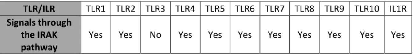
The IRAK pathway has important roles in innate immunity. When pathogens invade our bodies, cells can recognize various pathogen-associated molecular patterns (PAMP) (70, 73, 97). These PAMPs include bacteria lipids, virus RNA, and virus DNA to name a few. PAMPs are recognized by TLRs. Additionally, cells can produce cytokines when stimulated by PAMPS, which alerts the immune system and other cells in the environment of the infection. One such cytokine is interleukin-1 Beta (IL-1β), recognized by the interleukin-1 receptor (ILR1) (19, 98). There are 13 TLRs and 38 ILRs in humans (66, 99). Of these TLRs and ILRs, many signal through the IRAK pathway.
Table 1. 1 List of TLRs that can signal through the IRAK pathway.
TLR/ILR TLR1 TLR2 TLR3 TLR4 TLR5 TLR6 TLR7 TLR8 TLR9 TLR10 IL1R
Signals through the IRAK
9
When the IRAK pathway is activated by either TLR or ILR activity, a signaling cascade is initiated that results in the transcription factor NFκB being released to change cellular transcription and create a pro-inflammatory environment (19, 22, 100). For fighting infection in an acute response, activation of NFκB is good for the human body (20, 22, 70, 71, 97, 101). However, chronic inflammation due to NFκB can induce conditions such as arthritis and cancer (21, 22, 61, 97, 100, 102, 103).
Figure I. 1: The IRAK pathway functions to activate NFκB.
10
104). MYD88 recruits interleukin associated protein 4 (IRAK4) to the complex, which is phosphorylated upon recruitment (19). Phosphorylation of IRAK4 allows for interleukin associated protein 1 (IRAK1) to be recruited into the complex (105, 106). This MYD88-IRAK4-IRAK1 complex is known as the Myddosome, and its crystal structures has been solved (107). Following Phosphorylation of the IRAK1 activation site by IRAK4, IRAK1 undergoes hyper auto-phosphorylation, which causes its release from the complex and association with TRAF6 and TAK1 (19, 108-110). These two proteins are activated and remove the suppression of the IKK complex, allowing the release of NFκB (108, 111).
There are many variables within the IRAK pathway. The kinase activity of IRAK1 is not required for IL-1β signal propagation in some cell types (112). IRAK1 can activate STAT3, directly resulting in IL-10 transcription, which is dependent on kinase activity (113). IRAK-M is a negative regulator of the signaling pathway, preventing the phosphorylation of IRAK1 and IRAK4, but IRAK-M is primarily seen in monocytes and macrophages and is not expressed in B-cells (19, 100, 114). IRAK1 and IRAK4 are the most commonly expressed of the IRAK proteins in cancer cells (100). In T-cell acute lymphoblastic leukemia, inhibition of IRAK1-4 kinase activity resulted in cell arrest but not death (100). IRAK1 or IRAKM
11
models to regulate type I Interferon production induced by TLR7 and TLR9 in distinct cell types (66). IRAK1 is known to bind and phosphorylate IRF7 in a TLR7 and TLR9 dependent manner, and the IRAK1 kinase activity is indispensable for phosphorylation of IRF7 (118). The IRAK pathway is complex and of its many players, each appear to have non-canonical functions. When the pathway is dysregulated, there can be extremely negative effects, which will be discussed in the next section.
IRAK-MYD88 and Lymphoma
In a subset of diffuse large B cell lymphomas, MYD88 L265P is an activating mutation that results in constitutive activation of the NFκB pathway and development of a more aggressive lymphoma (119-122). The L265P mutation is referred to as a driver mutation and 30% activated B-cell like (ABC), 60% central nervous system, 77% testicular DLBL, and upwards of 90% Waldenstrom’s macroglobulinemia (WM) have this mutation (120, 123-125). The mutated MYD88 results in an increase in IRAK1 phosphorylation and NFκB activation (126). It should be noted that these numbers are contested by some research groups, who suggest that the percentage of MYD88 L265P cases are lower (127). Cells with the MYD88 L265P mutation also display increased levels of JAK/STAT signaling and activation of the Bruton's Tyrosine Kinase (BTK) pathway (119, 124).
Treating these L265P-mutant cells with an IRAK4 inhibitor was effective at reducing cellular proliferation, demonstrating that targeting the IRAK pathway could potentially serve as a drug target for certain lymphomas such as ABC-DLBCL (128). However, the members of the IRAK pathway have unique functions that depend on specific cell types and
12
of MYD88 in WM decreased the BTK signaling that was associated with the MYD88 L265P mutation (129). In this case, MYD88 was shown to act synergistically with CD79B
mutations in the activation of NFκB, resulting in BTK inhibitor resistance (120).
The IRAK pathway is also activated in other systems such as acute myeloid leukemia (AML) in which IRAK1 is overexpressed and hyper-phosphorylated at the T209 activation site (22, 130). In head and neck cancer, IRAK1 promoted cell survival and the inhibition of IRAK1 resulted in cell death (131). An analysis from the cancer genome atlas, performed in 2017, demonstrated that IRAK1 has a role in cellular dysregulation and contributes to several other cancers such as melanoma, lung cancer, and breast cancer, by stimulating downstream proinflammatory pathways (132, 133). We have covered how two separate IRAK pathway members, MYD88 and IRAK1, have a role in cancer development and will next turn to the interplay between the IRAK pathway, KSHV, and PEL.
The Interaction between KSHV and the IRAK Pathway
This section will highlight the interplay between IL-1β and KSHV, as well as discuss the IRAK pathway as studied in PEL. IL-1β is a cytokine that relays its signal through the IRAK pathway, activating NFκB (19). KSHV is known to produce a pro-inflammatory environment by activating the NFκB pathway during latency (134). Activation of cells by IL-1β also activates the NFκB pathway.
13
group to explore this pathway, eventually culminating in this thesis work. Preliminary research involving inhibitors and shRNA suggested that IRAK1 was essential for PEL survival (135). However, with the advancement of CRISPR and a greater understanding of the field, our hypothesis has changed to conclude that the IRAK pathway is important for the development of PEL but can be circumvented once the virus is established and the disease has progressed.
In KSHV-associated multicentric Castleman disease (MCD), KSHV can induce clinical flair-ups of IL-1β production (141). Yet, KSHV encodes for microRNAs that target both IRAK1 and MYD88, helping the virus to control this inflammatory pathway and it was demonstrated that KSHV RTA was able to degrade the TRAF protein, blocking IL-1β signaling during reactivation of the virus (40, 76, 142-144). IL-1β has a protective effect on serum-starved AIDS-KS cells, protecting them from apoptosis (145). These apparent contradictions suggest that the interplay between KSHV and IL1 signaling is complex and not yet fully understood.
PEL primarily occurs in HIV infected individuals, and it has been shown that HIV infection raises the levels of IL-1β, which can enhance KSHV infection (146-148). Closer examination of KS demonstrated that IL-1β induced an autocrine growth factor loop leading to uncontrolled cellular growth (149). Increased IL-1β levels are also associated with
14
In the case of PEL, the scientific community has long held the view that NFκB was crucial for PEL survival and that vFLIP was largely responsible for activating NFκB. Inhibitor studies using the IKK inhibitor, BAY11-7082 , gave supporting evidence to this view as did shRNA studies on vFLIP (58, 61, 64). However, as inhibitors were further studied, the off-target effects of BAY11-7082 were seen to be mainly responsible for PEL cell death (150, 151). The traditional viewpoint that one protein, such as NFκB, could be a silver-bullet therapeutic target has been rebutted as a greater understanding of tumor
plasticity developed (18, 25). In 2018 Manzano et al. group released a genome-wide CRISPR knockout screen conducted in BCBL-1 cells, a PEL tumor line, that demonstrated no
individual member of the NFκB pathway was required for PEL survival (12). The result of this screen aligns with the plasticity model of cancer and is reshaping the view on the NFκB/IRAK pathway within the KSHV/PEL field. Multitarget therapeutics will most likely be more effective at treating PEL. Resistance to multitarget therapy would be less likely to develop, which would explain the effectiveness of BAY 11 off-target effects. Of the
downstream genes turned on by NFκB activation, two of the most important for PEL are IL-10 and IL-6.
Interleukins in KSHV Infection and Disease
15
promoting lymphocyte migration and is expressed at high levels in human serum in various disease such as KS. Dysregulation of IL-10 and IL-6 occurs across all four KSHV-related diseases: KS, PEL, KICS, and MCD as discussed below.
KSHV is responsible for the increase of IL-10 and IL-6 expression in PEL (155-157). In one study, 19 PEL patients were compared with 20 HIV-associated DLBCL cases, and the PEL samples had elevated IL-10 and IL-6 levels when compared to the DLBCL (158). When comparing classic KS with AIDS-associated KS, higher levels of IL-10 were observed in AIDS-associated KS. Of the AIDS-associated KS, the highest levels of IL-10 were in
individuals with the most aggressive disseminated KS lesions (159). In KS lesions, the TLR4 pathway is constitutively active and results in increased IL-6 levels and STAT3 activation (160). Both KICS and MCD are characterized by high levels of IL-6 and IL-10 (161). In one study conducted on KICS, IL-10 and IL-6 levels were evaluated for all ten individuals (males that were HIV positive). These patients had an increased risk of death, lower hemoglobin and albumin, and displayed increased C-reactive protein levels (162).
MCD can be divided into HIV negative and HIV positive cases, with better outcomes for those who are HIV negative (163). Approximately 50% of MCD patients have
16
KSHV encodes a homolog of IL-6, vIL-6, which shares 25% homology with IL-6. vIL-6 binds directly to the gp130 receptor without the need of the IL-6R co-receptor that is required for 6 signal relay (170). KSHV v6 can activate cells that do not respond to IL-6 (171). N-linked glycosylation is required for optimal vIL-IL-6 function (172, 173). Two KSHV proteins, vIL-6 and vGPCR, are linked to upregulation of Angiopoietin-2, a secreted proangiogenic and lymphangiogenic (174). KSHV RTA binds to and activates the IL-10 promoter in collaboration with human specificity proteins 1 and 3 (175). KSHV vFLIP can block lytic replication by inhibiting RTA, yet RTA activation of vIL-6 is not inhibited (176). Epigenetic modifications are important for the function of vIL-6 because, during latency in PEL, the vIL-6 promotor is in an open chromatin formation but it differs in each viral episome (177, 178). KSHV encodes a viral endonuclease, SOX, which degrades cellular mRNA. Notably, IL-6 mRNA is protected from this degradation through a 3’ untranslated region (179). Tumor associated macrophages have been seen to play an important role in PEL disease progression (180).
In PEL cells IL-10 has a role in HSV1 induced reactivation of KSHV (181). EBV has a homolog of IL-10 (vIL-10) (182, 183). Infection of endothelial cells with KSHV activates IL-10, IL-6 and IL-13, which results in the development of monocytes into tumor-associated macrophages (180). Binding of KSHV to dendritic cells (DC) resulted in
increased levels of IL-10, IL-6, and IL-23, which activated STAT3 and blocked autophagy in the DC cells. This could allow for the establishment of KSHV and the development of KSHV associated diseases (184).
17
STAT5 (185, 186). Using bioinformatics, KSHV miR-K12-3 and miR-K12-7 were shown to target the 3'UTR of the transcription factor C/EBPβ, which regulates IL-6 and IL-10. Follow-up experiments revealed that these two miRNAs reduce expression of the C/EBPβ protein (187). During development of tumorigenesis, the KSHV miR-K12-1 activates NFκB and thus indirectly STAT3 (188). KSHV miR-K-10b and miR-K-12-12* have elevated levels in patients with sepsis. These miRNAs were also elevated postoperative but returned to normal after 7 days. KSHV miR-K-10b and miR-K-12-12* acted against TLR8 and are thought to have a function in increased IL-10 and IL-6 levels (189). Exomes are known to carry host and KSHV miRNAs. When exosomes were harvested from PEL effusions and used to treat endothelial cells, an increase in IL-6 and cellular migration was observed (190).
When mice were generated with inducible vFLIP in endothelial cells, KICS symptoms developed including increased IL-6 and IL-10 levels (191). The mouse KSHV homolog, murine gamma-herpesvirus 68 (MHV68) M2 protein, was shown to activate IRF4 and, in turn, IL-10, resulting in enhanced proliferation and survival (192, 193). When mice where engineered to express vIL-6, MCD symptoms quickly appeared, however, when vIL-6 mice were made in a IL-6 KO background, no symptoms appeared, leading to the conclusion that both vIL-6 and IL-6 are required for development of MCD (194).
18
survival rate to around 85% (197, 198). Tocilizumab, a monoclonal antibody against IL-6, has also been used to successfully treat MCD (199). Adding liposomal doxorubicin with rituximab causes a decrease in KSHV viral load, vIL-6, IL-6, C-reactive protein, and serum immunoglobulin (200). In 2014 the FDA approved siltuximab for the treatment of MCD (201). Even when KSHV MCD is successfully treated, the patients remain at risk of developing NHL (201).
CRISPR in PEL
Clustered Regularly Interspaced Short Palindromic Repeat (CRISPR) is a bacterial defense system that degrades invading phage DNA (202). The mechanism of CRISPR relies on RNA “PAM and guides” that recognize the invading phage genome coupled with a CRISPR-associated endonuclease (Cas) protein that cuts the target DNA, thus neutralizing the phage threat. This technique has been modified for use in mammalian cells by making guide RNAs (gRNA) that can target specific DNA sequences and when used with expression of a Cas9 protein, produce cuts in the DNA (202). The CRISPR technique is now extensively utilized and has been applied to create knockouts as well as gene knock in cell lines and model organisms.
19
with CRISPR, clonal populations are generated. For short-term effects and whole genome CRISPR screens, this third technique can be useful. The PEL cell lines that we studied in this thesis are derived from B-cells, and as such, we determined that the two-step system
described in the first technique would provide the best results since B-cells can be challenging to work with.
One of the first CRISPR experiments applied to KSHV was conducted by knocking out cellular genes encoding for the ALX/FPR proteins in U2OS cells and infecting these lines with KSHV to observe whether these proteins functioned in infection (203). This method, however, was not conducted in PEL cells, and U2OS cells are easily transduced and transfected unlike PEL, which can be difficult to manipulate. Previous research in the
Dittmer lab transfected CRISPR plasmids that targeted the Epstein-Barr virus (EBV) genome into KSHV/EBV co-infected PEL cell lines (34). Using this method, Bigi et al demonstrated that, in KSHV/EBV co-infected PEL cells, the loss of the EBV genome resulted in decreased viability of PEL cells (34).
20
At the time of writing of this thesis, the most recent application of CRISPR in PEL was using a one-part system with the Cas9 and two guides that target the KSHV protein, ORF57, transduced into BCBL-1 cells (206). Similar to what Bigi et al. showed when targeting EBV, after ORF57 was eliminated from BCBL-1 cells, the KSHV episome was destabilized (34, 206). Therefore, we see that although CRISPR is being used to study PEL, the techniques and methods are different, varying in the delivery system, host versus virus targets, and clonal versus pooled read outs following the CRISPR. The research in this thesis advances the study of PEL using CRISPR. In conclusion, CRISPR is a powerful tool to examine cellular pathways, and we adapted it to study the IRAK pathway in the context of PEL in this thesis.
21
CHAPTER 2: IRAK SIGNALING IN KSHV INDUCED PEL1 Importance
100% of Primary effusion lymphoma (PEL) cases are associated with Kaposi
Sarcoma-associated herpesvirus (KSHV). PEL cell lines, such as BCBL-1, are the workhorse for understanding this human oncovirus and the host pathways that KSHV dysregulates. Understanding their function is important for developing new therapies as well as identifying high-risk patient groups. The MYD88/IRAK pathway, which has pro-growth functions in other B cell lymphomas, has not been fully explored in PEL. By performing CRISPR/Cas9 KO studies targeting the IRAK pathway in PEL, we were able to determine that established PEL cell lines can circumvent the loss of IRAK1, IRAK4, and MYD88; however, the
deletion clones are deficient in IL-10 production. Since IL-10 suppresses T cell function, this suggests that the IRAK pathway may serve a function in vivo and during early-stage
development of PEL.
Introduction
Primary Effusion Lymphoma (PEL) is a currently incurable B cell lymphoma. The medium survival time is estimated at 6 months following diagnosis. Kaposi’s Sarcoma-associated herpesvirus (KSHV) is the etiological agent of PEL. Unlike the Epstein-Barr virus (EBV), KSHV infection of primary human cells in culture does not cause transformation.
1 Jedediah Seltzer, Razia Moorad, Jason Schifano, Justin Landis, and Dirk P. Dittmer. 2020. Interleukin-1
22
This suggests that host cell mutations are required to explain the PEL phenotype, in addition to virus infection (207). As with all other cancers, these host mutations can be inborn, existing prior to KSHV infection, or they can develop under selection during tumor
evolution. Tumor suppressor genes, such as p53 and Rb, exemplify the former and the latter are demonstrated by BCL6 and c-Myc, which become activated during germinal center passage of B cells and contribute to Burkitt lymphoma (10).
PEL is observed primarily in end-stage AIDS patients, although isolated cases of PEL have also been reported in HIV-negative patients (reviewed in (36)). Most PEL cases arise in males. PEL manifests as effusions in body cavities such as the peritoneum. Several PEL effusions gave rise to culture-adopted cell lines, such as BC-1 (208) and BCBL-1 (209). During initial outgrowth, these cell lines depended heavily on autologous human serum but later adapted to growth in solely fetal bovine serum. PEL persists in a highly inflammatory environment, often with concurrent microbial infections. Markers of inflammation, such as IL-10, precede lymphoma development in AIDS patients, and IL1-β is one of the cytokines that is elevated in KS lesions as well as in KSHV-associated multicentric Castleman’s disease (MCD) (141, 210-213). It remains unknown how this inflammatory
microenvironment shapes PEL tumor initiation and growth. This represents a gap in our knowledge of KSHV biology. Understanding the impact of predisposing genome variants in inflammatory signaling pathways may uncover novel targets of intervention and/or
prevention for PEL.
The Interleukin 1 Receptor Associated Kinase 1 (IRAK1) functions in the
23
recognizes a ligand, the receptors dimerize, resulting in the recruitment of the adapter protein MYD88. MYD88 then recruits Interleukin 1 Receptor Associated Kinase 4 (IRAK4), which undergoes auto-phosphorylation and activation. IRAK4 recruits IRAK1 to form a large complex composed of multimerized MYD88, IRAK4 and IRAK1 proteins, termed the “Myddosome” (215). There are two additional Interleukin 1 Receptor Associated Kinases: IRAK2 and IRAKM. IRAK2 has recently been shown to have limited kinase activity and IRAKM is an inhibitory protein, which is not expressed in PEL (103, 117, 135). IRAK4 phosphorylates IRAK1, which induces IRAK1 auto-phosphorylation at T209 and release from the “Myddosome” (19, 216). Phosphorylated IRAK1 activates TRAF6/TAK1, translocating p65/RELA into the nucleus and activating multiple NFκB responsive genes. IRAK1 is then either degraded or marked for nuclear transport through K48 or K63 ubiquitination respectively (19, 115, 217). IRAK1 is necessary and sufficient for IL-1β signaling in most cells, including B cells (71, 130). There are, however, cell lineages that deviate from this rule, where IRAK2 can substitute for IRAK1 (100), or where the presence of the IRAK1 protein is required but not its kinase activity (19). Additionally, MYD88 can signal through other downstream adaptors in addition to IRAK1/4, depending on the nature of the trigger (216, 218, 219).
MYD88 sometimes harbors a mutation, L265P, pertinent to B cell lymphoma. L265P results in constitutive activation of the IRAK pathway in a fraction – 44-75% subtype
specific – of diffuse large B cell lymphoma cases, and over 85% of Waldenstrom's
24
demonstrating that the IRAK pathway could potentially serve as a drug target for WM (128, 225-227). In this study, we set out to understand the IRAK pathway in PEL, and to test the hypothesis that this pathway could, as in WM, be exploited as a therapeutic target.
Methods
Cell Culture: Suspension cell lines were cultured in RPMI 1640 (Gibco), and adherent cells were cultured in DMEM (Gibco). Both mediums were supplemented with 100 U/mL
penicillin-streptomycin (Gibco), 2 mM L-glutamine (Gibco), and 10% “Fetalgro” Bovine Serum (VWR). Stable Cas9-transduced BCBL-1 cells were maintained in 10 µg/mL Blasticidin. Cells were maintained at 37°C in 5% CO2 and passaged for no more than 3 months at a time. All cell lines were obtained from the ATCC. For STR typing, cell pellets were submitted to Genetica DNA Laboratories, Burlington, NC using the PowerPlex®16HS assay (Promega) to provide results for 16 genetic test sites. Results were compared to reference data from ATCC. All cells underwent periodical mycoplasma testing (Lonza, LT07-701).
Lentivirus production: Lentivirus particles were produced in 293T cells using ViraPower (Thermo-Fisher, #K497500) or purchased from Millipore Sigma Sanger clone library. All plasmids used in the lab are assigned a unique identification number known as a pDD number. CRISPR plasmids were obtained from Millipore Sigma: pDD2160-61 (IRAK1, #HS5000019451-2), pDD2162-63 (MYD88, #HS5000001249-50), or from Addgene:
25
according to the manufacturer protocol. Virus particles were harvested 48-72 hours post-transfection, filtered, aliquoted, and stored at -80°C until use.
Stable Cas9 cell line generation: A Cas9 expression plasmid was obtained from Addgene (#52962). This Cas9 is under constitutive expression in mammalian systems. 293T cells were transfected with the Cas9 plasmid and ViraPower mix as described in previous section. Cas9 lentivirus were harvested and BCBL-1, BC-1 and BJAB cells were inoculated with the Cas9 lentivirus. Following spinfection positive Cas9 stable cells were selected using 10 μg/mL Blasticidin selection. Stable Blasticidin resistant cells were probed by western blot for Cas9 expression and used in all CRISPR experiments.
Spinfection procedure: For purchased particles, 50,000 cells were plated in 24-well plates with 100 µL of serum free media. 200 µL of particles, MOI of 5 (Particle titers determined by manufacturer using a p24 assay) were added with 10 µg/mL polybrene. The plates were centrifuged for 90 minutes at 1,500 RPM (1000 x g). Media was changed 18 hours following centrifugation. Selection media, containing 2.5 µg/ml of puromycin, was added 24 hours post centrifugation. Half of the cells were plated into three 1 mL colony formation assays for single cell clone selection; see colony formation assay methods. Trypan Blue (Millipore Sigma, #T8154) cell counting was used for all growth proliferation assays and live/dead cell counting.
CRISPR Knockout: For CRISPR KO, we used the same spinfection protocol as mentioned previously and performed a second spinfection with a second guide on the same target cell population. Following the second spinfection, 5,000 cells were plated in a 1%
methylcellulose medium with 2 μg/mL puromycin to select for single cell clones. After three
26
Flanking PCR and gel analysis on a Perkin Elmer LabChip GX Touch HT instrument validated CRISPR cutting. PCR primer sequences for CRISPR validation are as follows, IRAK1-F: CCTCTGGCCTCACCTGGA, IRAK1-R:
5’-CAGAACGCTGACCTGGAGTG, FB: 5’-TGGTGTGCGGTCTGAAGC, IRAK1-RB: CTTCGCTTCGAGAGCCTCA, MYD88-F:
5’-GCTGAACTAAGTTGCCACAGGA, MYD88-R: 5’-GAGCTTACCTGGAGAGAGGC, IRAK4-F: ACTGGAAAAAGTCCCACTTCTGA, IRAK4R:
ACTTTCTTACAGCCTAAGCCAGA, IRAK4-FB:
ACTGGCTGAAAAGAGAAGTATTTGC, and IRAK4-RB: 5’-GGCAACCCAGTTGTTGACAT.
Western blotting: One million cells were harvested; lysed with 100 μL “RIPA” buffer (150 mM NaCl, 1% Triton X, 0.1% SDS, 1% Na-Deoxycholate, 50 mM Tris pH 7.4, H2O); supplemented with protease inhibitor cocktail (Millipore Sigma, P8340), 30 mM beta glycerol phosphate, 50 mM NaF, 1 mM Sodium Orthovanadate, and Benzonase Nuclease (Millipore Sigma, 712053); and incubated for 30 minutes on ice, with 15 seconds of
27
developed using Pierce ECL western blotting substrate (Thermo-Fisher, #32106) on film (Genesee, #30-810L) or with Clarity ECL (Bio-Rad, #1705061) on a ChemiDoc (Bio-Rad) or iBright (Thermo-Fisher) imaging device. For the p-NFκB (p65) western blots, samples were harvested 10 minutes after stimulation with 1 ng/mL of TNF-α or IL-1β.
Colony Formation Assay (CFA): Cells were plated in 6-well dishes in triplicate using 10% FBS, complete RPMI media, 1% methylcellulose, and 2 μg/mL of puromycin to select for
stable knockouts. Light images of wells were obtained 3 weeks after plating, using 10X magnification. The number of colonies was quantified using ImageJ software.
NFκB reporter assay:3 μg of Pglo44 NFκB driven luciferase reporter plasmid (Promega),
pDD3209, was nucleofected into 1 million cells using 100 µL Ingenio electroporation solution (Mirus, #MIR50117) and the Lonza 4D nucleofector. Cells were stimulated with 1 ng/mL TNF-α, IL-1β, or PBS 24 hours post-transfection. The luciferase values were
measured after 6 hours. ONE-Glo (Promega, #E6120) firefly reagent was used, and activity was measured using a FLUOstar Optima plate reader (BMG Labtech). We used the same set-up as above to test the effects of inhibitors on luciferase production. 15 minutes before stimulation with IL-1β, IRAK inhibitors were added to the plate at 100, 50, and 25 μM. The NFκB reporter assay was performed at 6 hours post IL-1β stimulation.
IRAK1 complementation: We obtained three IRAK1 expression plasmids from Origene: pDD1951 (#RC221544, PEL phenotype full length IRAK1), pDD1952 (#RC224107, IRAK1 isoform B), pDD1953 (#RC204869, IRAK1 isoform C), and the empty vector control
28
plasmid was nucleofected into 1 million cells using 100 µL Ingenio solution (Mirrus) and the Lonza 4D nucleofector. Western blots were performed 48 hours post transfection. For the complementation experiment to test NFκB signaling, we co-transfected the expression plasmid pDD3209 with Pglo44 NFκB plasmid (Promega), stimulated 24 hours after nucleofection, and viewed as described in the reporter method above. WT, ΔIRAK1,
ΔIRAK4, and ΔMYD88 cell lines were all tested in this manner. All plasmids were sequence verified by complete plasmid sequencing on the Ion Torrent S5 platform at the UNC
Lineberger Vironomics Core (https://www.med.unc.edu/vironomics/).
Inhibitor EC50 assays: For generating IRAK, NFκB, BTK, and MYD88 inhibitor EC50 values, serial dilutions of the inhibitors were added to 96 well plates containing 5,000 cells per well and incubated for 48 hours. After 48 hours, cell viability assays were carried out using CellTiter-Glo® 2.0 Luminescent Cell Viability Assay (Promega, #G9242) per manufacturer instructions. Luminescence was measured at 560 nm using the FLUOstar Optima (BMG Lab Tech). EC50 values were calculated using the R package DRC (version 3.5.3). Inhibitors used in this study were as follows: inh1 (CAS No: 1042224-63-4), inh2 (CAS No: 928333-30-6), inh3 (CAS No: 1012343-93-9), inh4 (CAS No: 1012104-68-5), inh6 (CAS No: 1042672-97-8), inh1-4 (CAS No: 509093-47-4), BAY11-7082 (CAS No: 19542-67-7), STAT2825 (CAS No: 894787-30-5), Acalabrutinib (CAS No: 1420477-60-6), AVL-292 (CAS No: 1202757-89-8), and Ibrutinib (CAS No: 936563-96-1).
29
RNA-sequencing analysis: 1 million cells were harvested, flash frozen in TRIzol (Invitrogen, 15596026), and shipped on dry ice to Novogene for Illumina-based RNA-sequencing. Raw sequence data fastq files were imported into CLC genomics workbench (Qiagen, version 12). Read alignment was performed using default parameters, where reads were annotated by their genes as well as transcripts from annotations on the Homo sapiens (hg38) sequence mRNA. The resultant gene expression (GE) tracks were used as input for further analysis and to generate figures in R (version 3.5.3 (2019-03-11)) and Bioconductor v3.10 using DESeq2(228). Heat maps illustrating the gene expression for the WT and clones treated with IL-1β and IL-1β + inhibitors were generated on CLC genomics, using the GE tracks as input. The Hierarchical clustering was performed by measuring the Euclidean distance between clusters, which were defined by their average linkage. Filtering was performed based on a fixed number of features, with a minimum of 10 and a maximum of 100 genes. All reads were submitted to SRA under PRJNA590509
https://www.ncbi.nlm.nih.gov/sra/PRJNA590509.
Exome sequencing and mutation calling analysis: DNA was extracted from 1 million cells using a MagNA Pure Compact nucleic acid isolation I large-volume kit (Roche 3730972001) and quantitated by Qubit 3.0 double-stranded DNA (dsDNA) high-sensitivity (HS) assay (Life Technologies). Barcoded exome sequencing libraries were prepared from 100 ng DNA
30
Statistics: Colony formation count data was transformed to the square root of the total number of colonies. The square root transformation is a variance-stabilizing transformation for Poisson distributed random samples. A Tukey-post Test was run on count data to obtain significance and confidence intervals for the data and to adjust for multiple comparisons. All calculations were performed using R version 3.5.3 (2019-03-11). Data and code is available under (https://bitbucket.org/ddittmer/r_irak2019/src/master/)
Data availability
RNA-seq data for WT and IRAK4 knockout cells are available here on the SRA database https://www.ncbi.nlm.nih.gov/sra/PRJNA590509. Twenty samples were sequenced, Accessions numbers are as SRX7194576-SRX7194595. The Exome-seq data of the IRAK pathway knockout cells were submitted to the SRA and the accession number is
PRJNA596731 https://www.ncbi.nlm.nih.gov/sra/PRJNA596731. Individual accession numbers are SRX7417321- SRX7417326 with detailed description on each sample.For volcano plots and RNA-seq analysis R was used to perform the analysis. All code used in
this study is deposited here (https://bitbucket.org/ddittmer/r_irak2019/src/master/).
Results
MYD88 is functional, but dispensable for PEL growth. PEL has a unique B cell signaling pathway network consistent with its presumed cell of origin (81, 229, 230). Unlike other B cell lymphomas, PEL does not express an active B cell receptor, CD79 or CD20 (Figure II. 1A). Furthermore, PEL does not express Burton’s tyrosine kinase (BTK). Rather, PEL expresses high levels of MYD88, TLR1, 3, 4, 6-10, CCR5, and IL-10. TLR 7/8
31
for the MYD88/IRAK4/IRAK1 Myddosome, the MYD88 coding regions for the BCBL-1, BC-1, BC3, BCP1, JSC1, and VG1 PEL lines as well as seven primary PEL patient samples were sequenced to test whether PEL contains activating mutations in MYD88 (Figure II. 1B). The MYD88 gene sequence was WT, and no L265P activating mutation was observed
(Figure II. 1C &D).
To test the hypothesis that MYD88 is essential for PEL survival, we utilized the two-part CRISPR system. First, we generated stable Cas9-expressing cells, BCBL-1-Cas9. Following selection for stable, constitutively expressing Cas9-positive cells, we infected these cells with lentiviral vectors encoding MYD88-targeting gRNAs. We generated two MYD88-deficient cell lines (ΔMYD88.1 and ΔMYD88.2) following two rounds of single cell cloning (Figure II. 2). Western blot analysis showed that MYD88 protein was no longer expressed (Figure II. 2A). The growth rates of the ΔMYD88 were the same as empty-vector infected sub clones that had undergone the same procedure, or as wild-type BCBL-1Cas9 cells (Figure II. 2B). There was no difference in the ability of the ΔMYD88 cells to form colonies in soft agar, a rigorous measure of single cell viability (Figure II. 2C&D). Flanking PCR was used to verify for genomic DNA editing. The PCR product was run on a LabChip GX Touch HT (Perkin Elmer) instrument and showed that the MYD88 gene was edited in the ΔMYD88.1 and ΔMYD88.2 cells compared to non-edited WT cells (Figure II. 2E). In summary, MYD88 was not mutated and is dispensable for growth of BCBL-1Cas9 cells.
32
multiple isoforms (19, 231). We determined that the three canonical splice variants of IRAK1 were transcribed in PEL. Additionally, we observed a previously unreported fourth IRAK1 isoform (Schifano, unpublished observation). A protein in size consistent with the full-length isoform was observed by western blot, but no smaller species (Figure II. 3A).
To test the hypothesis that IRAK1 was essential for PEL survival, we again used CRISPR to generate IRAK1 knockout BCBL-1-Cas9 cell lines, ΔIRAK1.1 and ΔIRAK1.2 (Fig. 3). Western blot verified the absence of IRAK1 protein (Figure II. 3A). Proliferation rates for knockouts were not significantly different from WT (Figure II. 3B), and neither was colony formation in semisolid medium. PCR verified deletion of IRAK1 (Figure II. 3E). The complete exomes of the parent and deletion clones were determined by NGS to explore possible off-target effects of CRISPR/Cas-9 mutagenesis. As expected, both private and common SNV were observed, but none in regions of known clinical or biological
significance, SRA PRJNA596731. We were able to obtain stable deletion clones of BCBL-1Cas9 cells, ΔIRAK4.1 and ΔIRAK4.2, that had no discernable growth disadvantage (Figure II. 4). Western blot showed an effective KO (Figure II. 4A), and cellular growth was
33
see a band shift like this for BC-1 IRAK4 westerns. We determined knocking out these three proteins in BC-1 didn’t hinder growth (Figure II. 5D-F), or the ability of BC-1 cells to form colonies in the CFA assay (Figure II. 5. G-I).
To test the hypothesis that BCBL-1Cas9 cells were capable of MYD88/IRAK signaling in the first place, control and deletion clones were exposed to IL-1β. The
phosphorylation status of NFκB (p65) was probed using phospho-NFκB specific antibodies. No NFκB phosphorylation was observed in any of the deletion clones, whereas WT BCBL -1Cas9 had a strong p-NFκB signal in response to IL-1β stimulation within 10 minutes of exposure to 1 ng/mL IL-1β (Figure II. 6-8A). Additionally MYD88/IRAK activation was evaluated by NFκB reporter assay. Wild-type BCBL-1Cas9 responded robustly to IL-1β (Figure II. 6-8 B&C). By contrast, IRAK1, IRAK4 and MYD88 deleted clones did not respond. Levels of TNF-α induced activation were similar across all cell lines, demonstrating that the luciferase plasmids were present and functional in these experiments, as TNF-α induced activation of NFκB is independent of the MYD88/IRAK pathway (Figure II. 6-8) (116). These experiments demonstrate that each of the three components of the IL-1β
pathway, MYD88, IRAK1 and IRAK4, are functional in PEL and are independently required to transmit IL-1β-initiated signals.
34
mutation in the non-PEL background. Empty vector plasmid pDD1957 was used as control. The kinase-dead IRAK1 isoforms were expressed at higher protein levels, presumably since auto-phosphorylation primes IRAK1 for ubiquitin-mediated degradation (Figure II. 9A) (112). Upon nucleofection of any of the four IRAK isoforms into IRAK1 KO cells, NFκB signaling was restored and active independent of IL-1β (Figure II. 9B). As expected, posthoc comparison shows a statistically significant (adjusted p≤0.0001) difference between wild -type BCBL-1 and IRAK1 KO cells. WT BCBL-1 cells responded to IL-1β stimulation, whereas IRAK1 KO cells did not. Introduction of any one of the IRAK1 expression plasmids is able to complement the IRAK1 deficiency, whereas the vector plasmid is not (adjusted p≤0.0001 for each pairwise comparison) (Figure II. 9B).The same phenotype was detected in IRAK4 and MYD88 KO cells (Figure II. 9C). These experiments demonstrate that the downstream targets of IRAK1 were intact in IRAK1, IRAK4 and MYD88 KO cells.
Furthermore, it was demonstrated that IRAK1 is downstream of MYD88 and IRAK4 in the signaling pathway, as expected, and that IRAK1 functions in one of the pathways that induce NFκB in PEL. Our IRAK1 results substantiates earlier observations that IRAK1 can induce
NFκB independently of its kinase activity under circumstances of ectopic expression (66, 214, 232-234).
35
inhibitors in PEL (Figure II. 10). In vitro kinase assays for 480 human kinases
(KINOMEscans) were evaluated on the six presumed IRAK inhibitors to validate the target specificity of the six IRAK inhibitors. Most of these compounds showed extensive off-target activity. Three of the IRAK inhibitors were selected for detailed characterization in PEL (Figure II. 10C). (i) The AMGEN compound "IRAK inhibitor-1-4" had the greatest in vitro specificity. KINOMEscan identified that only four proteins were inhibited by greater than 80% at 250 nM concentration of inhibitor: IRAK1, CLK4, CLK1 and IRAK4. “IRAK
inhibitor 1-4” was the least effective at killing BCBL-1 cells (Figure II. 10A). Despite having a high EC50 value of over 200 μM “IRAK Inhibitor 1-4” reduced IL-1β activation at levels far below the EC50 value when the cells would not be killed from the inhibitor treatment (Figure II. 10B). (ii) Compound “IRAK inhibitor-1" was less specific in vitro. In addition to IRAK4, 19 other kinases were inhibited by greater than 80% at 250 nM (Figure II. 10C): FLT3, JAK3, MKNK2, GAK, MEK5, BIKE, RIOK3, PHKG2, RIOK, AAK1, PIP5K2B, PDGFRB, RSK4, PHKG1, KIT, ROCK1, PFCDPK1, RSK1 and SIK2. (iii) Compound “IRAK inhibitor-4", had no strong hits on the KINOMEscan (Figure II. 10C). IRAK1 and IRAK4 were inhibited to ~20% at 250 nM. Both compounds reduced IL-1β induced luciferase activity; however, it was not possible to separate the specific IRAK4 inhibition
from effects on other targets or cell death caused by the inhibitors, as the EC50 for IL-1β signaling inhibition was equal or above the EC50 for non-specific growth inhibition (Figure II. 10A&B).
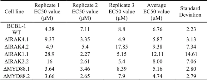
36
compared to the ΔMYD88 (5.16 ± 2.79 μM n=4) ΔIRAK1 (8.0 ± 7.6 μM n=4) or ΔIRAK4
(14.07 ± 16.6 μM n=4) cell lines (Table 2. 1). Likewise, none of the IRAK inhibitors showed any significant change in EC50 comparing WT to ΔIRAK4 BCBL-1CAS9 as seen in (Table 2. 2) The EC50s for ΔIRAK4 BCBL-1 were Inh1 (15.08 ± 16.1 μM n=4), Inh4 (5.1 ± 3.5 μM n=4) and inh1-4 (231 ± 236 μM n=4). This suggests that any anti-proliferative activity for these compounds in PEL was independent of their inhibition of IRAK4 kinase activity. Baseline IRAK signaling is elevated in PEL, which contributes to PEL-specific paracrine phenotypes and limits viral replication. To determine the consequences of
MYD88/IRAK1/IRAK4 ablation genome-wide, RNA-seq analysis of ΔIRAK4 and WT BCBL-1Cas9 was conducted under several treatment conditions. Each RNA-seq experiment was conducted across multiple independent deletion clones. There was minimal change between harvesting biological replicates three weeks apart from the same clone (pairwise Spearman correlation across all mRNAs 0.9722, 0.9675, and 0.9671 for n=3 biological replicates). The combined results are summarized in a heat map representation (Figure II. 11D). First, we observed differences in baseline gene transcription between WT and
ΔIRAK4 BCBL-1Cas9 cells. Two independently derived ΔIRAK4 cell lines were examined and common transcriptional changes were quantitated using false-discovery rate adjusted p-values for individual genes. Changes are visualized by Volcano plot (Figure II. 11A), which verifies the functional inactivation of IRAK4-dependent immune signaling. When treated with IL-1β, WT cells responded with upregulation of canonical IL-1β-responsive transcripts Figure II. 11B), and by contrast, ΔIRAK4 cells did not respond (Figure II. 11C).
37
HSP90AA1, RPS12, SELPLG, SLC16A3, SULT1A1, and YWHAQ (Figure II. 11A & D). IPA suggested that this gene pattern of upregulated cells is linked to aberrant cellular growth and cancer. The transcription of these genes was abrogated by deletion of IRAK4 as seen in two different clones (Figure II. 11D). Hence, we conclude (i) that IRAK signaling is
constitutively active in PEL and (ii) that these transcripts mediate the constitutive phenotype of IRAK4 in PEL. Both clones also upregulated a number of transcripts that were not
transcribed in WT cells to compensate for the loss of the IRAK phenotype (Figure II. 11D). These would be expected to compensate for loss of constitutive IRAK signaling; however,
we did not observe any transcripts that were common among both KO clones. Rather, each ΔIRAK4 clone compensated for loss of IRAK4 activity with a unique set of transcriptional adaptations. The significance of these adaptive transcriptional changes is unclear.
IL-10 was one of the transcripts differentially regulated between WT and IRAK4 KO cells. IL-10 is expressed at high levels in BCBL-1 cells and many other PEL cell lines (90, 212), and is central to the biology of PEL and KSHV infection in vivo, e.g. in KICS (38). ELISA verified the transcriptional phenotype for secreted IL-10 protein across all IRAK KO cell lines (Figure II. 11F, G). IL-10 levels were lower in the IRAK1, IRAK4, and MYD88 cell lines as compared to wild-type BCBL-1Cas9 cells, demonstrating the robustness of the profiling results.
38 Discussion
All PEL cases require KSHV for survival. Dual-infected PEL cases require both KSHV and EBV, presumably because EBV stabilizes the KSHV episome (15, 34, 238). In addition, PEL also acquired multiple genetic changes in the host genome, analogous to EBV-transformed BL (239, 240). PEL’s rarity has provided a challenge to developing definitive genomic analyses. Within these limitations, however, several SNVs in the protein coding regions of genes were identified, present across multiple PEL cases and cell lines at a higher frequency than in the general population (135). One of these SNVs occurred on the IRAK1 gene, which is located on the X chromosome and transcribed in PEL. IRAK1, IRAK4 and MYD88 constitute the Myddosome, which mediates IL-1β and TLR signaling. MYD88 is
mutationally activated in a large proportion of diffuse large B-cell lymphomas, such as WM, resulting in constitutive activation of the MYD88/IRAK pathway and driving proliferation of these cancers (121, 223, 241). This phenomenon prompted us to explore the biological function of MYD88/IRAK signaling in PEL.
MYD88 was not mutated in any of the PEL cases analyzed and upon CRISPR/Cas-9 facilitated deletion of MYD88, the BCBL-1Cas9 PEL cell line rapidly acquired
39
11F&G). This suggests that IRAK signaling was constitutively active in PEL, at least the part of the IRAK signaling cascade that is independent of kinase activities - as has been
previously observed (19, 66, 233). Another result of these experiments demonstrated that current tool compounds that target IRAK1/4 or MYD88 derived their anti-proliferative efficacy in PEL largely from off-target effects (Fig. 10). This does not invalidate the utility of these inhibitors but suggests a different mechanism of action. Since, in PEL, IRAK1 induced both kinase independent as well as kinase dependent targets, it remains unclear what the direct benefit of targeting IRAK kinase activity in PEL would be.
Another important piece of information to consider when examining data is in the differences in experimental designs. Small molecule inhibitor, siRNA, and shRNA screens measure acute phenotypes, often within hours after addition. These agents act effectively on dividing as well as on non-dividing cells since they target mRNA and protein and do not depend on homologous recombination. CRISPR/Cas9 requires each cell to traverse multiple replication/division cycles. Our approach of evaluating single cell clones differed from bulk CRISPR/Cas9 studies that were designed to measure a relative enrichment within the total population. Consistent with our results, Manzano et al. (12) reported a CRISPR/Cas9 screen in which PEL cells survived after deletion of NFκB, which is a downstream target of IRAK1 as well as a multitude of other signaling pathways. NFκB is also a target of the KSHV vFLIP and based on pharmacological studies was believed to be a driver for PEL growth (55, 56, 60). Yet, within a few generations, PEL clones that were independent of NFκB emerged.
40
rapamycin (92). In this study RNA-seq analysis of the ΔIRAK4 clones demonstrated that the cells adapted other means to support survival, which was the phenotype that the
CRISPR/Cas9 experiments select for (Fig. 11), but they did not adapt a means to transmit the IL-1β signal, since IL-1β responses were not selected for during the CRISPR/Cas9 process (Fig. 4).
It is important to remember that BCBL-1, like all cancer cell lines, has been selected for continuous growth in culture. Therefore, vulnerabilities that exist in patients may not manifest themselves under ideal growth conditions. For example, one would expect PDX or direct xenograft models (242) to be more susceptible to agents that modulate autocrine and/or paracrine signaling pathways or that augment the host immune response. IL-1β is present at high levels in KS lesions (243), precedes NHL development in AIDS patients, and likely has its most important role in modulating the PEL microenvironment in vivo (45). One would expect PEL (or KSHV-infected precursors to PEL) to respond to some pro-inflammatory cytokines, such as IL-1β, by releasing IL-10, dampening T cell activity and counteracting viral clearance. If this is the case it may explain the prevalence of the IRAK1 SNV seen in PEL patients as this SNV results in higher levels of NFκB signaling (136, 244). This leads us to suspect a continued importance of the IRAK pathway in PEL, especially at the onset of infection.
Acknowledgements
41
the members of the UNC Vironomics Core, Femi Villamor,
42 Tables
Table 2. 1: MYD88 inhibitor in IRAK pathway knockouts.
Results from treating IRAK pathway knockout cells with the MYD88 st2825 dimerization inhibitor are represented below in μM. Three biological replicates,
each the average of 4 technical replicates are shown.
Cell line EC50 value Replicate 1 (μM)
Replicate 2 EC50 value
(μM)
Replicate 3 EC50 value
(μM)
Average EC50 value
(μM)
Standard Deviation BCBL-1
WT 4.38 7.11 8.8 6.76 2.23
ΔIRAK4.1 9.37 3.35 4.9 5.87 3.13
ΔIRAK4.2 4.9 5.4 17.85 9.38 7.34
ΔIRAK1.1 28.9 2.27 5.15 12.11 14.61
ΔIRAK2.2 16 2.61 5.4 8.00 7.06
ΔMYD88.1 3.64 3.46 8.39 5.16 2.80
ΔMYD88.2 3.66 2.65 7.9 4.74 2.79
Table 2. 2: IRAK inhibitors in IRAK pathway knockouts.
IC50 values for three IRAK inhibitors conducted in WT and IRAK4 knockout BCBL-1 cell lines. Represented is the average and standard deviation of 4 biological replicates.
IRAK inhibitor-1
EC50 value (μM) IRAK inhibitor-4 EC50 value (μM) IRAK inhibitor-1-4 EC50 value (μM) BCBL-1 WT 14.93 ± 14.9 5.5 ± 0.5 275.4 ± 52