TITLE
REGULATION OF HUMAN CYTOMEGALOVIRUS MAJOR IMMEDIATE EARLY GENE
EXPRESSION DURING LYTIC REPLICATION
Kyle C. Arend
A dissertation submitted to the faculty at the University of North Carolina at Chapel Hill in partial fulfillment of the requirements for the degree of Doctor of Philosophy in the Department of
Microbiology and Immunology in the School of Medicine
Chapel Hill 2018
Approved by: Nathaniel Moorman Mark Heise
ii
COPYRIGHT
iii
ABSTRACT
Kyle C. Arend: Regulation of Human Cytomegalovirus Major Immediate Early Gene Expression During Lytic Replication
(Under the direction of Nathaniel J. Moorman)
Human cytomegalovirus (HCMV) is a significant cause of disease in immune-compromised adults and immune naïve newborns. No vaccine exists to prevent HCMV infection, and current antiviral therapies have toxic side effects that limit the duration and intensity of their use. Expression of the HCMV major immediate early (MIE) proteins, IE1 and IE2, is critical for the establishment of lytic infection and reactivation from viral latency. Defining the mechanisms controlling IE1 and IE2 expression is therefore important for understanding how HCMV regulates its replicative cycle. In Chapter 2 we identified several novel transcripts encoding full-length IE1 and IE2 proteins during HCMV lytic replication. While the canonical MIE mRNA was the most abundant transcript at immediate early times, the novel MIE transcripts accumulated to equivalent levels as the known MIE transcript later in infection and were found associated with polyribosomes. These results expand our understanding of the sequences controlling IE1 and IE2 expression by defining novel transcriptional units controlling the
iv
v
TABLE OF CONTENTS
LIST OF FIGURES ... vii
LIST OF TABLES ... ix
Chapter 1: Introduction ... 1
Herpesviridae Family ... 1
Human Herpesviruses ... 2
HCMV Disease and Impact and on Human Health ... 3
HCMV Genetic Structure and Expression ... 6
Cascade of Viral Gene Expression during Lytic Replication ... 6
Major Immediate Early Protein Functions ... 8
Major Immediate Early Promoter-Regulatory Region ... 9
Post-transcriptional Regulation of Major Immediate Early mRNAs ...11
Virus-Induced Changes in Kinase Activity and Cell Signaling Pathways ...13
References ...15
Chapter 2: Multiple Transcripts Encode Full-length Human Cytomegalovirus IE1 and IE2 Proteins During Lytic Infection ...27
Introduction ...27
Materials and Methods ...29
Results ...36
vi
References ...52
Chapter 3: The 5’ Untranslated Region of the Major Immediate Early mRNA is Necessary for Efficient Human Cytomegalovirus Replication ...57
Introduction ...57
Materials and Methods ...59
Results ...63
Discussion ...72
References ...76
Chapter 4: Kinome Profiling Identifies Druggable Targets For Novel Human Cytomegalovirus (HCMV) Antivirals ...81
Introduction ...81
Materials and Methods ...85
Results ...92
Discussion ... 104
References ... 109
Chapter 5: Discussion ... 115
Summary of Chapters ... 115
Role of Alternative MIE Promoters During HCMV Lytic Replication ... 116
5’ UTR Post-Transcriptional Regulation of MIE mRNAs ... 124
MIB/MS Kinome Profiling of HCMV-Infected Cells and Drug Repurposing ... 126
vii
LIST OF FIGURES
Figure 2.1 - Multiple transcription start sites give rise to mRNAs
containing MIE exon 2 ...37
Figure 2.2 - Transcripts originating from each MIE transcription start site give rise to mature IE1 and IE2 mRNAs ...39
Figure 2.3 - Temporal analysis of MIE transcript abundance ...41
Figure 2.4 - DNA sequences surrounding the novel MIE transcription start sites have promoter activity ...42
Figure 2.5 - The core promoter region of the MIEP is not necessary for IE1 and IE2 expression outside the context of HCMV infection ...44
Figure 2.6 - Removal of MIE intron A delays HCMV replication ...46
Figure 2.7 - Deletion of intron A results in decreased IE1 and IE2 mRNA and protein expression ...47
Figure 3.1 - The MIE 5’UTR increases reporter gene expression outside the context of infection ...65
Figure 3.2 - The MIE 5’UTR increases the translation of a luciferase reporter gene ...66
Figure 3.3 - Mutations in the MIE 5’UTR decrease HCMV infectivity ...67
Figure 3.4 - Mutations in the MIE 5’UTR decrease HCMV replication ...68
Figure 3.5 - The MIE 5’UTR is required for efficient IE1 and IE2 protein accumulation during infection ...70
Figure 3.6 - The MIE 5’UTR is necessary for efficient association of the IE1 and IE2 mRNAs with polysomes ...71
viii
Figure 4.1 - Cartoon showing workflow of kinome profiling of HCMV infected cell ...93
Figure 4.2 - Host kinome changes quantified over a time course of HCMV infection ...95
Figure 4.3 - HCMV infection alters multiple novel signaling pathways ...97
Figure 4.4 - Analysis of potential HCMV antivirals ...99
ix
LIST OF TABLES
Table 3.1 – Primers and Oligonucleotide Sequences………...75
1
Chapter 1:
Introduction
Herpesviridae Family
The Herpesviridae family was originally classified based on their distinct viral particle morphology but in recent years have also been classified by specific genetic signatures. The original classification recognized four main components of herpesvirus virions including the core, capsid, tegument, and envelope. The envelope of the virion is comprised of a lipid bilayer of cellular membranes acquired by the virus as it transits from the nucleus to the cell surface. Viral glycoproteins protrude from the envelope and act as adaptors for virus binding to cellular receptors. A largely unstructured and asymmetric proteinaceous layer, called the tegument, lies between the envelope and the capsid. After virion endocytosis and dissolution of the virus envelope, tegument proteins are released into the cytoplasm where they assist in evading the host immune system, transporting the viral capsid to the nucleus, unpackaging the genome from the capsid at the nuclear membrane, and transactivating viral gene expression to initiate the lytic viral replication cycle. The capsid is an icosadeltahedral structure made of capsomere subunits that self-assemble and encase the DNA inside the core. The innermost layer of the virion, the virion core, consists of a single linear double-stranded DNA genome that varies in size based on the subclass of herpesvirus. Following capsid migration to the nuclear
2
infectious virions. Latent genomes are maintained in reservoir cell populations throughout the host lifetime. Importantly, latent genomes can be reactivated by cellular or environmental stimuli to reinitiate the lytic replication cycle resulting in recurrent or potentially persistent cycles of herpesvirus infection.(1)
Human Herpesviruses
3
name “cytomegaly” meaning “abnormal enlargement of a cell” from the significant increase in cell size observed by microscopy during lytic replication in cell culture, which results from viral disruption of cytoskeletal structures.(6)
HCMV Disease and Impact and on Human Health
HCMV is serious health concern that draws relatively little public attention, though current estimates suggest nearly 70-100% of the world population is seropositive for HCMV.(7, 8) A potential reason for this lapse in public concern is that HCMV infections are usually
asymptomatic or cause mild flu-like disease symptoms that resolve without treatment, in
individuals with properly functioning immune systems.(9, 10) Yet, regardless of the appearance of clinical symptoms, HCMV-infected individuals produce high titers of virus that are shed through bodily fluids for days to weeks after the initial infection.(11, 12) The lack of clinical symptoms in HCMV-infected individuals combined with persistent virus shedding enables HCMV to transmit itself throughout the population in a seemingly “silent” manner.(13) While a competent immune system can limit active viral replication and mitigate disease symptoms, it cannot eradicate HCMV from the host. Instead, after primary infection, HCMV establishes a lifelong latent infection in CD14+ peripheral blood monocytes, CD34+ bone-marrow derived monocytes, and dendritic cells.(14-17) Latent infection of monocytes allows the virus to evade detection by the host immune system while also disseminating throughout the body as monocytes shuttle to different sites in response to immune stimuli. Although the exact mechanism of viral reactivation is unknown, it is clear that the transition of HCMV genomes from a latent to lytic viral replication cycle is closely correlated with monocyte differentiation.(17-23) The latent viral state and
4
Although HCMV infections are not of grave concern for healthy individuals,
immunocompromised individuals experience significant rates of morbidity and mortality following HCMV primary infection or reactivation of latent infections.(16, 24) In particular, individuals who have received solid organ transplants, chemotherapy, or developed AIDS from a prior HIV infection are most severely at risk for life-threatening complications associated with HCMV disease.(10, 11, 25-29) HCMV not only presents a serious health risk for immunocompromised individuals but it is also a significant health concern for neonates, whom prior to birth, have not yet developed competent immune systems. Both primary HCMV infection of an HCMV-naïve mother and reactivation of latent HCMV during pregnancy can result in congenital infection of neonates. Statistically speaking, HCMV continues to be the leading cause of infectious congenital birth defects worldwide despite the fact that most congenital HCMV infections present as asymptomatic. Developmental disabilities such as blindness, deafness, and mental disability are most common among symptomatic HCMV-infected neonates, however death can also occur in severe cases.(12, 24, 30-34)
5
due to their toxicity issues. Lastly, nearly all approved HCMV antivirals target the viral DNA polymerase resulting in the emergence of HCMV strains with mutations in the viral DNA polymerase that confer resistance to the entire array of approved HCMV antivirals.(39) These issues associated with currently available HCMV antiviral drugs highlight the need for continued development of safer and more effective therapeutics. Furthermore, the development of
antivirals that inhibit HCMV replication through novel mechanisms and/or targets is becoming increasingly important in order to counter the emergence of HCMV strains resistant to current antiviral drugs.
6
HCMV Genetic Structure and Expression
HCMV has the largest genome of any known human virus. The HCMV genome is a linear double-stranded DNA genome that ranges between 230-240 kilobase pairs (kbp) in size, depending on the specific virus strain. The genome consists of a long and short unique
sequence each flanked by a set of inverted repeat sequences. The long and short unique regions encode the vast majority of proteins synthesized during infection and are fairly
conserved between different virus strains. In contrast, the inverted tandem repeats are markedly different between patient isolates and tissue-culture adapted strains of HCMV. Furthermore the inverted repeat regions show very little protein coding capacity suggesting that their importance for viral replication in vivo is not reliant on the production of viral protein products. Their
importance for HCMV replication remains poorly understood at this time.(1, 53)
The HCMV genome was initially thought to encode about ~200 open reading frames (ORFs).(54) However recent technological advances have revealed that the protein coding capacity of the HCMV genome is much more extensive than previously appreciated. Most recently the use of ribosome profiling has shown that the HCMV genome encodes over 750 peptide-coding regions during lytic viral replication.(55) While the function of the majority of the HCMV ORFs is unknown, the prevailing hypothesis is that the extensive viral protein repertoire facilitates HCMV replication in a wide range of cellular environments. HCMV infects fibroblasts, macrophages, as well as epithelial, endothelial, smooth muscle, neuronal, and glial cells that each have different gene expression profiles and pose unique challenges that HCMV must overcome to successfully replicate.(2-5, 56-59)
Cascade of Viral Gene Expression during Lytic Replication
7
classification of genes into the IE, E, and L phases is not meant to indicate that their expression is limited only to that phase. Instead, IE genes continue to be expressed at E and L phases, and similarly that E genes continue to be expressed throughout the L phase of viral gene
expression.(1, 61-63)
The IE genes are expressed first, and are defined by their transcription in the absence of de novo protein synthesis.(64-67) Thus, IE genes do not require newly expressed viral proteins for their transcription. However, viral proteins in the tegument of incoming virions greatly facilitate IE transcription, particularly pp71.(68-70) HCMV IE genes are transcribed from only four regions of the genome that map to the UL122-123, UL36-37, IRS1/TRS1, and US3 regions of the HCMV genome. Collectively the IE proteins allow HCMV to evade detection by the host immune system, dysregulate the cell cycle, modulate cell metabolism, inhibit apoptosis, and trans-activate transcription from a wide range of host and viral early promoters.(71) The most highly expressed IE proteins are encoded by the UL122-123 genomic region, otherwise referred to as the major immediate early (MIE) gene region.
The promoters of viral E genes are trans-activated by viral IE proteins, and thus require prior IE gene expression.(64) E gene expression is much more widespread than IE genes, and E genes are dispersed across nearly the entire HCMV genome.(63) In general, viral E genes encode proteins that help produce a cellular environment conducive for viral DNA replication.(72) Furthermore, the machinery necessary for replicating the viral genome (e.g. the viral DNA polymerase encoded by UL54 and the polymerase processivity factors encoded by UL44) are expressed as E genes.(73-75)
8
cycle.(63) Late genes typically encode proteins that are structural constituents of the virion and/or are responsible for orchestrating virion packaging and egress from the cell.(76)
Major Immediate Early Protein Functions
Robust MIE gene expression is required for expression of E viral genes suggesting that the MIE protein products play an important role in activating E promoters. The MIE gene
contains a complex promoter-regulatory region, the major immediate early promoter (MIEP), five exons, and four introns.(77, 78) Transcription from the MIEP produces a 5.1kb MIE primary
transcript that is alternatively spliced into at least four different mRNAs. The two predominant mRNAs are a 1.95kb IE1 mRNA (exon 1-2-3-4) and a 2.2kb IE2 mRNA (exons 1-2-3-5) that are translated into the IE1-72kDa and IE2-86kDa proteins, respectively.(78-83) Two additional low abundance mRNAs, a 0.65kb IE1 mRNA and a 1.7kb IE2 mRNA, arise from additional splicing events in either exon 4 or exon 5, respectively. These minor MIE mRNAs are translated into the IE1-38kDa and IE2-55kDa protein, but little is known about their role in HCMV replication.(81, 84)
9
typically activate promoters synergistically.(95, 97-101) IE1-72kDa mainly promotes transcription by antagonizing histone deacetylation that would otherwise silence viral gene expression.(102) IE1-72kDa interacts with histone deacetylase 3 (HDAC3) and prevents HDAC3 from removing histone acetylation marks around target promoters. IE1-72kDa also disrupts nuclear domain 10 (ND10) bodies during IE times of infection that would normally recruit HDACs and other
chromatin remodeling enzymes to the viral genome, leading to suppressive nucleosome formation.(103-108) The ability of IE2-86kDa to transactivate promoters derives primarily from its interactions with components of the basal transcription machinery. IE2-86kDa binds to TATA binding protein (TBP) and transcription factor IIB (TFIIB), as well as numerous transcription factors including SP1, Tef-1, c-Jun, JunB, ATF-2, Egr-1, NF-kB, CBP, CHD-1, p300, and phosphorylated CREB.(98, 99, 109-118)
Major Immediate Early Promoter-Regulatory Region
The MIE genomic region is highly transcribed immediately after HCMV infection of permissive cells. As noted above, multiple alternatively spliced MIE mRNAs originate from a strong, constitutively active MIEP. The MIEP is regulated in part by a complex promoter-regulatory region that contains multiple binding sites for cis-promoter-regulatory elements that recruit components of the transcription initiation complex (i.e. transcription factors) or stabilize complex formation to enhance transcription from the MIEP. Three regions of the MIEP were found to play important roles during lytic replication: the core promoter, the proximal enhancer and the distal enhancer.(119-121) The core promoter and proximal enhancer are essential for HCMV replication. The core promoter contains a prototypical TATA box that binds TBP and recruits the RNA polymerase II transcription initiation complex. The core promoter also contains a cis-repression sequence (crs) that overlaps the MIEP transcription start site.(122-125) The crs provides a
10
contains several copies of repeat sequences with binding sites for several transcription factors including: NF-kB, CREB/ATF, AP-1, SP1, retinoic acid receptor, and NF-1.(126) Despite the requirement for the proximal enhancer for HCMV replication, the roles of the individual binding site(s) and cis-acting regulatory factor(s) are unknown. In contrast to the other two regions, the distal enhancer is only necessary for HCMV replication after low MOI infections. The distal enhancer includes binding sites for serum response factor, NF-kB, CREB/ATF, YY-1, and SP1.(127, 128) Removal of the distal enhancer delays viral replication due to decreased MIE gene expression and results in an aberrant small-plaque phenotype, even after high MOI infection. This suggests the redundancy of binding sites in the proximal and distal enhancers is required to promote robust MIE transcription and efficient HCMV replication.(129) Together, transcription factor binding sites in the core promoter, and proximal and distal enhancers endow the HCMV MIEP with the strongest known mammalian promoter activity. In addition, complex interplay between the cis-regulatory elements likely allows complex and dynamic regulation of MIE gene expression in specific settings.(130, 131)
In addition to host transcription factors, HCMV proteins also regulate transcription from the MIEP. In particular, the viral tegument proteins pp71, pTRS1, and pUL69 enhance MIEP activity, and stimulate the initial burst of MIE gene expression.(69, 88, 132-134) The IE1-72kDa and IE2-86kDa proteins also autoregulate the MIEP.(71, 80, 82, 119) In contrast to their abilities to
synergistically activate cellular and viral early promoters, the IE1-72kDa and IE2-86kDa proteins have opposing functions in MIEP regulation. The IE1-72kDa protein activates the MIEP similar to other viral promoters by antagonizing histone deacetylation, but can also directly bind the MIEP at NF-kB consensus binding sites to enhance transcription.(135) Conversely, the IE2-86kDa protein negatively regulates transcription from the MIEP by directly binding a 14bp
11
polymerase II recruitment and thus blocks transcription initiation from the MIEP.(138, 139) In summary, transcription from the MIEP is regulated at multiple levels including chromatin remodeling, recognition of cis-regulatory elements by cellular and viral factors, and autoregulation by IE1-72kDa and IE2-86kDa proteins.
In Chapter 2, I describe the discovery of multiple novel MIE mRNAs that initiate transcription from regions of the MIE gene both 5’ and 3’ of the MIEP. In addition, I detail the role of two of the novel MIE promoters in HCMV lytic replication. Collectively, these experiments identify alternative promoter usage as an additional layer of transcriptional regulation for the MIE genomic region during HCMV lytic replication.
Post-transcriptional Regulation of Major Immediate Early mRNAs
While promoters regulate the transcription of an mRNA, 5’ and 3’ untranslated regions (5’UTRs and 3’UTRs, respectively) flanking the coding determinant sequence also influence protein levels by regulating mRNA translation.(140) The 5’ UTR is defined as the sequence that extends from the transcription start site to just before the translation initiation codon of a protein coding region. 5’UTRs play an especially important regulatory role, as they directly impact translation initiation, the rate limiting step in protein synthesis.(141) 5’UTRs can either enhance or repress translation of downstream coding regions. One general example of 5’ UTR positive regulation is the recruitment of eukaryotic translation initiation factors (eIF4E, eIF4G, and eIF4A) to the 5’-terminal 7-methylguanosine (m7G) mRNA cap. Together these initiation factors
12
Despite well-established roles for 5’ UTRs in regulating eukaryotic mRNA translation, relatively little is known about the importance of 5’ UTRs for the translation efficiency of HCMV mRNAs during lytic replication. HCMV mRNAs are entirely reliant on cellular ribosomes for the synthesis of viral polypeptides. However, HCMV-infected cells do not shut off cellular protein synthesis but instead globally upregulate the synthesis of translation initiation factors, the formation of polyribosomes, and the rate of protein synthesis compared to uninfected cells. (145-147) The global increase in protein synthesis of HCMV-infected cells is facilitated by a wide-range of virus-induced cellular changes including increased ribosome biogenesis(148), accumulation of PABP and eIF4F complex subunits(147, 149, 150), and activation of cellular pathways that increase energy production and biosynthetic metabolites.(151-157) Nevertheless, by not antagonizing host protein synthesis viral mRNAs must compete with host mRNAs for access to ribosomes. The mechanisms that HCMV utilizes to ensure efficient translation of viral mRNAs at IE, E, and L stages of infection are still unknown.
Although less-studied, post-transcriptional events are also thought to regulate the
expression of MIE proteins. In particular, the rate of MIE protein synthesis and the association of MIE mRNAs with polyribosomes changes throughout infection despite fairly persistent
13
In Chapter 3, I show that the MIE 5’ UTR increases reporter gene expression in transfection assays, but not in a cell-free system. I also demonstrate that the MIE 5’ UTR increases reporter mRNA association with polyribosomes and enhances the translation efficiency of the mRNA compared to synthetic 5’ UTR sequences. In addition, I show that mutation of the canonical MIE 5’ UTR in recombinant viruses causes decreased viral infectivity, virion production, and expression of viral proteins compared to the wild-type virus. Furthermore, I demonstrate that the defects in recombinant viruses are caused by decreased translation efficiency of IE1 and IE2 mRNAs. These experiments identify the canonical MIE 5’ UTR sequence as a crucial component of the MIE mRNAs that post-transcriptionally regulates MIE protein expression
.
Virus-Induced Changes in Kinase Activity and Cell Signaling Pathways
Kinases are cellular enzymes that phosphorylate targets to modulate their activity, localization, stability, and interaction with other cellular components. Kinase networks evolved to coordinate cellular processes and maintain homeostasis. Furthermore the arrangement of kinases in signaling cascades allows for rapid, exponential activation of cellular pathways in response to intracellular and extracellular signaling molecules.(163, 164)
14
15
REFERENCES
1.
Mocarski, E., Shenk, T., and Pass, R. F. (2013) Cytomegaloviruses. In: Knipe, D. M., and Howley,
P. M., eds. Fields virology, 6th Ed., Wolters Kluwer/Lippincott Williams & Wilkins Health, Philadelphia, PA
2. Landini, M. P., and Jahn, G. (2000) The eclectic cellular tropism of cytomegalovirus. Pathologica 92, 51-7
3. Plachter, B., Sinzger, C., and Jahn, G. (1996) Cell types involved in replication and distribution of human cytomegalovirus. Adv Virus Res 46, 195-261
4. Sinzger, C., and Jahn, G. (1996) Human cytomegalovirus cell tropism and pathogenesis. Intervirology 39, 302-19
5. Sinzger, C., Grefte, A., Plachter, B., et al. (1995) Fibroblasts, epithelial cells, endothelial cells and smooth muscle cells are major targets of human cytomegalovirus infection in lung and
gastrointestinal tissues. J Gen Virol 76, 741-50
6. Craig, J. M., Macauley, J. C., Weller, T. H., and Wirth, P. (1957) Isolation of intranuclear inclusion producing agents from infants with illnesses resembling cytomegalic inclusion disease. Proc Soc Exp Biol Med 94, 4-12
7. Jeon, J., Victor, M., Adler, S. P., et al. (2006) Knowledge and awareness of congenital cytomegalovirus among women. Infect Dis Obstet Gynecol, 1-7
8. Cannon, M. J., Schmid, D. S., and Hyde, T. B. (2010) Review of cytomegalovirus seroprevalence and demographic characteristics associated with infection. Rev Med Virol 20, 202-13
9. Zanghellini, F., Boppana, S. B., Emery, V. C., et al. (1999) Asymptomatic primary cytomegalovirus infection: Virologic and immunologic features. J Infect Dis 180, 702-7
10. Sissons, J. G., and Carmichael, A. J. (2002) Clinical aspects and management of cytomegalovirus infection. J Infect 44, 78-83
11. Sia, I. G., and Patel, R. (2000) New strategies for prevention and therapy of cytomegalovirus infection and disease in solid-organ transplant recipients. Clin Microbiol Rev 13, 83-121
12. Stagno, S., Reynolds, D. W., Pass, R. F., and Alford, C. A. (1980) Breast milk and the risk of cytomegalovirus infection. N Engl J Med 302, 1073-6
13. Manicklal, S., Emery, V. C., Lazzarotto, T., et al. (2013) The "silent" global burden of congenital cytomegalovirus. Clin Microbiol Rev 26, 86-102
14. Bego, M. G., and St Jeor, S. (2006) Human cytomegalovirus infection of cells of hematopoietic origin: HCMV-induced immunosuppression, immune evasion, and latency. Exp Hematol 34, 555-70
15. Sinclair, J., and Sissons, P. (2006) Latency and reactivation of human cytomegalovirus. J Gen Virol 87, 1763-79
16. Sinclair, J. (2008) Human cytomegalovirus: Latency and reactivation in the myeloid lineage. J Clin Virol 41, 180-5
16
18. Docke, W. D., Prosch, S., Fietze, E., et al. (1994) Cytomegalovirus reactivation and tumour necrosis factor. Lancet 343, 268-9
19. Meier, J. L. (2001) Reactivation of the human cytomegalovirus major immediate-early regulatory region and viral replication in embryonal NTERA2 cells: Role of trichostatin a, retinoic acid, and deletion of the 21-base-pair repeats and modulator. J Virol 75, 1581-93
20. Goodrum, F. D., Jordan, C. T., High, K., and Shenk, T. (2002) Human cytomegalovirus gene expression during infection of primary hematopoietic progenitor cells: A model for latency. PNAS 99, 16255-60
21. Reeves, M. B., and Sinclair, J. H. (2010) Analysis of latent viral gene expression in natural and experimental latency models of human cytomegalovirus and its correlation with histone modifications at a latent promoter. J Gen Virol 91, 599-604
22. Keyes, L. R., Hargett, D., Soland, M., et al. (2012) Hcmv protein luna is required for viral reactivation from latently infected primary cd14(+) cells. PLoS One 7, e52827
23. O'Connor, C. M., and Murphy, E. A. (2012) A myeloid progenitor cell line capable of supporting human cytomegalovirus latency and reactivation, resulting in infectious progeny. J Virol 86, 9854-65
24. Griffiths, P. D. (2012) Burden of disease associated with human cytomegalovirus and prospects for elimination by universal immunisation. The Lancet Infectious Diseases 12, 790-98
25. Fietze, E., Prosch, S., Reinke, P., et al. (1994) Cytomegalovirus infection in transplant recipients. The role of tumor necrosis factor. Transplantation 58, 675-80
26. Ode-Hakim, S., Kern, F., Fietze, E., et al. (1995) Association between CMV-related graft injury and circulating cytokine-producing CD8+ memory cells. Transplant Proc 27, 951-3
27. Mesaric, B., Begovac, J., Ugrinovic, N., et al. (1998) Cytomegalovirus retinitis in patients with human immunodeficiency virus infection. Lijec Vjesn 120, 106-10
28. Drew, W. L. (2003) Cytomegalovirus disease in the highly active antiretroviral therapy era. Curr Infect Dis Rep 5, 257-65
29. Kim, H. C., Hwang, E. A., Park, S. B., et al. (2012) Historical comparison of prophylactic ganciclovir for gastrointestinal cytomegalovirus infection in kidney transplant recipients. Transplant Proc 44, 710-2
30. Revello, M. G., and Gerna, G. (2002) Diagnosis and management of human cytomegalovirus infection in the mother, fetus, and newborn infant. Clinical Microbiology Reviews 15, 680-715
31. Pannuti, C. S., Vilas-Boas, L. S., Angelo, M. J., et al. (1985) Congenital cytomegalovirus infection. Occurrence in two socioeconomically distinct populations of a developing country. Rev Inst Med Trop Sao Paulo 27, 105-7
32. Sissons, J. G., Carmichael, A. J., McKinney, N., et al. (2002) Human cytomegalovirus and immunopathology. Springer Semin Immunopathol 24, 169-85
17
34. Ludwig, A., and Hengel, H. (2009) Epidemiological impact and disease burden of congenital CMV in Europe. Eurosurveillance 14, 1-7
35. Littler, E., Stuart, A. D., and Chee, M. S. (1992) Human cytomegalovirus UL97 open reading frame encodes a protein that phosphorylates the antiviral nucleoside analogue ganciclovir. Nature 358, 160-2
36. Sullivan, V., Talarico, C. L., Stanat, S. C., et al. (1992) A protein kinase homologue controls phosphorylation of ganciclovir in human cytomegalovirus-infected cells. Nature 359, 85
37. Crumpacker, C. S. (1996) Ganciclovir. N Engl J Med 335, 721-9
38. Mercorelli, B., Sinigalia, E., Loregian, A., and Palu, G. (2008) Human cytomegalovirus DNA replication: Antiviral targets and drugs. Rev Med Virol 18, 177-210
39. Chou, S., Lurain, N. S., Thompson, K. D., et al. (2003) Viral DNA polymerase mutations associated with drug resistance in human cytomegalovirus. J Infect Dis 188, 32-9
40. Khanna, R., and Diamond, D. J. (2006) Human cytomegalovirus vaccine: Time to look for alternative options. Trends Mol Med 12, 26-33
41. Zhong, J., and Khanna, R. (2007) Vaccine strategies against human cytomegalovirus infection. Expert Rev Anti Infect Ther 5, 449-59
42. Lilja, A. E., and Mason, P. W. (2012) The next generation recombinant human cytomegalovirus vaccine candidates-beyond gB. Vaccine 30, 6980-90
43. Chiurchiu, S., Calo Carducci, F. I., Rocchi, F., et al. (2013) Is HCMV vaccine an unmet need? The state of art of vaccine development. Int J Immunopathol Pharmacol 26, 15-26
44. Xia, L., Su, R., An, Z., et al. (2017) Human cytomegalovirus vaccine development: Immune responses to look into vaccine strategy. Hum Vaccin Immunother
45. Bonaros, N., Mayer, B., Schachner, T., et al. (2008) CMV-hyperimmune globulin for preventing cytomegalovirus infection and disease in solid organ transplant recipients: A meta-analysis. Clin Transplant 22, 89-97
46. Revello, M. G., Lazzarotto, T., Guerra, B., et al. (2014) A randomized trial of hyperimmune globulin to prevent congenital cytomegalovirus. N Engl J Med 370, 1316-26
47. Nigro, G. (2017) Hyperimmune globulin in pregnancy for the prevention of congenital cytomegalovirus disease. Expert Rev Anti Infect Ther, 1-10
48. Andreoni, K. A., Wang, X., Huong, S. M., and Huang, E. S. (2001) Human CMV-IgIV (cytogam) neutralizes human cytomegalovirus (hcmv) infectivity and prevents intracellular signal
transduction after hcmv exposure. Transpl Infect Dis 3 Suppl 2, 25-30
49. Yamani, M. H., Avery, R., Mawhorter, S. D., et al. (2005) The impact of cytogam on cardiac transplant recipients with moderate hypogammaglobulinemia: A randomized single-center study. J Heart Lung Transplant 24, 1766-9
18
51. Lianghui, G., Shusen, Z., Tingbo, L., et al. (2004) Deferred versus prophylactic therapy with gancyclovir for cytomegalovirus in allograft liver transplantation. Transplant Proc 36, 1502-5
52. Bonaros, N. E., Kocher, A., Dunkler, D., et al. (2004) Comparison of combined prophylaxis of cytomegalovirus hyperimmune globulin plus ganciclovir versus cytomegalovirus hyperimmune globulin alone in high-risk heart transplant recipients. Transplantation 77, 890-7
53. Yu, D., Silva, M. C., and Shenk, T. (2003) Functional map of human cytomegalovirus AD169 defined by global mutational analysis. PNAS 100, 12396-401
54. Chee, M. S., Bankier, A. T., Beck, S., et al. (1990) Analysis of the protein-coding content of the sequence of human cytomegalovirus strain AD169. Curr Top Microbiol Immunol 154, 125-69
55. Stern-Ginossar, N., Weisburd, B., Michalski, A., et al. (2012) Decoding human cytomegalovirus. Science 338, 1088-93
56. Liu, R., Baillie, J., Sissons, J. G., and Sinclair, J. H. (1994) The transcription factor YY1 binds to negative regulatory elements in the HCMV major immediate early enhancer/promoter and mediates repression in non-permissive cells. Nucleic Acids Res 22, 2453-9
57. Wiley, C. A., Schrier, R. D., Denaro, F. J., et al. (1986) Localization of cytomegalovirus proteins and genome during fulminant central nervous system infection in an AIDS patient. J Neuropathol Exp Neurol 45, 127-39
58. Sinzger, C., Plachter, B., Grefte, A., et al. (1996) Tissue macrophages are infected by human cytomegalovirus in vivo. J Infect Dis 173, 240-5
59. Schmidbauer, M., Budka, H., Ulrich, W., and Ambros, P. (1989) Cytomegalovirus (CMV) disease of the brain in aids and connatal infection: A comparative study by histology,
immunocytochemistry and in situ DNA hybridization. Acta Neuropathol 79, 286-93
60. DeMarchi, J. M., Schmidt, C. A., and Kaplan, A. S. (1980) Patterns of transcription of human cytomegalovirus in permissively infected cells. J Virol 35, 277-86
61. Wathen, M. W., and Stinski, M. F. (1981) Temporal patterns of HCMV transcription: Mapping the viral RNAs synthesized at immediate early, early, and late times after infection. J Virol 41, 462
62. Chua, C. C., Carter, T. H., and St Jeor, S. (1981) Transcription of the human cytomegalovirus genome in productively infected cells. J Gen Virol 56, 1-11
63. Demarchi, J. M. (1981) Human cytomegalovirus DNA: Restriction enzyme cleavage maps and map locations for immediate-early, early, and late RNAs. Virology 114, 23-38
64. Wathen, M. W., Thomsen, D. R., and Stinski, M. F. (1981) Temporal regulation of human
cytomegalovirus transcription at immediate early and early times after infection. J Virol 38, 446-59
65. Stinski, M. F., Thomsen, D. R., Stenberg, R. M., and Goldstein, L. C. (1983) Organization and expression of the immediate early genes of human cytomegalovirus. J Virol 46, 1-14
66. Jahn, G., Knust, E., Schmolla, H., et al. (1984) Predominant immediate-early transcripts of human cytomegalovirus AD169. J Virol 49, 363-70
19
68. Saffert, R. T., and Kalejta, R. F. (2006) Inactivating a cellular intrinsic immune defense mediated by Daxx is the mechanism through which the human cytomegalovirus pp71 protein stimulates viral immediate-early gene expression. J Virol 80, 3863-71
69. Schierling, K., Stamminger, T., Mertens, T., and Winkler, M. (2004) Human cytomegalovirus tegument proteins ppUL82 (pp71) and ppUL35 interact and cooperatively activate the major immediate-early enhancer. J Virol 78, 9512-23
70. Feng, X., Schroer, J., Yu, D., and Shenk, T. (2006) Human cytomegalovirus pUS24 is a virion protein that functions very early in the replication cycle. J Virol 80, 8371-8
71. Stinski, M. F., and Meier, J. L. (2007) Immediate-early viral gene regulation and function. In: Arvin, A., Campadelli-Fiume, G., Mocarski, E., Moore, P. S., Roizman, B., Whitley, R., and Yamanishi, K., eds. Human herpesviruses: Biology, therapy, and immunoprophylaxis, Cambridge
72. White, E. A., and Spector, D. H. (2007) Early viral gene regulation and function. In: Arvin, A., Campadelli-Fiume, G., Mocarski, E., Moore, P. S., Roizman, B., Whitley, R., and Yamanishi, K., eds. Human herpesviruses: Biology, therapy, and immunoprophylaxis, Cambridge
73. Kerry, J. A., Priddy, M. A., Jervey, T. Y., et al. (1996) Multiple regulatory events influence human cytomegalovirus DNA polymerase (UL54) expression during viral infection. J Virol 70, 373-82
74. Weiland, K. L., Oien, N. L., Homa, F., and Wathen, M. W. (1994) Functional analysis of human cytomegalovirus polymerase accessory protein. Virus Res 34, 191-206
75. Ertl, P. F., and Powell, K. L. (1992) Physical and functional interaction of human cytomegalovirus DNA polymerase and its accessory protein (ICP36) expressed in insect cells. J Virol 66, 4126-33
76. Anders, D. G., Kerry, J. A., and Pari, G. (2007) DNA synthesis and late viral gene expression. In: Arvin, A., Campadelli-Fiume, G., Mocarski, E., Moore, P. S., Roizman, B., Whitley, R., and Yamanishi, K., eds. Human herpesviruses: Biology, therapy, and immunoprophylaxis, Cambridge
77. Stenberg, R. M., Thomsen, D. R., and Stinski, M. F. (1984) Structural analysis of the major immediate early gene of human cytomegalovirus. J Virol 49, 190-9
78. Stenberg, R. M., Witte, P. R., and Stinski, M. F. (1985) Multiple spliced and unspliced transcripts from human cytomegalovirus immediate-early region 2 and evidence of common transcription initiation site. J Virol 56, 665-75
79. Hermiston, T. W. M., C.L.; Witte, P.R.; Stinski, M. F. (1987) Identification and characterization of the HCMV IE region 2 gene that stimulates gene expression from an inducible promoter. J Virol 61, 3214-21
80. Stenberg, R. M. D., A.S.; Fortney, J.; Nelson, J.A. (1989) Regulated expression of early and late RNAs and proteins from the human cytomegalovirus immediate-early gene region. J Virol 63, 2699-708
81. Baracchini, E., Glezer, E., Fish, K., et al. (1992) An isoform variant of the cytomegalovirus immediate-early auto repressor functions as a transcriptional activator. Virology 188, 518-29
82. Stenberg, R. M., and Stinski, M. F. (1985) Autoregulation of the human cytomegalovirus major immediate-early gene. J Virol 56, 676-82
83. Kerry, J. A., Sehgal, A., Barlow, S. W., et al. (1995) Isolation and characterization of a
20
84. Shirakata, M., Terauchi, M., Ablikim, M., et al. (2002) Novel immediate-early protein IE19 of human cytomegalovirus activates the origin recognition complex I promoter in a cooperative manner with IE72. J Virol 76, 3158-67
85. Marchini, A., Liu, H., and Zhu, H. (2001) Human cytomegalovirus with IE-2 (UL122) deleted fails to express early lytic genes. J Virol 75, 1870-8
86. Heider, J. A., Bresnahan, W. A., and Shenk, T. E. (2002) Construction of a rationally designed human cytomegalovirus variant encoding a temperature-sensitive immediate-early 2 protein. PNAS 99, 3141-6
87. Greaves, R. F., and Mocarski, E. S. (1998) Defective growth correlates with reduced accumulation of a viral DNA replication protein after low-multiplicity infection by a human cytomegalovirus IE1 mutant. J Virol 72, 366-79
88. Bresnahan, W. A. S., T.E. (2000) UL82 virion protein activates expression of IE viral genes in HCMV infected cells. PNAS 97, 14506-11
89. Plachter, B., Britt, W., Vornhagen, R., et al. (1993) Analysis of proteins encoded by IE regions 1 and 2 of human cytomegalovirus using monoclonal antibodies generated against recombinant antigens. Virology 193, 642-52
90. Wiebusch, L., and Hagemeier, C. (1999) Human cytomegalovirus 86-kilodalton IE2 protein blocks cell cycle progression in G(1). J Virol 73, 9274-83
91. Murphy, E. A., Streblow, D. N., Nelson, J. A., and Stinski, M. F. (2000) The human
cytomegalovirus IE86 protein can block cell cycle progression after inducing transition into the S phase of permissive cells. J Virol 74, 7108-18
92. Noris, E., Zannetti, C., Demurtas, A., et al. (2002) Cell cycle arrest by human cytomegalovirus 86-kDa IE2 protein resembles premature senescence. J Virol 76, 12135-48
93. Petrik, D. T., Schmitt, K. P., and Stinski, M. F. (2006) Inhibition of cellular DNA synthesis by the human cytomegalovirus IE86 protein is necessary for efficient virus replication. J Virol 80, 3872-83
94. Zhu, H., Shen, Y., and Shenk, T. (1995) HCMV IE1 and IE2 proteins block apoptosis. J Virol 69, 7960-70
95. Pizzorno, M. C., O'Hare, P., Sha, L., et al. (1988) Trans-activation and autoregulation of gene expression by the immediate-early region 2 gene products of human cytomegalovirus. J Virol 62, 1167-79
96. Hermiston, T. W., Malone, C. L., and Stinski, M. F. (1990) Human cytomegalovirus immediate-early two protein region involved in negative regulation of the major immediate-immediate-early promoter. J Virol 64, 3532-6
97. Klucher, K. M., Sommer, M., Kadonaga, J. T., and Spector, D. H. (1993) In vivo and in vitro analysis of transcriptional activation mediated by the human cytomegalovirus major immediate-early proteins. Mol Cell Biol 13, 1238-50
21
99. Scully, A. L., Sommer, M. H., Schwartz, R., and Spector, D. H. (1995) The human
cytomegalovirus IE2 86-kilodalton protein interacts with an early gene promoter via site-specific DNA binding and protein-protein associations. J Virol 69, 6533-40
100. Malone, C. L., Vesole, D. H., and Stinski, M. F. (1990) Transactivation of a human cytomegalovirus early promoter by gene products from the immediate-early gene IE2 and augmentation by IE1: Mutational analysis of the viral proteins. J Virol 64, 1498-506
101. Stenberg, R. M., Fortney, J., Barlow, S. W., et al. (1990) Promoter-specific trans activation and repression by human cytomegalovirus immediate-early proteins involves common and unique protein domains. J Virol 64, 1556-65
102. Nevels, M., Paulus, C., and Shenk, T. (2004) Human cytomegalovirus immediate-early 1 protein facilitates viral replication by antagonizing histone deacetylation. PNAS 101, 17234-9
103. Korioth, F., Maul, G. G., Plachter, B., et al. (1996) The nuclear domain 10 (ND10) is disrupted by the human cytomegalovirus gene product IE1. Exp Cell Res 229, 155-8
104. Ahn, J. H., and Hayward, G. S. (1997) The major immediate-early proteins IE1 and IE2 of human cytomegalovirus colocalize with and disrupt PML-associated nuclear bodies at very early times in infected permissive cells. J Virol 71, 4599-613
105. Ahn, J. H., Brignole, E. J., 3rd, and Hayward, G. S. (1998) Disruption of PML subnuclear domains by the acidic IE1 protein of human cytomegalovirus is mediated through interaction with pml and may modulate a ring finger-dependent cryptic transactivator function of PML. Mol Cell Biol 18, 4899-913
106. Ahn, J. H., and Hayward, G. S. (2000) Disruption of PML-associated nuclear bodies by OE1 correlates with efficient early stages of viral gene expression and DNA replication in human cytomegalovirus infection. Virology 274, 39-55
107. Tavalai, N., Papior, P., Rechter, S., et al. (2006) Evidence for a role of the cellular ND10 protein pml in mediating intrinsic immunity against human cytomegalovirus infections. J Virol 80, 8006-18
108. Tavalai, N., and Stamminger, T. (2011) Intrinsic cellular defense mechanisms targeting human cytomegalovirus. Virus Res 157, 128-33
109. Hagemeier, C., Walker, S., Caswell, R., et al. (1992) The human cytomegalovirus 80-kilodalton but not the 72-kilodalton immediate-early protein transactivates heterologous promoters in a TATA box-dependent mechanism and interacts directly with TFIID. J Virol 66, 4452-6
110. Caswell, R., Hagemeier, C., Chiou, C. J., et al. (1993) The human cytomegalovirus 86k
immediate early (IE) 2 protein requires the basic region of the TATA-box binding protein (TBP) for binding, and interacts with TBP and transcription factor TFIIB via regions of IE2 required for transcriptional regulation. J Gen Virol 74 ( Pt 12), 2691-8
111. Furnari, B. A., Poma, E., Kowalik, T. F., et al. (1993) Human cytomegalovirus immediate-early gene 2 protein interacts with itself and with several novel cellular proteins. J Virol 67, 4981-91
112. Jupp, R., Hoffmann, S., Stenberg, R. M., et al. (1993) Human cytomegalovirus IE86 protein interacts with promoter-bound TATA-binding protein via a specific region distinct from the autorepression domain. J Virol 67, 7539-46
22
114. Sommer, M. H., Scully, A. L., and Spector, D. H. (1994) Transactivation by the human cytomegalovirus IE2 86-kilodalton protein requires a domain that binds to both the TATA box-binding protein and the retinoblastoma protein. J Virol 68, 6223-31
115. Lang, D., Gebert, S., Arlt, H., and Stamminger, T. (1995) Functional interaction between the human cytomegalovirus 86-kilodalton IE2 protein and the cellular transcription factor CREB. J Virol 69, 6030-7
116. Schwartz, R., Helmich, B., and Spector, D. H. (1996) CREB and CREB-binding proteins play an important role in the IE2 86-kilodalton protein-mediated transactivation of the human
cytomegalovirus 2.2-kilobase RNA promoter. J Virol 70, 6955-66
117. Yoo, Y. D., Chiou, C. J., Choi, K. S., et al. (1996) The IE2 regulatory protein of human
cytomegalovirus induces expression of the human transforming growth factor beta1 gene through an Egr-1 binding site. J Virol 70, 7062-70
118. Wu, J., O'Neill, J., and Barbosa, M. S. (1998) Transcription factor SP1 mediates cell-specific trans-activation of the human cytomegalovirus DNA polymerase gene promoter by immediate-early protein IE86 in glioblastoma U373 MG cells. J Virol 72, 236-44
119. Meier, J. L., and Stinski, M. F. (1996) Regulation of human cytomegalovirus immediate-early gene expression. Intervirology 39, 331-42
120. Thomsen, D. R., Stenberg, R. M., Goins, W. F., and Stinski, M. F. (1984) Promoter-regulatory region of the major immediate early gene of human cytomegalovirus. PNAS 81, 659-63
121. Boshart, M., Weber, F., Jahn, G., et al. (1985) A very strong enhancer is located upstream of an immediate early gene of human cytomegalovirus. Cell 41, 521-30
122. Pizzorno, M. P. H., G.S. (1990) The IE2 gene products of HCMV specifically down-regulate expression from the MIEP through target sequence located near the cap site. J Virol 64, 6154-65
123. Cherrington, J. M., Khoury, E. L., and Mocarski, E. S. (1991) Human cytomegalovirus IE2 negatively regulates alpha gene expression via a short target sequence near the transcription start site. J Virol 65, 887-96
124. Liu, B. H., T.W.; Stinski, M.F. (1991) A cis-acting element in the MIEP of HCMV required for negative regulation by IE2. J Virol 65, 897-903
125. Macias, M. P. H., L.; Lashmit, P.E.; Stinski, M.F. (1996) Cellular or viral protein binding to a cytomegalovirus promoter transcription initiation site - effects on transcription. J Virol 70, 3628-34
126. Isomura, H., Tsurumi, T., and Stinski, M. F. (2004) Role of the proximal enhancer of the major immediate-early promoter in human cytomegalovirus replication. J Virol 78, 12788-99
127. Meier, J. L., and Pruessner, J. A. (2000) The human cytomegalovirus major immediate-early distal enhancer region is required for efficient viral replication and immediate-early gene expression. J Virol 74, 1602-13
23
129. Isomura, H., and Stinski, M. F. (2003) The human cytomegalovirus major immediate-early enhancer determines the efficiency of immediate-early gene transcription and viral replication in permissive cells at low multiplicity of infection. J Virol 77, 3602-14
130. Baskar, J. F., Smith, P. P., Nilaver, G., et al. (1996) The enhancer domain of the human cytomegalovirus major immediate-early promoter determines cell type-specific expression in transgenic mice. J Virol 70, 3207-14
131. Meier, J. L., and Stinski, M. F. (1997) Effect of a modulator deletion on transcription of the human cytomegalovirus major immediate-early genes in infected undifferentiated and differentiated cells. J Virol 71, 1246-55
132. Winkler, M., Rice, S. A., and Stamminger, T. (1994) UL69 of human cytomegalovirus, an open reading frame with homology to ICP27 of herpes simplex virus, encodes a transactivator of gene expression. J Virol 68, 3943-54
133. Baldick, C. J. M., A.; Patterson, C.E.; Shenk, T. (1997) HCMV pp71 enhances infectivity and mie transcription. J Virol 77, 4400-08
134. Romanowski, M. J., and Shenk, T. (1997) Characterization of the human cytomegalovirus IRS1 and TRS1 genes: A second immediate-early transcription unit within IRS1 whose product antagonizes transcriptional activation. J Virol 71, 1485-96
135. Cherrington, J. M., and Mocarski, E. S. (1989) Human cytomegalovirus IE1 transactivates the alpha promoter-enhancer via an 18-base-pair repeat element. J Virol 63, 1435-40
136. Lang, D., and Stamminger, T. (1993) The 86kDa IE2 protein of hcmv is a sequence specific DNA-binding protein that interacts directly with the autoregulatory response element. J Virol 67, 323-31
137. Lang, D., and Stamminger, T. (1994) Minor groove contacts are essential for an interaction of the human cytomegalovirus IE2 protein with its DNA target. Nucleic Acids Res 22, 3331-8
138. Jupp, R., Hoffmann, S., Depto, A., et al. (1993) Direct interaction of the human cytomegalovirus IE86 protein with the cis repression signal does not preclude TBP from binding to the TATA box. J Virol 67, 5595-604
139. Wu, J., Jupp, R., Stenberg, R. M., et al. (1993) Site-specific inhibition of RNA polymerase II preinitiation complex assembly by human cytomegalovirus IE86 protein. J Virol 67, 7547-55
140. Mignone, F., Gissi, C., Liuni, S., and Pesole, G. (2002) Untranslated regions of mRNAs. Genome Biol 3, REVIEWS0004
141. Jackson, R. J., Hellen, C. U., and Pestova, T. V. (2010) The mechanism of eukaryotic translation initiation and principles of its regulation. Nat Rev Mol Cell Biol 11, 113-27
142. Gingras, A. C., Raught, B., and Sonenberg, N. (1999) eIF4 initiation factors: Effectors of mRNA recruitment to ribosomes and regulators of translation. Annu Rev Biochem 68, 913-63
143. Hinnebusch, A. G., Ivanov, I. P., and Sonenberg, N. (2016) Translational control by 5'-untranslated regions of eukaryotic mRNAs. Science 352, 1413-6
144. Morris, D. R., and Geballe, A. P. (2000) Upstream open reading frames as regulators of mRNA translation. Mol Cell Biol 20, 8635-42
24
146. Stinski, M. F. (1977) Synthesis of proteins and glycoproteins in cells infected with human cytomegalovirus. J Virol 23, 751-67
147. Walsh, D., Perez, C., Notary, J., and Mohr, I. (2005) Regulation of the translation initiation factor eIF4F by multiple mechanisms in human cytomegalovirus-infected cells. J Virol 79, 8057-64
148. Tirosh, O., Cohen, Y., Shitrit, A., et al. (2015) The transcription and translation landscapes during human cytomegalovirus infection reveal novel host-pathogen interactions. PLoS Pathog 11, e1005288
149. McKinney, C., Perez, C., and Mohr, I. (2012) Poly(A) binding protein abundance regulates eukaryotic translation initiation factor 4F assembly in human cytomegalovirus-infected cells. PNAS 109, 5627-32
150. Perez, C., McKinney, C., Chulunbaatar, U., and Mohr, I. (2011) Translational control of the abundance of cytoplasmic poly(A) binding protein in human cytomegalovirus-infected cells. J Virol 85, 156-64
151. Landini, M. P. (1984) Early enhanced glucose uptake in HCMV infected cells. J Gen Virol, 1229-32 152. Munger, J., Bajad, S. U., Coller, H. A., et al. (2006) Dynamics of the cellular metabolome during
human cytomegalovirus infection. PLoS Pathog 2, e132
153. Munger, J., Bennett, B. D., Parikh, A., et al. (2008) Systems-level metabolic flux profiling identifies fatty acid synthesis as a target for antiviral therapy. Nat Biotechnol 26, 1179-86
154. Chambers, J. W., Maguire, T. G., and Alwine, J. C. (2010) Glutamine metabolism is essential for human cytomegalovirus infection. J Virol 84, 1867-73
155. McArdle, J., Schafer, X. L., and Munger, J. (2011) Inhibition of calmodulin-dependent kinase kinase blocks human cytomegalovirus-induced glycolytic activation and severely attenuates production of viral progeny. J Virol 85, 705-14
156. Yu, Y., Clippinger, A. J., and Alwine, J. C. (2011) Viral effects on metabolism: Changes in glucose and glutamine utilization during human cytomegalovirus infection. Trends Microbiol 19, 360-7
157. Yu, Y., Maguire, T. G., and Alwine, J. C. (2014) ChREBP, a glucose-responsive transcriptional factor, enhances glucose metabolism to support biosynthesis in human cytomegalovirus-infected cells. PNAS 111, 1951-6
158. Goins, W. F., and Stinski, M. F. (1986) Expression of a human cytomegalovirus late gene is posttranscriptionally regulated by a 3'-end-processing event occurring exclusively late after infection. Mol Cell Biol 6, 4202-13
159. Stenberg, R. M., Depto, A. S., Fortney, J., and Nelson, J. A. (1989) Early and late RNA and protein from HCMV MIE gene region. J Virol 63, 2699-708
160. Jacobson, A., and Peltz, S. W. (1996) Interrelationships of the pathways of mRNA decay and translation in eukaryotic cells. Annu Rev Biochem 65, 693-739
25
162. Mangus, D. A., Evans, M. C., and Jacobson, A. (2003) Poly(A)-binding proteins: Multifunctional scaffolds for the post-transcriptional control of gene expression. Genome Biol 4, 223
163. Manning, G., Whyte, D. B., Martinez, R., et al. (2002) The protein kinase complement of the human genome. Science 298, 1912-34
164. Ubersax, J. A., and Ferrell, J. E., Jr. (2007) Mechanisms of specificity in protein phosphorylation. Nat Rev Mol Cell Biol 8, 530-41
165. Hertel, L., Chou, S., and Mocarski, E. S. (2007) Viral and cell cycle-regulated kinases in cytomegalovirus-induced pseudomitosis and replication. PLoS Pathog 3, e6
166. Moorman, N. J., Cristea, I. M., Terhune, S. S., et al. (2008) Human cytomegalovirus protein UL38 inhibits host cell stress responses by antagonizing the tuberous sclerosis protein complex. Cell Host Microbe 3, 253-62
167. Spencer, C. M., Schafer, X. L., Moorman, N. J., and Munger, J. (2011) Human cytomegalovirus induces the activity and expression of acetyl-coenzyme a carboxylase, a fatty acid biosynthetic enzyme whose inhibition attenuates viral replication. J Virol 85, 5814-24
168. Roy, S., and Arav-Boger, R. (2014) New cell-signaling pathways for controlling cytomegalovirus replication. Am J Transplant 14, 1249-58
169. Vincent, H. A., Ziehr, B., and Moorman, N. J. (2016) Human cytomegalovirus strategies to maintain and promote mRNA translation. Viruses 8, 97
170. Johnson, R. A., Wang, X., Ma, X. L., et al. (2001) Human cytomegalovirus up-regulates the phosphatidylinositol 3-kinase (PI3-K) pathway: Inhibition of PI3-K activity inhibits viral replication and virus-induced signaling. J Virol 75, 6022-32
171. Moorman, N. J., and Shenk, T. (2010) Rapamycin-resistant mTORC1 kinase activity is required for herpesvirus replication. J Virol 84, 5260-9
172. Clippinger, A. J., Maguire, T. G., and Alwine, J. C. (2011) Human cytomegalovirus infection maintains mTOR activity and its perinuclear localization during amino acid deprivation. J Virol 85, 9369-76
173. Du, G., Dutta, N., Lashmit, P., and Stinski, M. F. (2011) Alternative splicing of the human cytomegalovirus major immediate-early genes affects infectious-virus replication and control of cellular cyclin-dependent kinase. J Virol 85, 804-17
174. McArdle, J., Moorman, N. J., and Munger, J. (2012) HCMV targets the metabolic stress response through activation of AMPK whose activity is important for viral replication. PLoS Pathog 8, e1002502
175. Ziehr, B., Vincent, H. A., and Moorman, N. J. (2016) Human cytomegalovirus pTRS1 and pIRS1 antagonize protein kinase R to facilitate virus replication. J Virol 90, 3839-48
176. Vincent, H. A., Ziehr, B., and Moorman, N. J. (2017) Mechanism of protein kinase R inhibition by human cytomegalovirus ptrs1. J Virol 91
177. Terry, L. J., Vastag, L., Rabinowitz, J. D., and Shenk, T. (2012) Human kinome profiling
26
178. Hutterer, C., Wandinger, S. K., Wagner, S., et al. (2013) Profiling of the kinome of
cytomegalovirus-infected cells reveals the functional importance of host kinases Aurora A, ABL and AMPK. Antiviral Res 99, 139-48
179. Duncan, J. S., Whittle, M. C., Nakamura, K., et al. (2012) Dynamic reprogramming of the kinome in response to targeted mek inhibition in triple-negative breast cancer. Cell 149, 307-21
27
Chapter 2: Multiple Transcripts Encode Full-length Human Cytomegalovirus IE1
and IE2 Proteins During Lytic Infection
1Introduction
The human cytomegalovirus (HCMV) IE1 and IE2 proteins are critical regulators of the viral replicative cycle. Both proteins are immediately expressed upon infection and together stimulate the expression of host and viral genes necessary for virus replication.(1) IE2 acts as a general transcription factor that broadly transactivates host genes and viral early and late genes to facilitate virus replication.(2-12) IE1 promotes transcription from the HCMV genome by
inhibiting histone deactylases (HDACs), which otherwise limit virus transcription by forming inhibitory chromatin structures on the viral genome.(13-15) Re-expression of IE1 and IE2 is also thought to be critical for the reactivation of quiescent HCMV genomes from latent infection. Thus, understanding the regulatory mechanisms controlling IE1 and IE2 expression is important to understand how HCMV regulates its replicative cycle.
The mRNAs encoding the 72kDa IE1 and 86kDa IE2 proteins are derived from a shared precursor RNA through alternative splicing.(16) The mature IE1 and IE2 transcripts share the same first three exons (exons 1 through 3), but differ in their final exon. Transcripts that include exon 4 encode the 72kDa IE1 protein, while transcripts that skip exon 4 and retain exon 5 encode the 86kDa IE2 protein.(17) Several IE1 and IE2 splice variants have been identified. For example IE1 transcript variants arise from alternative splice donor and acceptor usage in exons
28
3 and 4 respectively.(16-19) Similarly, IE2 transcript variants arise from alternative splicing within exon 5.(17) The proteins encoded by these alternatively spliced transcripts differ in function from full-length IE1 and IE2, highlighting the role of alternative splicing in expanding the functions of proteins encoded by the major immediate early (MIE) locus.
In addition to alternative splicing, multiple promoters direct the transcription of several distinct RNAs from the MIE locus. The most studied promoter in this region is the major immediate early promoter (MIEP), whose activity is regulated by multiple cellular and viral factors such as CREB/ATF, NFB, SP1, pp71, pp65 and pTRS1.(20-32) In addition, the IE1 and IE2 proteins themselves regulate MIEP activity. While IE1 stimulates MIEP activity, IE2 binds a cis-repression sequence (crs) located adjacent to the MIEP transcription start site, inhibiting transcription initiation.(33-37) Alternative promoters in the MIE locus also control expression of IE1 and IE2 transcript variants in specific contexts. A promoter proximal to the IE2-specific exon 5 drives the expression of several truncated IE2 isoforms during the late stage of infection.(38-40) An additional promoter located 5’ of the MIEP is active in latently infected human monocytes, suggesting that MIE transcription may be driven by different promoters during lytic and latent infections.(41)
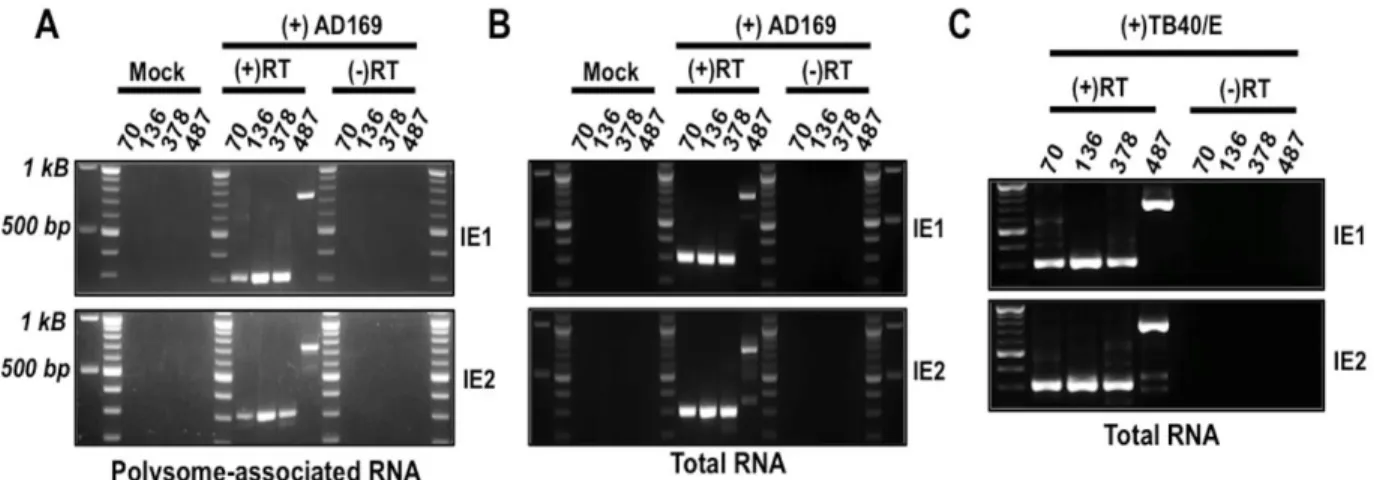
In this study we identified several previously unrecognized MIE transcripts encoding full-length IE1 and IE2 proteins that are expressed during HCMV lytic infection. The novel IE1 and IE2 transcripts differed from the known MIE transcript in their transcription start sites, and thus their 5’ untranslated regions (5’UTRs). Sequences surrounding each transcription start site possessed promoter activity that increased during HCMV infection. Each transcript was
29
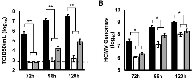
initiated in the first intron (intron A) of the known MIE transcript. Mutant viruses lacking intron A or the region surrounding one of the “intronic” promoters expressed lower levels of IE1 and IE2 mRNA and protein, and replicated less efficiently than the parental virus. Together our data show that multiple transcripts encode full length IE1 and IE2 proteins during HCMV infection, and suggest that multiple transcription units act in concert to regulate IE1 and IE2 expression during lytic replication.
Materials and Methods
Cells and Viruses. Primary human foreskin fibroblasts (HFFs), MRC-5 fibroblasts and
Hela cells were grown in DMEM with 10% FBS and penicillin and streptomycin. Unless
otherwise indicated, BAC-derived HCMV AD169 strain containing a GFP reporter driven by the SV40 promoter was used for all infections.(42) Cell free virus was titered by the TCID50 method on MRC-5 fibroblasts.
Construction of Recombinant Viruses. Recombinant viruses were constructed in the
AD169 genomic background using bacterial artificial chromosome (BAC)-mediated recombineering, as previously described.(43-46) Briefly, the HCMV∆UTR378 BAC was constructed by amplifying a kanamycin cassette flanked by FRT sites with primers HCMV∆UTR378-FRTKanF (5'-TGGTGACGATACTTTCCATTACTAATCCATAACA
30
genome FJ527563.1). The deletion of intron A was accomplished using a two-step recombination approach. In the first step the KanSacB gene was amplified with primers HCMV∆IntronA – KanSacBF (5'-GCGGCCGGGAACGGTGCATTGGAACGCGGA
TTCCCCGTGCCAAGAGTGACAATTCGAGCTCGGTACCCGG-3') and HCMV∆IntronA – KanSacBR (5'-AGGGTCCATCTTTCTCTTGGCAGAGGACTCCATCGTGTCAAGGAC GGTGACATCCCGGGAAAAGTGCCACC-3'), which contained 50 nucleotide homology arms flanking intron A. Recombination with the AD169 genome resulted in the replacement of intron A with the KanSacB cassette. Recombinants were selected by growth on LB plates containing kanamycin and chloramphenicol. For the second step, the primers Exon1/2Fusion F/R (5'-CGGCCGGGAACGGTGCATTGGAACGCGGATTCCCCGTGCCAAGAGTGACTCACCGTCCT TGACACGATGGAGTCCTCTGCCAAGAGAAAGATGGACCCT-3' and 5'-AGGGTCCATC TTTCTCTTGGCAGAGGACTCCATCGTGTCAAGGACGGTGAGTCACTCTTGGCACGGGGAA TCCGCGTTCCAATGCACCGTTCCCGGCCG-3', respectively) were used to amplify a region spanning the exon1/exon2 splice junction using IE2 cDNA as a template. The KanSacB cassette was then removed by a second round of recombineering using the PCR product above. The recombinants were grown on LB agar with chloramphenicol and 6% sucrose to select against colonies that retained the SacB gene. Sucrose-resistant colonies were then screened for kanamycin sensitivity to ensure loss of the kanamycin cassette. The resulting recombinant, HCMVIntronA contained a seamless fusion of exons 1 and 2. (nucleotides 173738-174564 deleted, reference genome FJ527563.1) The absence of genomic
rearrangements was confirmed by restriction digest of the recombinant BAC DNAs at each step. No errors were detected when the 500 base pairs on either side of each mutation were
31
libraries were sequenced on an Illumina MiSeq. No changes other than the intended mutations were detected in the recombinant genomes.
Quantitative real-time PCR analysis. Total RNA was extracted using Trizol according
to the manufacturer’s directions as described previously.(47, 48) Briefly, cell pellets were resuspended in Trizol, extracted with chloroform and RNA was precipitated with isopropanol. The RNA was resuspended in water and quantified on a NanoDrop spectrophotometer. For quantitative reverse transcriptase PCR (qRT-PCR), cDNA was generated from 0.5 µg total RNA using the High Capacity cDNA Reverse Transcription Kit (ThermoFisher) and random hexamer primers. The abundance of each transcript was determined using a real-time PCR machine (Biorad) and SYBR Select Master Mix (Roche). The abundance of each product was
determined by comparison to a standard curve generated from qPCR analysis of tenfold serial dilutions of a DNA standard specific for each primer pair. For comparative analysis of MIE transcript abundance, each of the UTR-specific primer pairs were confirmed to i) only amplify the specific UTR sequence, and not amplify the other MIE UTRs or HCMV BAC DNA (data not shown) ii) have similar real time PCR efficiencies (>95% efficiency for each primer pair) and iii) have a similar linear range of detection of the appropriate DNA standard (linear between 108 and 102 copies, R2 >.97 for all experiments included in this work). Where indicated
phosphonoacetic acid (PAA; Sigma) was used at a final concentration of 200 µg/mL. Primer sequences used for qRT-PCR were: IE1 qPCR F/R (5’-CAAGTGACCGAGGATTGCAA-3’ and 5’-CACCATGTCCACTCGAACCTT-3’), IE2 qPCR F/R (5’-TGACCGAGGATTGCAACGA-3’ and 5’-CGGCATGATTGACAGCCTG-3’), GAPDH qPCR F/R (5’-CTGTTGCTGTAGCCAAATTCGT-3’ & 5’-ACCCACTCCTCCACCTTTGAC-(5’-CTGTTGCTGTAGCCAAATTCGT-3’), UTR70 qPCR TAGCTGACAGACTAACAGAC-3’), exon2-3 qPCR (5'-GGTCACGGGTGTCTCGGGCCGT-3'), UTR136 qPCR
32
UTR136/378 qPCR Reverse CCTTGACACGATGGAGTCCT-3’), UTR487 qPCR F/R (5’-GCATTATGCCCAGTACATGACC-3’ and 5’-GAAATCCCCGTGAGTCAAACC-3’).
For gene-specific reverse transcription reactions, primers specific for IE1 (exon 4) or IE2 (exon 5) were used in the cDNA synthesis step: exon 4 RT (5’-TCCTTTTTAGCACGGGCCTT-3’), exon 5 RT (5’-CGCATCCACCTCACTCTTCA-3’). The cDNA was then amplified by PCR using the following reaction conditions: 94oC for 5 minutes, thirty cycles of (94oC for 30 seconds, 55oC for 30 seconds, 72oC for 5 minutes), followed by a 7 minute extension at 72oC. The
following PCR primers were used: exon 4 Out (5’-TGAATTTCTCTTCCGTCTGG-3’); exon 4 In (5’-AACCTTAATCTGTTTGACGA -3’); exon 5 Out (5’-CTGCAAGAGTGGGTTGTCAG -3’); exon 5 In (5’CTGGGCGAGGATGTCACCGA 3’); UTR70 Out (5’ACAGACTAACAGACTGTTCC -3’); UTR70 In (5’-TCCATGGGTCTTTTCTGCAG --3’); UTR136 Out (5’-TGACCTCCATAGAAG ACACC -3’); UTR136 In (5’-TTCCCCGTGCCAAGAGTGAC -3’); UTR378 Out (5’-CGCTGAC GCATTTGGAAGAC -3’); UTR378 In TGTTCTGATAAGAGTCAGAG-3’); UTR487 Out (5’-AAGTACGCCCCCTATTGACG -3’); UTR487 In (5’-CCCCTATTGACGTCAATGAC-3’). PCR products were analyzed on agarose gels and visualized with ethidium bromide on an imager (Biorad). PCR products were also sequenced to confirm the specificity of the PCR reaction.
HCMV DNA was quantified essentially as described previously.(49) Briefly, infected cell pellets were lysed in DNA extraction buffer (400 mM NaCl, 10 mM Tris-HCl pH: 8.0, 10 mM EDTA) and digested with proteinase K (10 mg/mL) overnight at 37oC. The lysates were
extracted with phenol-chloroform, digested with RNase A (10 mg/mL) for one hour at 37oC, and again extracted with phenol-chloroform. DNA was precipitated with isopropanol and
(5'-33
GTGTCCCATTCCCGACTCG-3' and 5'-TTCACAACGTCCACCCACC-3'), and GAPDH qPCR F/R (listed above).
Plasmid construction. To generate the promoter reporter constructs, the five hundred
nucleotides from -450 to +50 bp flanking each transcription start site were amplified by PCR and cloned into the HindIII and NotI sites of the pGL3-Basic vector (Promega) using Gibson cloning (NEB). Primer sequences used were: pGL3 Basic UTR70 F/R
(TCTTACGCGTGCTAGCCCGGCTATCGCCGATAGAGGCGACATCAAG-3' and 5'-TACCAACAGTACCGGAATGCCAAGGCGGGCCATTTACCGTCATTG-3'); pGL3 Basic UTR136 F/R (TCTACGCGTGCTAGCCCGGGCCGTATGTTCCCATAGTAACGCC-3' and 5'-TACCAACAGTACCGGAATGCCAGTGTCTTCTATGGAGGTCAAAACAG-3'); pGL3 Basic UTR378 F/R (5'-TCTTACGCGTGCTAGCCCGGGCATCGCCTGGAGACGCCATCCAC-3' and 5'-TACCAACAGTACGGAATGCCAATTCGCGTGGAGATCCCACGTTATG-3'); pGL3 Basic UTR487 F/R (5'-TCTTACGCGTGCTAGCCCGGGCACCACCGTCCCCAGTGCCCGCAG-3' and 5'-TACCAACAGTACCGGAATGCCACAGAAAAGACCCATGGAAAGGAAC-3'). The inserted region was sequenced to confirm the absence of mutations. To generate the plasmid
pSVHMIEP, the nucleotides -94 to +64 relative to the canonical transcription start site in the MIEP were deleted from the pSVH vector ((50); generously provided by Q. Tang) using PCR-mediated mutagenesis and Gibson cloning. Primers pSVHMIEP F/R
(GGCACCAAAATCAACGGGACTTTCCATAGAAGACACCGGGACCGATCA-3' and 5'-GGAAAGTCCCGTTGATTTTGGTGCC-3') were designed to i) flank the region to be deleted and ii) contain 20 nucleotides of sequence complementary to the other primer. The plasmid was amplified by PCR and the PCR product was circularized in a subsequent Gibson reaction. The clones were sequenced to confirm the absence of spurious mutations.