Investigating the Etiology of Primary Failure of Eruption (PFE): A Comprehensive Phenotypic and Genetic Analysis
Heather Morgan Hendricks
A thesis submitted to the faculty of the University of North Carolina at Chapel Hill in partial fulfillment of the requirements for the degree of Master of Science in the School of Dentistry
(Orthodontics).
Chapel Hill 2015
Approved by:
Sylvia A. Frazier-Bowers
Sompop Bencharit
Kimon Divaris
©2015
ABSTRACT
Heather Morgan Hendricks: Investigating the Etiology of Primary Failure of Eruption (PFE): A Comprehensive Phenotypic and Genetic Analysis
(Under the direction of Sylvia A Frazier-Bowers)
The genetic basis of PFE (OMIM ID: 125350), a diagnosis that conveys a poor prognosis
in the eruption/ function of teeth, was interrogated. Treatment with a continuous archwire
worsens the condition. Two aims tested the hypothesis that PTH1R mutations result in loss of
function and that multiple genes cause PFE: to determine 1) the fate of a functional PTH1R
mutation and 2)PFE contribution by BMP2 and TNFSF-11. Methods: We used IFA and
transfected COS7 cells with either the WT or 1092delGPTH1R mutation sequence to compare
the fate of the expressed protein and performed mutational analysis of BMP2 and TNFSF-11
with PCR and sequencing. Results: Sequencing revealed 3 intronic SNPs in TNFSF-11; in silico /functional studies showed expression alterations and structural changes in mutant vs WT
ACKNOWLEGEMENTS
I would like to thank my committee members; Dr. Bencharit, Dr. Divaris, and Dr. Wright
for their guidance, expertise, and support in connection with the completion of my Master’s
Thesis. This experience has left a lasting impression towards my personal and professional
development that I will treasure and reflect upon for years to come. I also want to acknowledge
and thank my family for their continued support and engagement. Lastly, I especially want to
thank Dr. Sylvia A. Frazier-Bowers for her unwavering dedication and profound support in my
endeavors. The council she has offered, and the example set forth is testament to the ideal of a
TABLE OF CONTENTS
LIST OF TABLES ... vii
LIST OF FIGURES ... viii
LIST OF CHARTS……….xi
INVESTIGATING THE ETIOLOGY OF PRIMARY FAILURE OF ERUPTION (PFE): A COMPREHENSIVE PHENOTYPIC AND GENETIC ANALYSIS ... 1
Background ………..……….………....1
Eruption Disorders and Diagnostic Approaches……..……….……….…2
PTH1R and PFE……….…………..4
PTH1R: A G-protein coupled receptor……….………..5
Previous PTH/PTHrp and PTH1R Studies……….………….…8
Epidemiology……….……….……….9
CHAPTER ONE: INVESTIGATING THE PROTEIN EXPRESSION OF WT AND MUTANT PTH1R IN COS7 CELLS ... 12
Introduction ... 12
Materials and Methods ... 16
Results ... 19
Discussion... 21
Conclusion ... 26
Introduction ... 27
Materials and Methods ... 31
Results ... 34
Discussion... 35
Conclusion ... 38
LIST OF TABLES
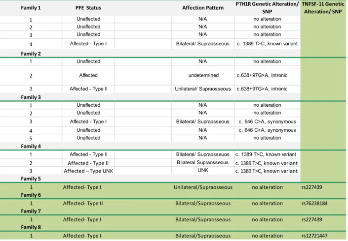
Table 2.1- Correlation of Genotype: phenotype of 4 families
with segregating PFE...39
Table 3.1- Mutational Analysis Results of PFE affected
LIST OF FIGURES
Figure 1- Figure 1- Clinical photograph of child in the mixed dention, presenting with a posterior
lateral open bite- secondary to a lateral tongue thrust………41
Figure 2- Patient with a functional familial PTH1R mutation,
originally diagnosed as having ankylosis………41
Figure 3- Frontal intraoral photos of (A) before and (B) after-orthodontic treatment in a patient
with a later confirmed case of PFE……….…42
Figure 4- Jansen’s Metapyseal Chondrodysplasia………..43
Figure 5- Bloomstrand’s Lethal Chondrodysplasia……….………43
Figure 6- The Human PTH1R gene is located on the
short (p) arm of chromosome 3……….……….….44
Figure 2.1- Bloomstrand’s Lethal Chondrodysplasia
exhibits a recessive PTHR1 mutative deactivation………..…44
Figure 2.2and 2.2b- PFE results from a autosomal dominant
PTH1R mutation. The classical presentation is a progressive
anterior-posterior lateral openbite……….…………45
Figure 2.3- Diagram of the pcDNA3.1 + vector used contain
the WT PTH1R insert………..………..…46
Figure 2.4- The pcDNA 3.1 (+) vector was fabricated to
accept a WT PTH1R insert. The new vector was named TG682 7260………47
Figure 2.5a- Nucleotide sequence segment of interest of WT PTH1R……….47
Figure 2.5b- Nucleotide sequence segment of interest of 1092delGPTH1R………..…47
Figure 2.6- 48 hour post transfection of COS7 cells with
pcDNA 3.0 (+GFP insert)……….48
Figure 2.7- Confocal Image of 24 hours post-transfection
Figure 2.8a- Immunoflourence assay (IFA) was utilized during visualization (Inverted digital microscope) of the PTH1R
protein in terms of localization and expression……….…50
Figure 2.8 b and 2.8c- 48 Hours Post-Transfection of WT PTH1R/COS7 cell (Inverted digital microscope)………50
Figure 2.9- 48 Hours Post-Transfection of 1092delG PTH1R/COS7 Cell (Inverted digital microscope)……….51
Figure 2.10- Chromatogram demonstrating the familial 1092delG in the PTH1R gene, linked to infraocclusion of primary teeth in an affected child……….51
Figure 2.11a- Panoramic radiograph demonstrating the involvement of both primary and permanent teeth in the affected patient who carries a 1092delG mutation in PTH1R………….…52
Figure 2.11b- Intraoral clinical photographs 12 of PFE presentation in a 7yo 9 mo patient whose 1092delG mutation……….……..52
Figure 2.12- Chromatogram demonstrating the familial 996_997insC mutation in the PTH1R gene……….……53
Figure 2.13- Exon:Domain Pairing of hPTH1R. Note that the functional exons are depicted, along with the denotation of the corresponding protein domains………...…54
Figure 2.14- WT, 572G>A, 996_997incC, 1353delA, and 1092delG PTH1R Protein sequences with Transmembrane, extracellular, and intracellular loops labeled……….….55
Figure 2.15-This chart denotes the transmembrane loops and the locations of the functional mutations in PTH1R, identified in the Frazier-Bowers Laboratory……….57
Figure 2.16a- In silico prediction of WT PTH1R………..….57
Figure 2.16b- In silico prediction of 572delA PTH1R mutation……….…57
Figure 2.16c- In silico prediction of 996_997insCPTH1R mutation………59
Figure 2.16e- In silico prediction- 1353-1G>A PTH1R mutation………..….….61
Figure 3.1- Initial radiograph of patient presenting with clinic signs
of Primary Failure of Eruption (PFE)……….…62
Figure 3.2- Patient with a functional familial PTH1R mutation,
initially diagnosed as having ankylosis………....62
Figure 3.3- Ingenuity Pathway Analysis (IPA) of the interaction of multiple genes with PTH1R.
The genes of interest were TNFSF-11 and BMP2………..…..63
Figure 3.4- Chromatograms exhibit three TFSF-11 SNPs A) c.501A>G (21% MAF), B) c.252T>A (3.4% MAF),
LIST OF CHARTS
Chart 3.1- Phenotypic patterning within the non-functional alteration cohort ……….….65
Chart 3.2- Individuals affected with either Type 1 or 2 PFE
within each SNP identified……….…….66
Chart 3.3- Distribution of Unilateral versus Bilateral Affection
INVESTIGATING THE ETIOLOGY OF PRIMARY FAILURE OF ERUPTION (PFE): A COMPREHENSIVE PHENOTYPIC AND GENETIC ANALYSIS
Introduction
Background and Significance
The process of normal tooth eruption is an integral part of normal tooth development
and a thorough understanding of the biologic underpinnings is critical for any dental
practitioner. Every member of the dental team needs to have a comprehensive and functional
understanding of the patterning and sequencing of eruption in order to provide optimal care.
The ability to develop a prioritized diagnosis and treatment plan as a result of this knowledge is
especially important for the practicing clinician and therefore makes expanding our
understanding of this biologic process (i.e. eruption) more critical. This knowledge also includes
the ability to understand when to refer a patient to a specialist, who may be better trained to
treat certain diagnoses. The literature has identified and described many different etiologies of
eruption disturbances that are not uncommon occurrences within the dental practice, however,
the critical step in diagnosing eruption disorders lies in first identifying its etiology out of the
wide range of possibilities.1, 7, 29 It is understood that eruption disorders may stem from local
and non-syndromic causes (cysts, ankylosis, lateral tongue pressure, supernumerary teeth,
thumb habit,) 1, 3 or may manifest as part of a genetic syndrome (Cleidocranial Dysplasia,
Eruption Disorders and Diagnostic Approaches
Three categories of eruption disorders exist within the broad spectrum of
local/non-syndromic causes.2 These are Mechanical Failure of Eruption (MFE), ankylosis, and the more
perplexing- Primary Failure of Eruption (PFE) (OMIM ID:125350). MFE is defined an eruption
failure due to a mechanical obstruction of a tooth’s eruption pathway- which can often be
treated successfully with the removal of the mechanical blockage due to a correct diagnosis.8,13
Ankylosis is defined as the occurrence when the cementum on the root of the tooth fuses
directly to the bone, thereby eliminating the periodontal ligament space (PDL) and halting
spontaneous eruption potential.3, 4 However the primary diagnosis is based on clinical
appearance; this often includes infraocclusion relative to the adjacent teeth. An important
adjunctive diagnostic approach is the diagnosis of ankylosis through radiographic examination;
this is based largely on identifying locations of an absence of a visible periodontal ligament
space. However, the diagnosis of ankylosis is frequently more subjectively based on the lack of
clinically-appreciable mobility, and a sharp sound noted upon tooth percussion with a metal
object.5 It is important to realize the limitations of these diagnostic techniques, in addition to
their applications. Currently, the only two ways to definitively diagnose an ankylosed tooth is to
apply orthodontic force and wait for the tooth to move, or to luxate the tooth and achieve
visual mobility. The ability to implement advanced imaging is a promising tool, however, when
evaluating a two dimensional radiograph, the appearance of PDL fusing to bone can either be
overstated or completely undetected; this is in part due to the ability, or inability, to
radiographically image mineralized tissue that falls within a specific range of density and size.
a Cone Beam CT scan of any size or resolution. Even though the large field of view CBCT scans
have an average resolution of 0.3-0.4 voxels 6, this is still an inadequate resolution to evaluate
whether a fusion of cementum and bone exists. The important goal in distinguishing ankylosis
from other eruption problems is that unlike PFE, the ankylosed tooth can be extracted and the
remaining teeth will likely be responsive to orthodontic treatment.4 The challenge is however
that ankylosis can often be confused with the more enigmatic Primary Failure of Eruption
(Figure 2). 2
Primary Failure of Eruption is poorly understood but clearly falls within the spectrum of
Local/Non-syndromic eruption disruptions. First described at UNC by Proffit and Vig (1981), PFE
is clinically marked by failure of permanent (adult) tooth eruption in the absence of mechanical
obstruction, and affects only the posterior dentition.7 The use of the term, primary, denotes
that the defect is located in the eruption mechanism of the tooth itself.8 PFE diagnosis has been
characterized by distinction of two different patterns of clinical presentation: Type 1 PFE and
Type 2 PFE. Type 1 PFE presentation consists of a progressive, anterior-posterior lateral open
bite that worsens distal to the most mesially positioned PFE-affected tooth; Type 2 PFE
presents with a more varied pattern of infra-eruption distal to the most mesially affected
tooth.18 An early possible treatment of isolated ankylosis of permanent first molars can be
managed via the extraction of the offending ankylosed tooth; this will allow for normal eruption
of the second and third molars. The failure of the second and third molars to fully erupt would
be pathognomonic for PFE.30 This selective extraction pattern is a potential treatment for a
complex surgical intervention and multiple tooth extractions required to restore form and
function (Figure 3).
From a treatment standpoint these cases are complicated; if traditional orthodontics is
attempted the condition only worsens (Figure 1). Taken together, this supports that the
diagnosis of PFE conveys a poor prognosis in the normal eruption and function of teeth. 8
Recent investigations that have focused on PFE and other eruption disorders have simply done
so from an etiologic perspective (i.e., no consideration as to how the etiology relates to the
phenotype and proposed management has been combined for a holistic picture of the
disorder). The discovery and association of PFE with the parathyroid one hormone receptor
gene, PTH1R (discussed below), opens a new dimension of investigation; the role of the PTH1R
gene alone may substantially contribute to our understanding of developmental processes
beyond tooth eruption.8, 9, 16
PTH1R and PFE
Since the original description of PFE, investigations of its etiology have been completed
by multiple researchers; a major commonality among these documented studies of PFE is the
identification of mutations in the PTH1R gene. Currently, autosomal dominant mutations in the
(PTH1R) gene have been identified as the only concrete cause of PFE.2,8, 9, 16, 17, 18, 30 This recent
genetic discovery associated with PFE represents a breakthrough, creating a paradigm shift in
diagnostic and clinical approaches. PFE and its connection with PTH1R not only reveals another
dimension of inquiry into the aspects of dental eruption pattering, but it obviates the gaps in
knowledge that exist in terms of how this gene may be integrated within the odontogenic code
systemic health.13,20 For example, the fact that PFE that solely expresses as a dental problem,
now understood to be a genetic disorder caused by PTH1R, is implicitly connected to systemic
processes that are also governed by this gene. Hence, the isolated genotype:phenotype
relationship between PTH1R and PFE presents an interesting conundrum.18 A deeper
understanding of the PTH1R gene may offer insights as to why this presents in clinically diverse
conditions; such as Jansen’s Metaphyseal Chondrodysplasia (OMIM ID: 156400).
Bloomstrand’s Lethal Chondrodysplasia (OMIM ID: 215045), Enchondromatosis (Ollier’s
Disease) (OMIM ID: 166000), osteoarthritis, and PFE.28 Briefly, Jansen’s is caused by autosomal
dominant heterozygous mutations in PTHR1 resulting in short-limbed dwarfism(Figure 4).28
Ollier’s disease is also correlated to an autosomal dominant mutation, which is characterized by
multiple enchondromas located primarily in the metaphyseal shafts of long bones; having the
ability to develop into chondrosarcomas. Bloomstand’s, however, exhibits a recessive PTHR1
mutative deactivation which results in early lethality, shortened limbs, and premature bone
maturity (Figure 5).28 The variation of clinical presentations of both dominant and recessive
autosomal mutations within the same gene make its inquiry that much more interesting, and
exhibits why PFE, having more relatively benign effects, could be a potential model of PTH1R
gene studies. Although studies completed in this project cannot fully answer these questions,
they provide the impetus for future studies that will unravel these mysteries.
PTH1R: A G-protein coupled receptor
While the varied functions and interactions of PTH1R are indeed intriguing, one of the
membrane-bound G-protein coupled receptors (GPCR), called Class II (Family B) receptors that contain
7-transmembrane helecies.11 In humans, PTH1R is located on chromosome 3 (3p22-p1.1), has 14
coding exons, 3 non-coding exons, and 3 promoter regions (Figure 6).28 Regardless of the
species, PTH1R function is based on two key interactions: 1) the interaction between the
c-terminus of the Parathyroid hormone(PTH)/Parathyroid hormone-related peptide (PTHrP) with
the N-terminus of the receptor, and 2) the interaction between the N-terminus of the receptor
with its transmembrane region.28 It is highly concentrated in the skeletal muscles, liver, kidneys,
and bone and has the ability to bind to multiple ligands. Depending on which ligand it binds to
(either PTH or PTHrp), it is responsible for a plethora of somatic functions (endochondral
ossification, tooth eruption, or osteogenesis regulation). The PTH1R molecule is also a rather
interestingly ‘promiscuous’ receptor, in that it can bind to a multitude of different G-proteins,
which alter the type of action initiated via its signal transduction.33
PTH1R primarily binds to the stimulatory subfamily of G-proteins (Gs), which in turn
stimulates the activation of adenylyl cyclase (AC) and the production of cAMP. cAMP then binds
to the regulatory subunit of protein kinase A (PKA), which results in a fully active catalytic
subunit.33 This subunit acts as an important mediator in gene transcription and also plays a
large part in gene transactivation, through the action of PKA phosphorylyzing the cAMP
response element binding protein (CREB). PTH1R can also bind to the subclass Gq/11 protein,
which results in the sequela of the activation of phospholipid C, inositol triphosphates (IP3), and
diacylglycerol (DAG).33 This results in the release of intracellular calcium, and the production of
protein kinase C (PKC). A third class of proteins that PTH1R can bind to is the subfamily on
cAMP. This pathway also activates the production of phospholipid C(PLC), resulting in increased
intracellular calcium levels, via either calcium-channel mediated influx or intracellular calcium
stores. Another interesting G-protein subfamily that PTH1R can bind to is the G12/13 group.33 This
pathway occurs primarily in osteoblastic cells, and imitates activity through the activation of
phospholipid D.33
It is known that a majority of DNA is either ultimately translated into a protein with a
primary, secondary, tertiary, and quaternary structure, or it indirectly affects protein function.
It is also known that the sequence of the DNA affects the structures of the protein, and
therefore expresses as numerous functions.10 Accordingly, alterations in the DNA sequence of
PTH1R can directly affect the structure and behavior of the resultant protein. The literature
has nicely illustrated that the 2nd and 3rd intracellular loops are the most important for
G-protein binding, and that the N-terminus and 3rd intracellular loop are most functionally
sensitive to mutations. The importance of the 2nd and 3rd loops in connection to their
responsibility surrounding G-protein binding has been used as a basis of interrogation in various
labs, and it offers support towards the concept of competitive binding for Gs and Gq/11.11,27, 28 It
is also important to note that the C-terminus is most robust in terms of G-protein interaction.
The fact that PHT1R has a direct pathogenic effect leading to PFE, and because of its relative
reduced mortality as compared to other conditions caused by mutations in PTH1R (e.g. lethal
dwarfism) it becomes an ideal model to further investigate the function of PTH1R at both ends
of the spectrum – systemic and somatic effect versus localized effect resulting from different
disorder; and that early detection and management of these situations provides the best
chance at a successful treatment outcome.1, 2 Inaccurate diagnosis will most certainly lead to
suboptimal treatment choices, which ultimately hold negative consequences for the patient.
Previous PTH/PTHrp and PTH1R Studies:
PTH1R, PTH, and PTHrP have been the focus of numerous inquiries; our understanding
of their functions and interactions are improving at a slow but steady pace. We have benefited
from functional studies of the PTH/PTHrP/PTH1R pathway. For instance, Ouyang et al have
shown that PTHrP plays an important role in cementum biomineralization.24 Not only is PTHrp
and PTH1R crucial in the maintenance of calcium homeostasis, but each compound is found in
significant concentrations in cementoblasts. While this demonstrates the important function of
PthrP and PTH1R in cementogenesis, it is important to note that periodontal ligament (PDL)
cells provide the osteoclast precursor cells needed to clear the eruption pathway.10 Studies
have shown that within PDL cells, PTHrP increases the relative level of TNFSF-11 expression
versus osteoprotegerin (OPG)through a cAMP/PKA-independent pathway, thereby increasing
the amount of osteoclastogenesis.24 Collectively, this information reveals an obvious and
intimate connection with the PTH1R pathway and tooth eruption, but the specific pathogenesis
that leads to an eruption disorder versus a systemic disease is still elusive.10
We can begin to understand the more enigmatic aspects of the role of PTH1R and tooth
eruption by starting with an understanding of the interaction of the receptor to the ligand.
Previous studies using mutated PTH1R and PTH revealed that the most important aspect of
PTH1R.28 Specifically, Shimizu and collaborators proved that a truncated ligand, as small as 14
amino acids, resulted in the production of similar basal cAMP levels- whether it interacted with
the mutated PTH1R that was missing its extracellular N-terminus, or if it bound to the WT
PTH1R. This further showed that binding and affinity assays with COS7 cells revealed that the
most important factor for the activation of the receptor was the interaction of its
transmembrane region with the ligand.26 Rickard completed an activity-based PTH1R assay
identifying a small molecular ligand for the receptor that acts as a weak, micromolar agonist.
This proposed allosteric mechanism offers a theoretical construct for future therapeutic
approaches for diseases linked to PTH1R disorders, such as osteoporosis or osteoarthritis.28
Epidemiology
The logical first step to further dissection and characterization of the clinical and
molecular defect is to take advantage of the already vast database of eruption disorders in the
UNC Department of Orthodontics. To date, no epidemiological studies have been completed
to provide the exact incidence of PFE, but it has been estimated to occur in almost 1% of the
world-wide population.30 The fact that this condition is often under-diagnosed and thereby
improperly treated- suggests a prevalence that is likely higher than its estimated 1%.9This
modest estimate does not account for the more common clinical problem – delayed tooth
eruption. Tooth eruption anomalies occur in a significant segment of the population causing
problems such as inability to chew and digest food properly in addition to esthetic issues that
affect the quality of life for affected individuals. While the effects of PFE are not nearly as
for PTH1R study will allow for the attainment of an improved understanding surrounding the
actions of PTH1R and PTH/PTHrP in the dental and somatic system.
The aims completed in this study further investigate the molecular genetic basis of
primary failure of eruption (PFE) as well as characterize the corresponding clinical presentation.
Our understanding of the pathogenesis of PFE in relationship to mutations in the PTH1R gene
will aid us in elucidating the mechanism of PTH1R in the somatic system, the pathogenesis of a
PTH1R mutation, and allow us to add to the body of knowledge defining the eruption process in
general. We report here the completion of two aims that tested the hypothesis that PTH1R
mutations result in a loss of function and that several potential genes cause PFE. Aim 1 utilized
functional studies to determine the consequence of a functional PTH1R mutation (mut)
compared to wildtype (WT), and Aim 2 evaluated the contribution of two candidate genes,
BMP2 and TNFSF-11.
The collective body of work presented here reveals that differences exist in the
expression (localization and intensity) of the PTH1R protein in the mutant vs WT. These studies
represent a promising prelude to definitive quantitative and functional studies. We also sought
to understand whether other genes known to be in the PTH1R pathway (Ingenuity Pathway
Analysis) were found to harbor mutations in individuals with cleared clinical phenotype of PFE.
This did reveal Single Nucleotide Polymorphisms (SNPs) in TNFSF-11 vs. BMP-2, however further
analyses, and more importantly WES, may reveal additional genes that contribute to the PFE
phenotype. Taken together, these findings and the future studies that will derive from it are of
paramount importance, because it enriches our current diagnostic regime of eruption disorders
precision of personal medicine; using genetic information in order to arrive at an accurate
CHAPTER ONE
INVESTIGATING THE PROTEIN EXPRESSION OF WT AND MUTANT PTH1R IN COS7 CELLS Introduction
Eruption disorders represent multifactorial, and therefore clinically challenging entities
to diagnose.2, 7, 8 The multifactorial aspect in combination with a limited understanding of the
etiology and mechanism that leads to this problem translates into an elusive phenomenon.
Specifically, the genetic basis of one category of eruption failure, Primary Failure of Eruption
(PFE) (OMIM ID:125350), has recently revealed the molecular basis of this disorder but the
specific pathogenesis has yet to be elucidated.2,9 Moreover, it is known that autosomal
recessive mutation in the very same gene that causes PFE, parathyroid hormone-1 receptor
gene (PTH1R), leads to lethal conditions and PFE, which is limited to the dental units.17 One of
the most fascinating aspects of PTH1R function is the juxtaposition of clinical presentations (i.e.
lethal versus localized). Moreover, the PTH1R gene, which is known to regulate calcium
homeostasis and bone metabolism, warrants further interrogation of this interesting receptor
and the associated biologic/clinical phenomenon.
PTH1R:A G-protein coupled receptor (GPCR)
PTH1R is a G-protein coupled receptor (GPCR) bound at the membrane of a cell and has
concentrated in the skeletal muscles, liver, kidneys, and bone.8 The function of PTH1R is
determined whether its activating ligand is PTH or PTHrP; these corresponding functions are
responsible for a broad spectrum of systemic processes (endochondral ossification, tooth
eruption, mammary gland development, or osteogenesis regulation). Moreover, the PTH1R
receptor is also a rather interestingly ‘promiscuous’ receptor, in that is can bind to many
different G-proteins, which alter the type of action initiated via its signal transduction
pathway.33
The PTH1R receptor consists of an N-terminus, C-terminus, 3 extracelluar loops, 3
intracellular loops, and 7 transmembrane (TM) loops. The literature has shown that the 2nd and
3rdintracellular loops (attach TM3/4 and TM5/6, respectively) are the most important for
efficient initial G-protein binding, and that the N-terminus and 3rd intracellular loop are most
functionally sensitive to mutations.33 The understanding that the 2nd and 3rd loops are
responsible for G-protein binding has been used as a basis of interrogation in various labs,
offering support for the concept of competitive binding for Gs and Gq/11.33 It is also important to
note that the C-terminus is most robust for G-protein interaction. The GPCR mechanism
represents an integral aspect of many drug delivery systems making the pharmacogenetic
impact of future PTH1R discoveries significant.10 Since PHT1R has been proven to be causative
in the development of PFE, the relatively milder phenotype resulting from a PTH1R mutation
provides the potential to use PFE as a “low-morbidity level” model to further investigate the
function of PTH1R. Accordingly, the localized defect observed in PFE versus the more severe
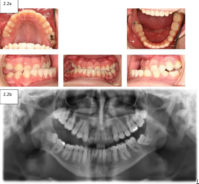
PTH1R (Figure 2.1 ((http://www.eurorad.org/eurorad/case.php?id=1463) and Figures 2.2a/b
((Frazier-Bowers et al 2013)). Based on the literature and our current findings, it is understood
that alterations in molecular pathways that underlie normal eruption can result in an eruption
disorder; and that early detection and management of these situations provides the best
chance at a successful treatment outcome.1, 2 Inaccurate diagnosis will most certainly lead to
suboptimal treatment choices, which ultimately hold negative consequences for the patient.
The remaining question of how an understanding of the mechanism that leads to PFE can
potentially improve diagnosis and treatment and these studies present a logical first step to
accomplishing this.
PTH/PTHrP and Dental Eruption
In order to elucidate the function of PTH1R, it is imperative to understand the
behaviors of its ligands. This is especially important in the inquiry of PFE and PTH1R because
while more is understood about PTH1R and its activators in the skeletal and endocrine systems,
less is known about its behavior in the dental tissues. PTHrP has been shown to be integral in
the process of tooth eruption by its presence and activity in the enamel organ, specifically the
stellate reticulum of the dental follicle.25 Studies have shown that the absence of PTHrP
production of the dental follicle cells (via conditional-PTHrP knockout mice), initially normally
developing teeth eventually become impacted and encapsulated by a bony crypt. This is due to
the failure of the formation of an eruption pathway, and fusion of the cementum to the
alveolar bone (ankylosis).25 It has been demonstrated that the cAMP/PKA pathway is largely
responsible for the varying effects of PTHrP and PTH regulation of mineralized tissues.28 Due to
supported that the signaling effect of PTHrP or PTH relies on the net outcome of cell’s
integration into multiple signaling pathways. This explains why the activation of the Gs
protein/cAMP/PKA pathway can result via PThrP or PTH, with each resulting in different
downstream pathways, yet yielding a constitutively singular result. When concerning the dental
unit, activation of the cAMP/PKA pathway (via Gs protein activation) by either ligand results in
the progression of tooth development and eruption- while the disruption of the same pathway
results in ankylosis (up-regulation of the biomineralization of cementoblasts) and a failure to
erupt-secondary to a blocked eruption pathway.10
Similarly, in the skeletal system, the targeted loss of PTH1R is accompanied by
impaired chondrocyte proliferation and accelerated maturation and calcification of
chondrocytes, which mimics the loss of Gs (activation of cAMP and the PKA pathway).10
Clinically, inactivating mutations in PTH1R results in hypocalcaemia and Bloomstrand’s
chondroplasia (OMIM ID: 215045). Constitutively activating mutations of PTH1R lead to
hypercalcemia and Jansen’s metaphyseal chondroplasia (OMIM ID: 156400). Loss of
parathyroid hormone (PTH) results in the aberrant formation of primary spongiosa of long bone
and defective mineralization.20 This is in contrast with PFE (OMIM ID: 125350), which has
numerous documented PTH1R mutations, yet it is isolated within the dental units, which
increases the intrigue of the interactions between PFE, PTH1R, and PTH/PTHrP. These findings
also point towards future studies involving further interrogation of PTH/PTHrP up/downstream
signaling pathways and specific functional assays.
We therefore seek to understand the functional consequence of specific mutations in
PFE result in a loss of function. We speculate that this loss of function could be connected to an
aberration in localization of the protein or G-binding. The studies presented in this report,
taken together, reveal that differences do exist between the expressive localization of the WT
versus mutant PTH1R. Further studies with specific binding assays will allow us to elucidate
more precise mechanistic flaws that lead to PFE.
Materials and Methods
Transformation and Plasmid Prep of E.Coli
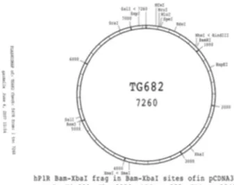
The pcDNA™3.1 (+) Mammalian Expression Vector (5.4kb) (Figure 2.3) was used to create
the construct containing the wild type human PTH1R sequence, named TG682 7260. TG682
7260 was generously donated from the Gardella laboratory (Massachusetts General Hospital,
Boston, Massachusetts) for the functional studies (Figure 2.4). Transformation of competent
Escherichia coli DH5α cells (purchased from Invitrogen) was completed for the TG682 7260.
Transformed E coli was grown to a logarithmic stage in LB broth (1.0% tryptone, 0.5% yeast
extract, and 1% NaCl) and LB agar (1.0% tryptone, 0.5% yeast extract, 1% NaCl and 1.5% agar)
medium were used. IPTG (100 mg/ml) and Ampicillin (100 mg/ml) for the preparation of agar
plates for growth, incubation was done at 37°C. After transforming the E. coli cells, 50 uL of the
cell suspension was plated overnight at 37°. A single clone was selected for mid-scale plasmid
preparation (NucleoBond® Xtra Midi, Macherey-Nagel GmbH & Co. KG), and was performed as
per the provided protocol.All media was prepared according to the manufacturer’s
Tissue Culture
The COS7 cell line was generously donated by Dr. Sompop Bencharit and cultured in
Dulbecco's modified Eagle's medium supplemented with 10% fetal calf serum and 100 U/ml
penicillin, and 100 µg/ml streptomycin. The cells were then typsinized and re-plated on 8-well
glass chamber slides in preparation for transient transfection.
Site-Directed Mutagenesis
Site-Directed Mutagenesis was performed (Genscript ©) on the TG682 WT PTH1R vector
to create the 1092delGPTH1R construct (Figures 2.5a and 2.5b). The mutated 1092delG vector
was then transfected into COS7 cells for comparison with the WT PTH1R.
Transfection of COS7 Cells
Exponentially growing COS7 cells were plated on a glass 8-well chamber slide.
Transfection was performed using FUGENE 6™ (Roche Applied Science) and Immunoflourence
assay (IFA) studies were performed according to standard protocols. Briefly, cells were grown
on a glass chamber slide, and grown to a density of 70% confluence prior to adding the DNA
mixture, including 0.1 mg/ml of the vector containing the WT-PTH1R or mutant insert. The cells
weretransfected with either: the vector containing the human functional mutation (1092del G),
or the wildtype PTH1R insert. The polyclonal anti-PTH1R antibody used for IFA was purchased
from Abcam ©, and the secondary FITC 546 goat anti-rabbit antibody (Life Technologies ©,
Thermo Fischer Brand Inc.) was generously donated from the Webster-Cyriaque laboratory.
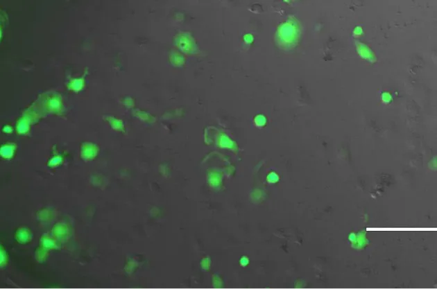
Transfection of COS7 cells with the pcDNA3.0 (+GFP insert) for 48 hours was completed as an
approximation of transfection efficiency. The transfection efficiency was estimated to be 70%
IFA Studies
Initial imaging of the WT PTH1R cells under a confocal microscope, illustrated that fact
that transport vesicles were of the most notable prominence, with little evidence of cell
membrane localization (Figure 2.7). This supported the selection of a 48-hours post transfection
standard to evaluate WT vs Mutant PTH1R localization to the cell membrane.
After 24 and 48 hours, the transfected COS7 cells were fixed for 15 minutes with -20 °C 80%
Acetone in PBS at 25°C. Nonspecific binding was blocked by incubation of the COS7 cells for 30
minutes in 1% albumin in PBS. The cells were incubated in primary FITC goat anti-rabbit
antibody for 10 hours, washed, then incubated with secondary AlexaFluor 546 (Life
Technologies, Grand Island, NY)) for 1 hour in the dark. The nuclei were stained with Hoechst
33342 DNA-dye at 1/10,000 for 1 min (Life Technologies, Grand Island, NY). After a final wash
within the chamber slides, cells were imaged using an inverted digital, wide-view, and confocal
microscope. Preparation for publication was completed using Photoshop® (Adobe) software as
described previously (Dibble, Bencharit et al, 2010).
In silico Analysis
In silico analysis was completed using software from Phyre ™ software11, followed by
PyMol ™ software for imaging (The PyMOL Molecular Graphics System, Version 1.7.4
Schrödinger, LLC.). This form of prediction analysis uses the known structure of a given protein
based on protein X-ray crystallography.11 The protein sequence containing the altered
sequence of interest was interrogated by the software that uses previously scanned protein
interactions to fabricate a best-fit-model. The resulting prediction was provided in the form of a
3D image of a protein.11
Results
Phenotypic presentation of 1092delG and 966_997insC PTH1R mutations
The 1092delG mutation,which resulted in a frameshift and truncated PTH1R protein, was
selected for the functional studies described here for many reasons (Figure 2.10). From a
clinical standpoint, this novel mutation was associated with the clinical finding of infraoccluded
primary teeth, and was identified in a small nuclear family (N=2). The non-syndromic index
case was a 7 years, 9 months old male who exhibited PFE affection of the permanent and
primary teeth in the form of a right lateral posterior open bite, mild Class lll skeletal
malocclusion, and an early-onset dilaceration of a PFE-affected 1st permanent molar (Figures
2.11a and 2.11b (Rhoads et al, 2013).29 Another mutation, the previously identified
966_997insCPTH1R mutation, was also selected to model the subsequent protein structure
(Figure 2.12 (Rhoads et al, 2013). Similar to the 1092delG, another mutation, 996_997insC, was
selected because of affection of the primary dentition with early-stage dilacerations of the
PFE-affected 1st permanent molar. 12 This frameshift mutation segregated in a family as Type I PFE in
affected individuals. Of those affected, two presented with one-jaw only patterning, and two
with a bilateral maxillary and mandibular pattern. Unaffected individuals in this family (n=5) did
not carry the 996_997insC mutation and did not reveal any signs of the dental eruption
phenotype, indicating an autosomal dominant inheritance with complete penetrance.12
Notably, systemic and/or skeletal problems were detected in this family. Five individuals
back and/or hip region around the second or third decade of life (Table 2.1).12 The functional
alterations identified in our study were not refined enough to decipher how the development
of osteoarthritis progression is related to the mutational pathology.
Variation in WT vs 1092delG PTH1R protein localization using IFA
There were observed differences between the expressionof the WT and Mutant PTH1R as
visualized by IFA. Although these differences were not specifically quantified, it was noted that
the WT PTH1R (Figures 2.8a, b, and c) bound and concentrated itself at the membrane in higher
levels than that of the mutant (Figure 2.9). This suggests that the mutant’s ability to fully bind
to the cell membrane is diminished due to the mutation. The intensity of the WT stain was
more pronounced, and the transport vesicles appeared more vibrant and concentrated, in
comparison to the mutant.
Genotype: Protein Sequence (1092delG and 996_997insCPTH1R mutations)
The 1092delGPTH1R mutation results in an a altered amino acid sequence extending
from the transmembrane-5 (TM5) loop through transmembrane-7 (TM7), before shifting back
into frame near the beginning of the c-terminus. The number of amino acids of the 1092delG
protein is approximately 81 amino acids (AAs) short of the WT, although the c-terminus is
conserved. The 996_997insC mutation resulted in a frameshift mutation that affected the
regions of the transmembrane-4 (TM4) and TM5 loops. The protein was truncated; however its
c-terminus was conserved. Most importantly, both of the aforementioned mutations alter the
amino acids that constitute the 3rd intracellular loop, which are central to G-protein interaction.
These important amino acids are Val 384, Leu385, Thr387, and Lys388.3 Taken together, these
sites for use in more specific, g-protein activation pathway interrogation. Figures 2.13, 2.14, and
2.15 illustrate the genotype: protein domain correlations, current database of PTH1R DNA:
protein mutations, and a graphical depiction of the mutated regions of the PTH1R protein.
In silico Predictions of WT PTH1R, 1092delG, and other functional PTH1R mutations
We submitted WT and mutated protein sequences identified in our lab (1092delG,
1353-1 G>A, 966_997insC, 572delA) into Phyre2 ™ and visualized the resultant proteins via PyMol ™
(Figures 2.16a-e). All mutated predictions resulted in an altered, truncated protein with a slight
alteration of the transmembrane c-terminus. However, within our current database of
functional PTH1R mutations, the 1092delG mutation is the only one that caused a downstream
alteration within the 6th TM loop. Additionally, while the c-terminus is conserved based on the
amino acid sequence, the c-terminus of the 1092delG protein is truncated and altered to the
greatest extent (Figure 2.15b). This is in comparison to the 996_997insCPTH1R mutation that
led to a downstream alteration in the 3rd intracellular loop, and had a moderately altered
c-terminus in comparison to the collection of current PTH1R mutations. All of the predictions
yielded a reported confidence of 100%.
Discussion
Reduction of Mutant PTH1R Cell Surface Presence
While the potential alteration in G-protein coupling is a compelling argument, the
functional study demonstrated that the mutated protein is less localized in the cell membrane
as compared to the WT. This reduction in cell membrane localization supports the postulation
that part of the consequence of the 1092delG mutation is the reduction of cell membrane
PTH/PTHrP. Future studies that interrogate downstream effectors can further confirm whether
there is an actual defect in the signal transduction pathway. An important place to begin such
queries would be the enzymes critical for the G-protein signaling cascades (ie, AC, PLC, PKC, and
PKA).
In silico Analysis
Computer modeling is a useful tool in the intellectual journey of improving our
understanding of the molecular and structural consequence of PTH1R mutations, however, it
has its limitations. In silico analysis, by its very nature, is a prediction of structure. It is a
structural interpretation based on a limited database, albeit a very educated one. The reliability
of an in silico analysis is centered on the reliability of its database, however, the absolute
validity of our prediction results are strengthened by our functional studies. Indeed, the gestalt
of our in silico analysis reveals differences in the protein structure that appear consistent with
the location of the mutation but more importantly these studies can be used to support further
and more pointed inquiries into the results of a PTH1R mutation.
PTH1R Mutation Potentially Affects G-Protein Binding
The in silico protein prediction of the WT v. the 1092delG, 1353-1 G>A, 966_997insC,
572delA predictions, resulted in truncated protein mutants with an altered intracellular
c-terminus. However, the most remarkable of the protein predictions is the structural alteration
of the 1092delG and 996_997insC PTH1R mutations. Although the c-terminus was conserved in
both cases, each mutation led to downstream alterations that spanned key locations within the
TM or intracellular loops; with each region being highly relevant for normal g-protein binding
intracellular loop, and the TM6 loop creates an altered G-protein binding pattern. Furthermore,
the fact that both of the identified mutations affected the very 4 amino acids critical for
efficient G-protein coupling increases the probability of functional disruption. This is
substantiated, in part, by our understanding that PTH1R can bind to different G-proteins, some
of which, have antagonistic effects within the somatic and cell system.28 It is plausible that the
TM, intracellular loop, and c-terminus alterations caused the PTH1R protein to bind to a
G-protein that 1) has greater antagonistic activity towards the eruption process; 2) did not allow
the receptor to bind to a protein at all, or 3) lead to an altered up or downstream regulatory
pathway.
Primary PFE Tooth Affection, 1092delG and 996_997insC
The observation that both mutations represent unique cases in our database with PFE
affection of primary teeth and the early stages of permanent 1st molar dilaceration is
significant. These mutations are associated with the infraocclusion of primary teeth, and based
on knowledge of the sequence they are the only mutations that affect key G-protein binding
sites within locations of PTH1R. Therefore, isolating G-protein uncoupling occurring within
these mutations could help identify the signaling pathway responsible for an integral part of
eruption. Moreover, the affection of primary teeth potentially represents a more extreme
manifestation of PTH1R malfunction; isolating the pathway of primary tooth eruption failure
will aid in elucidating the pathway to permanent tooth eruption and eruption failure.
G-Protein Conformational Change Alterations and Ligand Binding
Since two mutations clearly affected TM3 and TM6 loops as discussed above, we
their altered G-protein affinity and basal signaling.3 Alterations in PTH1R conformation can also
result in varying affinities for different ligands. Furthermore, PTH1R exists in two different
G-protein related conformations: 1) RG, in which the receptor is bound to it G-G-protein, and 2) Ro,
in which the receptor is not coupled to a G-protein.28 PTH and PTHrP have been shown to bind
similarly to the G-protein coupled PTH1R conformations, however PTH has a much greater
binding affinity, than PTHrP in relation to the uncoupled G-protein PTH1R conformation. This
confers that when PTH1R is in its Ro conformation, PTH has much greater signal production
capability than PTHrP.33 Depending on the GPCR conformation, PTH and PTHrP will have
different initial binding affinities to the receptor, as well as varying stability during its existence
as the ligand-receptor complex. This concept is important to ponder when evaluating the
potential effects of the 1092delG mutation and can help explain the different modes of ligand
activity (endocrine v. paracrine). For instance, if the conformation of the receptor, due to the
frameshift mutation, creates an inability able to bind effectively to the G-protein or the
required ligand- a breakdown in the classical pattern of PTH1R function may be imminent.28
Dimeric Oligomerization Activation/Deactivation of the Homodimeric PTH1R Complex
In the 1092delG mutation we can consider the findings of Xu et al. In their study, it was
determined that the crystal structure of the ligand-free PTH1R extracellular domain (ECD) forms
an α-helix, which mimicks PTH/PTHrP by effectively occupying the peptide binding groove of its
opposing protomer.27 PTH binding was shown to disrupt receptor oligomerization via functional
studies, as predicted by their model and a receptor reduced to a monomeric state by mutations
in the ECD retained wild-type PTH binding and cAMP signaling ability. PTH1R therefore forms a
monomeric PTH1R is able to couple with, and activate, a G-protein.27 These findings are highly
relevant to the results of our functional studies which showed that the diminished activity of
one mutant PTH1R copy is likely tied to a decrease in G-protein activation. This also aligns with
the varying degrees and types of PFE presentation because we suspect that if the overall
activation of the coupled G-protein is attenuated, it is possible that the threshold for normal
eruptive function is not met- which can result in the apparent temporal and spatial variations of
eruption failure.
Future Studies
The functional studies described in this paper suggest that the mechanism of the clinical
affection is likely partially due to the aberrant expression or localization of the PTH1R protein,
but also represents a situation where a potentially large percentage of the mutation’s affect is
loss of function; such as altered ligand binding or alterations in G-protein coupling. Based on
these results we predict that this functional defect in the protein may act to disrupt the
membrane binding, intracellular signaling, and G-protein binding; however, future studies are
needed to interrogate downstream targets.
The use of bioluminescence (BRET) and fluorescence resonance energy transfer (FRET)
techniques that would enable us to investigate the receptor oligomerization and interaction in
living cells is one way to extend our interrogation. Reducing the amount of DNA used to
transfect the COS7 cells in order to produce increased contrast of protein expression will
provide a clearer image of localization. Additionally, the use of more specific confocal imaging,
RT-PCR, specific binding assays, and quantitative Western blots are additional potential
Conclusions
1. The 1092delG PTH1R mutation results in the reduction of the protein’s ability to localize and
bind to the cell membrane.
2. Identified amino acid alterations in the PTH1R transmembrane region, specifically the 3rd
intracellular loop, affects the functional aspect of protein function.
3. Two (2) functional mutations affected TM6 and the third intracellular loops are specifically
related to the eruption failure of primary teeth.
4. Further studies are needed to determine the functional effect of a PTH1R mutation, such as a
RT-PCR, fractionation, a quantitative western blot, functional binding assays, bioluminescence
(BRET), fluorescence resonance energy transfer (FRET) techniques, and transfection at a lower
CHAPTER TWO
MUTATIONAL ANALYSIS OF TNFSF-11 AND BMP2 IN PFE AFFECTED PATIENTS Introduction
The presentation of eruption disorders among the human population presents the
dental practitioner with a plethora of differential diagnoses, connected to a diverse set of
phenotypes. Primary Failure of Eruption (PFE) (OMIM ID: 125350) was first described by Proffit
and Vig, and is defined as a failure of the eruption mechanism itself7, which cannot be
explained by a syndrome or a mechanical interference.8 A hallmark of PFE is that these teeth do
not respond favorably to orthodontic traction and, in fact, attempted orthodontic treatment
often results in a worsened malocclusion and increased open bite due to intrusion of adjacent
teeth.1, 2, 9 PFE clinically presents as the infra-occlusion of affected teeth, resulting in a
posterior open bite malocclusion (Figure 3.1 (Frazier Bowers et al, 2009)). Typically, all teeth
distal to the most mesially affected tooth also fail to erupt.9 The diagnosis of PFE is critical as it
dictates that treatment with continuous archwires should be avoided. Some successful
treatment has been reported by multiple individual tooth osteotomies or selective individual
tooth extractions followed by implant restorations to restore a functional occlusion.8 The
PTH1R and PFE
Our current understanding of eruption disorders has been strengthened by human
genetic studies which have highlighted mutations in parathyroid hormone receptor 1 (PTH1R)
as a causative factor for familial cases of PFE.1, 2, 8, 9 A study of nine family members revealed
PTH1R as an autosomal dominant mutation associated with a PFE phenotype. All family
members with PFE had a mutation in the PTH1R gene in this study, while those without PFE
lacked this mutation.8PTH1R mutations associated with PFE have in silico predictions that
result in the formation of truncated proteins with a somewhat conserved c-terminus.
Haploinsufficiency has been suggested to be part of the underlying cause of PFE, in which
insufficient amounts of functional receptors are formed from the unaffected allele.
Non-syndromic PFE patients do not exhibit any peripheral signs of the disease, and it may be
hypothesized that this mutation causes a disruption confined to alveolar bone in the
epithelial-to-mesenchymal (EMT) crosstalk signaling pathways that are necessary for normal bone
resorption and apposition in tooth eruption.8, 9 Taken together, this implicates genetic
mutations in PTH1R as diagnostic and causative of PFE and is important in the context that
many patients diagnosed with PFE and confirmed by the presence of a mutation in PTH1R were
initially misdiagnosed with ankylosis.1, 2 Both PFE and ankylosis preferentially affect molars and
posterior teeth making them even more difficult to distinguish from one another.2
PFE: A Diagnostic Challenge
The contrast between ankylosis and PFE are just examples of the complexity and
treatment, rely heavily on accurate diagnosis and treatment planning. The fact that we have a
method to genetically and clinically diagnose individual disorders is a promising new frontier in
medicine, due to the fact that the connection between the etiology and the clinical
presentation of patients are not always as obvious. This can be seen specifically, in one family,
in which five members carried the same mutation in PTH1R, but two affected individuals
carrying this mutation were diagnosed incorrectly with ankylosis through percussion testing
(see Figure 3.2).8 This scenario further reveals a gap in the knowledge and points to the
difficulty in clinically distinguishing PFE from other eruption disorders (i.e. ankylosis or
mechanical failure of eruption - MFE). PFE is a poorly understood problem of tooth eruption
that with more investigation will improve our understanding of the biology of normal tooth
eruption, but also the diagnosis of eruption failure.
Mutational Analysis Studies of PTH1R
The foundation for the mutational studies described herein was based on the database
of functional and non-functional PTH1R mutations previously identified and described in the
Frazier-Bowers’ Lab.12 Studies in this laboratory revealed that while six of twelve families with
PFE segregate mutations in PTH1R, the remaining cohort lack functional mutations in PTH1R,
further suggesting other gene(s) as causative of PFE.12,18,29 These findings also revealed a novel
inheritance pattern in PFE, incomplete penetrance and therefore forms the basis for an
Phenotypic Analysis of PFE
PFE has been analyzed from a genetic and clinical perspective. Clinical analysis of
photos and radiographs evaluated the following: jaw-affection, bilateral or unilateral affection,
super- or infraosseous tooth position, and the clinical Type of PFE. Broadly, PFE is divided into
two subtypes, Type 1 and Type 2. Type 1 PFE presents as a progressive, anterior-posterior
lateral openbite that worsens distal to the most mesially positioned PFE-affected tooth; Type 2
PFE presents with a more varied pattern of infra-eruption distal to the most mesially affected
tooth.17 The affected members in each family group had varied clinical PFE presentations. This
variation was noted as both between and within familial segregating mutations. Of those
families with functional mutations, four families were classified as Type I PFE, while the other
two families were classified as either Type II or having a mixture of Type I and Type II
presentations. All of the affected individuals who provided records were found to have an
eruption failure that manifested during the supraosseous phase (i.e. the teeth had emerged
partially through the bone), and unilateral versus bilateral eruption failure (i.e. manifesting on
one side of the dental arches) occurred equally.
The Interaction of BMP2, TNFSF-11, and PTHrP in Tooth Eruption
PTHrP has been shown to be integral to tooth eruption through the observation of
increased concentration of PTHrP levels in the dental follicle and stellate reticulum at the
initiation of eruption.5 The coronal region of the dental follicle regulates osteoclastogenesis and
the apical region is responsible for bone formation. CFS-1, localized in the superior region of the
dental follicle, up-regulates osteoclast precursor cells and down-regulates osteoprotegerin
the stellate reticulum cells in a paracrine manner, up-regulates TNFSF-11 expression in dental
follicle cells.5 This same hormone results in the production of vascular endothelial growth factor
(VEGF), which up-regulates the producing of RANK receptors on osteoclast precursors.5 Taken
together, this results in more available RANK receptors interacting with increased levels of
TNFSF-11, leading to osteoclastogenesis and eruption pathway clearance. The apical region of
the dental follicle has a relatively higher concentration of BMP2, which is responsible for
alveolar bone formation via osteoblasts. PTHrP causes the apical dental follicle cells to
up-regulate BMP2 production.5 Taken together, it was determined that BMP2 and TNFSF-11 were
plausible candidate genes to investigate in terms for their potential role in PFE and eruption.
Ingenuity Pathway Analysis (IPA)
Ingenuity Pathway Analysis (IPA) was performed (Figure 3.3) to inform our understanding
of high-priority candidate genes for PFE based on interactions with PTH1R. IPA revealed that
multiple genes involved in craniofacial development interact with PTH1R (e.g. PTHRP, TNFSF-11,
TGFB1, BMP2, BMP6). As a result of this analysis and based on the literature, we chose to
interrogate the role of BMP2 (20p12.3) and TNFSF-11 (13q14.11) (hRANKL) in the development
of PFE. Our rationale to study these particular candidate genes was strengthened by the
intimate connection between the periodontal ligament (PDL), TNFSF-11, PTH1R, and
PTHrP/PTH.28
Materials and Methods
Ascertainment of Families and Diagnosis
Approval for this study was granted by the Biomedical Institutional Review Board (IRB)
in the case of minors, provided their consent to participate in this study. Typically, the index
case was identified through a referring orthodontist. Fifteen cases were selected for analysis
of BMP2 and TNFSF-11 based on the negative results of these probands following sequencing of
the PTH1R gene. Probands lacking functional mutations in the PTH1R gene, despite clinical
signs of PFE, ranged in age from 6 – 68 years. Previously collected phenotypic data was
analyzed for characterization of non-functional genetic alterations. All probands (N=15) had
pre-treatment clinical photographs, panoramic and cephalometric radiographs following the
initial clinical assessment, as described below.
A positive PFE phenotype diagnosis was made based on at least one infraoccluded first
molar using clinical data (ie radiographs, and/or examination at minimum). A clinical interview
was completed for each affected individual and/or their family members to determine general
health status and the elimination of any syndromic patients. Index cases (individuals of interest)
were identified based on their affection with PFE, as determined from radiographic and clinical
presentation, as well as their lack of a functional PTH1R mutation. DNA was extracted using
Oragene Salivary Kit and using the manufacturer’s protocol.
Phenotypic Analysis
Clinical (phenotypic) information was collected and reviewed for the individuals
harboring a nonfunctional PTH1R, TNFSF-11, or BMP2 mutation. The records assessed included
a minimum of a panoramic radiograph and intraoral clinical photographs. The following
information was gathered:
1) Unilateral or bilateral presentation of infraoccluded teeth
3) Presence or absence of any other abnormal or noteworthy findings, including the
significant involvement of primary teeth
Additionally, the type of PFE, Type I or II (determined by the degree of eruption of the second
molars) was recorded.
The classification of PFE was recorded as either Type I or II, as previously described in
the literature.2, 17 These types are distinguished based on the timing of onset. Briefly, Type 1
PFE is characterized by a progressive posterior open bite, in which all teeth distal to the most
mesial infra-occluded tooth are affected and do not erupt into occlusion. Type 2 PFE exhibits
greater eruption potential, although still inadequate, for the more distal teeth, such as second
molars.
A phenotypic analysis was completed for each individual that was clinically diagnosed
with PFE in our cohort of inquiry. With the use of clinical photographs and radiographs, PFE
affection was described as either unilateral or bilateral, and PFE pattern was described as either
Type 1 or Type 2. All of the current PFE cases within this cohort are of European decent.
Mutational Analysis of BMP2 and TNFSF-11
Mutational analysis was performed following extraction and purification of DNA from
saliva (Oragene, DNA Genotek, Toronto, Canada) for all individuals in this study. We amplified
and sequenced all coding exons of BMP2 (3 exons- only 2 coding exons) and TNFSF-11 (4
exons), included identifying common synonymous SNPs and non-synonymous (deleterious)
mutations. Sequencing was performed on 15 probands using designed primer sets. To include
splice junctions in our analysis, primer sets were designed to delineate regions that included a
performed using HotStart polymerase chain reaction (PCR) buffer and enzyme mix (Life
Technologies/Invitrogen, Bethesda, MD) under the following conditions: 10 min 95°C
activation/premelt, followed by 35 cycles of 30 s at 94°C melt, 30 s at 60°C anneal, and 3 min of
72°C extension. PCR products were purified using ExoSaplt (USB, Cleveland OH), and sequenced
at the University of North Carolina at Chapel Hill Genome Analysis Core facility. Sequences were
compared to wild type TNFSF-11 and BMP2 (accession NM_003701 and accession
NC_000020.10) from Genbank release GRCh37 using the BLAST algorithm.
Ingenuity Pathway Analysis (IPA)
Based on Ingenuity Pathway Analyses (Ingenuity Systems, Inc.) several candidate genes
have documented interactions with one another. The interactions that are presented in
connection with an IPA are varied and vast. Therefore, it was determined to focus primarily on
TNFSF-11 and BMP2 due to the strong association with PTH1R in the pathway analysis, as well
as their involvement with the eruption process.
Results
Mutational Analysis
Analysis of both BMP2 and TNFSF-11 candidate genes in PFE-affected patients who did
not carry a mutation in PTH1R (N=15) resulted in the identification of 3 intronic SNPs in
TNFSF-11, without any compelling genetic alterations in BMP2, to-date. Two SNPs were located within
Exon 5 and one was located within Exon 8.
The intronic SNPs in TNFSF-11 have been previously documented in NCBI: c.387+14G>A
(21% minor allele frequency), c.533-34T>A (3.4% minor allele frequency), and c.220-75T>G
(reported by NCBI - dbSNP) is the minor allele frequency for each SNP ID in a default global
population. Global MAF distinguishes common polymorphism from rare variants. The MAF is
actually the second most frequently occurring allele value. The current default global
population is generated from the 1000 Genomes phase 1 genotype data, which has a
collection of 1094 worldwide individuals (released in the May 2011) (NCBI). The relatively rare
allele of at least 2 SNPs identified in association with PFE, c.533-34T>A (3.4% minor allele
frequency), and c.220-75T>G (.042% MAF) suggest the possibility of a functional role of these
SNPS that do not represent common variants.
Phenotypic Analysis
After a comprehensive analysis of index cases, (Table 3.1) it was determined that 75% of
reported TNFSF-11 SNP-containing genotypes of sequenced individuals presented with a
bilateral, 2-jaw, supraosseous affection; of which both the premolars and molars were affected.
A total of 75% of PFE affected individuals presented with a Type I pattern, and the remaining
with a Type II pattern. However, only 50% of the bilaterally affected side had a Type I
presentation. Out of our cohort of PFE affected, non-functional alterations; the majority (78%)
presented with a bilateral affection, and 56% of individuals presented with Type 1 PFE (Chart
3.1). It was also noted, that no particular trend in affection appears to sequester with any SNP in
particular (Chart 3.2 and 3.3).
Discussion
BMP2 and TNSFS-11 were prioritized for this project because BMP2 is highly active in the
basal region of the dental follicle and is responsible for root formation and apical bone