Pathogenic
Role
of
Type
I
Interferons
in
HIV-Induced
Immune
Impairments
in
Humanized
Mice
LishanSu1
Publishedonline:4May2019
Abstract
PurposeofReview Recentfindingsonthecriticalpathogenicroleoftype1interferons(IFN-I)inHIV-1persistenceinhumanized micesuggestthatinhibitingIFN-IsignalingtransientlywillreverseHIV-induced inflammatorydiseasesandrescueanti-HIV immunitytocontrolHIV-1reservoirs.
RecentFindingsInbothhumanizedmiceandinmonkeys,IFN-Isignalingisfunctionallydefinedtoplayanimportantrolein suppressingearlyHIV-1andSIVinfection.Duringpersistentinfectioninhumanizedmice,however,IFN-Isignalingisrevealed toinduceTcelldepletionandimpairment.Interestingly,inHIV-infectedmicewitheffectivecombinationantiretroviraltherapy (cART),blockingIFN-IsignalingreversesHIV-inducedinflammation,rescuesanti-HIVTcells,andreducesHIV-1reservoirs.
SummaryThesefindingsfunctionallydefinetheroleofIFN-IinHIV-1reservoirpersistenceandsuggestthatblockingIFN-I signalingwillprovideanoveltherapeuticstrategyto(i)reverseinflammation-associateddiseasesinHIVpatientsundercART, (ii)rescuehostanti-HIVimmunity,and(iii)reduceorcontrolHIV-1reservoirs.
Keywords IFNAR.ISG.pDC.Inflammation.T cellexhaustion .HIV-1reservoirs
Introduction
HIV-1 persistence is associated with hyper-inflammatory ac-tivation [1]. Despite efficient suppression of HIV-1 replication and increased survival with highly active or combination an-tiretroviral therapy (HAART or cART, respectively), HIV-1 rebounds in all patients post-cART cessation due to the “cART-resistant” viral reservoir (latent or low-replicating HIV infection) in lymphoid tissues [2,3]. In addition, some cART-treated patients with effective HIV-1 suppression fail to reverse hyper-inflammatory or hyper-immune activation and achieve full immune recovery [1]. The mechanism underlying those“immune non-responder (INR)”patients remains un-clear. Although type 1 interferons (IFN-I) are reduced under ART [4], low levels of IFN-I persist and IFN-stimulated-genes
(ISGs) are still upregulated in peripheral blood cells or lym-phoid organs [5,6], which may contribute to increased clinical complications and mortality in cART/HIV-1 patients [1]. Persistent hyper-inflammation has also been associated with pathogenesis in non-human primates (NHP) with SIV infec-tion, but the underlying cellular and molecular effectors re-main elusive. Strong correlations have been established be-tween persistently activated IFN signaling with HIV-1 [7] or SIV disease progression [8,9]. First, IFN and ISG responses persist in HIV-1 infection and pathogenic SIV infections in Asian macaque species, but resolve to baseline in non-pathogenic SIV infections of African monkeys [10, 11]. Second, HIV-infected patients that do not exhibit disease de-spite high plasma virus have paradoxically low levels of ISG expression [12]. Therefore, there is a strong correlation be-tween HIV-1 pathogenesis and IFN-signaling gene signature. Due to the limitation of human studies, however, the function-al role of IFN-I in HIV-1 is not clearly defined. To functionfunction-ally define the role of IFN-I in HIV-1 persistence and pathogene-sis, several recent studies have been reported in HIV-1-infected human patients, and in SIV-HIV-1-infected NHP models. In earlier studies, administration of recombinant IFN-I showed little or no beneficial effects in HIV-1 patients [13–15]. In fact, it may have accelerated HIV-associated
* Lishan Su [email protected]
immunological diseases in those HIV-1 patients treated with IFN-I [16–18]. Consistently, recent studies with peg-IFN in HIV-1 patients under HAART showed unclear or minimal effect on the persistence of HIV-1 reservoirs during HAART [19–21], but enhanced HIV-1-associated CD4 depletion [19], although lower HIV-1 replication was detected in the IFNα -treated group after stopping HAART [19]. In SIV-infected monkeys under cART, pegylated-IFN has shown no effect on SIV replication or T cell function [22].
Several recent reports have attempted to define the role of IFN-I signaling in SIV-infected NHP models, by modulating IFN-I activities before and during SIV infection. Using a re-combinant human IFN-1ant that binds IFNAR2 but not IFNAR1 (thus antagonistic to wild type human IFN-I [23]), blocking IFN-I signaling prior to and during acute SIV infec-tion in monkeys elevated SIV replicainfec-tion and accelerated AIDS progression, confirming an important role of IFN-I in controlling early SIV infection [24]. In contrast, administra-tion of IFN-α2a initially upregulated expression of antiviral genes and prevented systemic infection. However, prolonged IFN-α2a treatment induced IFN-I desensitization, increased SIV infection, and accelerated disease progression. Thus, ear-ly IFN-I signaling during acute SIV infection is critical to suppress SIV replication, but its persistence may be detrimen-tal and accelerate SIV disease progression. In a similar study, an antibody (AGS-009) that neutralizes 11/13 of IFN-α sub-types was infused 1 day prior to SIV infection. No obvious effect on ISG expression was detected, but high-dose AGS-009 treatment induced a slight increase in acute-phase viral replication. Early blockade of IFN-αduring acute infection, interestingly, decreased the level of activated CD4+ and CD8+ T cells during chronic infection phase, but accelerated progression to AIDS [25]. This study again indicates that IFN-I signaling during acute SIFN-IV infection plays a critical role to modulate SIV disease progression. One caveat in this study is that AGS-009 only neutralizes 11 of 13 IFN-αsubtypes, not other IFN-I types including two IFNαsubtypes and IFNβ.
When administered during chronic SIV infection, IFN-1ant significantly reduced expression of ISGs, but showed no signif-icant effect on SIV replication or SIV-induced inflammatory cytokines [26]. In ART-suppressed chronically SIV-infected an-imals, IFN-1ant only marginally inhibited the low ISG expres-sion, and showed no effect on SIV infection [26]. In addition, IFN-I blockade showed no effect on T cell activation and ex-haustion markers, or any adverse effect on the host. The conclu-sion from this study is weakened by the fact that the recombi-nant IFN-1ant, which binds IFNAR2 but not IFNAR1, still has some low IFN-I activity to induce antiviral ISGs in human cells, and is thus only partially antagonistic to wild type IFN-I [23]. Indeed, two doses of IFN-1ant in SIV/ART-treated animals re-sulted in reduced expression of ISGs, but three doses showed no such effect [26]. Its antagonistic effect is likely only obvious when endogenous IFN-I is high, but not in SIV/ART-treated
animals with persistent but low IFN-I signaling. A more com-plete blockade of all IFN-I signaling with IFNAR-blocking mAb is required to clearly define the role of IFN-I signaling during chronic SIV infection with and without ART.
Role of IFN-I Signaling in HIV-1 Infection,
Pathogenesis, and Therapy in Humanized
Mice
Humanized mice transplanted with human immune tissues or cells have served as robust models to study HIV-1 infection [27–29]. Humanized mice transplanted with HSC or both HSC and thymus fragments (hu-HSC, hu-HSC/TEC [30], or BLT [31] mice) have enabled investigation of HIV-1 persis-tence and human immune responses to HIV-1 infection, as well as human immunology and immunopathology [32,33].
Humanized mice have been used to functionally define the role of Treg [34,35] and of plasmacytoid dendritic cell (pDC) [36–40] in HIV immuno-pathogenesis; as well as in human vaccine evaluation [41–43]. Importantly, HIV-1 infection re-sults in immune activation, correlating with T cell depletion and functional impairment in lymphoid organs of humanized mice [40]. As in human patients, cART could efficiently in-hibit HIV-1 replication, but HIV rebounds rapidly post-cART discontinuation [44••,45–47]. Furthermore, humanized mice have been used to study HIV-1 latency [46,47] and HIV-1 therapy by broadly neutralizing antibodies (bnAb) [48,49], and by IFNAR-blocking mAb [44••,50,51••,52] for HIV-1 reservoir reduction or elimination. Therefore, humanized mice are proven relevant and robust to study HIV-1 persistence, pathogenesis, and therapy [32,33].
HIV-1 infection and pathogenesis in vivo, a monoclonal anti-body (mAb) that blocks IFNAR1, thus all IFN-I signaling in humanized mice, was developed. Similarly as pDC depletion, HIV-1 replication was significantly elevated when the anti-IFNAR1 antibody was used to completely block IFN-I signal-ing in humanized mice [44••,53•]. Therefore, IFN-I expression from pDCs plays a critical role to suppress acute phase HIV-1 replication in humanized mice.
Persistent IFN-I Signaling Contributes to HIV-1 Disease and Persistence In VivoWith depletion of pDCs during persistent HIV-1 infection in humanized mice, HIV-1 replication was also elevated in blood and lymphoid tissues. Consistent with higher viral loads, human T cells expressed higher levels of activation markers such as CD38 and HLA-DR. Surprisingly, depletion of pDCs could rescue human immune cells from HIV-induced depletion in the presence of increased HIV-1 replication [38,39]. When tested with the IFNAR1-blocking mAb during persistent HIV-1 infection, the HIV-1 replication level in the IFNAR1-blocked group was also enhanced, asso-ciated with complete suppression of ISG expression. Phenocopying the pDC depletion study, human T cells were rescued in number and functions in anti-IFNAR1 mAb-treated mice. Hence, blocking IFNAR1, like pDC depletion, also re-verses HIV-1 disease progression during persistent HIV-1 in-fection in humanized mice [44••,50,51••,52,53•]. Besides
human T cells, pDC depletion or IFNAR blockade also re-versed HIV-induced immunopathology in other cell types and organs including innate lymphoid cell type 3 (ILC3) and hematopoietic progenitor cells (HPCs) in the bone marrow [37, 38]. In an independent study, an IFNAR2-blocking mAb also achieved similar ISG inhibition or rescue of human T cells was observed in humanized mice [51••]. Distinct from
the IFNAR1-blocking mAb, however, the IFNAR2-blocking mAb reduced HIV-1 replication in humanized mice without ART. It is not clear if the difference on HIV-1 replication is due to the different antibodies that target IFNAR1 or IFNAR2. Other factors such as different HIV-1 isolates may also have contributed to the difference. Nonetheless, these two studies have independently concluded that persistent IFN-I signaling during chronic HIV-1 infection contributes critically to the depletion and impairment of human T cells in humanized mice. This is consistent with recent findings reporting that IFN-I plays a detrimental role during chronic LCMV infection and blocking IFN-I signaling by an IFNAR1 antibody could enhance antiviral immune response and lead to early clearance of LCMV infection [54,55].
Blocking IFNAR in Humanized Mice with HIV-1/cART Reverses T Cell Immune Exhaustion, Rescues Anti-HIV T Cell Activity, and Reduces HIV-1 Reservoirs HIV-1 infection in humanized mice can be effectively suppressed by combination ART with two RT inhibitors and one integrase inhibitor as in human
patients. As in HIV-1 patients, cART fails to fully suppress HIV-induced inflammation such as ISG expression. In addition, HIV-1 reservoirs are detected by cell-associated HIV-1 DNA or by virus outgrowth assays (VOA). When cART is stopped, HIV-1 rebounds rapidly to pre-cART levels in all animals [44••,46,47,52]. Therefore, humanized mice provide a highly relevant and robust model to study HIV-1 reservoir persistence, immunopathogenesis, and therapy. In humanized mice infected with HIV-1 and treated with cART drugs, IFNAR blockade fully reversed inflammation, rescued human T cells, and re-duced exhaustion markers such as PD1/TIM3 induction on CD8 T cells. Importantly, 3 weeks of IFNAR mAb treatment rescued anti-HIV T cell functions. Thus, effective cART led to recovery of human T cells but failed to reverse aberrant immune hyper-activation and T cell exhaustion, and inhibition of IFN-I signaling in combination with cART rescued human T cell functions [44••,50,51••,52,53•]. Consistently, both levels of
cell-associated HIV DNA and cells with infectious HIV-1 were reduced in mice treated with IFNAR-blocking antibodies [44••,
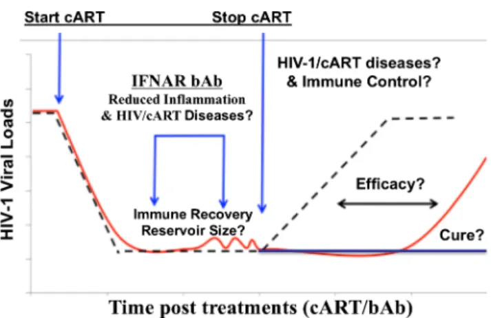
51••,52]. When cART was stopped, a significant delay of HIV-1 rebound was observed and the virus rebounded to a lower level [44••]. Therefore, IFNAR blockade provides a new prom-ising HIV-1 cure strategy to reduce or control HIV-1 reservoirs (Fig.1).
Conclusion and Perspectives
Recent findings in humanized mice have revealed the dual roles of IFN-I signaling in HIV-1 infection and pathogenesis:
it plays a critical role to suppress acute HIV-1 infection and to set the anti-viral program. During chronic infection, however, persistent IFN-I signaling contributes to HIV-associated hy-per-inflammation, immune exhaustion, and HIV-1 persis-tence. Intriguingly, blocking IFNAR in cART-treated human-ized mice rescued anti-HIV immunity and reduced HIV-1 res-ervoirs [44••,50,51••].
In ART-suppressed chronically SIV-infected animals, IFN-1ant showed no effect on SIV replication or on T cell activa-tion and exhausactiva-tion [26]. The pitfall of that study is that the recombinant IFN-1ant, which binds IFNAR2 but not IFNAR1, still possesses some IFN-I activity to induce antivi-ral ISGs in human cells [23]. Its antagonistic effect is likely only obvious when endogenous IFN-I is high, but not in SIV/ ART-treated animals with persistent but low IFN-I signaling. A more complete blockade of IFN-I signaling with IFNAR-blocking mAb, which blocks signaling of all type I inter-ferons, is required to clearly define the role of IFN-I during chronic SIV infection with and without ART.
It is important to point out the limitations of the humanized mouse models, including limited life span, incomplete human immune functions, and lymphoid organs. The relatively short life span of humanized mice will prohibit prolonged HIV-1 infection and cART; thus, the model may be limited to inves-tigation of HIV-1 reservoirs during short-term cART in vivo. The defects in human immune functions such as lymphoid structure and B cell IgG response, and low reconstitution in gut-associated lymphoid tissues, should be carefully consid-ered when interpreting experimental results for specific ques-tions. The findings in humanized mice with IFNAR-blocking mAb thus should be confirmed in the SIV/NHP models with and without cART. If verified, blocking IFN-I under cART will provide a novel strategy to treat HIV/cART-associated inflammatory diseases in HIV-1 patients, as well as to rescue anti-HIV T cells to control HIV-1 reservoirs (Fig. 1). Regarding the safety of transiently blocking IFN-I signaling in humans, clinical trials in lupus patients have shown that treatment with an IFNAR1 antibody in healthy people for 84 days or in lupus patients for 48 weeks is clinically safe in human subjects [56,57].
Acknowledgements The author acknowledges the contributions of the current and former Su laboratory members.
Funding Information The author’s relevant research has been supported by grants from NIH (AI127346 and AI134631) and by the Lineberger Comprehensive Cancer Center at UNC-Chapel Hill.
Compliance with Ethics Guidelines
Conflict of Interest Dr. Su reports grants from NIH (AI127346 and AI134631). In addition, UNC/Dr. Su has filed a patent (Modulation of Type I Interferons to Reactivate HIV-1 Reservoir and Enhance HIV-1 Treatment, pending).
Human and Animal Rights and Informed Consent This article does not contain any studies with human or animal subjects performed by the author.
References
Papers of particular interest, published recently, have been highlighted as:
• Of importance ••Of major importance
1. Deeks SG. HIV infection, inflammation, immunosenescence, and aging. Annu Rev Med. 2011;62:141–55.
2. Ho YC, Shan L, Hosmane NN, Wang J, Laskey SB, Rosenbloom DI, et al. Replication-competent noninduced proviruses in the latent reservoir increase barrier to HIV-1 cure. Cell. 2013;155:540–51. 3. Lorenzo-Redondo R, Fryer HR, Bedford T, Kim EY, Archer J,
Kosakovsky Pond SL, et al. Persistent HIV-1 replication maintains the tissue reservoir during therapy. Nature. 2016;530:51–6. 4. Buimovici-Klein E, Lange M, Sonnabend J. Decline of endogenous
alpha-interferon with zidovudine. Lancet. 1992;339:1123. 5. Dunham RM, Vujkovic-Cvijin I, Yukl SA, Broadhurst MJ, Loke P,
Albright RG, et al. Discordance between peripheral and colonic markers of inflammation during suppressive ART. J Acquir Immune Defic Syndr. 2014;65:133–41.
6. Fernandez S, Tanaskovic S, Helbig K, Rajasuriar R, Kramski M, Murray JM, et al. CD4+ T cell deficiency in HIV patients responding to antiretroviral therapy is associated with increased expression of interferon-stimulated genes in CD4+ T cells. J Infect Dis. 2011;204:1927–35.
7. Buimovici-Klein E, Lange M, Klein RJ, Cooper LZ, Grieco MH. Is presence of interferon predictive for AIDS? Lancet. 1983;2:344. 8. Harris LD, Tabb B, Sodora DL, Paiardini M, Klatt NR, Douek DC,
et al. Downregulation of robust acute type I interferon responses distinguishes nonpathogenic simian immunodeficiency virus (SIV) infection of natural hosts from pathogenic SIV infection of rhesus macaques. J Virol. 2010;84:7886–91.
9. Campillo-Gimenez L, Laforge M, Fay M, Brussel A, Cumont MC, Monceaux V, et al. Nonpathogenesis of simian immunodeficiency virus infection is associated with reduced inflammation and recruit-ment of plasmacytoid dendritic cells to lymph nodes, not to lack of an interferon type I response, during the acute phase. J Virol. 2010;84:1838–46.
10. Bosinger SE, Li Q, Gordon SN, Klatt NR, Duan L, Xu L, et al. Global genomic analysis reveals rapid control of a robust innate response in SIV-infected sooty mangabeys. J Clin Invest. 2009;119:3556–72.
11. Jacquelin B, Mayau V, Targat B, Liovat AS, Kunkel D, Petitjean G, et al. Nonpathogenic SIV infection of African green monkeys in-duces a strong but rapidly controlled type I IFN response. J Clin Invest. 2009;119:3544–55.
12. Rotger M, Dalmau J, Rauch A, McLaren P, Bosinger SE, Martinez R, et al. Comparative transcriptomics of extreme phenotypes of human HIV-1 infection and SIV infection in sooty mangabey and rhesus macaque. J Clin Invest. 2011;121:2391–400.
13. Lane HC, Kovacs JA, Feinberg J, Herpin B, Davey V, Walker R, et al. Anti-retroviral effects of interferon-alpha in AIDS-associated Kaposi’s sarcoma. Lancet. 1988;2:1218–22.
15. Hutchinson V, Cummins JM. Low-dose oral interferon in patient with AIDS. Lancet. 1987;2:1530–1.
16. Vakharia DD, Szebenyi SE, Gutterman JU, Rich SA. Interferon-alpha-induced human lupus inclusions and p36 protein in cancer and AIDS. J Interferon Cytokine Res. 1996;16:709–15.
17. Gori A, Caredda F, Franzetti F, Ridolfo A, Rusconi S, Moroni M. Reversible diabetes in patient with AIDS-related Kaposi’s sarcoma treated with interferon alpha-2a. Lancet. 1995;345:1438–9. 18. Deyton LR, Walker RE, Kovacs JA, Herpin B, Parker M, Masur H,
et al. Reversible cardiac dysfunction associated with interferon alfa therapy in AIDS patients with Kaposi’s sarcoma. N Engl J Med. 1989;321:1246–9.
19. Azzoni L, Foulkes AS, Papasavvas E, Mexas AM, Lynn KM, Mounzer K, et al. Pegylated interferon alfa-2a monotherapy results in suppression of HIV type 1 replication and decreased cell-associated HIV DNA integration. J Infect Dis. 2013;207:213–22. 20. Moron-Lopez S, Gomez-Mora E, Salgado M, Ouchi D, Puertas
MC, Urrea V, et al. Short-term treatment with interferon alfa dimin-ishes expression of HIV-1 and reduces CD4+ T cell activation in patients coinfected with HIV and hepatitis C virus and receiving antiretroviral therapy. J Infect Dis. 2016;213:1008–12.
21. Strouvelle VP, Braun DL, Vongrad V, Scherrer AU, Kok YL, Kouyos RD, et al. No effect of pegylated interferon-alpha on total HIV-1 DNA load in HIV-1/HCV coinfected patients. J Infect Dis. 2018;217:1883–8.
22. Palesch D, Bosinger SE, Mavigner M, Billingsley JM, Mattingly C, Carnathan DG, et al. Short-term pegylated interferon alpha2a treat-ment does not significantly reduce the viral reservoir of simian immunodeficiency virus-infected, antiretroviral therapy-treated rhesus macaques. J Virol. 2018;92:e00279–18.
23. Levin D, Schneider WM, Hoffmann HH, Yarden G, Busetto AG, Manor O, et al. Multifaceted activities of type I interferon are re-vealed by a receptor antagonist. Sci Signal. 2014;7:ra50. 24. Sandler NG, Bosinger SE, Estes JD, Zhu RT, Tharp GK, Boritz E,
et al. Type I interferon responses in rhesus macaques prevent SIV infection and slow disease progression. Nature. 2014;511:601–5. 25. Carnathan D, Lawson B, Yu J, Patel K, Billingsley JM, Tharp GK,
et al. Reduced chronic lymphocyte activation following interferon alpha blockade during the acute phase of simian immunodeficiency virus infection in rhesus macaques. J Virol 2018;92:e01760–17. 26. Nganou-Makamdop K, Billingsley JM, Yaffe Z, O’Connor G,
Tharp GK, Ransier A, et al. Type I IFN signaling blockade by a PASylated antagonist during chronic SIV infection suppresses spe-cific inflammatory pathways but does not alter T cell activation or virus replication. PLoS Pathog. 2018;14:e1007246.
27. McCune JM, Namikawa R, Kaneshima H, Shultz LD, Lieberman M, Weissman IL. The SCID-hu mouse: murine model for the anal-ysis of human hematolymphoid differentiation and function. Science. 1988;241:1632–9.
28. Namikawa R, Kaneshima H, Lieberman M, Weissman IL, McCune JM. Infection of the SCID-hu mouse by HIV-1. Science. 1988;242: 1684–6.
29. Traggiai E, Chicha L, Mazzucchelli L, Bronz L, Piffaretti JC, Lanzavecchia A, et al. Development of a human adaptive immune system in cord blood cell-transplanted mice. Science. 2004;304: 104–7.
30. Kalscheuer H, Danzl N, Onoe T, Faust T, Winchester R, Goland R, et al. A model for personalized in vivo analysis of human immune responsiveness. Sci Transl Med. 2012;4:125ra130.
31. Wege AK, Melkus MW, Denton PW, Estes JD, Garcia JV. Functional and phenotypic characterization of the humanized BLT mouse model. Curr Top Microbiol Immunol. 2008;324:149– 65.
32. Zhang L, Su L. HIV-1 immunopathogenesis in humanized mouse models. Cell Mol Immunol. 2012;9:237–44.
33. Garcia JV. In vivo platforms for analysis of HIV persistence and eradication. J Clin Invest. 2016;126:424–31.
34. Jiang Q, Zhang L, Wang R, Jeffrey J, Washburn ML, Brouwer D, et al. FoxP3+ CD4+ regulatory T cells play an important role in acute HIV-1 infection in humanized Rag2−/-gammaC−/−mice in vivo. Blood. 2008;112:2858–68.
35. Nunoya J, Washburn ML, Kovalev GI, Su L. Regulatory T cells prevent liver fibrosis during HIV type 1 infection in a humanized mouse model. J Infect Dis. 2014;209:1039–44.
36. Zhao J, Cheng L, Wang H, Yu H, Tu B, Fu Q, et al. Infection and depletion of CD4+ group-1 innate lymphoid cells by HIV-1 via type-I interferon pathway. PLoS Pathog. 2018;14:e1006819. 37. Li G, Zhao J, Cheng L, Jiang Q, Kan S, Qin E, et al. HIV-1 infection
depletes human CD34+ CD38- hematopoietic progenitor cells via pDC-dependent mechanisms. PLoS Pathog. 2017;13:e1006505. 38. Zhang Z, Cheng L, Zhao J, Li G, Zhang L, Chen W, et al.
Plasmacytoid dendritic cells promote HIV-1-induced group 3 in-nate lymphoid cell depletion. J Clin Invest. 2015;125:3692–703. 39. Li G, Cheng M, Nunoya J, Cheng L, Guo H, Yu H, et al.
Plasmacytoid dendritic cells suppress HIV-1 replication but con-tribute to HIV-1 induced immunopathogenesis in humanized mice. PLoS Pathog. 2014;10:e1004291.
40. Zhang L, Jiang Q, Li G, Jeffrey J, Kovalev GI, Su L. Efficient infection, activation, and impairment of pDCs in the BM and pe-ripheral lymphoid organs during early HIV-1 infection in human-ized rag2(−)/(−)gamma C(−)/(−) mice in vivo. Blood. 2011;117: 6184–92.
41. Cheng L, Wang Q, Li G, Banga R, Ma J, Yu H, et al. TLR3 agonist and CD40-targeting vaccination induces immune responses and reduces HIV-1 reservoirs. J Clin Invest. 2018;128:4387–96. 42. Cheng L, Zhang Z, Li G, Li F, Wang L, Zhang L, et al. Human
innate responses and adjuvant activity of TLR ligands in vivo in mice reconstituted with a human immune system. Vaccine. 2017;35:6143–53.
43. Graham JP, Authie P, Yu CI, Zurawski SM, Li XH, Marches F, et al. Targeting dendritic cells in humanized mice receiving adoptive T cells via monoclonal antibodies fused to Flu epitopes. Vaccine. 2016;34:4857–65.
44.••Cheng L, Ma J, Li J, Li D, Li G, Li F, et al. Blocking type I interferon signaling enhances T cell recovery and reduces HIV-1 reservoirs. J Clin Invest. 2017;127:269–79.Using a monoclonal antibody to bind and block IFN-α/βreceptor 1 (IFNAR1) in humanized mice persistently infected with HIV-1 under effec-tive ART, this study reports that IFNAR1 blockade fully re-verses HIV-induced hyper-inflammation and rescues anti-HIV-1 T cells to reduce the size of anti-HIV-1 reservoirs in lymphoid tissues. It reveals that low levels of IFN-I signaling in ART-treated hosts contribute to HIV-associated inflammation and immune dysfunction that promotes HIV-1 persistence.
45. Choudhary SK, Rezk NL, Ince WL, Cheema M, Zhang L, Su L, et al. Suppression of human immunodeficiency virus type 1 (HIV-1) viremia with reverse transcriptase and integrase inhibitors, CD4+ T cell recovery, and viral rebound upon interruption of therapy in a new model for HIV treatment in the humanized Rag2−/−{gamma}c −/−mouse. J Virol. 2009;83:8254–8.
46. Denton PW, Olesen R, Choudhary SK, Archin NM, Wahl A, Swanson MD, et al. Generation of HIV latency in humanized BLT mice. J Virol. 2012;86:630–4.
47. Marsden MD, Kovochich M, Suree N, Shimizu S, Mehta R, Cortado R, et al. HIV latency in the humanized BLT mouse. J Virol. 2012;86:339–47.
49. Bournazos S, Klein F, Pietzsch J, Seaman MS, Nussenzweig MC, Ravetch JV. Broadly neutralizing anti-HIV-1 antibodies require Fc effector functions for in vivo activity. Cell. 2014;158:1243–53. 50. Deeks SG, Odorizzi PM, Sekaly RP. The interferon paradox: can
inhibiting an antiviral mechanism advance an HIV cure? J Clin Invest. 2017;127:103–5.
51.••Zhen A, Rezek V, Youn C, Lam B, Chang N, Rick J, et al. Targeting type I interferon-mediated activation restores immune function in chronic HIV infection. J Clin Invest. 2017;127:260–8.Using a monoclonal antibody that binds IFN-α/β receptor 2 (IFNAR2) in HIV-infected humanized mice, this study demon-strates that in vivo blockade of IFNAR2/IFN-I signaling during chronic HIV infection, in the absence or presence ART, reduces HIV-driven T cell exhaustion and restores HIV-specific CD8 T cell function to lead to decreased viral replication and reduces the persistently infected HIV reservoir in ARTtreated mice.
52. Cheng L, Ma J, Li G, Su L. Humanized mice engrafted with human HSC only or HSC and thymus support comparable HIV-1 replica-tion, immunopathology, and responses to ART and immune thera-py. Front Immunol. 2018;9:817.
53.• Cheng L, Yu H, Li G, Li F, Ma J, Li J, et al. Type I interferons suppress viral replication but contribute to T cell depletion and dysfunction during chronic HIV-1 infection. JCI Insight 2017;2: e94366.Using a monoclonal antibody to block IFN-α/β
receptor 1 (IFNAR1) during persistent HIV-1 infection in hu-manized mice, this report documents that IFNAR1 blockade increases viral replication, correlated with elevated T cell acti-vation. Surprisingly, IFNAR1 blockade rescues both total hu-man T cell and HIV-specific T cell numbers despite elevated HIV-1 replication and immune activation.
54. Teijaro JR, Ng C, Lee AM, Sullivan BM, Sheehan KC, Welch M, et al. Persistent LCMV infection is controlled by blockade of type I interferon signaling. Science. 2013;340:207–11.
55. Wilson EB, Yamada DH, Elsaesser H, Herskovitz J, Deng J, Cheng G, et al. Blockade of chronic type I interferon signaling to control persistent LCMV infection. Science. 2013;340:202–7.
56. Furie R, Khamashta M, Merrill JT, Werth VP, Kalunian K, Brohawn P, et al. Anifrolumab, an anti-interferon-alpha receptor monoclonal antibody, in moderate-to-severe systemic lupus erythe-matosus. Arthritis Rheumatol. 2017;69:376–86.
57. Tummala R, Rouse T, Berglind A, Santiago L. Safety, tolerability and pharmacokinetics of subcutaneous and intravenous anifrolumab in healthy volunteers. Lupus Sci Med. 2018;5: e000252.