Differential contribution of
cis
-regulatory elements to
higher order chromatin structure and expression of
the
CFTR
locus
Rui Yang
1,2, Jenny L. Kerschner
1,2,†, Nehal Gosalia
1,2,†, Daniel Neems
3, Lidija K. Gorsic
1,2,
Alexias Safi
4, Gregory E. Crawford
4, Steven T. Kosak
3, Shih-Hsing Leir
1,2and
Ann Harris
1,2,*1Human Molecular Genetics Program, Lurie Children’s Research Center, Chicago, IL 60614, USA,2Department of
Pediatrics, Northwestern University Feinberg School of Medicine, Chicago, IL 60611, USA,3Department of Cell and Molecular Biology, Northwestern University Feinberg School of Medicine, Chicago, IL 60611, USA and4Division of
Medical Genetics, Department of Pediatrics and Center for Genomic and Computational Biology, Duke University Medical School, Durham, NC 27708, USA
Received July 13, 2015; Revised November 21, 2015; Accepted November 24, 2015
ABSTRACT
Higher order chromatin structure establishes do-mains that organize the genome and coordinate gene expression. However, the molecular mechanisms controlling transcription of individual loci within a topological domain (TAD) are not fully understood. The cystic fibrosis transmembrane conductance reg-ulator (CFTR) gene provides a paradigm for investi-gating these mechanisms.CFTRoccupies a TAD bor-dered by CTCF/cohesin binding sites within which are cell-type-selectivecis-regulatory elements for the locus. We showed previously that intronic and ex-tragenic enhancers, when occupied by specific tran-scription factors, are recruited to theCFTRpromoter by a looping mechanism to drive gene expression. Here we use a combination of CRISPR/Cas9 edit-ing of cis-regulatory elements and siRNA-mediated depletion of architectural proteins to determine the relative contribution of structural elements and en-hancers to the higher order structure and expression of the CFTRlocus. We found the boundaries of the
CFTR TAD are conserved among diverse cell types and are dependent on CTCF and cohesin complex. Removal of an upstream CTCF-binding insulator al-ters the interaction profile, but has little effect on
CFTRexpression. Within the TAD, intronic enhancers recruit cell-type selective transcription factors and deletion of a pivotal enhancer element dramatically
decreasesCFTRexpression, but has minor effect on its 3D structure.
INTRODUCTION
The role of the architectural proteins CCCTC-binding fac-tor (CTCF) and cohesin complex in organizing the higher order structure of the genome and facilitating coordinated gene expression is now well-established (1–6). Moreover, genome-wide studies using a variety of novel approaches showed the integrated functions of cell-selective transcrip-tion factors within the topological domains established by the architectural proteins. Though the global view of chro-matin organization and its inter-relationship with gene ex-pression is now becoming clearer, the details, which can be provided by in-depth investigation of gene clusters or sin-gle gene loci, require further analysis. Much has been learnt from analysis ofcis-regulatory elements and cell-specific 3D structures within the globin and homeobox (HOX) gene clusters (7–15). In contrast there are few single gene loci that are as well studied as these gene clusters, however one exception is the cystic fibrosis transmembrane conductance regulator (CFTR) gene (reviewed in (16,17)). CFTR is a large (∼190 kb) gene lying within a single transcriptional unit flanked by CTCF- and cohesin complex-binding ele-ments. The gene is expressed primarily in specialized ep-ithelial cells, though the relative abundance of the tran-script shows substantial variation in different tissues. Muta-tions inCFTRcause the devastating inherited disorder cys-tic fibrosis (CF). TheCFTR locus adopts a looped struc-ture which is limited by architectural proteins, but within
*To whom correspondence should be addressed. Email: [email protected] †These authors contributed equally to the paper as second authors.
Present address: Jenny L. Kerschner, UNC Lineberger Comprehensive Cancer Center and Department of Biochemistry and Biophysics, University of North Carolina School of Medicine, Chapel Hill, NC 27599, USA.
C
The Author(s) 2015. Published by Oxford University Press on behalf of Nucleic Acids Research.
which intronic and extragenic cis-regulatory elements are brought into close association with the gene promoter by additional chromatin tethers (18–21). TheCFTRpromoter appears to lack tissue-specific regulatory elements and cell-type control is provided by the recruitment of different sets ofcis-elements in airway and intestinal epithelial cells. To date the best-characterizedCFTR cis-elements are en-hancers, which bind diverse transcription factors in the in-dividual differentiated cell types. In intestinal epithelial cells enhancers in introns 1 and 11 of the gene are critical in driving gene expression. A transcriptional network includ-ing forkhead box A1/A2 (FOXA1/A2), hepatocyte nuclear factor 1 (HNF1) and caudal-type homeobox 2 (CDX2) is recruited to these elements (22,23). In contrast, in some air-way epithelial cells open chromatin regions at−44 kb and
−35 kb 5to theCFTRtranslational start site encompass elements that respond to antioxidants and interferon regu-latory factors, respectively (24–26). Another cell type that expresses abundantCFTRmRNA is the male genital duct epithelium, where primary cells show a combination of the
cis-regulatory elements of both airway and intestinal epithe-lium.
Here we take a global view of theCFTRlocus and the role of individual classes ofcis-regulatory elements in coor-dinating its higher order chromatin structure and gene ex-pression. To achieve this we used CRISPR/Cas9-mediated deletion of a CTCF-binding insulator element 5to the lo-cus and a pivotal intronic enhancer, followed by an unbiased chromosome conformation capture technique, 4C-seq. Our data reveal a mechanism coordinating regulatory elements across the locus, which senses structural perturbations and apparently responds to maintain normal gene expression levels. However, the impact of loss of a key intronic en-hancer onCFTRtranscription cannot be rescued by struc-tural changes in the locus.
MATERIALS AND METHODS
Cell culture
Caco2 (27) and Calu3 (28) cell lines were obtained from ATCC and grown in DMEM (Dulbecco’s Modified Eagle’s medium) with 10% FBS (fetal bovine serum). For all ex-periments with Caco2 cells they were harvested 48 h post-confluence, a time at which CFTR expression is close to maximum levels (29). Human bronchial epithelial (HBE) cells were donated by Dr Scott Randell (UNC) and cul-tured in Bronchial Epithelial Cell Growth Medium, Lonza. Adult human epididymis cells (caput) were described previ-ously (30). Skin fibroblasts (GM08333) from Coriell Insti-tute were grown in Ham’s F-10 media supplement with 15% FBS.
Transient siRNA depletion experiments
40 nM StealthTM CTCF and RAD21 siRNAs (Life
Tech-nologies, LT) (6) along with non-targeting medium GC negative control siRNAs were forward transfected with RNAiMAXTM (LT) into Caco2 cells, 48 h after plating.
Cells were harvested 72 h after transfection at which time they were 48 h post-confluent.
Reverse transcription quantitative PCR (RT-qPCR)
Total RNA was extracted with TRIzol and cDNA prepared with the TaqMan reverse transcription kit (LT). CFTR
mRNA levels were assayed using a primer/probe set span-ning exons 5 and 6 (29). The data were normalized to 18s rRNA as an endogenous control. To verify that the deletion of the intron 11 enhancer had not caused aberrant splic-ing of theCFTR transcript RT-PCR was performed with primers B1R and B1L (31) and products visualized on a 1% agarose gel.
Chromatin immunoprecipitation (ChIP)
ChIP was performed as previously described (18,22). Anti-bodies were specific for FOXA2 (Santa-Cruz Biotechnol-ogy sc-6554x) and goat IgG (sc-2028), CTCF (Millipore 07–729), RNAP II (sc-9001x) and rabbit IgG (Millipore12– 370). Primer sequences used for qPCR can be found in Sup-plementary Table S1.
DNase-seq
DNase-seq libraries were prepared from Caco2, Calu3, adult human epididymis and HBE cells as previously de-scribed (32,33). The first 20 bases of sequencing reads were used for mapping to the hg19 genome using Bowtie (34), allowing a maximum of 2 mismatches. The aligned reads were then processed with Homer software (35) to generate tag density files and peak files. DNAse-seq data are available at GEO (GSE74709).
4C-seq
4C-seq libraries were generated from cultured cells as de-scribed previously (36). NlaIII and DpnII or Csp6I were used as the primary or secondary restriction enzymes, re-spectively. Enzyme pairs and primer sequences used to gen-erate 4C-seq libraries for each viewpoint are shown in Sup-plementary Table S2. Some of the primers contain a 2 nu-cleotide barcode at the 3end of P5 linker to enable mul-tiplexing of libraries generated from same viewpoint in the same Hi-Seq flow cell. Samples were sequenced on Illumina Hi-Seq machines using standard protocols. The sequencing data were processed using the 4Cseqpipe protocol (37). All 4C-seq images were generated using default parameters of the pipeline.
Fluorescencein situhybridization (FISH) and data analysis
image will be bi-modal and describes two classes of pixels; foreground and background. This method was performed on the bi-modal histogram generated by the collection of intensity values from every z-plane in the complete image stack. Nuclei that were close together and thus not well re-solved by this method were further split by visual inspec-tion and manual segmentainspec-tion. Individual FISH signals were identified through manual intensity based segmenta-tion with a focus on minimizing the total number of pixels in each signal. The FISH signal/genomic location was then calculated as the intensity weighted centroid of the thresh-olded signal. Given that Caco2 have three copies of Chr7, k-means clustering was performed for three clusters with the centroids of all the FISH signals within a single nucleus. The resultant clusters describe loci on the same chromo-some and were used to measure the Euclidian distance in three dimensions between loci of interest.
CRISPR guide design, CRISPR/Cas9 transfection and screening
Two pairs of gRNAs flanking the −20.9 kb CTCF site and the intron 11 enhancer core were identified using the CRISPR Design Program (http://crispr.mit.edu) (Supple-mentary Table S3). gBlocks from Integrated DNA Tech-nologies (Iowa), were amplified with the gRNA primers and Phusion polymerase and cloned into pSCB (Agilent). Caco2 cells were seeded onto 6-well plates and transfected after 48 h with 0.11 pmol each of pMJ290 (wild-type Cas9 plasmid tagged with GFP) (Addgene, plasmid #42234), 5 gRNA, and the 3gRNA using Lipofectamine 2000 (LT). 48 h after transfection, cells were trypsinized and single cell suspensions were generated for fluorescence-activated cell sorting of GFP positive cells. Single cells were plated into individual wells of a 96-well plate and clones expanded for screening. Effective deletion of each cis-regulatory element was confirmed by PCR of genomic DNA using primers flanking the gRNA PAM sites and sequencing.
RESULTS
The 3D structure of theCFTRlocus shows a cell-type-specific conformation
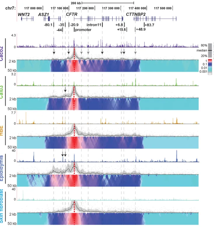
In order to characterize the 3D structure of the activeCFTR
locus in epithelial cells from different tissues involved in CF pathology, we used 4C-seq (Figures1and2). Epithelial cell lines from intestine (Caco2 adenocarcinoma) and lung (Calu3 adenocarcinoma), primary human bronchial epithe-lial cells (HBE) and primary human epididymis (caput) ep-ithelial cells were investigated. Skin fibroblasts, which do not expressCFTRprovided a negative control representing the inactive gene. 4C-seq libraries were generated at least twice for each cell line and from two independent donor cultures for primary cells.CFTR expression levels in each cell type were established previously (18,24) and verified in primary cells by RT-qPCR (not shown). Open chromatin for each cell type was mapped by DNase-seq and is shown above each panel in Figures 1 and 2. To identify poten-tially novelcis-regulatory elements we first interrogated in-teractions genome-wide with an NlaIII restriction fragment
viewpoint at theCFTRpromoter (Figure1, dotted red ver-tical line). Each panel in Figure1shows a different cell type and black lines mark the main trend of relative interactions across the locus, while domainograms (39) underneath show interaction frequency with multiple scales. Relative inter-actions are always strongest close to the viewpoint due to the physical proximity of the restriction fragments (20,37). Specific long-range interactions are indicated by peaks of the main trend, together with a local increase in intensity of the domainogram. In Caco2 cells, consistent with previ-ous 3C results (18,20,21), strong interactions are evident be-tween theCFTRpromoter and−80.1 kb,−20.9 kb, introns 10/11, and several elements at the 3end of the gene (indi-cated by black arrows). In addition to these sites, the 4C-seq data reveal novel regions interacting with the promoter including within introns 2, 4, the 3 end of the gene, and +48.9 kb downstream of the last exon (marked by gray ar-rows). In contrast, in Calu3 cells, no interactions were seen between the CFTR promoter and any intronic elements, though those with the −80.1 kb and +48.9 kb sites, and the 3 end of the gene, are evident. In addition, a strong interaction is seen between the promoter and an element at−35 kb 5to the gene (black arrow) in Calu3 cells. This element encompasses an airway-selective enhancer for the
CFTRpromoter that responds to interferon regulatory fac-tors (IRF1/2) (25). In HBE cells, the promoter interacts with the same region of intron 2 that is seen to interact in Caco2 cells and also with multiple sites between intron 4 and the middle of intron 10. Elements immediately 3 to the gene and the +48.9 kb element are also brought into close association with the promoter in HBE cells, though the intron 11 intestinal enhancer is not. Specific interac-tions upstream of the promoter are less evident in these cells. TheCFTR 4C profile in primary epididymis cells is similar to that of HBE cells within the locus and 3 to it. However, unique to the epididymis are the extensive strong interactions of upstream sequences with the promoter, par-ticularly elements at−80.1,−44,−35 and−20.9 kb to the gene. Weak interactions are also detected far upstream of the−80.1 kb CTCF binding site, within the Wingless-Type MMTV Integration Site Family Member 2 (WNT2) locus, which are not seen in other cell types. The high interaction frequency seen at−44 and−35 kb corresponds to two DHS that we observed previously using DNase-chip in fetal pri-mary epididymis cells (18) and DNase-seq analysis of adult epididymis caput cells (indicated by arrows). However, these elements may not be occupied by the same factors in epi-didymis and airway cells. Though the DHS associated with the strong intestinal enhancer in intron 11 is also evident in fetal and adult epididymis, only weak interactions were seen between theCFTRpromoter and this site in the epi-didymis 4C-seq profile. In comparison to the epithelial cells with activeCFTRloci, the locus is inactive in skin fibrob-lasts, consistent with the absence of significant interactions across the region, though the topological domain encom-passing the gene is evident.
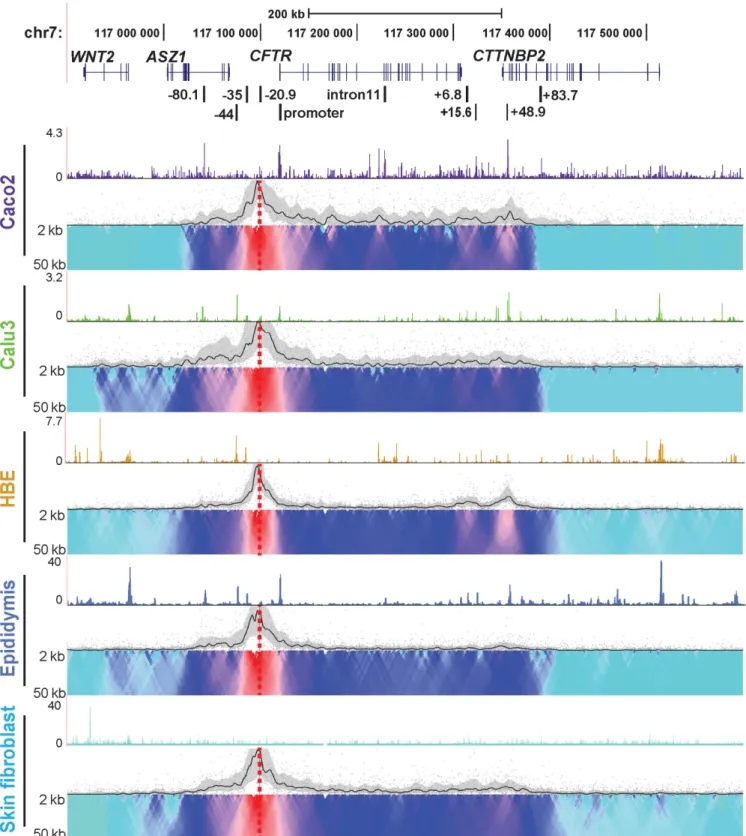
kb CTCF binding sites. These interactions were strongest in Caco2 and HBE, though also present in Calu3 and weakly in primary epididymis cells, suggesting a higher order chro-matin structure that was independent of cell type. Also of note is that interactions decreased dramatically upstream of
−80.1 kb and downstream of +48.9 kb (shown by the do-mainogram) in all cell types, with both promoter and−20.9 kb viewpoint. This is indicative of a potential topological domain or sub-domain at theCFTRlocus. In cell types with promoter: enhancer interactions at−35 and−44 kb (Calu3 and epididymis), a low level of interaction is also seen up-stream of the−80.1 kb site. Caco2 cells show unique in-teracting regions with the−20.9 kb viewpoint in intron 1, intron 4 and intron 10 which may reflectcis-regulatory el-ements specific to intestinal cells. In all cases, there was a strong correlation between 4C interactions and regions of open chromatin shown by DNase-seq. Some interactions are seen between the −20.9 kb bait and the WNT2gene in Calu3 airway cells and primary human epididymis cells. This peak of interaction corresponds to a cell type selec-tive CTCF binding site (ENCODE data), though it is much weaker than interactions with other CTCF sites across the locus and inCTTNBP2on the 3side.
TheCFTR locus is demarcated by a topological domain in which cell-type-specific chromosome looping occurs
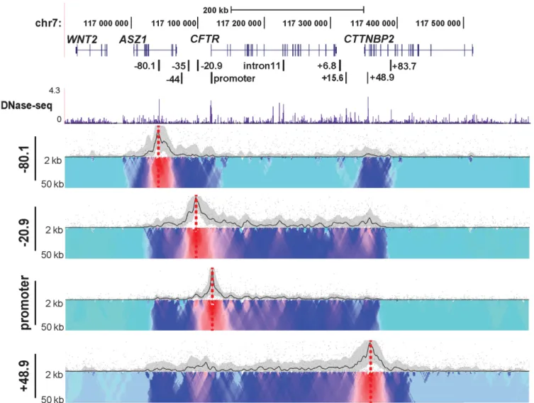
Topological domains (TADs) are usually conserved across cell types and their borders are enriched for CTCF sites (41). To further characterize role of CTCF in the chromatin structure at theCFTRlocus, we designed additional 4C-seq viewpoints at the−80.1 and +48.9 kb CTCF binding sites flanking the locus (21). 4C-seq data from each viewpoint in Caco2 cells are shown in Figure3. A lack of interactions with all viewpoints beyond the −80.1 and +48.9 kb sites, suggested that these two CTCF sites define the boundary of a TAD or sub-TAD at theCFTRlocus. To verify this inter-pretation, we used published human fibroblast Hi-C data (http://www.3dgenome.org) to define the TADs surround-ingCFTRand observed an identical domain to that illus-trated by our 4C-seq data (Supplementary Figure S1). The 4C data also showed that the two boundary CTCF bind-ing sites at−80.1 and +48.9 kb, interact strongly with each other, but show relatively weaker associations with the gene body in the middle of the TAD. In contrast, the−20.9 kb CTCF binding site viewpoint interacts strongly with both the boundary CTCF sites and with intronic elements, which also associate with the promoter. These data reveal two lev-els of chromosome looping involving CTCF at the CFTR
locus: the first maintains the overall 3D structure of the topological domain, while the second facilitates recruitment ofcis-regulatory elements to the gene promoter. To verify the interaction between the boundary CTCF sites, we per-formed FISH using BAC probes (CTD-2100H12 covers the
−80.1 kb site and CTD-3014G19 encompasses the +48.9 element). A BAC (CTD-2329K1) mapping downstream of the +48.9 kb element, at an equal genomic distance as the
−80.1 to +48.9 kb interval, was chosen as a control probe (Supplementary Figure S2A). Quantification of the FISH signals (Supplementary Figure S2B) in 94 nuclei shows that the−80.1 and +48.9 kb sites are significantly closer together
than are the +48.9 kb site and the control probe (Supple-mentary Figure S2C).
CTCF/cohesin complex are required for maintaining the 3D chromatin structure and proper regulation of theCFTRlocus
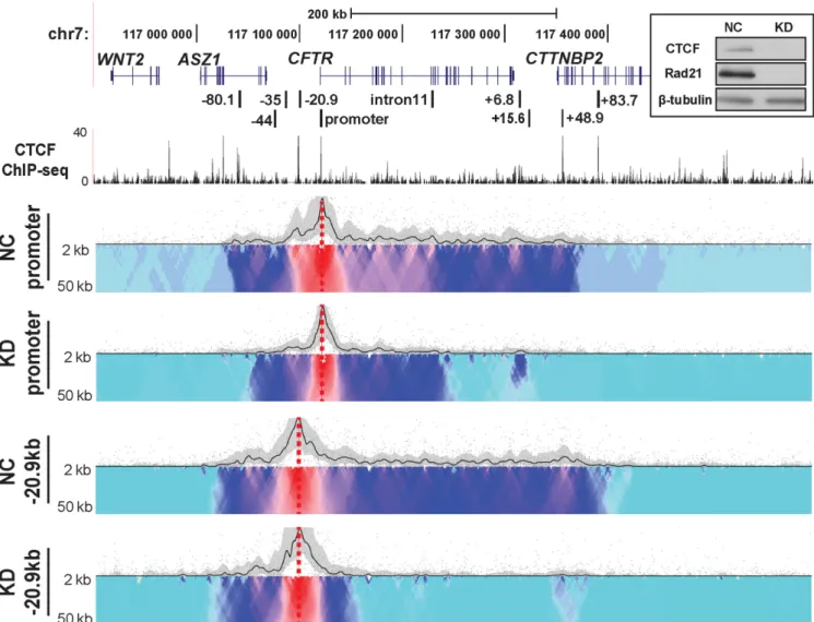
We previously showed that CTCF and cohesin complex had distinct roles in modulating the higher order structure of theCFTRgene (21). While both were involved in maintain-ing the 3D structure of the locus, cohesin had an additional role in stabilizing the interactions between the gene pro-moter and specificcis-regulatory elements. Moreover, loss of both CTCF and Rad21 (a subunit of the cohesin com-plex) increasedCFTRexpression levels (21). Having estab-lished by 4C-seq that the interaction of CTCF binding sites governed the structural organization of the activeCFTR lo-cus, we next investigated the direct contribution of these architectural proteins. CTCF and Rad21 were simultane-ously depleted in Caco2 cells by siRNA (Figure4inset) and the impact on theCFTRlocus interaction profile was mea-sured by 4C-seq (Figure4). Using viewpoints at either the promoter or −20.9 kb in comparison to negative control siRNA, the CTCF/Rad21 siRNAs caused a dramatic and global decrease of interactions across the whole topologi-cal domain. The interactions between the promoter view-point and CTCF sites at−80.1, −20.9 and 48.9 kb were either greatly reduced or lost completely, as were associ-ations with intronic regions 3 to the middle of the locus. Intronic regions 5to and including intron 11 still showed some interactions with the promoter but at a greatly re-duced frequency. The downstream boundary of the topo-logical domain was barely evident using either the promoter or the−20.9 kb viewpoint, though overall the loss of as-sociations with the latter was greater. The interaction pro-file upstream of the−20.9 kb viewpoint was less affected by CTCF/cohesin depletion, implicating other factors in maintenance of these structures. We showed previously that CTCF/RAD21 depletion in Caco2 cells does not alter ex-pression of the genes immediately adjacent toCFTRon the 5and 3side (21).
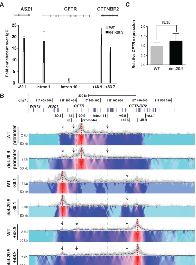
Loss of a CTCF-binding insulator element at−20.9 kb en-hances CTCF recruitment at adjacent sites, maintaining lo-cus architecture andCFTRexpression
To determine the contribution of a single CTCF binding site to the higher order structure and expression ofCFTR, the−20.9 kb insulator (40) was removed from the endoge-nous locus in Caco2 cells. CRISPR/Cas9 technology (42) with two flanking guides was used to remove an∼1.2 kb fragment encompassing the−20.9 kb element from all three
Figure 3. Chromatin structure at theCFTRlocus in Caco2 cells analyzed from multiple viewpoints. 4C-seq data and DNase-seq data presentation in Caco2 cells as described in Figure1. Viewpoints are CTCF-binding sites at−80.1,−20.9 and +48.9 kb and at the promoter.
and 3 to the locus at +48.9 kb. These data suggested that loss of a critical CTCF site caused a re-distribution of the architectural proteins to maintain the 3D structure of the active locus. Consistent with the CTCF ChIP data are 4C-seq results (Figure5B) showing changes in looping inter-actions across the locus after removal of the−20.9 kb site. Though the overall TAD remained intact, as would be pre-dicted since this element is not at the TAD boundary, inter-actions between CTCF binding sites at−80.1 and +48.9 kb and with the gene promoter were altered. With the +48.9 kb viewpoint, a reduction in interactions with−20.9 kb re-gion was accompanied by increased interactions with−80.1 kb and 3end ofCFTRin deletion clones (indicated by ar-rows). With the−80.1 kb viewpoint, interactions were in-creased across the locus and with the +6.8 kb CTCF site, in the del-20.9 clones (indicated by arrows). The promoter viewpoint showed that deletion of−20.9 kb increased inter-actions with the−80.1 kb site and−35 kb sites but reduced interactions with specific, but not all enhancer elements in the 5half of the locus (indicated by arrows). Next we per-formed RT-qPCR to measureCFTRexpression levels in the
−20.9 kb deletion clones in comparison to WT clones and showed that the minor increase (1.25-fold) was not signifi-cant (Figure5C). This suggests that the−20.9 kb site pri-marily plays a structural role at the locus and in its absence other CTCF sites can at least partially compensate for its function.
To confirm that the alteration in CTCF occupancy after removal of the−20.9 kb element was specific to theCFTR
locus we examined CTCF binding at the gel-forming mucin gene cluster on chromosome 11. We previously reported the role of CTCF in the organization of this multi-gene cluster (43). No difference in CTCF occupancy was observed by ChIP at two critical CTCF binding sites in the region (sites IV and IX) in WT and−20.9 kb deletion clones (Supple-mentary Figure S5).
Deletion of a strong enhancer in intron 11 has little impact on the 3D architecture, but repressesCFTRgene expression
con-Figure 4. Depletion of CTCF and cohesin complex has a dramatic impact on chromatin structure at theCFTRlocus. 4C interaction profiles of theCFTR
locus in Caco2 cells after CTCF/Rad21 knockdown (KD). Top right inset. Efficacy of CTCF/Rad21 KD: western blots of cell lysates from Caco2 cells co-transfected with CTCF, RAD21-specific or negative control (NC) siRNAs, probed with antibodies to CTCF, RAD21 and-tubulin. 4C-seq viewpoints are at theCFTRpromoter and−20.9 kb in negative control or CTCF/Rad21 double knockdown (KD) Caco2 cells. CTCF ChIP-seq data combined from multiple cell types are from ENCODE (62).
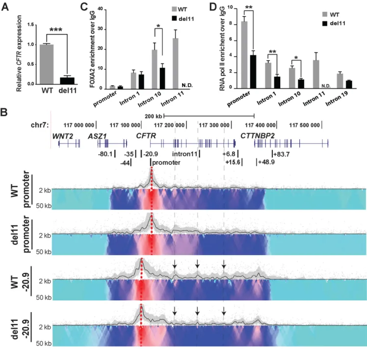
tribute to higher order chromatin structure and gene ex-pression we again used CRISPR/Cas9 technology to re-move it from Caco2 cells. Approximately 1.2 kb encompass-ing the bindencompass-ing sites for critical transcription factors driv-ing the intron 11 enhancer (FOXA1/2, HNF1 and CDX2 (18,22,23)) were removed as shown in Supplementary Fig-ure S3C. Deletion of DHS11 (del11) from all threeCFTR
alleles in targeted Caco2 cell clones was verified by PCR of genomic DNA (Supplementary Figure S3C and D). Three wild-type (non-targeted) and three del11 clones were used for further analysis. CFTRexpression in these clones was measured by RT-qPCR and deletion of the enhancer re-duced transcript levels by more than 80% (Figure6A). To verify that the deletion of DHS11 did not interfere with the normal splicing ofCFTRmRNA, we performed RT-PCR with primers spanning from exon 6 through exon 13 of the transcript (44). The results show no differences in splicing between WT and del11 clones (Supplementary Figure S4). 4C–seq was next performed on both WT and del11 clones
and though no substantial changes in the 3D structure of the locus were evident with a promoter viewpoint, interac-tions with−20.9 kb became weaker in some intronic regions marked by arrows (Figure6B). Del11 clones also showed minor changes in the interaction profile between both view-points and the intron 4 to intron 10 regions. Since chromatin structure of the locus did not show major changes after loss of DHS11, we examined whether del11 altered recruitment of critical transcription factors to other elements in the lo-cus. FOXA2, a pioneer transcription factor (45), regulates
Figure 6. Deletion of the intron 11 enhancer from theCFTRlocus has little impact on chromatin structure but substantially reduces gene expression. (A) RT-qPCR analysis showingCFTRexpression relative to 18s RNA in WT (gray) and del11 (black) Caco2 clones. Values are mean±S.E.M.,n=3, ***P<
0.001. (B) 4C-seq interaction profiles of WT and del11 Caco2 clones using viewpoints at the promoter and−20.9 kb. (C) FOXA2 ChIP in WT (gray) and del11 (black) Caco2 clones, followed by qPCR with primers for the promoter and introniccis-regulatory elements inCFTR. (D) RNA pol II ChIP in WT (gray) and del11 (black) Caco2 clones, followed by qPCR. ChIP results are pooled from three independent experiments from two WT and two deletion clones. Data present the mean±S.E.M., *P<0.05, **P<0.01.
the mechanism of del11 induced loss ofCFTRexpression, we measured enrichment of RNA polymerase II (RNAP II) across the locus by ChIP (Figure6D). In addition to occu-pancy at theCFTRpromoter in WT Caco2 cells, RNAP II is enriched at the activecis-regulatory elements in intron 1, 10 and 11. A regulatory site in intron 19 that is not utilized in Caco2 cells is also bound by RNAP II, illustrating the active transcriptional state of the locus. The del11 clones show a significant reduction in RNAP II occupancy at the promoter, intron 1, 10 and 11, suggesting a global loss of the
polymerase across theCFTRlocus. These data suggest that the intron 11 enhancer participates in recruitment of RNAP II to theCFTRpromoter as well as to intronic regions.
DISCUSSION
boundaries at−80.1 kb and +48.9 kb flanking theCFTR
gene, irrespective of its expression levels. Consistent with the original description of the domain structure of the mam-malian genome by Hi-C and 5C techniques (41,47), we show that the boundaries of the TAD at theCFTRlocus are cell type independent and enriched for CTCF and cohesin com-plex (21). Our data also suggest that there are two discrete but interrelated types of chromatin looping at the locus: the first establishes and maintains the TAD, while the sec-ond brings enhancers and othercis-regulatory elements into close association with the gene promoter and the CTCF structural network. The latter loops are likely formed hier-archically within the topological domain and are cell-type dependent. Consistent with this model is the general lack of intra-domain interactions within theCFTRTAD in the fi-broblast cells, which do not express the gene and the unique loop footprints seen in intestinal, airway and epididymis ep-ithelial cells, all of which express abundantCFTR. Further evidence is provided by the observation that disruption of the looping interactions by depletion of CTCF Rad21 alters
CFTRexpression.
CTCF and cohesin complex occupy many of the same sites genome-wide and both mediate long-range interac-tions (3,4,6,48–52). Our data provide direct evidence for the maintenance of higher order chromatin structures be-ing dependent on CTCF and cohesin complex, and are in agreement with observations at the-globin locus (14), at a complex gene cluster (Hox) through development (53) and genome-wide (5,14,54). The high-resolution studies on a single locus (CFTR) that are described here contribute a more detailed level of understanding of the underlying mechanisms. We show that depletion of CTCF/Rad21 not only destroys the TAD structure, but also disrupts the intra-domain enhancer-promoter interactions. Looking in more detail at the two levels of chromatin looping, for example in Caco2 cells, the CTCF/cohesin sites at either end of the TAD ((21), site I,−80.9 kb and site IV, +48.9 kb), seem to isolate theCFTR locus from regulatory signals from out-side the domain. Moreover, CTCF/RAD21 depletion has little or no impact on interactions between elements within the TAD and adjacent regions outside it. This suggests that either minimal occupancy of these proteins is required to maintain the boundaries, other proteins may compensate for their loss or that the TAD has inherent structural fea-tures based on DNA sequence alone. It is of interest that structural disruption of TADs directly impacts promoter enhancer interactions and is associated with disease pheno-types (55).
In contrast, though the CTCF/cohesin site at−20.9 kb has enhancer blocking insulator activity (40) and interacts with the CTCF sites at the TAD boundaries (−80.1 kb and +48.9 kb), it also has a pivotal role in looping within the TAD, which recruits cis-regulatory elements to the gene promoter. siRNA-mediated depletion of CTCF/Rad21 at this site may be largely responsible for the global loss of in-teractions of intronic sites with the gene promoter. This sug-gestion is supported by the observation that deletion of the
−20.9 kb element decreases interactions between theCFTR
promoter and specific intronic regions of the gene. It is of in-terest that despite this loss of intragenic loopingCFTRgene expression is not significantly affected by the deletion. There
are many potential explanations for this observation. These include an inbuilt redundancy of CTCF/cohesin binding sites, so that increased CTCF occupancy and interactions with other nearby sites (for example immediately adjacent to−20.9 kb, or within intron 1 ofCFTR) could compen-sate for the loss of the −20.9 kb site. Alternatively, re-moval of the−20.9 kb site facilitates interaction between the CFTR promoter and 5enhancers at−35 and−44 kb that are not normally active in Caco2 cells (25,26). The 4C -seq data would support this suggestion (Figure5), however the critical factors that drive these 5 enhancers in airway cells may not be present in intestinal cells. It is also pos-sible that removal of the−20.9 kb CTCF site destabilizes theCFTRlocus so facilitating interactions outside the do-main, though these appear generally to be weak. Alterna-tively, recruitment of critical transcription factors to the in-tronic enhancers may transmit signals to the gene promoter by looping-independent mechanisms.
The high-resolution structure/function data on CFTR
presented here greatly advance our understanding of the cell-type specific regulatory mechanisms for this complex gene. Previously, a strong intestinal enhancer element was identified in intron 11 ofCFTRgene in Caco2 cells, which was shown to interact with the gene promoter by 3C assays (18,22). The unbiased 4C-seq data presented here not only confirmed the 3C data, but also identified multiple addi-tional interacting regions that could contribute to the gene regulation. Some of these regions may contain novel en-hancers, as suggested by histone modifications (56), while others may be bystanders in the looping mechanism. Con-sistent with this suggestion are the results of deleting the intron 11 enhancer in Caco2 cells. Removal of this site re-pressesCFTRexpression by more than 80%, but does not abolish global interactions between the 5half of the locus and the gene promoter, implicating additional sites in driv-ing the loopdriv-ing. The deletion also results in increased in-teractions with other sites within and outside the gene. Our observation that removal of the intron 11 enhancer reduces RNAP II occupancy at the gene promoter by only∼50% likewise predicts the interaction of multiple intronic en-hancers with theCFTRpromoter. Indeed, we reported the involvement of several critical intronic enhancers in Caco2 cells in previous work (18). It is also of note that RNAP II occupancy is reduced to the same extent (∼50%) at several other sites across the locus suggesting that deletion of the intron 11 enhancer does not impactCFTRtranscript elon-gation.
Finally, we integrated the cell type dependent chromatin structures we observed at the CFTR locus with our open chromatin data, ChIP-seq data for both transcription fac-tors (TFs) and histone modifications (56). In Caco2 cells, interacting regions identified by 4C show a high degree of overlap with open chromatin regions revealed by DNase-seq data (Figures1and2). Similarly, these regions coincide with peaks of occupancy by TFs, which participate in the network activatingCFTRexpression in intestinal epithelial cells. Important TFs include FOXA2, CDX2, HNF1 and HNF4␣ (22,23,46,58), (Yang et al. submitted). These ob-servations suggest a strong relationship between the regula-tory network and 3D chromatin structure. This suggestion is supported by our previous results showing that depletion of FOXA2 concurrently repressesCFTRexpression and in-teractions between the gene promoter and intronic regula-tory elements (23). It also coincides with data from pluripo-tent stem cells, showing that loss of important transcription factors disrupted pluripotency-specific long range interac-tions (59–61). The correlation between cell-specific genomic interactions and open chromatin is also seen in Calu3 (56) and human primary epididymis cells (Figure1). In conclu-sion, theCFTRgene provides a paradigm for understand-ing detailed regulatory mechanisms at a sunderstand-ingle locus, and can inform studies on other loci. These mechanisms involve the interplay of architectural proteins, transcription factors binding to cis-regulatory elements and chromatin modifi-cations, to establish and facilitate dynamic responses of the 3D chromatin structure.
SUPPLEMENTARY DATA
Supplementary Dataare available at NAR Online.
ACKNOWLEDGEMENT
We thank Dr Scott Randell and colleagues for HBE cells; also Dr Wouter de Laat and Dr Omer Schwartzman for help with 4C experiments and data analysis; and Dr P Faber and staff at the University of Chicago Genomics Core.
FUNDING
National Institutes of Health [R01HD068901 to A.H.); NIGMS New Innovator Award [DP2 OD008717 to S.T.K.]; Cystic Fibrosis Foundation (Harris 11G0 and 14P0). Fund-ing for open access charge: National Institutes of Health and others.
Conflict of interest statement.None declared.
REFERENCES
1. Bell,A.C., West,A.G. and Felsenfeld,G. (1999) The protein CTCF is required for the enhancer blocking activity of vertebrate insulators.
Cell,98, 387–396.
2. Giles,K.E., Gowher,H., Ghirlando,R., Jin,C. and Felsenfeld,G. (2010) Chromatin boundaries, insulators, and long-range interactions in the nucleus.Cold Spring Harb. Symp. Quant. Biol.,75, 79–85. 3. Hadjur,S., Williams,L.M., Ryan,N.K., Cobb,B.S., Sexton,T.,
Fraser,P., Fisher,A.G. and Merkenschlager,M. (2009) Cohesins form chromosomal cis-interactions at the developmentally regulated IFNG locus.Nature,460, 410–413.
4. Nativio,R., Wendt,K.S., Ito,Y., Huddleston,J.E., Uribe-Lewis,S., Woodfine,K., Krueger,C., Reik,W., Peters,J.M. and Murrell,A. (2009) Cohesin is required for higher-order chromatin conformation at the imprinted IGF2-H19 locus.PLoS Genet.,5, e1000739.
5. Zuin,J., Dixon,J.R., van der Reijden,M.I., Ye,Z., Kolovos,P., Brouwer,R.W., van de Corput,M.P., van de Werken,H.J.,
Knoch,T.A., van,I.W.F.et al.(2014) Cohesin and CTCF differentially affect chromatin architecture and gene expression in human cells.
Proc. Natl. Acad. Sci. U.S.A.,111, 996–1001. 6. Wendt,K.S., Yoshida,K., Itoh,T., Bando,M., Koch,B.,
Schirghuber,E., Tsutsumi,S., Nagae,G., Ishihara,K., Mishiro,T.et al.
(2008) Cohesin mediates transcriptional insulation by CCCTC-binding factor.Nature,451, 796–801. 7. Bau,D., Sanyal,A., Lajoie,B.R., Capriotti,E., Byron,M.,
Lawrence,J.B., Dekker,J. and Marti-Renom,M.A. (2011) The three-dimensional folding of the alpha-globin gene domain reveals formation of chromatin globules.Nat. Struct. Mol. Biol.,18, 107–114. 8. Berlivet,S., Paquette,D., Dumouchel,A., Langlais,D., Dostie,J. and
Kmita,M. (2013) Clustering of tissue-specific sub-TADs accompanies the regulation of HoxA genes in developing limbs.PLoS Genet.,9, e1004018.
9. Forrester,W.C., Epner,E., Driscoll,M.C., Enver,T., Brice,M., Papayannopoulou,T. and Groudine,M. (1990) A deletion of the human beta-globin locus activation region causes a major alteration in chromatin structure and replication across the entire beta-globin locus.Genes Dev.,4, 1637–1649.
10. Gavrilov,A.A. and Razin,S.V. (2008) Spatial configuration of the chicken alpha-globin gene domain: immature and active chromatin hubs.Nucleic Acids Res.,36, 4629–4640.
11. Higgs,D.R., Vernimmen,D. and Wood,B. (2008) Long-range regulation of alpha-globin gene expression.Adv. Genet.,61, 143–173. 12. Hughes,J.R., Lower,K.M., Dunham,I., Taylor,S., De Gobbi,M.,
Sloane-Stanley,J.A., McGowan,S., Ragoussis,J., Vernimmen,D., Gibbons,R.J.et al.(2013) High-resolution analysis of cis-acting regulatory networks at the alpha-globin locus.Philos. Trans. R. Soc. Lond. B, Biol. Sci.,368, 20120361.
13. Kim,A. and Dean,A. (2003) A human globin enhancer causes both discrete and widespread alterations in chromatin structure.Mol. Cell. Biol.,23, 8099–8109.
14. Splinter,E., Heath,H., Kooren,J., Palstra,R.J., Klous,P., Grosveld,F., Galjart,N. and de Laat,W. (2006) CTCF mediates long-range chromatin looping and local histone modification in the beta-globin locus.Genes Dev.,20, 2349–2354.
15. Vernimmen,D., Marques-Kranc,F., Sharpe,J.A., Sloane-Stanley,J.A., Wood,W.G., Wallace,H.A., Smith,A.J. and Higgs,D.R. (2009) Chromosome looping at the human alpha-globin locus is mediated via the major upstream regulatory element (HS -40).Blood,114, 4253–4260.
16. Gillen,A.E. and Harris,A. (2012) Transcriptional regulation of CFTR gene expression.Front. Biosci.,4, 587–592.
17. Gosalia,N. and Harris,A. (2015) Chromatin dynamics in the regulation of CFTR expression.Genes,6, 543–558.
18. Ott,C.J., Blackledge,N.P., Kerschner,J.L., Leir,S.H., Crawford,G.E., Cotton,C.U. and Harris,A. (2009) Intronic enhancers coordinate epithelial-specific looping of the active CFTR locus.Proc. Natl. Acad. Sci. U.S.A.,106, 19934–19939.
19. Ott,C.J., Suszko,M., Blackledge,N.P., Wright,J.E., Crawford,G.E. and Harris,A. (2009) A complex intronic enhancer regulates expression of the CFTR gene by direct interaction with the promoter.J. Cell. Mol. Med.,13, 680–692.
20. Gheldof,N., Smith,E.M., Tabuchi,T.M., Koch,C.M., Dunham,I., Stamatoyannopoulos,J.A. and Dekker,J. (2010) Cell-type-specific long-range looping interactions identify distant regulatory elements of the CFTR gene.Nucleic Acids Res.,38, 4325–4336.
21. Gosalia,N., Neems,D., Kerschner,J.L., Kosak,S.T. and Harris,A. (2014) Architectural proteins CTCF and cohesin have distinct roles in modulating the higher order structure and expression of the CFTR locus.Nucleic Acids Res.,42, 9612–9622.
22. Kerschner,J.L. and Harris,A. (2012) Transcriptional networks driving enhancer function in the CFTR gene.Biochem. J.,446, 203–212. 23. Kerschner,J.L., Gosalia,N., Leir,S.H. and Harris,A. (2014)
Chromatin remodeling mediated by the FOXA1/A2 transcription factors activates CFTR expression in intestinal epithelial cells.
24. Zhang,Z., Ott,C.J., Lewandowska,M.A., Leir,S.H. and Harris,A. (2012) Molecular mechanisms controlling CFTR gene expression in the airway.J. Cell. Mol. Med.,16, 1321–1330.
25. Zhang,Z., Leir,S.H. and Harris,A. (2013) Immune mediators regulate CFTR expression through a bifunctional airway-selective enhancer.
Mol. Cell. Biol.,33, 2843–2853.
26. Zhang,Z., Leir,S.H. and Harris,A. (2015) Oxidative stress regulates CFTR gene expression in human airway epithelial cells through a distal antioxidant response element.Am. J. Respir. Cell Mol. Biol.,52, 387–396.
27. Fogh,J., Wright,W.C. and Loveless,J.D. (1977) Absence of HeLa cell contamination in 169 cell lines derived from human tumors.J. Natl. Cancer Inst.,58, 209–214.
28. Shen,B.Q., Finkbeiner,W.E., Wine,J.J., Mrsny,R.J. and
Widdicombe,J.H. (1994) Calu-3: a human airway epithelial cell line that shows cAMP-dependent Cl- secretion.Am. J. Physiol.,266, L493–L501.
29. Mouchel,N., Henstra,S.A., McCarthy,V.A., Williams,S.H., Phylactides,M. and Harris,A. (2004) HNF1alpha is involved in tissue-specific regulation of CFTR gene expression.Biochem. J.,378, 909–918.
30. Leir,S.H., Browne,J.A., Eggener,S.E. and Harris,A. (2015) Characterization of primary cultures of adult human epididymis epithelial cells.Fertil. Steril.,103, 647–654.
31. Chalkley,G. and Harris,A. (1991) Lymphocyte mRNA as a resource for detection of mutations and polymorphisms in the CF gene.J. Med. Genet.,28, 777–780.
32. Bischof,J.M., Ott,C.J., Leir,S.H., Gosalia,N., Song,L., London,D., Furey,T.S., Cotton,C.U., Crawford,G.E. and Harris,A. (2012) A genome-wide analysis of open chromatin in human tracheal epithelial cells reveals novel candidate regulatory elements for lung function.
Thorax,67, 385–391.
33. Boyle,A.P., Davis,S., Shulha,H.P., Meltzer,P., Margulies,E.H., Weng,Z., Furey,T.S. and Crawford,G.E. (2008) High-resolution mapping and characterization of open chromatin across the genome.
Cell,132, 311–322.
34. Langmead,B., Trapnell,C., Pop,M. and Salzberg,S.L. (2009) Ultrafast and memory-efficient alignment of short DNA sequences to the human genome.Genome Biol.,10, R25.
35. Heinz,S., Benner,C., Spann,N., Bertolino,E., Lin,Y.C., Laslo,P., Cheng,J.X., Murre,C., Singh,H. and Glass,C.K. (2010) Simple combinations of lineage-determining transcription factors prime cis-regulatory elements required for macrophage and B cell identities.
Mol. Cell,38, 576–589.
36. Splinter,E., de Wit,E., van de Werken,H.J., Klous,P. and de Laat,W. (2012) Determining long-range chromatin interactions for selected genomic sites using 4C-seq technology: from fixation to computation.
Methods,58, 221–230.
37. van de Werken,H.J., Landan,G., Holwerda,S.J., Hoichman,M., Klous,P., Chachik,R., Splinter,E., Valdes-Quezada,C., Oz,Y., Bouwman,B.A.et al.(2012) Robust 4C-seq data analysis to screen for regulatory DNA interactions.Nat. Methods,9, 969–972.
38. Otsu,N. (1979)IEEE Trans. Syst. Man. Cybern. IEEE, Vol.9, pp. 62–66.
39. de Wit,E., Braunschweig,U., Greil,F., Bussemaker,H.J. and van Steensel,B. (2008) Global chromatin domain organization of the Drosophila genome.PLoS Genet.,4, e1000045.
40. Blackledge,N.P., Carter,E.J., Evans,J.R., Lawson,V., Rowntree,R.K. and Harris,A. (2007) CTCF mediates insulator function at the CFTR locus.Biochem. J.,408, 267–275.
41. Dixon,J.R., Selvaraj,S., Yue,F., Kim,A., Li,Y., Shen,Y., Hu,M., Liu,J.S. and Ren,B. (2012) Topological domains in mammalian genomes identified by analysis of chromatin interactions.Nature,485, 376–380.
42. Mali,P., Yang,L., Esvelt,K.M., Aach,J., Guell,M., DiCarlo,J.E., Norville,J.E. and Church,G.M. (2013) RNA-guided human genome engineering via Cas9.Science,339, 823–826.
43. Gosalia,N., Leir,S.H. and Harris,A. (2013) Coordinate regulation of the gel-forming mucin genes at chromosome 11p15.5.J. Biol. Chem.,
288, 6717–6725.
44. Hull,J., Shackleton,S. and Harris,A. (1994) Analysis of mutations and alternative splicing patterns in the CFTR gene using mRNA derived from nasal epithelial cells.Hum. Mol. Genet.,3, 1141–1146.
45. Sekiya,T., Muthurajan,U.M., Luger,K., Tulin,A.V. and Zaret,K.S. (2009) Nucleosome-binding affinity as a primary determinant of the nuclear mobility of the pioneer transcription factor FoxA.Genes Dev.,23, 804–809.
46. Gosalia,N., Yang,R., Kerschner,J.L. and Harris,A. (2015) FOXA2 regulates a network of genes involved in critical functions of human intestinal epithelia cells.Physiol. Genomics,47, 290–297.
47. Nora,E.P., Lajoie,B.R., Schulz,E.G., Giorgetti,L., Okamoto,I., Servant,N., Piolot,T., van Berkum,N.L., Meisig,J., Sedat,J.et al.
(2012) Spatial partitioning of the regulatory landscape of the X-inactivation centre.Nature,485, 381–385.
48. Hou,C., Dale,R. and Dean,A. (2010) Cell type specificity of chromatin organization mediated by CTCF and cohesin.Proc. Natl. Acad. Sci. U.S.A.,107, 3651–3656.
49. Kurukuti,S., Tiwari,V.K., Tavoosidana,G., Pugacheva,E., Murrell,A., Zhao,Z., Lobanenkov,V., Reik,W. and Ohlsson,R. (2006) CTCF binding at the H19 imprinting control region mediates maternally inherited higher-order chromatin conformation to restrict enhancer access to Igf2.Proc. Natl. Acad. Sci. U.S.A.,103, 10684–10689. 50. Parelho,V., Hadjur,S., Spivakov,M., Leleu,M., Sauer,S.,
Gregson,H.C., Jarmuz,A., Canzonetta,C., Webster,Z., Nesterova,T.
et al.(2008) Cohesins functionally associate with CTCF on mammalian chromosome arms.Cell,132, 422–433.
51. Rubio,E.D., Reiss,D.J., Welcsh,P.L., Disteche,C.M., Filippova,G.N., Baliga,N.S., Aebersold,R., Ranish,J.A. and Krumm,A. (2008) CTCF physically links cohesin to chromatin.Proc. Natl. Acad. Sci. U.S.A.,
105, 8309–8314.
52. Stedman,W., Kang,H., Lin,S., Kissil,J.L., Bartolomei,M.S. and Lieberman,P.M. (2008) Cohesins localize with CTCF at the KSHV latency control region and at cellular c-myc and H19/Igf2 insulators.
EMBO J.,27, 654–666.
53. Narendra,V., Rocha,P.P., An,D., Raviram,R., Skok,J.A., Mazzoni,E.O. and Reinberg,D. (2015) Transcription. CTCF establishes discrete functional chromatin domains at the Hox clusters during differentiation.Science,347, 1017–1021.
54. Hou,C., Zhao,H., Tanimoto,K. and Dean,A. (2008)
CTCF-dependent enhancer-blocking by alternative chromatin loop formation.Proc. Natl. Acad. Sci. U.S.A.,105, 20398–20403. 55. Lupianez,D.G., Kraft,K., Heinrich,V., Krawitz,P., Brancati,F.,
Klopocki,E., Horn,D., Kayserili,H., Opitz,J.M., Laxova,R.et al.
(2015) Disruptions of topological chromatin domains cause pathogenic rewiring of gene-enhancer interactions.Cell,161, 1012–1025.
56. Fossum,S.L., Mutolo,M.J., Yang,R., Dang,H., O’Neal,W.K., Knowles,M.R., Leir,S.H. and Harris,A. (2014) Ets homologous factor regulates pathways controlling response to injury in airway epithelial cells.Nucleic Acids Res.,42, 13588–13598.
57. Harris,A. and Coleman,L. (1989) Ductal epithelial cells cultured from human foetal epididymis and vas deferens: relevance to sterility in cystic fibrosis.J. Cell Sci.,92, 687–690.
58. Verzi,M.P., Shin,H., He,H.H., Sulahian,R., Meyer,C.A., Montgomery,R.K., Fleet,J.C., Brown,M., Liu,X.S. and
Shivdasani,R.A. (2010) Differentiation-specific histone modifications reveal dynamic chromatin interactions and partners for the intestinal transcription factor CDX2.Dev. Cell,19, 713–726.
59. Apostolou,E., Ferrari,F., Walsh,R.M., Bar-Nur,O., Stadtfeld,M., Cheloufi,S., Stuart,H.T., Polo,J.M., Ohsumi,T.K., Borowsky,M.L.
et al.(2013) Genome-wide chromatin interactions of the Nanog locus in pluripotency, differentiation, and reprogramming.Cell Stem Cell,
12, 699–712.
60. de Wit,E., Bouwman,B.A., Zhu,Y., Klous,P., Splinter,E., Verstegen,M.J., Krijger,P.H., Festuccia,N., Nora,E.P., Welling,M.
et al.(2013) The pluripotent genome in three dimensions is shaped around pluripotency factors.Nature,501, 227–231.
61. Wei,Z., Gao,F., Kim,S., Yang,H., Lyu,J., An,W., Wang,K. and Lu,W. (2013) Klf4 organizes long-range chromosomal interactions with the oct4 locus in reprogramming and pluripotency.Cell Stem Cell,13, 36–47.