RAS SIGNALING AND THERAPEUTIC RESISTANCE IN MELANOMA
Bingying Zhou
A dissertation submitted to the faculty of the University of North Carolina at Chapel Hill in partial fulfillment of the requirements for the degree of Doctor of Philosophy in the Department of
Pharmacology.
Chapel Hill 2016
ABSTRACT
BINGYING ZHOU: RAS signaling and therapeutic resistance in melanoma (Under the direction of Adrienne D. Cox)
Increasing appreciation of the complexity of RAS signaling in cancer has led to a renewed wave of RAS research. I have focused on two key areas: the role of wild-type RAS isoforms in RAS-mutant cancers, and mechanisms of resistance to molecularly targeted therapies directed against RAS effector pathways. Melanoma, the most aggressive form of skin cancer, presents an excellent model to study RAS signaling; ~90% of melanomas have a driver mutation in NRAS (26%) or BRAF (63%), thus hyper-activating the canonical RAS-RAF-MEK-ERK effector signaling pathway. An army of small molecule inhibitors has emerged to target this pathway, and several are FDA-approved for melanoma treatment. However, all targeted therapies face the challenge of resistance. Most validated mechanisms of resistance to these inhibitors involve either reactivation of ERK signaling or other bypass routes that result in cancer cell survival.
In my investigation of resistance mechanisms in BRAF-mutant melanoma. I found that CK2α was sufficient to drive resistance to inhibitors of BRAF (BRAFi) and of MEK (MEKi). CK2α
inhibitor SCH772984. These findings support a new mechanism whereby a kinase-independent scaffolding function of CK2α promotes resistance to RAF- and MEK-targeted therapies.
Another pressing issue is understanding the biological activities of wild-type RAS isoforms in RAS-mutant tumors. Most studies have investigated KRAS-mutant cancers, but little is known about NRAS-mutant cancers. NRAS-mutant melanomas comprise the second largest subgroup of melanoma patients, and no targeted therapy is approved for these patients. Exploring the roles of wild-type RAS isoforms in NRAS-mutant melanoma cells, I found that WT KRAS is essential for their proliferation and survival. Interestingly, depletion of KRAS resulted in a unique cellular morphology and in signaling outcomes distinct from those due to silencing of NRAS or HRAS. Moreover, KRAS knockdown stabilized p53 protein, which was accompanied by an increase in p53 target genes. Intriguingly, by using reverse phase protein array analysis, I found that KRAS knockdown severely impaired phosphorylation of ribosomal protein S6, dependent on S6K1 (p70 S6K) activity, but independent of Akt-mTOR, or ERK-p90RSK activity. These results may shed light on the potential efficacy of pan-RAS inhibition in NRAS-mutant cancers.
Together, my findings uncover a novel kinase-independent scaffolding function of CK2α
ACKNOWLEDGEMENTS
First of all, I would like to express my deepest gratitude to my advisor, Dr. Adrienne Cox, for her guidance, enthusiasm, encouragements and unwavering support in both research and life, providing me with an inspiring and loving atmosphere for completing my Ph.D. work.
I am also thankful for current and past members of the Cox Lab for making this lab a wonderful place. Special thanks to Lauren and Jim, who helped me get started in the lab, and provided lots of help during the first few years of my graduate life.
I would also like to express my sincere thanks to Dr. Channing Der, who provided me with many ideas, insightful suggestions, reagents, and the opportunity to join Der Lab meetings. I enjoyed my interactions with all Der Lab members, who are a cheerful and scientifically resourceful group.
I am grateful to my thesis committee, Dr. Adrienne Cox, Dr. Channing Der, Dr. Gary Johnson, Dr. Ben Major and Dr. Lee Graves for their constructive feedback on my projects.
statistical analysis on NRAS-mutant melanoma cell lines. Dr. Channing Der generously shared equipment, reagents, and ideas throughout the entire course of my study.
TABLE OF CONTENTS
CHAPTER I: INTRODUCTION ... 1
RAS is a small GTPase ... 1
RAS isoforms are structurally similar but functionally distinct ... 2
The roles of wild-type RAS in RAS-mutant cancers remain controversial ... 5
RAS effector pathway signaling is frequently deregulated in cancer ... 9
The RAS-RAF-MEK-ERK “pathway” is really a “web” ... 12
CK2 modulates RAF-MEK-ERK pathway activity and vice versa ... 16
RAS-RAF-MEK-ERK signaling is critical to RAS- and RAF-driven cancers ... 17
Resistance to targeted therapies remains a major challenge in treating RAS- or RAF-driven cancers ... 20
Melanoma is an excellent disease model for studying RAS-RAF-MEK-ERK signaling and therapeutic resistance ... 24
Rationale and objectives for this project ... 26
Define a novel mode of resistance to RAF-MEK-ERK pathway inhibition ... 26
Define the roles of wild-type RAS isoforms in the presence of oncogenic NRAS ... 27
CHAPTER II: CK2α MAINTAINS ERK ACTIVITY TO PROMOTE RESISTANCE TO INHIBITORS OF THE RAF-MEK-ERK PATHWAY IN BRAF-MUTANT MELANOMA ... 35
Overview ... 35
Introduction ... 36
Experimental procedures ... 38
Cell culture and reagents ... 38
Plasmid constructs and gateway cloning ... 38
Lentivirus production and infection ... 39
Pharmacologic growth inhibition (GI50) assay ... 40
Clonogenic assay ... 41
Site-directed Mutagenesis ... 41
RNA isolation, reverse transcription and real-time PCR ... 42
Co-immunoprecipitation ... 42
Results ... 43
CK2α expression is upregulated in a subset of melanomas ... 43
CK2α promotes resistance to inhibitors of BRAF and MEK in BRAF-mutant melanoma cells ... 43
CK2α depletion sensitizes melanoma cells to BRAF inhibition ... 44
CK2α sustains ERK phosphorylation under conditions of RAF-MEK-ERK pathway inhibition ... 45
CK2α regulates DUSP6 protein levels in a kinase-dependent manner ... 46
CK2α-mediated maintenance of ERK phosphorylation and pathway inhibitor resistance does not require its kinase function ... 47
CK2α(WT) and CK2α(K68M) bind equally well to the RAF-MEK-ERK scaffold protein KSR1 ... 47
ERK inhibition avoids CK2α-mediated resistance to RAF-MEK-ERK pathway blockade .... 48
Discussion ... 48
Chapter III: AN ESSENTIAL ROLE FOR WILD-TYPE KRAS IN NRAS-MUTANT MELANOMA ... 60
Overview ... 60
Introduction ... 61
Methods and Materials ... 65
Cell culture and reagents ... 65
shRNA constructs ... 65
Lentivirus production and infection ... 66
Western blotting ... 66
RNA isolation, reverse transcription and real-time qPCR ... 68
2D clonogenic assay ... 69
Soft agar assay ... 69
Cell cycle analysis ... 69
Annexin-V/PI staining ... 70
Reverse-phase protein array analysis ... 70
Results ... 71
KRASWT is essential for cell survival and proliferation in NRAS-mutant melanoma cells .... 71
RAS isoforms play divergent roles in NRAS-mutant melanoma ... 72
KRAS depletion increases p53 stability ... 73
KRAS depletion impairs phosphorylation of ribosomal protein S6 ... 74
Discussion ... 76
Chapter IV: CONCLUSIONS AND FUTURE DIRECTIONS ... 89
Summary and Conclusions ... 89
Future Directions ... 91
Is CK2α-mediated resistance to RAF/MEK inhibition clinically relevant? ... 91
How does kinase-inactive CK2α promote resistance to inhibitors of RAF or MEK? ... 93
How does CK2α regulate DUSP6 protein levels? ... 95
Why is KRASWT more critical to cell survival than oncogenic NRAS in NRAS-mutant melanoma? ... 96
What are the functional consequences of the suppression of rpS6 phosphorylation that is induced upon KRAS silencing? ... 98
How do RAS isoforms differentially engage effector signaling? ... 99
How does KRAS control rpS6 phosphorylation? ... 101
REFERENCES ... 109
LIST OF FIGURES
Figure 1.1. The RAS GTPase cycle. ... 28
Figure 1.2. Overall domain structure of RAS isoforms. ... 29
Figure 1.3. RAS effector signaling pathways. ... 30
Figure 1.4. The PI3K-AKT-mTOR-S6K pathway. ... 31
Figure 1.5. Scaffolds and negative feedback signaling fine-tune RAS-RAF-MEK-ERK signaling outcome. ... 32
Figure 1.6. Mechanisms of resistance to BRAF inhibition lead to ERK1/2 reactivation. ... 33
Figure 1.7. BRAF and NRAS mutations in skin cutaneous melanoma. ... 34
Figure 2.1. CK2α protein expression is elevated in melanoma cell lines compared to normal human melanocytes (NHM). ... 52
Figure 2.2. Ectopic CK2α promotes resistance to inhibitors of BRAF and MEK. ... 53
Figure 2.3. Suppression of endogenous CK2α increases sensitivity to the BRAF inhibitor vemurafenib. ... 54
Figure 2.4. Overexpressed CK2α accelerates ERK rebound or sustains ERK phosphorylation in response to RAF-MEK-ERK pathway inhibition. ... 55
Figure 2.5. CK2α decreases protein stability of the ERK phosphatase DUSP6 in a kinase-dependent manner. ... 56
Figure 2.6. CK2α-mediated maintenance of ERK phosphorylation upon pathway inhibition and resistance to BRAFi/MEKi are both kinase-independent. ... 57
Figure 2.7. Both wild-type and kinase-inactive CK2α interact with the RAF-MEK-ERK scaffold protein KSR1. ... 58
Figure 2.8. ERK inhibitor SCH772984 is insensitive to overexpression of CK2α. ... 59
Figure 3.1. KRASWT is essential for cell proliferation and survival in NRAS-mutant melanoma cells. ... 81
Figure 3.2. RAS isoforms perform divergent functions in NRAS-mutant melanoma cells. ... 82
Figure 3.3. KRAS depletion increases p53 protein stability and activation of its target genes. .. 83
Figure 3.4. KRAS, but not NRAS or HRAS depletion leads to severe impairment of rpS6 phosphorylation at Ser235/236. ... 84
Figure 3.6. RAS isoforms differentially regulate cell morphology and downstream signaling. ... 86
Figure 3.7. KRASWT depletion specifically upregulates PUMA mRNA. ... 87
Figure 3.8. rpS6 phosphorylation is decreased upon KRAS depletion in
NSCLC cells and does not cause p53 protein upregulation. ... 88
Figure 4.1. CK2alpha (CSNK2A1) mRNA expression is associated with
melanoma progression. ... 103
Figure 4.2 CK2alpha inhibition sensitizes CK2alpha-overexpressing A375 cells to BRAF
inhibition by vemurafenib. ... 104
Figure 4.3. Opposing effects of CK2alpha and CK2beta on DUSP6 protein expression. ... 105
Figure 4.4. Proliferation of fast-dividing and slow-dividing NRAS-mutant
melanoma cells depend differentially on NRAS and KRAS. ... 106
Figure 4.5. AKT and ERK phosphorylation is differentially regulated by RAS isoforms. ... 107
Figure 4.6. ERK pathway signaling controls residual rpS6 phosphorylation
LIST OF ABBREVIATIONS AND SYMBOLS
2D two-dimensional
3D three-dimensional
4E-BP1 Eukaryotic translation initiation factor 4E-binding protein 1 AKT protein kinase B
AML acute myeloid leukemia Arf ADP ribosylation factor
ATCC American Type Culture Collection C-terminus carboxyl-terminus
CAAX cysteine-aliphatic-aliphatic-unconserved amino acid Cdc42 cell division cycle 42 small GTPase
cDNA complementary deoxyribonucleic acid Chk1 checkpoint kinase 1
CK1 casein kinase 1 CK2 casein kinase 2
COSMIC catalogue of somatic mutations in cancer CRD cysteine-rich domain
DMEM-H high glucose Dulbecco's modified Eagle medium DMSO dimethyl sulfoxide
eNOS endothelial nitric oxide synthase ERK extracellular signal-regulated kinase Ets E-twenty six transcription factor FBS fetal bovine serum
FDA Food and Drug Administration FGFR fibroblast growth factor receptor
FLAG an octapeptide fusion tag consisting of eight amino acids, DYKDDDDK Flt3 Fms-Related tyrosine kinase 3
FRS2 fibroblast growth factor receptor substrate 2 FTI farnesyltransferase inhibitor
GAP GTPase-activating protein GDP guanine diphosphate
GEF guanine nucleotide exchange factor GFP green fluorescent protein
GI50 growth inhibitory 50, concentration of drug to cause 50% reduction in proliferation GIST gastrointestinal stromal tumor
Grb2 growth factor receptor-bound protein 2 GSK-3 glycogen synthase kinase 3
GTP guanine triphosphate GTPase guanosine triphosphatases
h hour
HA hemagglutinin
HRAS Harvey rat sarcoma viral oncogene homolog HVR hypervariable region
kDa kilodalton
KRAS Kirsten rat sarcoma viral oncogene homolog KSR1/2 kinase suppressor of RAS 1/2
LCCC Lineberger Comprehensive Cancer Center MAPK mitogen-activated protein kinase
MEK mitogen-activated protein kinase kinase
min minute
MKK4 MAP kinase kinase 4 ml milliliter
MP1 MEK partner 1
mRNA messenger ribonucleic acid Mst1 macrophage stimulating 1
mTOR mechanistic target of rapamycin or mammalian target of rapamycin mTORC mTOR complex
MTT 3-[4,5-dimethylthiazol-2-yl]-2,5-diphenyltetrazolium bromide Myc v-Myc avian myelocytomatosis viral oncogene homolog NF1 neurofibromin 1
NHM normal human melanocytes
NRAS neuroblastoma rat sarcoma viral oncogene homolog NSCLC non-small cell lung cancer
N-terminus amino-terminus
NT non-targeting
PDGFR platelet-derived growth factor receptor PDK1 phosphoinositide-dependent protein kinase1 PI3K phosphatidylinositol 3-kinase
PI propidium iodide
PIP2 phosphatidylinositol-4,5-bisphosphate PIP3 phosphatidylinositol-3,4,5-trisphosphate PKA cAMP-dependent protein kinase
PKC protein kinase C
PLCε Phospholipase C epsilon
PTEN phosphatase and tensin homolog PVDF polyvinylidene difluoride
Rab rat brain small GTPase
Rac Ras-related C3 botulinum toxin substrate Raf rapidly accelerated fibrosarcoma kinase
Ral RAS-like
RalA RAS-like A RalB RAS-like B
RalBP1 Ral-binding protein 1
RalGDS Ral guanine nucleotide dissociation stimulator RalGEF Ral guanine nucleotide exchange factor Ran RAS-related nuclear protein
RAS rat sarcoma viral oncogene homolog RASSF RAS-association domain family RB1 retinoblastoma 1
RBD RAS binding domain
Rho Ras homology RhoA Ras homology A
RIPA radioimmunoprecipitation assay RNA ribonucleic acid
RPPA reverse phase protein array rpS6 ribosomal protein S6 RTK receptor tyrosine kinase
qPCR quantitative polymerase chain reaction S6K1 p70 ribosomal S6 kinase 1
S6K2 p70 ribosomal S6 kinase 2
SEER Surveillance, Epidemiology, and End Results SEM standard error of the mean
SDS sodium dodecyl sulfate SH2 Src homology 2
SHP2 SH2-domain-containing protein tyrosine phosphatase-2 shRNA short hairpin ribonucleic acid
SOS son of sevenless SPRY Sprouty
STAT3 signal transducer and activator of transcription-3 SV40 simian vacuolating virus 40
t1/2 half-life
TBK1 TANK-binding kinase 1
TBST Tris buffered saline with Tween-20 TCF Tissue Culture Facility
TCGA The Cancer Genome Atlas
Tiam1 T-cell invasion and metastasis gene 1 TPA 12-O-tetradecanoylphorbol-13-acetate TRC The RNAi Consortium
VEGFR vascular endothelial growth factor receptor
WT wild-type
γH2AX H2AX phosphorylated on Ser139
µm micron
µM micromolar
CHAPTER I: INTRODUCTION
RAS is a small GTPase
RAS (rat sarcoma viral oncogene homolog) genes were initially identified in acute transforming retroviruses as genetic sequences responsible for their oncogenic properties (Barbacid, 1987). The identification of mutationally activated human RAS genes as potently transforming oncogenes in several contemporary studies (Der et al., 1982; Goldfarb et al., 1982; Parada et al., 1982; Pulciani et al., 1982; Santos et al., 1982; Shih and Weinberg, 1982) has spawned decades of extensive research into RAS structure, biochemistry, biology, and pathobiology (Cox and Der, 2010; Malumbres and Barbacid, 2003).
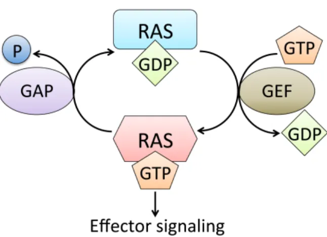
GEFs, thereby promoting nucleotide (GDP) release from RAS. Due to the ten-fold higher intracellular concentrations of GTP than GDP, RAS transiently binds to GTP, after which GAPs quickly return RAS to its resting state (Figure 1.1). Structural studies have uncovered two critical stretches of RAS that undergo significant conformational changes when RAS cycles between GTP- and GDP-bound states. Termed switch I (amino acids 30-38) and switch II (amino acids 60-76), these regions are also crucial to RAS interaction with its regulators and effectors. Oncogenically activating mutations in RAS, predominantly occurring at residues G12, G13, and Q61, impair both intrinsic and GAP-mediated GTP hydrolysis, rendering mutant RAS constitutively active (Cox et al., 2014). Analysis of the COSMIC (catalogue of somatic mutations in cancer) database reveals that somatic mutations in the three RAS genes are the most frequent mutations of oncogenes in human cancers (Cox et al., 2014). In the following section, the specific mutational profiles of the RAS isoforms will be introduced.
RAS isoforms are structurally similar but functionally distinct
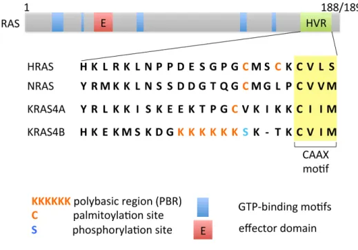
identity, differing only at their C-termini (hypervariable regions), which are critical for their respective lipid modifications, and hence for their membrane targeting and subcellular localizations (Cox et al., 2014) (Figure 1.2).
Although highly similar in structure, the RAS isoforms have been shown to carry out overlapping but still very distinct functions (Castellano and Santos, 2011; Newlaczyl et al., 2014). Genetic knockout studies in mice suggested that KRAS is the most important isoform during development, since Kras-ablated mice die during embryogenesis (Johnson et al., 1997; Koera et al., 1997). In contrast, Nras gene function was shown to be dispensable for normal mouse development, growth, and fertility (Umanoff et al., 1995), yet was later found to be important for antiviral immune response and T-cell function in mice (Perez de Castro et al., 2003). Neither did abrogration of Hras result in any developmental defects (Esteban et al., 2001). However, it reduced the numbers of papillomas formed compared with wild-type littermates after 20 weeks of 12-O-tetradecanoylphorbol-13-acetate (TPA) treatment (Ise et al., 2000). Strikingly, Hras (-/-)/Nras(-/-) double knockout mice were viable, and displayed normal development, growth, fertility, and neuronal development (Esteban et al., 2001). Both wild-type (WT) NRAS and KRAS were found to be required for SV40 TAg-induced transformation in mouse embryonic fibroblasts, during which they performed unique functions by engaging different signaling pathways (Fotiadou et al., 2007). Specifically, wild-type NRAS regulated adhesion through RAF and RhoA, whereas KRAS coordinated motility through AKT and Cdc42.
stability, suggesting tissue-specific regulatory components as major players in shaping signaling outcomes. Another study compared the effects of the effector loop mutation P24G in RAS isoforms in the context of the G12V activating mutation. As expected, P24G diminished both RAF-MEK-ERK and PI3K-AKT pathway activity. However, whereas HRASG12V/P34G retained its ability to induce transformation of NIH 3T3 cells and to activate RalGDS-RalA and Rac signaling, both NRASG12V/P34G and KRASG12V/P34G did not (Oliva et al., 2004). A comparison of leukemogenic potentials between NRASG12D, KRASG12D and HRASG12V in a bone marrow transplantation mouse model revealed different strengths and distinct phenotypes induced by different RAS mutants (Parikh et al., 2007). It was suggested that differential PI3K-AKT pathway activation underlies the phenotypic differences among the RAS isoforms. In mice conditionally expressing G12D mutants of KRAS or NRAS, KRASG12D led to hyperproliferation of the colon epithelium in a MEK-dependent manner. In comparison, NRASG12D did not affect growth of the epithelium, but conferred resistance to dextran sodium sulfate (DSS)-induced apoptosis (Haigis et al., 2008). Further analysis showed that NRAS formed a signaling complex with RAF-1 and STAT3, which activates STAT3 signaling independent of ERK activity (Wang et al., 2013c). In immortalized, non-transformed Ink4a/Arf deficient melanocytes, NRASG12V exhibited superior tumorigenicity compared to KRASG12V, which was attributed to AKT activation that prevents GSK-3-mediated phosphorylation of Myc at T58 (Whitwam et al., 2007), a site commonly associated with proteasomal degradation of Myc protein. A retrospective analysis to assess the efficacy of cetuximab plus chemotherapy in chemotherapy-refractory metastatic colorectal cancer in >1,000 human tumor DNA samples concluded that, while KRASmut has a negative effect on outcome, NRAS mutations are significantly associated with a low response rate (De Roock et al., 2010).
the isoform that had attracted most attention in the early RAS years, is in fact the least frequently mutated isoform (3%) in human cancers. KRAS, on the contrary, is the most commonly mutated isoform (86%), and is almost the exclusive isoform mutated in some of the most deadliest cancers, such as pancreatic ductal adenocarcinoma and lung adenocarcinoma. NRAS is the second most frequently mutated isoform in human cancer (11%), and has been, for both historical reasons and its low occurrence in the most dreaded cancers, the “neglected” isoform in most RAS studies to date. In addition to the overall mutation frequencies of RAS isoforms, there are two other mutational biases. One is the preferential mutation of a specific isoform in a given disease. For example, although rare in all cancers, HRAS is the predominantly mutated isoform in bladder cancer (57%) and in head and neck squamous cell carcinoma (86%). NRAS, which is rarely found in pancreatic ductal adenocarcinoma or lung adenocarcinoma, is the major oncogenic isoform in cutaneous melanoma (94%) and acute myeloid leukemia (59%). The other bias is a codon-specific mutation signature. As alluded to earlier, most RAS mutations (98%) occur at codons G12, G13, or Q61. While 83% of KRAS mutations occur at G12, 62% of NRAS mutations occur at Q61. Yet in cutaneous melanoma, Q61 mutations account for 87.5% of all NRAS mutant cases (Burd et al., 2014). These observations are strong evidence for distinct functionalities of RAS isoforms.
The roles of wild-type RAS in RAS-mutant cancers remain controversial
Loss of the wild-type RAS allele in RAS-mutant cancers is frequently observed, and suggests a tumor suppressor role of the wild-type counterpart of oncogenic RAS. When using the Kras2LSLMx1-Cre (KM) mouse model to study KrasG12D-induced leukemia, Bergo and co-workers found that all T-cell acute lymphoblastic leukemia (T-ALL) tumors in bone marrow-transplanted mice showed loss of the wild-type Kras2 allele, indicating a tumor suppressive role of wild-type Kras (Staffas et al., 2015). Zhang et al. showed that, in a fraction of endogenous oncogenic KRAS-induced hematopoietic malignancies including acute T-cell lymphoblastic leulemia/lymphoma (T-ALL) and myeloproliferative neoplasm (MPN), wild-type Kras expression is lost by epigenetic or genetic mechanisms (Kong et al., 2016). Loss of endogenous wild-type Hras has also been found at high frequencies in carcinogen-induced skin tumors (Bremner and
Balmain, 1990). Thymic lymphomas have also been demonstrated to lack the normal allele of Nras in carcinogen-induced mouse models (Guerrero et al., 1985).
However, there are also studies that have demonstrated tumor-promoting roles of wild-type RAS (Diaz et al., 2004; Maruyama et al., 2001; Matallanas et al., 2011; Tsunematsu et al., 1994). By comparing signaling events in an isogenic colorectal cancer cell line pair HCT-116 (KRASG13D/WT) and Hke3 (KRAS-/WT) with or without selective siRNA-based silencing selectively targeting KRASWT, Matallanas et al. demonstrated that KRASG13D activates the proapoptotic MST2 pathway. whereas KRASWT antagonizes this activation (Matallanas et al., 2011). This indicates that, in these colorectal cancer cells, wild-type KRAS supports mutant KRAS-induced transformation. Collectively, while the vast majority of studies demonstrate an oncosuppressive role of the wild-type RAS allele in tumors driven by oncogenic activation of the same RAS isoform, it is also true that tissue and/or cellular specificity can influence the role that wild-type RAS plays in tumors.
inhibitor)-mediated radiosensitization in cell lines that express oncogenic KRAS, the authors found that wild-type HRAS, although not wild-type NRAS, contributes to radiation survival in most pancreatic and colorectal carcinoma cell lines tested (Cengel et al., 2007). Similarly, Panayotou et al. reported that ectopic expression of KRASG12V in the Caco-2 colorectal cancer cell line increased expression and activity of endogenous HRAS, and that oncogenic KRAS partly exerted its effects, such as enhanced invasiveness, through wild-type HRAS (Ikonomou et al., 2012). Likewise, Keller and co-workers demonstrated that the presence of an oncogenic KRAS allele resulted in elevated levels of GTP-bound NRAS in two human colorectal cancer cell lines, HCT 116 and DLD-1, compared to their isogenic counterparts in which the mutant KRAS allele was disrupted by homologous recombination (Keller et al., 2007). Detailed mechanistic insight was first provided by Lim et al., who showed that activation of eNOS (endothelial nitric oxide synthase) by phosphorylation of S1177 promotes C118 S-nitrosylation and activation of endogenous wild-type RAS proteins, suggesting that an oncogenic RASmut-PI3K-AKT-eNOS-RASWT pathway is required for tumor initiation and maintenance (Lim et al., 2008). They pointed out in particular that the activation of the other wild-type RAS family members by eNOS may serve as an important means to diversify RAS signaling beyond that of oncogenic RAS. In agreement with their speculation, they found that loss of wild-type HRAS does not inhibit oncogenic HRASG12V-mediated oncogenesis in TtH cells expressing either scramble or HRAS shRNA in addition to RNAi-resistant oncogenic HRASG12V. In fact, the wild-type counterparts of the oncogenic RAS proteins are often deleted in cancers (Li et al., 2003; Wan et al., 2006), suggesting that, in contrast to the non-counterpart wild-type isoforms, these play a tumor-suppressive role.
A later study by the same group provided additional insights into RAS protein interactions (Grabocka et al., 2014), where wild-type HRAS or NRAS depletion in mutant KRAS cancer cells resulted in hyperactivation of both ERK-p90 RSK and PI3K-AKT pathways, thereby phosphorylating Chk1 at an inhibitory site, S280. Consequently, the G2 DNA damage checkpoint was inhibited, leading to increased sensitivity of KRAS-mutant cells to DNA damaging agents, such as irinotecan, a topoisomerase I inhibitor that is FDA-approved for the treatment of colorectal cancer. Furthermore, oncogenic and wild-type RAS isoforms have been reported to be responsible for regulating different aspects of signal transduction, with oncogenic RAS modulating basal mitogen-activated protein kinase (MAPK) pathway signaling, and wild-type isoforms controlling response to growth factor signaling (Young et al., 2013). However, surprisingly little is known about whether NRAS-mutant cancers require KRASWT or HRASWT for tumor initiation and/or maintenance. It is therefore an aim of my thesis work to understand the roles of wild-type RAS isoforms in a NRAS mutant setting.
RAS effector pathway signaling is frequently deregulated in cancer
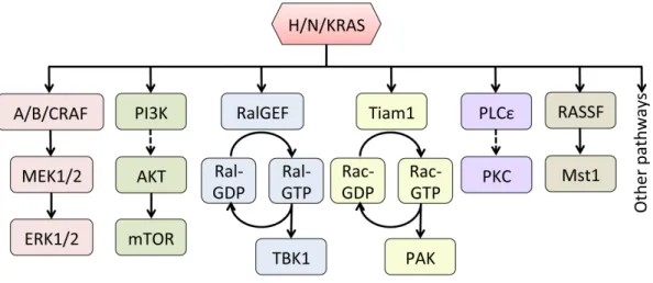
There are at least 11 identified direct effectors of RAS, including RAF, PI3K, RalGDS, Tiam1, PLCε, etc. (Figure 1.3). Among all downstream pathways, the RAF-MEK-ERK and the PI3K-AKT-mTOR pathway are by far the best studied. The RAF-MEK-ERK pathway, the first RAS effector pathway delineated, has been shown in various contexts to contribute significantly to tumor development (Blasco et al., 2011; Collisson et al., 2012; De Luca et al., 2012; Khosravi-Far et al., 1995; Stang et al., 1997), and thus has been a major focus of targeted therapies (Roberts and Der, 2007; Samatar and Poulikakos, 2014; Santarpia et al., 2012). A detailed discussion of this crucial pathway is provided in the following section.
progression (Vanhaesebroeck et al., 2010). In 1994, the p110 catalytic subunits of class I PI3K (phosphatidylinositol-4,5-bisphosphate 3-kinase) were identified as direct downstream targets of RAS (Kodaki et al., 1994; Rodriguez-Viciana et al., 1994). PI3K is a lipid kinase that phosphorylates the 3’-hydroxyl group of PIP2 (phosphatidylinositol-4,5-bisphosphate), converting the latter into PIP3 (phosphatidylinositol-3,4,5-trisphosphate), which then recruits AKT to the plasma membrane to further transmit signaling. On the other hand, PIP3 is negatively regulated by dephosphorylation by the tumor suppressor, PTEN (phosphatase and tensin homolog). Upon translocation to the plasma membrane, AKT can be phosphorylated by PDK1 on its activation loop (T308) (Wick et al., 2000), which is sufficient to activate the mammalian
target of rapamycin complex 1 (mTORC1) by direct phosphorylation on Ser2448 (Nave et al.,
1999). Activation of mTORC1 results in increased protein synthesis and cell survival upon
phosphorylation of two key effectors. Phosphorylation of 4E-BPs (elF4E-binding proteins)
terminates their binding to elF4E and relieves the block on translation initiation (Aoki et al.,
2001; Nave et al., 1999), whereas phosphorylation of p70 ribosomal S6 kinases (S6K1 and
S6K2) (Dufner and Thomas, 1999; Saitoh et al., 2002) activates their major target, ribosomal
protein S6 (rpS6). While mTORC1 signals downstream of PI3K-AKT, another mTOR complex,
mTORC2, contributes to the full activation of AKT by phosphorylating it on Ser473 (Zhang et al.,
Counter, 2005), resulting in epic efforts to develop small molecule inhibitors against components of this pathway. To date, at least 53 inhibitors of PI3K-AKT-mTOR inhibitors have reached clinical evaluation (Cox et al., 2014). While showing minimal promise as monotherapeutics, they potentially yield powerful synergism when combined with inhibitors of the RAF-MEK-ERK pathway (Engelman et al., 2008; Ewald et al., 2014; Jin et al., 2011; Lasithiotakis et al., 2008; Meier et al., 2007; Pitts et al., 2014).
Another major RAS downstream effector pathway is the RalGEF-RalA/RalB axis. RalA and RalB are two highly related (82% overall amino acid sequence identity) small GTPases of the RAS subfamily (Gentry et al., 2014). The importance of Ral activity has been established in many human cancers, including non-small cell lung cancer (NSCLC), melanoma, ovarian cancer, colorectal cancer, bladder cancer, pancreatic cancer, etc. (Coltart et al., 1990; Guin and Theodorescu, 2015; Lim et al., 2006; Mishra et al., 2010; Smith et al., 2007; Wang et al., 2010; Wang et al., 2013a; Zipfel et al., 2010). In contrast to the RAF-MEK-ERK pathway or the PI3K-AKT-mTOR pathway, Ral downstream signaling is less well characterized, and components of this pathway are not mutated in cancer. The first effector identified for Ral was RalBP1 (Cantor et al., 1995; Jullien-Flores et al., 1995; Park and Weinberg, 1995), which is also the most studied. Xenograft studies have demonstrated that RalBP1 plays an important role in several tumor types, such as pancreatic (Leake et al., 2012), prostate (Wu et al., 2010), colorectal (Mollberg et al., 2012), bladder (Wu et al., 2010), and glioblastoma (Wang et al., 2013b). Other well-established effectors of Ral include Sec5 and Exo84 (Moskalenko et al., 2002), which are subunits of the octomeric exocyst complex, and have been implicated in fostering oncogenic RAS-mediated tumorigenesis (Issaq et al., 2010).
Tiam1-RAC-PAK signaling (Baker et al., 2014; Dummler et al., 2009), RASSF-Mst1/2 signaling (Chao et al., 2015; Maruyama et al., 2008; Mezzanotte et al., 2014; Zhou et al., 2014), and PLCε-PKC signaling (Dowling et al., 2016; Leonard et al., 2015; Martins et al., 2014).
The RAS-RAF-MEK-ERK “pathway” is really a “web”
phosphorylating and activating MEK (or MKK, mitogen-activated protein kinase kinase). Two highly-related MEK1 and MEK2 proteins share 80% sequence identity, and are the only well-validated RAF substrates. Activated MEK then propagates the signal to its downstream effectors ERK1 and ERK 2 (extracellular signal-regulated kinase), which have 86% sequence identity, and are the only well-validated MEK substrates. ERK1/2, however, has over 200 substrates, distributed throughout the cell (Roskoski, 2012). The most thoroughly studied nuclear substrate of ERK1/2 is Elk1 (Yoon and Seger, 2006), a member of the ternary complex factor subfamily of Ets (E-twenty six)-domain transcription factors, which play a major role in inducing the expression of immediate early genes. The best-known cytoplasmic substrates of ERK1/2 are p90 RSK family kinases, which are known to regulate cell growth, motility, and survival (Anjum and Blenis, 2008).
showed that the MEK1/MEK2 heterodimer is negatively regulated by ERK-mediated phosphorylation of MEK1 on Thr292, a residue not present in the proline-rich sequence of MEK2 (Catalanotti et al., 2009). This phosphorylation blocks the ability of PAK to phosphorylate S298 and of Rac-PAK signaling to enhance MEK1-ERK complex formation (Eblen et al., 2004). The mechanism by which the other phosphorylation site, Thr398, attenuates MEK-ERK signaling, however, is less well understood. ERK, and its immediate substrate p90 RSK-2, are both capable of phosphorylating son of sevenless 1 (SOS1) at several residues, thereby interfering with SOS1 binding to Grb2, an adaptor protein that couples RTKs to RAS, leading to the ultimate downregulation of RAS signaling (Corbalan-Garcia et al., 1996; Douville and Downward, 1997). Further downstream, activation of ERK1/2 signaling induces the transcriptional upregulation of some negative regulators of the pathway, including dual-specificity MAP kinase (MAPK) phosphatases (MKPs or DUSPs) and Sprouty (SPRY) proteins. ERK1/2 signaling drives the expression of a variety of DUSP proteins, including the ERK-specific phosphatases DUSP5 (nuclear) and DUSP6/MKP3 (cytoplasmic) (Kidger and Keyse, 2016; Zhang et al., 2010), thus providing a straightforward means of controlling its own activity. The importance of DUSPs in controlling RAF-MEK-ERK signaling is exemplified by the frequent loss of DUSP6 in EGFR- and KRAS-driven non-small cell lung cancers (Zhang et al., 2010) and by the demonstration that the loss of DUSP5 accelerates HRAS-driven skin cancer in mice (Rushworth et al., 2014).
Understanding such negative feedback regulation is important for predicting the consequences of pharmacologically inhibiting elements of this pathway for cancer treatment, as well as for unraveling the consequences of signaling from wild-type RAS isoforms in the presence of oncogenic RAS.
Scaffolding proteins, such as kinase suppressor of RAS (KSR), also play an important role in shaping RAS-RAF-MEK-ERK signaling (Kolch, 2005). A variety of scaffold proteins including KSR1/2, IQGAP1, MP1, β-Arrestin1/2 participate in regulation of the RAF-MEK-ERK kinase cascade (Morrison and Davis, 2003). The human genome encodes two KSRs, KSR1 and KSR2. Sequence analysis indicates that KSR1/2 belong to the protein-serine/threonine kinase family. These proteins were considered catalytically inactive owing to the absence of critical conserved amino acid residues. Emerging studies have shown otherwise, but the bulk of evidence still supports a kinase-independent function of KSR. KSR1 binds all modules of the RAF-MEK-ERK pathway, but, whereas MEK is associated constitutively, RAF and ERK might bind in a stimulus-dependent manner (Morrison, 2001). KSR1-/- mice were found to be less susceptible to oncogene-induced tumors than their wild-type counterparts (Kortum and Lewis, 2004; Lozano et al., 2003; Nguyen et al., 2002), supporting a role for KSR1 in promoting the proliferative and transforming functions of the RAF-MEK-ERK pathway. As discussed in Chapter 2, I have found a previously unrecognized role for a KSR-interacting protein in modulating responses to pharmacological inhibition of the RAF-MEK-ERK kinase cascade.
CK2 modulates RAF-MEK-ERK pathway activity and vice versa
Amongst all the molecules that can modulate RAF-MEK-ERK activity, protein kinase CK2 is not one that is commonly thought of. However, there have been multiple reports of CK2 affecting the signaling output of the RAF-MEK-ERK pathway. Protein kinase CK2, formerly known as “casein kinase 2”, is a widely expressed, constitutively active kinase that phosphorylates nearly 300 protein substrates, and plays important roles in cellular survival, proliferation and differentiation (Pinna, 2002; Pinna and Allende, 2009; Trembley et al., 2009). The CK2 tetrameric holoenzyme is composed of two regulatory (β) and two catalytic (α or α’) subunits. There is substantial evidence to suggest that the subunits can also function independently of the tetramer (Hanif et al., 2010).
demonstrated that, upon EGFR activation, ERK2 directly binds CK2α via the ERK2 docking groove and phosphorylates CK2α at Thr360 and Ser362, thereby enhancing CK2α activity toward α-catenin phosphorylation (Ji et al., 2009). Furthermore, CK2 has been characterized as an integral component of the KSR1 scaffolding complex, and has been shown to be critical to the maximal activation of this pathway (Ritt et al., 2007). Aside from fine-tuning the signaling amplitude of the RAF-MEK-ERK pathway, CK2 has been reported to regulate ERK nuclear translocation and translation of nuclear targets of ERK, which, in essence, can also affect the signaling efficiency of the pathway (Plotnikov et al., 2011). Mechanistically, CK2 can phosphorylate Ser244 and Ser246 in the nuclear translocation signal (NTS) of ERK, allowing ERK binding to Imp7 (importin 7), a protein that is responsible for the nuclear import of many proteins.
Although the interactions between CK2 and the RAF-MEK-ERK pathway are sophisticated and relatively poorly defined, the following can be concluded from the above studies related to CK2 and RAF-MEK-ERK signaling: 1) CK2 is both a target and a regulator of RAF-MEK-ERK signaling, and 2) there are multiple ways by which CK2 and components of the RAF-MEK-ERK pathway can interact with and modulate each other's activity.
RAS-RAF-MEK-ERK signaling is critical to RAS- and RAF-driven cancers
Hyperactivation of the RAS-RAF-MEK-ERK pathway occurs very frequently in solid tumors, and is often the result of activating mutations in upstream receptor tyrosine kinases, RAS, or BRAF. For example, it is known that EGFR-mutant non-small cell lung cancers constitutively express active ERK (Balko et al., 2009; Britson et al., 2009). Activating RAS mutations or loss of the RasGAP NF1 can cause activation of the pathway at the level of RAS. RAS, especially KRAS, gain-of-function mutations occur at extremely high frequencies in
Genome Atlas Research, 2014a), and endometrial adenocarcinomas (Cancer Genome Atlas Research et al., 2013), whereas NF1 loss-of-function mutations are more commonly seen in melanomas (Cancer Genome Atlas, 2015), glioblastomas (Verhaak et al., 2010), and lung squamous cell carcinomas (Cancer Genome Atlas Research, 2014a). The next module in the tier, RAF, specifically BRAF, is frequently mutated in melanomas (Cancer Genome Atlas, 2015) and in papillary thyroid carcinomas (Cancer Genome Atlas Research, 2014b). In sharp contrast, MEK and ERK mutations are very rarely found in human cancers.
Given the frequency of mutational activation of the pathway and its importance in tumor maintenance, it is therefore not surprising that considerable efforts have been made to develop targeted therapies against RAS-driven cancers that are directed against the RAF-MEK-ERK kinase cascade (Samatar and Poulikakos, 2014). Attempts to target active RAS in the early days focused on targeting RAS membrane association, using FTIs to block RAS obligate lipid modification by farnesylation. However, despite their preclinical efficacy in tumor models, FTIs proved unsuccessful in the clinic for a variety of reasons including their failure to block alternative prenylation of KRAS and NRAS (Berndt et al., 2011). The next big step was a leap towards designing inhibitors that target RAS downstream signaling, in particular the RAF-MEK-ERK and PI3K-AKT-mTOR cascades. Here, I will focus on inhibitors of the RAF-MEK-RAF-MEK-ERK pathway.
RAS-MEK-ERK signaling can be manifested by the appearance of an array of secondary lesions, including papillomas, squamous cell carcinomas, keratoacanthomas, and basal cell carcinomas, in those parts of the skin without BRAF mutations (Holderfield et al., 2014a; Su et al., 2012).
ERK inhibitors are also emerging, prompted by the observation that ERK reactivation is a major cause for resistance to RAF or MEK inhibition (Lito et al., 2013). Three ERK inhibitors (BVD-523, GDC-0994, MK-8353/SCH-900353) have reached clinical evaluation (Cox et al., 2014), and SCH772984, an analog of MK-8353, has been well characterized in preclinical models (Morris et al., 2013). Whether ERK inhibitors will be superior to RAF or MEK inhibitors remains to be determined.
Resistance to targeted therapies remains a major challenge in treating RAS- or RAF-driven cancers
A common theme of targeted therapies is the emergence of drug resistance, despite remarkable initial tumor responses. The study of resistance mechanisms is necessary for uncovering new therapeutic targets or rationalizing drug combinations that forestall or delay tumor relapse (Zhou and Cox, 2015). Among all classes of inhibitors, the resistance mechanisms for BRAF inhibition are best characterized (Hartsough et al., 2014; Lito et al., 2013; Spagnolo et al., 2015) (Figure 1.6). Resistance is broadly divided into two not mutually exclusive categories: intrinsic and acquired resistance. Approximately 50% of BRAF-mutated melanoma patients show no response (~15% of them) or little response (~35% of them), as defined by a degree of tumor shrinkage sufficient to meet the RECIST criteria for a partial response to BRAF inhibition (intrinsic resistance) (Chapman et al., 2011; Flaherty et al., 2010; Sosman et al., 2012). The other half of treated patients initially display response to therapy (>30% tumor shrinkage), but eventually develop secondary tumors that display acquired resistance to these inhibitors.
encodes MEK1, have also been shown to confer RAF inhibitor resistance. However, the significance of other MEK mutations needs further interrogation, because although P124S and I111S mutations in MEK1 have been detected in pre-treatment samples, they do not provide resistance to RAF inhibitors (Shi et al., 2012a).
Mechanisms of acquired resistance that attenuate the dependence of tumor cells on ERK usually involve the activation of other parallel pathways that bypass the need for ERK signaling. For example, increased AKT pathway signaling, resulting from the concurrent loss of the PTEN and RB1 tumor suppressors, is capable of diminishing RAF-MEK-ERK dependence in BRAF-mutant melanomas (Xing et al., 2012). Upregulation of RTK signaling is another means
by which tumor cells become less dependent on ERK signaling. Using a panel of kinase-'addicted' human cancer cell lines, Settleman and co-workers found that most cells can be rescued from drug sensitivity by simply exposing them to one or more RTK ligands (Wilson et al., 2012). Girotti et al. demonstrated that BRAF inhibitor-mediated activation of EGFR-SFK (Src family kinase)-STAT3 signaling mediated drug resistance in patients with BRAF-mutant melanoma (Girotti et al., 2013). Villanueva, Herlyn and colleagues reported an IGF-1R/PI3K-dependent survival mechanism in the development of resistance to BRAF inhibitors (Villanueva et al., 2010).
resistance to MEK inhibitor RO4927350 (Wang et al., 2011). The clinical relevance of MEK1 mutations as a mechanism of acquired resistance to MEK1/2 inhibitors was highlighted by the identification of a MEK1P124L mutation in a metastastic focus of a patient post-selumetinib relapse that was undetectable in pre-treatment samples (Emery et al., 2009). Additionally, concurrent MEK2Q60P mutation and BRAFV600E amplification was found to promote resistance to trametinib (Villanueva et al., 2013). Another type of MEK inhibitor resistance mechanism involves amplification of the driving oncogene. For example, resistance to MEK inhibitor selumetinib was driven by analogous mechanisms in two separate colon cancer cell lines (Little et al., 2011). In drug-resistant COLO-205 (BRAFV600E) clones, increased abundance of BRAF was the result of BRAF amplification. Similarly, in drug-resistant HCT-116 (KRASG13D) clones, elevated KRAS abundance was due to KRAS amplification (Little et al., 2011). In the same vein, BRAF amplifications were found in two additional colorectal cancer cell lines (COLO201 and
COLO206F) with BRAFV600E mutations when chronically exposed to selumetinib (Corcoran et al., 2010). Increased abundance of activated KRAS confers resistance to MEK inhibitor CI-1040 was demonstrated in C26 murine colon cancer cells, which harbor KRASG12V (Wang et al., 2005).
Since ERK inhibitors have only recently reached the clinic, there is limited data on the mechanisms of resistance to ERK inhibition. A study by Jha et al. sought to preemptively define modes of resistance to ERK inhibition (Jha et al., 2016). Chronic exposure of the HCT-116 (KRASG13D) cell line to ERK inhibitor SCH772984 led to emergence of a G186D mutation in ERK1, which impairs binding to the ERK inhibitor.
Melanoma is an excellent disease model for studying RAS-RAF-MEK-ERK signaling and therapeutic resistance
Melanoma is a malignancy that arises from melanocytes, the melanin-producing cells that reside in a number of different anatomic sites including skin, mucosal epithelia, and meninges (Sullivan et al., 2015). For historical reasons, unless otherwise unspecified, melanoma normally refers to “skin cutaneous melanoma”, which is the most common form of the disease, and also a focus of this dissertation.
The incidence rate of melanoma has been steadily rising over past few decades (Siegel et al., 2015). Well-known as a very aggressive disease, it accounts for 75% of all skin cancer deaths. According to the SEER (Surveillance, Epidemiology, and End Results) statistics of the National Cancer Institute, melanoma is currently the sixth most common cancer in the United States, with an estimated 73,870 new cases and 9,940 deaths in 2015. Although the 5-year survival rate for localized melanoma is 98.3%, once metastasized, it plummets to 16.6%.
Advances in deep-sequencing technologies have contributed dramatically to the in-depth understanding of melanoma. Since the publication of the first melanoma genome in 2010 (Pleasance et al., 2010), subsequent large-scale sequencing studies have discovered numerous previously unknown melanoma-associateed genes involved in the regulation of the RAS-RAF-MEK-ERK and other signaling pathways (Cancer Genome Atlas, 2015; Hodis et al., 2012; Krauthammer et al., 2012; Stark et al., 2012). It is now widely accepted that, based on genomic classification, melanomas are divided into four major subtypes: mutant BRAF, mutant RAS, mutant NF1, and Triple-WT (wild-type) (Cancer Genome Atlas, 2015) (Figure 1.6). Focal
indicating that the vast majority of melanomas have hyperactivation of the RAS-RAF-MEK-ERK pathway.
Melanoma is distinct in its “choice” of RAS mutation. As introduced earlier, while most cancers preferentially harbor mutations in KRAS, melanomas exhibit a strong bias towards NRAS mutations (94%), compared to KRAS (2%) or HRAS (4%) (Cox et al., 2014).
Furthermore, in contrast to acute myeloid leukemia (AML), which also favors NRAS mutations, melanomas preferentially harbor mutations at codon Q61, rather than at G12 or G13 (Burd et al., 2014; Johnson et al., 2014). Most studies on the roles of wild-type RAS isoforms in RAS-mutant cancers to date have focused on KRAS mutant disease, likely due to the prevalence of mutations in this isoform (Grabocka et al., 2014; Jeng et al., 2012; Lim et al., 2008; Weyandt et al., 2015). Owing to their distinct biological functions in cancers (Castellano and Santos, 2011), observations from KRAS-mutant cancers may not apply to cancers with another mutated RAS isoform. Therefore, interrogating the contributions of wild-type RAS in different mutant-isoform system, such as NRAS-mutant diseases, is desired. Hence, NRAS-mutant melanoma is a naturally occurring disease model that is highly suitable for the study of RAS isoform interactions in a new mutant-isoform setting. Moreover, NRAS mutations are an independent prognostic indicator for worse clinical outcomes (Devitt et al., 2011; Jakob et al., 2012; Mann et al., 2013), and this subgroup does not have access to any FDA-approved targeted therapy. There is thus a strong urge to better understand this subpopulation of melanoma patients so as to improve their prognosis.
BRAF-mutant melanoma patients, on the contrary, are currently treated with 4 out of all
for tumor relapse, and there are many possibilities as to how a secondary tumor develops. Therefore, because of the extensive use of targeted agents in this genetic subtype, and due to the inevitable occurrence of tumor relapse, BRAF-mutant melanoma presents a suitable disease model to study therapeutic resistance to inhibitors of the RAS-RAF-MEK-ERK pathway.
Rationale and objectives for this project
Define a novel mode of resistance to RAF-MEK-ERK pathway inhibition
Considerable efforts have been invested in identifying resistance mechanisms of BRAF-mutant melanomas to BRAF inhibition (Hartsough et al., 2014; Lito et al., 2013; Spagnolo et al., 2015). Interestingly, the literature also provides some evidence that a given resistance mechanism can occur in the same or in different tumor types treated with different inhibitors of the same pathway (Corcoran et al., 2010; Duncan et al., 2012; Emery et al., 2009; Johannessen et al., 2010; Johannessen et al., 2013; Little et al., 2011; Nazarian et al., 2010; Shi et al., 2012a). Therefore, I hypothesized that some mechanisms of resistance can be shared by different drugs that target the same pathway, or even by different diseases that show hyperactivity of the same pathway.
Define the roles of wild-type RAS isoforms in the presence of oncogenic NRAS
Figure 1.4. The PI3K-AKT-mTOR-S6K pathway. The direct RAS effector phosphatidylinositol-4,5-bisphosphate 3-kinase (PI3K) converts PIP2 into PIP3, the latter recruiting AKT to the plasma membrane. PIP3 can be dephosphorylated to PIP2 by phosphatase and tensin homolog (PTEN). Upon translocation to the plasma membrane, AKT can be phosphorylated by PDK1 on its activation loop (T308), which is sufficient to activate the mammalian target of rapamycin complex 1 (mTORC1) by direct phosphorylation on Ser2448. Activation of mTORC1 results in increased protein synthesis and cell survival due to phosphorylation of its effectors, such as 4E-BPs (elF4E-binding proteins) and p70 ribosomal S6 kinases (S6K1 and S6K2), and
Figure 1.5. Scaffolds and negative feedback signaling fine-tune RAS-RAF-MEK-ERK signaling outcome. The kinase suppressor of RAS 1 (KSR1) scaffold protein facilitates the assembly of RAF, MEK and ERK to enhance signaling efficiency. Rapid and direct feedback by inhibitory phosphorylation on SOS, BRAF, CRAF and MEK1 by ERK1/2, and in some cases, the ERK1/2 effector p90 RSK, limits signal transduction through the pathway. ERK1/2 activation induces the transcriptional expression of ERK-specific dual-specificity phosphatases (DUSPs) and Sprouty (SPRY) proteins. Nuclear DUSP5 and cytoplasmic DUSP6 directly
CHAPTER II: CK2α MAINTAINS ERK ACTIVITY TO PROMOTE RESISTANCE TO INHIBITORS OF THE RAF-MEK-ERK PATHWAY IN BRAF-MUTANT MELANOMA
Overview
The protein kinase CK2 is a pleiotropic and constitutively active kinase that plays crucial roles in cellular proliferation and survival. Overexpression of CK2, particularly the alpha catalytic subunit (CK2α, CSNK2A1), has been implicated in a wide variety of cancers and is associated with poorer survival and with resistance to both conventional and targeted anticancer therapies. Here, we found that CK2α protein is elevated in melanoma cell lines compared to normal human melanocytes. We then tested the involvement of CK2α in drug resistance to FDA-approved single-agent targeted therapies for melanoma. In BRAF-mutant melanoma cells, ectopic CK2α decreased sensitivity to vemurafenib (BRAFi), dabrafenib (BRAFi), and trametinib (MEKi), by a mechanism distinct from that of mutant NRAS. Conversely, knockdown of CK2α
sensitized cells to inhibitor treatment. CK2α-mediated RAF-MEK kinase inhibitor resistance was tightly linked to its maintenance of ERK phosphorylation. We found that CK2α post-translationally regulates the ERK-specific phosphatase DUSP6 in a kinase dependent-manner, decreasing its abundance. However, we unexpectedly showed, by using a kinase-inactive mutant of CK2α, that RAF-MEK inhibitor resistance did not rely on CK2α kinase catalytic function, and both wild-type and kinase-inactive CK2α maintained ERK phosphorylation upon inhibition of BRAF or MEK. That both wild-type and kinase-inactive CK2α bound equally well to
the RAF-MEK-ERK scaffold KSR1 suggested that CK2α increases KSR facilitation of ERK phosphorylation. Accordingly, CK2α did not cause resistance to direct inhibition of ERK by the ERK1/2-selective inhibitor SCH772984 (Morris et al., 2013). Our findings support a kinase-independent scaffolding function of CK2α that promotes resistance to RAF- and MEK-targeted therapies.
Introduction
The protein kinase CK2 (casein kinase 2 or II), is a highly conserved, ubiquitously expressed, pleiotropic, and constitutively active serine/threonine kinase that has crucial roles in cell survival, proliferation and differentiation (Pinna, 2002; Pinna and Allende, 2009; Trembley et al., 2009). The CK2 holoenzyme consists of two regulatory (beta) and two catalytic (alpha or alpha’) subunits, the latter of which can also function independently of the tetramer (Hanif et al., 2010). Although CK2 itself does not appear to be a proto-oncogene, its upregulation has been shown to promote growth and prevent apoptosis, both of which promote cancer (Trembley et al., 2009). Indeed, overexpression of CK2 at the transcript and/or protein level has been observed in many cancers (Ortega et al., 2014), including multiple myeloma (Piazza et al., 2006), chronic lymphocytic leukemia (Martins et al., 2010), breast (Giusiano et al., 2011), colorectal (Lin et al., 2011) and liver cancers (Zhang et al., 2015), etc., and is correlated with poorer patient survival (Bae et al., 2015; Lin et al., 2011; Ortega et al., 2014). Similarly, CK2 exhibited 2.5-fold higher catalytic activity in metastatic melanoma than in dermal nevi (Mitev et al., 1994).
gemcitabine induced MKK4/JNK signaling, resulting in cell death (Kreutzer et al., 2010). Further, CK2 displayed elevated protein expression and activity in chronic myeloid leukemia cells that are resistant to the small molecule kinase inhibitor imatinib (Borgo et al., 2013). Either reduction of CK2α expression or pharmacological inhibition of CK2α kinase activity restored imatinib sensitivity, possibly through suppressing Akt activity (Borgo et al., 2013). However, the role of CK2α in drug resistance, particularly to targeted therapeutics, has remained underexplored.
Inhibitors of BRAF and MEK, members of the RAF-MEK-ERK kinase cascade, have achieved remarkable overall response rates in advanced melanomas harboring BRAF V600 mutations, yet a significant proportion of patients is intrinsically resistant to such therapies, and those who respond almost inevitably develop resistance over a matter of months (Menzies and Long, 2014). Considerable efforts have been invested in identifying resistance mechanisms of BRAF-mutant melanomas to BRAF inhibition, and some have demonstrated clinical relevance
(Lito et al., 2013; Spagnolo et al., 2014; Sullivan and Flaherty, 2013; Wang et al., 2015). In a recent whole-kinome siRNA screen for kinases that could induce resistance to ERK kinase inhibitors in pancreatic ductal adenocarcinoma cells, we identified CK2α as a synthetic lethal partner of ERK inhibition (Hayes et al., 2016). We postulated that kinase inhibitor resistance mechanisms can be shared by diseases that show hyperactivity of the same pathway. Given that the RAF-MEK-ERK pathway is strongly activated in both pancreatic cancer and melanoma, we sought to determine whether CK2 also plays a role in resistance to inhibition of this pathway in melanoma.
due to these decreased levels of DUSP6. Instead, we found that CK2α-mediated maintenance of ERK phosphorylation and drug resistance were kinase-independent. The ability of both wild-type and kinase-inactive CK2α to bind to the key RAF-MEK-ERK pathway scaffold protein KSR1, that is required for optimal ERK phosphorylation and activation, supports a kinase-independent scaffolding role for CK2α in facilitating optimal ERK signaling under conditions of pathway inhibition. That CK2α overexpression did not cause resistance to a direct ERK inhibitor is further evidence that ERK inhibition may overcome resistance mechanisms that shorten the effectiveness of blocking upstream kinases in the RAF-MEK-ERK pathway.
Experimental procedures Cell culture and reagents
A375, A2058, Sbc12A, Malme-3 and 293T cell lines were grown in DMEM-H supplemented with 10% FBS (HyCloneTM, Thermo Scientific) and 1% gentamycin/kanamycin (Tissue Culture Facility (TCF), Lineberger Comprehensive Cancer Center (LCCC), University of North Carolina at Chapel Hill (UNC-CH)). SK-MEL-28 and RPMI-7951 were grown in MEM-Alpha (Gibco) supplemented with 10% FBS and 1% gentamycin/kanamycin. Normal human melanocyte pellets were kindly provided by Dr. William Kaufmann, UNC-CH. The BRAFi vemurafenib (PLX4032) was a generous gift from Gideon Bollag (Plexxikon). The BRAFi dabrafenib (GSK2118436) MEKi trametinib (GSK1120212) were purchased from Selleckchem. MG132 was purchased from Calbiochem (#474790).
Plasmid constructs and gateway cloning
manufacturer’s protocol. A set of five shRNAs targeting CK2α (CSNK2A1) in pLKO.1 vector was purchased from the Lenti-shRNA Core Facility at UNC-CH. Target sequences are indicated below.
shRNA TRC Clone ID Target sequence
#1 TRCN0000000606 5’-GCTGCATTTAGGTGGAGACTT-3’
#2 TRCN0000000607 5’-CGTAAACAACACAGACTTCAA-3’
#3 TRCN0000000608 5’-CAAGAATATAATGTCCGAGTT-3’
#4 TRCN0000000609 5’-AGAATTTGAGAGGAGGTCCCA-3’
#5 TRCN0000000610 5’-CCAAGAATATAATGTCCGAGT-3’
Lentivirus production and infection
To produce lentivirus, 293T cells were transfected with pHAGE vector-based GFP, CK2α
or NRAS(Q61K), in combination with psPAX2 and pMD2.G, at a ratio of 4:3:1. After overnight transfection, the culture medium was changed to DMEM-H supplemented with 20% FBS. Thirty-six hours later, viral supernatants were harvested and filtered through a sterile 0.45 µm filter to
remove cell debris. Cleared supernatants were aliquotted and frozen at -80°C until use. Cells
were infected with 500 µl virus in 5 µg/ml polybrene (Millipore) overnight. Selection of
transduced cells in puromycin (1 µg/ml or 2 µg/ml for A375 or SK-MEL-28 cells, respectively)
was complete at 48 h after infection.
Western blotting
Lysates were depleted of cell debris by centrifugation at maximum speed (4°C, 10 min), then
proteins were quantified by Bradford assay (DCTM Protein Assay, Bio-Rad), normalized, reduced, denatured at 95 °C for 5 min, and resolved by SDS gel electrophoresis. Proteins were
transferred to PVDF membranes (Millipore, #IPFL00010) and probed with primary antibodies recognizing pERK (Cell Signaling, #4370), ERK (Cell Signaling, #9102), pMEK (Cell Signaling, #9154), pRSK (Cell Signaling, #9344), DUSP6 (Abcam, ab54940), beta-actin (Sigma, A5316), CK2α (Santa Cruz, sc-373894), FLAG-tag (Sigma, F3165; Novus, NBP1-06712SS), or GFP (Roche, 11814460001). A rabbit antibody to the phosphorylated CK2α substrate EEF1D was a generous gift from David W. Litchfield (Western University). A rabbit-anti KSR1 antibody (Morrison Lab) was used to detect KSR1 following immunoprecipitation. After incubation with the appropriate secondary anti-mouse (GE Healthcare, NA931V) or anti-rabbit (GE Healthcare, NA934V) antibody, proteins were detected by chemiluminescence (Thermo Scientific, #34075). Blots were developed by exposure to X-ray film, or by the ChemiDocTM MP Imaging System (Bio-Rad) for quantification.
Pharmacologic growth inhibition (GI50) assay
Growth inhibition assays were performed as described previously (Johannessen et al., 2013; Nazarian et al., 2010) with minor modifications. Cultured cells were seeded into 96-well plates (2,000 cells per well). Sixteen hours after seeding (baseline), serial dilutions of inhibitors were prepared in DMSO and added to cells, yielding final drug concentrations ranging from 1 nM to 10 µM for vemurafenib, dabrafenib and SCH772984, 0.1 nM to 1 µM for trametinib, with
the final volume of DMSO not exceeding 1%. Cells were incubated for 72 h following addition of drug. To measure cell proliferation, 5 mg/ml MTT (Sigma-Aldrich, #M5655) was added 1:10 into wells, and incubated at 37°C for 4 h. Formazan products were solubilized using acidified
background subtraction at 650 nm. Percent cell growth under each condition was calculated as follows: Cell growth (%)=100×(T-T0)/(C-T0), where T0 is absorbance at baseline, T is absorbance of drug-treated wells at 72 h, C is absorbance of DMSO-treated wells at 72 h. A minimum of three replicates was performed for each cell line and drug combination. Data from growth inhibition assays were modeled using a non-linear regression curve fit with a sigmoidal dose-response (GraphPad Prism, v5.0c). The resulting curves were displayed, and GI50 values were also generated, using GraphPad Prism.
Clonogenic assay
Clonogenic assays were performed as described previously (Franken et al., 2006) with slight modifications. Briefly, cells were plated in duplicate wells at 100 cells/well in 6-well plates, and allowed to adhere for 3 h at 37°C, after which culture medium was carefully removed and
replaced with medium containing either DMSO vehicle control or inhibitor. Two weeks following drug treatment, cells were washed once with PBS, then fixed and stained with crystal violet/paraformaldehyde for 30 min at room temperature. Stain was decanted, and plates were carefully rinsed with distilled water, until background staining of the wells was minimized. Finally, plates were air-dried, and colonies were counted manually using a cell counter.
Site-directed Mutagenesis
Site-directed mutagenesis of CK2α was performed using QuikChange II XL Site-Directed
Mutagenesis Kit (Agilent). A forward primer 5’-
nucleotides showing the site of mutagenesis. Reaction conditions strictly followed the manufacturer’s protocol.
RNA isolation, reverse transcription and real-time PCR
Total RNA was isolated from cells using Thermo RNA kit (Thermo Scientific), and then 0.5 µg total RNA was reverse transcribed to generate cDNA using iScriptTM cDNA Synthesis Kit
(Bio-Rad) according to the manufacturer’s instructions. Real-time PCR was performed using the SsoFastTM EvaGreen® Supermix (Bio-Rad) on the Bio-Rad CFX-96 Real-Time PCR System. Beta-actin was used for normalization. DUSP6 was amplified using forward primer 5’-CGACTGGAACGAGAATACGG-3’ and reverse primer 5’-TTGGAACTTACTGAAGCCACCT-3’.
Co-immunoprecipitation
Results
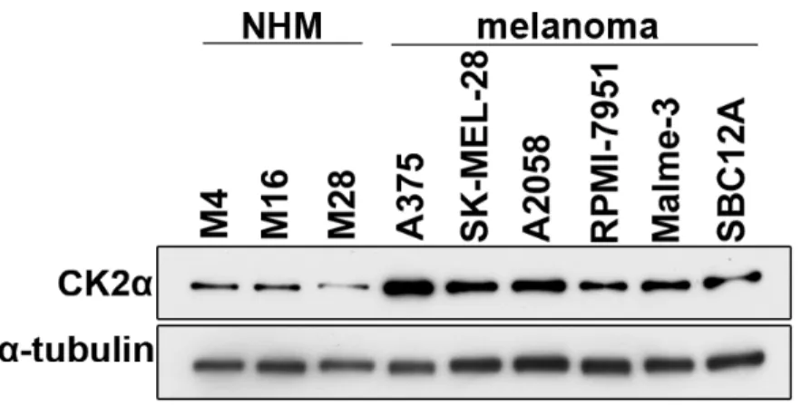
CK2α expression is upregulated in a subset of melanomas
To examine the expression of CK2α in melanoma, we first surveyed the Cancer Genome Atlas (TCGA) skin cutaneous melanoma dataset for CK2α mRNA expression through cBioPortal (http://www.cbioportal.org). We found that the CK2α transcript is upregulated in a subset of those tumors (15% of 278 samples), and that 90% of that subset also harbor mutations in BRAF, NRAS and/or NF1 that lead to hyperactivation of ERK. Next, we measured CK2α protein expression in a panel of normal human melanocytes (NHMs) and melanoma cell lines (5 BRAF-mutant: A375, SK-MEL-28, A2058, RPMI-7951, Malme-3; and 1 NRAS-BRAF-mutant: Sbc12A). Compared to NHM, all of the melanoma cell lines had higher levels of CK2α protein expression (Figure 2.1). Like the melanoma patient samples evaluated by TCGA, BRAF- and NRAS-mutant melanoma cell lines also exhibit ERK hyperactivation (Shields et al., 2007; Solit et al., 2006).
CK2α promotes resistance to inhibitors of BRAF and MEK in BRAF-mutant melanoma cells