A CELLULAR COMPARISON OF BONE INDUCTIVE PROPERTIES OF TRABECULAR METAL vs TITANIUM IN HEALTHY AND OSTEOPENIA/OSTEOPOROSIS SUBJECTS
Elizabeth L. Campbell
A thesissubmitted to thefacultyof theUniversityofNorth Carolina at Chapel Hill in partial fulfillment of the requirements for the degreeofMaster of Sciencein the School of Dentistry
(Periodontology)
Chapel Hill 2017
Approved by:
Thiago Morelli
Steven Offenbacher
©2017
Elizabeth L Campbell
ABSTRACT
ELIZABETH L. CAMPBELL : A Cellular Comparison of Bone Inductive Properties of Trabecular Metal vs Titanium in Healthy and Osteopenia Subjects.
(Under the direction of Thiago Morelli, Steven Offenbacher and Antonio Moretti)
In this study, we examined the histologic healing associated with initial osseous
healing comparing trabecular metal vs. standard titanium in healthy and
osteopenia/osteoporosis subjects. We proposed to add to this body of knowledge in
two major ways. First, we identified the temporal differences in cellular
recruitment and activation during early healing and early osteogenesis, and the bone to
implant contact (BIC). This was done by placing test cylinders (approx. 2.9-3 x 5 mm)
using standard titanium or trabecular metal. Both total BIC and BIC percentage was
calculated for both trabecular and titanium cylinders harvested 4 weeks after
placement. Secondly, we recognized that osseous healing can vary considerably in
the face of chronic health problems including osteopenia/ osteoporosis. This
chronic condition is known to impair bone repair and osseous regeneration. The use
of trabecular metal may have the strategic advantage of demonstrating superior
osseous healing in these compromised individuals as compared to titanium. This
project was designed to examine the differences in the histologic BIC, not only
comparing trabecular metal to titanium, but also to examine these differences in
ACKNOWLEDGEMENTS
I want to thank my husband most of all for always being so supportive and going above
and beyond. You are the love of my life and I am forever grateful for your love and support.
I literally could not have made it to the end without you.
I also want to thank my amazing mentor, Dr. Thiago Morelli, who has been an
inspiration throughout this entire journey. I appreciate all the support he has given me. I
want to give a special thanks to Dr. Antonio Moretti for being so supportive throughout my
entire residency, even after a couple very large set backs. I cannot thank you enough for
standing by me, and all the words of encouragement. Words cannot truly describe how much
I appreciate everything that you have done for me. Thank you to Dr. Offenbacher for
generously taking time out of your busy schedule to work with me on this project. I have
learned so much under your guidance. Dr. Silvana Barros, thank you for you guidance and
hard work. Also thank you Dr. Sompop Bencharit for all your efforts with this study.
I am so grateful for everyone who helped me along this journey and made this entire
project possible. Acela, you have been my role model throughout residency and like a sister
to me, I am forever indebted for your kindness and guidance.
Thank you for the entire Go Health team, especially S.T Phillips and Wendy Lamm.
I want to express my deepest gratitude to all my friends and colleagues who supported
and encouraged me every step of the way. My fellow residents, David and Krystal, you both
I want to thank the UNC Core Facility and Wallace from CHANL and the amazing
staff that have been nothing but kind, helpful and accommodating. I am forever thankful to
Victoria Madden and Kristen White, you both were so amazing, and I am so grateful for the
time you spent working with me throughout this project.
I would also like to thank Zimmer for their generous contribution and their willingness
to fund this project. Their support is immensely appreciated.
Lastly I want to thank my amazing parents; Tommy and Kathy, and my wonderful
brother, Michael. I am so grateful for your continued support and unwavering love and
TABLE OFCONTENTS
LIST OFTABLES ... viii
LIST OF FIGURES ... ix
LIST OF ABBREVIATIONS ... x
CHAPTER 1: OSTEOPOROSIS AND OSTEOPENIA, BONE AND TISSUE ENGINEERING AND THE ROLE OF DENTAL IMPLANTS IN AFFECTED PATIENTS ... 1
Introduction ... 1
Section 1.1 Bone and Tissue Engineering: Gross Structure, Formation ... 2
Section 1.2 Bone Metabolism ... 4
Section 1.3 Bone Metabolic biological pathways and gene expression ... 5
Section 1.4 Osteoporosis: Definition, types and pathophysiology ... 7
Section 1.5 Osteoporosis Diagnosis, Prevalence and Burden ... 9
Section 1.6 Osteoporosis Treatment ... 12
Section 1.7 Dental Implants and Osseointegration ... 23
Section 1.8 Outcomes and Factors Affecting Dental Implants ... 25
Section 1.9 Implications of Osteoporosis and Dental Implant Therapy ... 26
Section 1.10 Porous Tantalum Trabecular Metal: Production and Biologic Influence ... 30
Summary ... 31
Introduction ... 33
Section 2.1 Materials and Methods ... 34
Clinical Relevance ... 34
Participants ... 34
Study design ... 38
Histology ... 43
Section 2.2 Statistical Analyses ... 44
Section 2.3 Results ... 44
Histology Data ... 45
Discussion ... 57
CHAPTER 3:CONCLUSION ... 62
Conclusions ... 62
LIST OFTABLES
Table 1.1: Molecular pathways of osseointegration ... 7
Table 1.2: Bone Mineral Density Diagnostic Categories ... 10
Table 1.3: Osteoporosis therapies, mechanism of action and safety concerns ... 23
Table 2.1: Patient Demographics ... 37
Table 2.2: GLM Analysis Data for Mean BIC Percentage ... 50
Table 2.3: Analysis of GEE Parameter Estimates for Mean BIC Percentage ... 51
Table 2.4: GLM Analysis Data for Mean BIC Total ... 53
LIST OF FIGURES
Figure 1.1: Management Chart for Osteoporosis ... 12
Figure 1.2: Chemical Structure of Bisphosphonates and Inorganic Pyrophosphate ... 14
Figure 1.3: The cellular and biochemical mechanisms of action of bisphosphonates ... 16
Figure 1.4: Effect of Binding Affinity of Bisphosphonates on their Uptake and Detachment from bone surfaces and their re-cycling ... 17
Figure 1.5: Effects of parathyroid hormone ... 21
Figure 1.6: Classification of bone quality and jaw shape ... 28
Figure 2.1: Inclusion and Exclusion criteria ... 36
Figure 2.2: Titanium and Tantalum Test Cylinders ... 39
Figure 2.3: Test cylinders on day of insertion ... 40
Figure 2.4: Trephine with Stent ... 41
Figure 2.5: Histologic images of 4x Tantalum (a) and Titanium (b) test cylinders in healthy patients ... 46
Figure 2.6: Histologic images of 20x Tantalum (a) and Titanium (b) test cylinders in healthy patients ... 46
Figure 2.7: Histologic images of 20x Tantalum (a) and Titanium (b) test cylinders in osteonpenia/osteoporotic patients ... 47
Figure 2.8: Histologic images of 20x Tantalum (a) and Titanium (b) test cylinders of osteopenia/osteoporotic patients ... 47
Figure 2.9: Cross-sectional slices of titanium and PTTM test cylinders ... 48
Figure 2.10: Mean BIC Percentage Comparison ... 49
Figure 2.11: Mean Total BIC Comparison ... 52
Figure 2.12: Mean Difference BIC Percentage (PTTM vs Titanium ... 54
LIST OF ABBREVIATIONS
ANOVA Analysis of variance
ATP Adenosine triphosphate
BFM Brightfield microscopy
BGLAP Bone Gamma-Carboxyglutamate protein
BIC Bone to implant contact: %
BMP Bone morphogenetic protein
BMD Bone Mineral Density
BOP Bleeding on probing
BRONJ Bisphosphonate-related osteonecrosis of jaws
BSAP Bone-specific alkaline phosphatase
CT Calcitonin
CTX C-telopeptide of Type I collagen
DALYs Disability Adjusted Life Years
DXA Dual-energy X-ray absorptiometry
EGFR Epidermal Growth Factor Receptor
ERT Estrogen replacement therapy
FGFR2 Fibroblast Growth Factor Receptor 2
FN1 Fibronectin 1
FRAX Fracture Risk Assessment Tool
GEE General estimating equations
GLM General linear model
IRB Institutional review board
MI Myocardial infarction
MRONJ Medication-related osteonecrosis of the jaws
MSC Mesenchymal stem cells
M-CSF Macrophage colony-stimulating factor
NIH National Institutes of Health
NTX N-terminal telopeptide
OC Osteocalcin
ONJ Osteonecrosis of jaws
OPG Osteoprotegerin
PCR Polymerase chain reaction
PINP Procollagen type I N-terminal propeptide
PHEX Phosphate Regulating Endopeptidase Homolog X-linked
PPi Inorganic Pyrophosphate
PTH Parathyroid hormone
PTTM Porous Tantalum Trabecular Metal
RANK Receptor activator of nuclear factor kappa B
RANKL Receptor activator of nuclear factor kappa B ligand
RNA Ribonucleic acid
SEM Scanning electron microscopy
SERM Selective Estrogen Receptor Modulator
Ta Tantalum
Tb Trabecular
TGF/BMP Transforming growth factor/Bone morphogenetic proteins
TGFB Transforming Growth Factor Beta
TNF-α Tumor necrosis factor alpha
TPTD Teriparatide
U.S. United States
VCAM1 Vascular Cell Adhesion molecule
VDR Vitamin D (1,25- Dihydroxy Vitamin D3) Receptor
VEGFB Vascular Endothelial Growth Factor B
VTE Venous thromboembolic events
WHO World Health Organization
CHAPTER 1: OSTEOPOROSIS/OSTEOPENIA, BONE AND TISSUE ENGINEERING AND THE ROLE OF DENTAL IMPLANTS IN AFFECTED
PATIENTS
Introduction
Osteoporosis is a debilitating metabolic bone disease that is a major health condition
that affects approximately 75 million people in the US, Europe and Japan(1). Ten million are
affected in the U.S. alone, and by the year 2025 it is predicted that a 50% increase in
prevalence will be seen due to the contribution of the aging population(2). It is defined by
the World Health Organization (WHO) as a bone density 2.5 SDs below the mean for young
adult women(3). Osteoporosis leads to an increased incidence of fracture, most notably hip
fractures; leading to an increased mortality and morbidity(3). In patients with hip fractures, 1
in 5 die, while of the total number with osteoporotic related hip fractures, 1/3 require nursing
home placement because they are functionally dependent(3). These fractures trigger
increased healthy care costs, estimated at $10-15 billion annually(3).
While osteoporosis is not directly a cause of tooth loss, most patients affected with
osteoporosis are elderly and may have an increased risk of tooth loss due to prolonged
exposure to various factors that are associated with tooth loss. The need for implant therapy
can increase as patient ages due to increased risk for tooth loss. These patients can benefit
immensely from rehabilitation using dental implants.
The use of dental implants in patients with osteoporosis is a debatable issue, caused by
altered ability for bone formation. Medications used to treat osteoporosis have been shown
to cause a decrease in risk of fracture, however, many lead to oral side effects such as
bisphosphonate related osteonecrosis of the jaw (BRONJ) or more recently known as
medication related osteonecrosis of the jaw (MRONJ). There are an abundance of clinical
studies reporting on the outcomes of implant therapy in patients with osteoporosis, but
several fail to differentiate between the effects of osteoporosis and its therapy. Many studies
have demonstrated successful osseointegration of dental implants in osteoporotic patients
with no contraindication for implant therapy(4-6). However, there are other studies which
show that osseointegration of implants in patients with osteoporosis could be negatively
affected(7, 8).
Section 1.1 Bone Tissue: Gross Structure, Formation
Bone is a mineralized and dynamic type of connective tissue that has many functions,
including: movement, protection, support, hematopoiesis and mineral reservoir(9). This
histologically unique tissue is comprised of a complex structure of both organic content and
inorganic components(9). The organic components are comprised mainly of type I collagen
and various other non-collagenous matrix proteins(9). Despite their location in the body, all
bones have similar basic structure. The variability in the overall size and shape of individual
bones begins during embryonic development and continues up to the pre-adult stage in
growth(9).
The overall organizational structure of bone includes internal and external components
that all interact with one another to form a delicate equilibrium. The external structure of
bone includes an outer layer of dense fibrous connective tissue membrane known as the
which are involved in the functionality of this tissue. The outer layer is known as the fibrous
periosteum and it is not in contact with the bone and acts as the location for muscle and
tendon attachment, along with aiding in nutritional support(9). Cells within the fibrous layer
are very densely packed together; lymphatics, vasculature neural tissue are also located in the
fibrous layer(9). The inner layer, known as the osteogenic periosteum, is the layer adjacent
to the cortical bone surface(9). The inner periosteum functions to maintain the osteogenic
potential of the bone during injury or growth. The internal structural elements include a
dense outer layer of compact bone which encapsulates the inner supporting network of
trabecular bone(9). The porous structure of the trabeculated bone is filled with marrow,
which is the vital tissue involved in hematopoiesis(9). A poorly demarcated layer of loose
connective tissue, known as the endosteum, lines the internal aspect of each bone(9). The
role of the endosteum is a physical barrier between the two bone layers; it also acts as a
source of osteogenic cells(9). The combination of all of these aspects makes up the
organizational components of bone.
Different cells that comprise the bone have different functions and in health work in
congruence with one another to maintain form and function, however these same cells can
function in disease when the balance is tipped. Histologically, both compact and trabecular
bones are identical and consist of microscopically unique lamellar types, each of which are
found in distinct locations with the bone structure. The osteon is the basic organizational
unit that consists of concentric lamellae, which form a cylinder of bone surrounding a central
vascular channel(9). Osteoblasts are of mesenchymal origin and are mononucleated cells
regulate cellular function and bone metabolism(9). Osteocytes are osteoblasts cells that have
become entombed in the mineral matrix they secrete while forming the structure. These
cells are located in lacunae and communicate with one another and other cellular types via
enclosed channels. These channels are known as canaliculi and allow for the osteocytes to
sense the biochemical and mechanical environmental changes within the bone and transmit
signals to induce a response(9). Osteoclasts are larger multinucleated bone cells responsible
for the bone degradation, a critical function in maintenance, repair and remodeling
processes(9). Osteoclasts originate from monocytes/macrophage lineages and differentiate
into functional cells when exposed to receptor activator of nuclear factor kappa-B ligand
(RANKL), macrophage colony-stimulating factor (M-CSF), and tumor necrosis factor alpha
(TNF-α)(10).
Section 1.2: Bone Metabolism
Because bone is a metabolically active tissue, it is in a constant state of flux in response
to specific functional and nutritional demands(9). During childhood, bone turnover is much
greater than adults, as one ages the rate of turnover rate decreases, yet remains balanced
during a state of health. Bone turnover rates for trabecular bone are 15% per year, while
cortical bone is approximately 5% per year(9). The cyclical nature of remodeling consists of
three consecutive phases: resorption, reversal and formation(11). The resorptive phase is
initiated with the migration of partially differentiated mononuclear pre-osteoclast and lasts
approximately 2 weeks(11). The main player in the resorptive phase is the osteoclast, which
dissolves the bone mineral, and enzymatically degrades extracellular matrix (ECM)
proteins(10). The initiation of the resorptive phase is systemically controlled via four main
estrogen(12). Calcitonin, PTH and 1,25 Vit D3 are all secreted with the intention to control
the levels of serum calcium to maintain a precise physiological range(12). With the
completion of the resorptive phase, mononuclear cells will appear on the surface of the bone
and prepare the surface for bone formation by osteoblasts(11). These reversal mononuclear
cells provide signals for the differentiation and migration of osteoblasts and can last up to 4
to 5 weeks(11). Specific factors are created by the differentiated osteoclasts and
mononuclear reversal cells, or released from the demineralized matrix trigger the initiation of
the formation phase(9). The formative phase main cellular constituent is the osteoblast,
which produces the organic bone matrix and aids in mineralization(13). As osteoblasts form
bone and mature, they produce more OPG and less RANKL, resulting in the inhibitory action
of osteoclasts(9). The formation phase can continue for up to 4 months(11). Mechanical
force also plays a role in bone remodeling and architecture through local and systemic
responses(14). The osteocyte, which a post-mitotic osteoblasts-derived cell, acts as a
mechanosensor and an endocrine cell, facilitating the signal transduction in reaction to
mechanical or metabolic stimuli(15).
Section 1.3: Bone Metabolic biological pathways and gene expression
The interplay of osteoblastic and osteoclastic function forms a delicate balance created
by a multitude of unique and different cytokines, growth factors, and hormones(9). In
disease states, however, the balance is tipped leading to pathologic bone formation or loss.
Although the transcriptional regulation and signaling pathways are not well understood, the
formation bone requires highly controlled biologic pathways stemming from the
process from mesenchymal stem cells or hematopoietic precursor cell into their respective
bone cells(9). Only select transcription factors will be discussed in this review. Osteoblastic
phenotyptic commitment is regulated by principal transcription factors, one of which is
transforming growth factor/ bone morphogenetic protein (TGF/BMP) and fibroblast growth
factors (FGF)(17). The transcription factors RunX2 and Osterix are essential for control of
commitment and differentiation of mesenchymal stem cells (MSCs) into osteoblasts(9). The
threshold of RunX2/Osterix will determine the pathway of commitment to either osteocyte or
chondrocyte cell lineage(17). Several hormones and cytokines are involved in a dosage and
time dependent fashion, influencing the progression of MSCs to an osteoblastic lineage either
directly or indirectly(18). Receptor-activated nuclear factor (RANK) and its ligand
(RANKL), expressed on the plasma membrane of osteoblast progenitor cells(9). RANKL
then binds to the RANK expressed on plasma membrane of osteoclast progenitor cells,
inducing the differentiation of osteoclasts(9). The osteoclasts also secrete a decoy to
RANKL, known as osteoprotegrin (OPG), which blocks the binding of RANK and RANKL,
creating a type of self-regulatory function(9). Localized control of the remodeling ensures
that bone is removed when damaged and replaced where needed(19). Fibroblast growth
factors (FGF) are growth factors that are involved in the regulation of cellular proliferation,
differentiation, survival, and migration of osteoblasts(20). An osteocytic marker,
phosphate-regulating gene with homolog X chromosome (PHEX), is one of the many
“mineralization-related genes” which are involved in the regulation of mineralization and phosphate
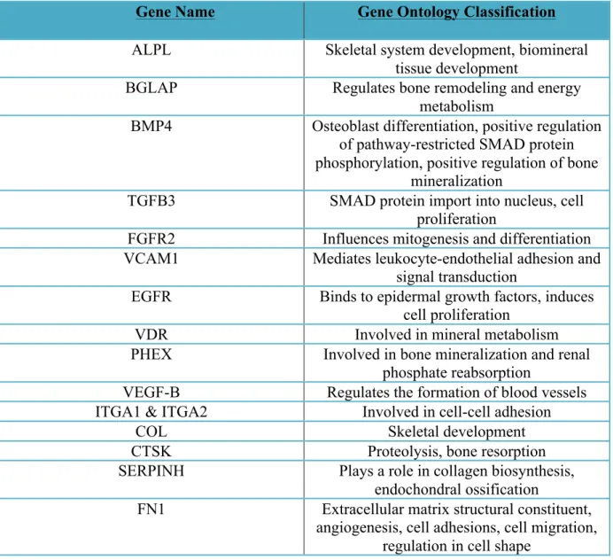
metabolism(21). The genes discussed in this section and their ontology classification is
Gene Name Gene Ontology Classification
ALPL Skeletal system development, biomineral
tissue development
BGLAP Regulates bone remodeling and energy
metabolism
BMP4 Osteoblast differentiation, positive regulation
of pathway-restricted SMAD protein phosphorylation, positive regulation of bone
mineralization
TGFB3 SMAD protein import into nucleus, cell
proliferation
FGFR2 Influences mitogenesis and differentiation
VCAM1 Mediates leukocyte-endothelial adhesion and
signal transduction
EGFR Binds to epidermal growth factors, induces
cell proliferation
VDR Involved in mineral metabolism
PHEX Involved in bone mineralization and renal
phosphate reabsorption
VEGF-B Regulates the formation of blood vessels
ITGA1 & ITGA2 Involved in cell-cell adhesion
COL Skeletal development
CTSK Proteolysis, bone resorption
SERPINH Plays a role in collagen biosynthesis,
endochondral ossification
FN1 Extracellular matrix structural constituent,
angiogenesis, cell adhesions, cell migration, regulation in cell shape
Table 1.1: Molecular assessment of osseointegration. Adapted from Thalji & Cooper 2013(22).
While the mechanisms involved in the local control and systemic control of bone remodeling
is not completely understood, the current known major mechanisms have been reviewed in
the most basic sense.
Section 1.4 Osteoporosis: Definition, types and pathophysiology
The National Institute of Health (NIH) defined osteoporosis as a “skeletal disorder
characterized by low mineral density, microarchitecture deterioration along with changes in
the mineral property of the bone causing enhanced fragility, ultimately leading to a higher
susceptibility for fracture(23). Bone strength, density and quality are all utilized as
measurements to evaluate the effect of bone metabolic diseases, specifically osteoporosis; all
are interdependent on one another. Bone strength is the echoes of the integration of bone
density and quality, while bone density is simply expressed in units: grams per area or
volume(3). Bone quality is the architecture, mineralization, damage accumulation, and
turnover (3). There are two types of osteoporosis, primary and secondary. Primary
osteoporosis most often occurs after the onset of menopause, however, it can occur in both
sexes and generally is seen later in life for males(3). Secondary osteoporosis is due to the
effects of specific medications such as glucocorticoids, diseases or disorders causing
malabsorption(3). Primary osteoporosis mainly affects the trabecular bone, while secondary
osteoporosis is characterized by the loss of cortical and trabecular bone(24). The
micro-architectural differences can be seen upon gross histologic evaluation and with more detail
utilizing scanning electron microscopy (SEM).
The pathophysiology of osteoporosis is multifactorial, but stems from the inability of
the formative bone phase to outpace the resorptive bone phase, leading to an overall loss of
mineral and detrimental changes in microstructure. In health, targeted remodeling is
completed in order to repair sites of micro-damage, while stochastic remodeling is completed
to maintain plasma homeostasis(25). If stochastic remodeling is excessive it can lead to
skeletal weakness through loss of bone mass(25). While remodeling favors the resorptive
phase, trabeculae become thinner and fewer in number as well as lose their connectivity(26).
leading to increased risk for fracture(27). An assessment of the remodeling rates can be done
with the use of markers of bone resorption: N-terminal telopeptide (NTX) or C-telopeptides
of type I collagen (CTX), and markers of bone formation: OC, procollagen type I N-terminal
propeptide (PINP) and bone-specific alkaline phosphatase (BSAP); can be used to evaluate
increased fracture risk, however, these are difficult apply clinically in patients(25).
Section 1.5: Diagnosis, Prevalence and Burden
The changes in bone mineral density (BMD) microarchitecture that lead to fractures
drastically impact the quality of life and can lead to temporary or permanent disability(28).
Therefore, the diagnosis of osteoporosis is extremely important in the clinical setting to
prevent fractures and decrease fracture risk. In a clinical setting, the utilization of patient
history and physical exam coupled with diagnostic tests are key to an accurate diagnosis and
evaluation for intervention. In many cases, the disease can be undiagnosed until the patient
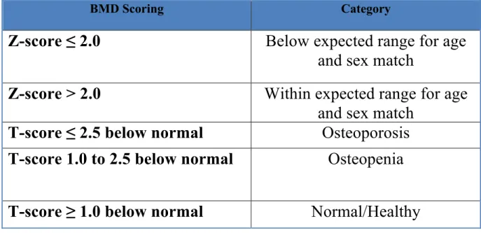
has suffered from a fracture. The diagnosis of osteoporosis is given when the BMD exceeds
more than 2.5 standard deviations (SD) below the normal mean for a young adult woman(3).
BMD is reported using T-scores, and a routine and noninvasive method for the measurement
of BMD is done by dual energy X-ray absorptiometry (DXA), it is also considered the gold
standard for BMD assessment(29). A BMD T-score between 1.0 and 2.5 SDs below the
young adult woman mean equates to a diagnosis of osteopenia, while a BMD T-score equal
to or above -1.0 is reflects as a normal score(30). A test for BMD will also produce a
Z-score that allows for the comparison of the patient’s BMD to that of a healthy age matched
individual. Both gender and ethnicity/race play a role in the prevalence and fracture
BMD Scoring Category
Z-score ≤ 2.0
Below expected range for age
and sex match
Z-score > 2.0
Within expected range for age
and sex match
T-score ≤ 2.5 below normal
Osteoporosis
T-score 1.0 to 2.5 below normal
Osteopenia
T-score ≥ 1.0 below normal
Normal/Healthy
Table 1.2: BMD Diagnostic Categories. Adapted from Leslie et al 2006 (31).
All ages, races and ethnicities can be affected by osteoporosis, however, the majority
are post-menopausal white women(3). Although both men and women show an age-related
decline in BMD towards the beginning of midlife, women have a much more pronounced
BMD loss after the onset of menopause, which occurs roughly around the age of 51 for most
women(3). Menopause is defined as the reduced secretion of estrogen and progesterone,
leading to the cessation of menstruation(32). It is diagnosed 12 months after amenorrhea not
resulting from a pathological cause, but can be the result of surgical intervention,
chemotherapy or radiation(32). With the depletion of estrogen, the inhibition of osteoclasts
is lost, along with decreased intestinal calcium absorption caused by the flux of calcium into
the plasma from bone resorption resulting in the reduction in parathyroid hormone levels(33).
The damaging effects of osteoporosis on BMD can be seen 5 to 7 years surrounding the onset
of menopause, whereby women lose approximately 12% of their total bone which is the
the effectiveness of the fracture risk assessment, WHO developed the Fracture Risk
Assessment Tool (FRAX)(34). FRAX utilizes risk factors that were identified from several
meta-analyses; its algorithms estimate a 10-year probability of fracture(34). Risk factors for
fracture include age (40-90 years), weight, height, sex, low femoral neck BMD, history of
previous fracture, parental history of hip fracture, current tobacco habits, use of
glucocorticoids, rheumatoid arthritis, alcohol intake and other causes attributed to secondary
osteoporosis(35). FRAX will then provide a numerical value which is the ten year
probability of a major osteoporotic fracture as a percentage and the decision for intervention
can be determined from the management chart seen in Figure 1.1(35). With osteoporosis
related fractures comes numerous medical related expenses, estimated at 15 to 20 billion
dollars yearly in the US which, much of the cost is paid by Medicaid and Medicare(36).
Osteoporosis related fractures account for more combined deaths and morbidity than any
single type of cancer, with the exception of lung cancer(37). The global burden of
osteoporosis is normally quantified by disability adjusted life years (DALYs), which factors
the years of life lost due to fracture and the disability of those who survive(38). It was
calculated that in 2000, an estimated 9 million osteoporosis related fractures accounted for a
total of 5.8 million DALYs(37). This number encompasses 0.83% of the world-wide burden
Figure 1.1: Management Chart for Osteoporosis. Dotted line shows the intervention threshold. Reproduced with permission.(39).
Section 1.6: Treatment of Osteoporosis
Osteoporosis is treated in a varying number of ways, including changes in lifestyle
approaches and pharmacological interventions. Lifestyle changes range from smoking
cessation, weight bearing exercises, calcium and vitamin D supplementation. However, if
the patient falls into an increased risk category based on their T/Z-scores or other risk
assessment tool, pharmacologic intervention is necessary. Prior to pharmacologic
intervention, the cost, risk of adverse effects versus benefit, and limitation of the medication
should all be considered(40). The main objective of each pharmacologic treatment modality
been utilized in the treatment of osteoporosis: antiresorptive agents and bone-forming agents.
Antiresorptive agents include Hormone Replacement Therapy (HRT), bisphosphonates,
calcitonin, parathyroid hormone (PTH), RANKL inhibitor, and selective estrogen receptor
modulators (SERMs)(41). The current bone-forming agent available on the market is
teriparatide. Both antiresorptive and bone-forming agents have a range of dosing frequencies
and routes of administration that are tailored specifically for each medication(41). With the
use of pharmacologic intervention, vertebral fracture risk is reduced by 30-70%, dependent
on the agent and level of adherence(41). The decision to prescribe a pharmacologic
intervention or utilize life style changes or dietary supplementation needs to be completed
after careful evaluation of the individuals’ current BMD score and relative risk.
Bisphosphonates historically have been one of the most frequently used osteoporotic
treatment medications. The chemical structure of bisphosphonate is similar to inorganic
pyrophosphate (PPi), allowing this type of drug to perform partially as a nonhydrolyzable PPi
analog(42). The chemical structure of several bisphosphonates and PPi can be seen in Figure
Figure 1.2: Chemical Structure of Bisphosphonates and Inorganic Pyrophosphate. Reproduced with permission. (43).
In general, bisphosphonates are very hydrophilic with poor lipophilicity relating to poor
oral bioavailability with only <1% is absorbed from the gastrointestinal tract after oral
administration(44). To offset the poor absorption, increased doses can be given as
bisphosphonates have a dose-dependent absorption(44). The overall mechanism of
bisphosphonates includes the localization of the drug to the bone where they bind to the
calcium in hydroxyapatite due to their high affinity for bone, as they are synthetic analogues
to PPi(45). The chemical structure of bisphosphonates greatly affects their function; the
hydroxyl groups are critical for the binding to calcium and the terminal functional group
been shown that bisphosphonates can prevent apoptosis of osteocytes and osteoblasts via the
rapid activation through phosphorylation of extracellular signal regulated kinase (ERK)
pathway(46). As remodeling occurs, the drug is incorporated into osteoclast, causing
reduced resorptive activity and eventually cell death(47). The ability to be preferentially
incorporated into the bone and obtain skeletal retention is based on the availability of
hydroxyapatite binding sites(45). The hydroxyapatite binding sites are more readily
available in bone metabolic disorders that favor resorption(45). Any drug that is not
incorporated into the skeleton is then rapidly cleared from circulation by renal excretion(45).
There are two types of bisphosphonate types, nitrogenous and non-nitrogenous. The
first generation bisphosphonates are non-nitrougenous and include: etidronate, clodronate
and tiludronate(45). These non-nitrogen containing bisphosphonates become incorporated
into newly formed adenosine triphosphate (ATP) molecules which accumulate inside
osteoclasts and are cytotoxic due to their inability to by hydrolyzed, leading ultimately to
cellular apoptosis(45). Second and third generation are nitrogen-containing and include:
alendronate, risedronate, ibandronate, pamidronate and zoledronic acid(45). These later
generation bisphosphonates bind and inhibit a key regulatory enzyme critical to the
production of cholesterols, isoprenoid lipids and other sterols(45). This inhibition leads to
interference in key cellular function in osteoclasts, leading to apoptosis(45). The mechanism
Figure 1.3: The cellular and biochemical mechanisms of action of bisphosphonates. Reproduced with permission. (43).
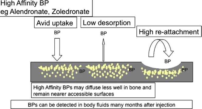
The route of administration of bisphosphonates is important for dosage, onset and risk
for potential side effects. The maximum effects of resorption suppression can be seen around
3 months after the initiation of the oral bisphosphonate(45). The rate of bone remodeling
dictates the half-life and due to the length of natural bone turnover the half-life can be at least
10 years(44). These long half-lives are the reason that a drug holiday would not allow for the
recovery of osteoclastic function and return of normal bone turnover(45). The effect of
binding affinity of different bisphosphonates and the relative uptake and detachment from the
Figure 1.4: Effect of Binding Affinity of Bisphosphonates on their Uptake and Detachment from bone surfaces and their re-cycling. Reproduced with permission. (43).
Bisphosphonates have been associated with various adverse side effects and safety concerns.
Some of the adverse side effects are based on the mode of administration; oral administration
has been associated with gastrointestinal effects(45). However, acute phase reactions
including fever, myalgia, arthralgia and headache are more common with intravenous (IV)
administration of bisphosphonates(45). One of the most significant adverse events with
severe oral implications is osteonecrosis of the jaw (ONJ), which is thought to be caused by
an over suppression of bone turnover(48). This over-suppression allows for the persistence
rigid and brittle, with an osteoporotic architecture(50). The majority (94%) of reported cases
have been in patients with intra-venous (IV) second and third generation bisphosphonate
therapy for the treatment for metastatic bone diseases(48). Of the areas in the mouth that are
affected, there is a higher propensity for the mandible to be affected (2:1 ratio) as compared
to the maxilla(48). More than half of the patients affected (60%) reported a dento-alveolar
surgical procedure prior to onset, however, spontaneous onset without injury or surgical
intervention can occur(51). The fragility of the mucosal barrier against trauma and increase
exposure to oral pathogens are reasons for increase susceptibility for osteonecrosis of the oral
cavity(45). The most important risk factors for BRONJ are the total dose, history of trauma,
dental surgery or dental infection.
The most abundant mineral in bone is calcium; bone acts as a reservoir for the storage
of over 98% of the body’s total calcium(52). The majority of peak bone mass is genetically
determined and acts as a significant risk determinant for future fracture risk(52). The main
advantageous effects of calcium supplementation in the increase of BMD and reduction in
fracture risk is most pronounced in the later postmenopausal years, while there is less of an
effect in the early post menopausal years(52). Research has shown that calcium supplements
alone improve BMD but are unsuccessful in their ability to reduce the risk for fracture(53).
Unfortunately, the use of calcium supplementation is not without risk as evidence shows
calcium supplementation has an increased risk of cardiovascular events such as myocardial
infarction (MI)(53).
Many practitioners have extensively used vitamin D and calcium supplementation as an
intervention for at-risk patients; however, many overlook this need prior to or during
bisphosphonates, due to limited dietary absorption and reduced intake, possible renal
impairment and higher incidence for limited sun exposure(45). Although currently there is
no consensus on the optimal 1,25 Vit D3, however 30ng/mL, and reduced vitamin D serum
levels cause a reduction in calcium absorption (10-15%) and phosphorus absorption (60%)
(54). Hypovitaminosis D can also lead to skeletal muscle weakness which can increase the
risk for falls, increasing the risk for fractures(54). With the drop in intestinal calcium
absorption there is an increase production in parathyroid hormone increasing the skeletal
leaching of calcium to supplement low serum levels(54). The metabolism of vitamin D in
the regulation of calcium, phosphorous and bone metabolism can be seen in Figure 1.8.
An endogenous polypeptide hormone used in the treatment of postmenopausal
osteoporosis through its affect on osteoclasts is calcitonin (CT)(55). With specific receptors
on osteoclasts, the binding of calcitonin causes the movement out of the resorptive pit with
the loss of the brush border; it also modifies the internal structure of osteoclasts by inhibiting
multiple cytoplasmic functions key to bone resorption(56). Calcitonin shortens the lifespan
of number of osteoclasts, it also blocks the union of mononuclear marrow progenitor cells
that become osteoclasts, thereby reducing the number of osteoclasts(57). More recently
research has shown that calcitonin aid in increasing the survival rate of osteoblasts and
osteocytes(46). Early calcitonin was administered via injection, leading to issues in
compliance and the need for the creation of suppository and nasal spray modes of
administration(58). The effectiveness of calcitonin on osteoporosis treatment has shown a
30% reduction in hip fractures(59). It has also been suggested that calcitonin may have an
calcitonin can occur and that intermittent administration may help to avoid tolerance(61).
Unlike bisphosphonates, the effects of calcitonin on osteoclasts is reversible and the
medication does not persist in the bone even after longlapses in medication
administration(62).
Teraparatide (TPTD) or recombinant parathyroid hormone is an anabolic therapy was
the first FDA approved medication for the treatment of osteoporosis that stimulates bone
formation rather than BMD reduction(63). TPTD is recommended for the treatment of
osteoporotic female patients with a high fracture risk and male patients at high fracture risk
due to primary or hypogonadal osteoporosis. Parathyroid hormone in the body controls
calcium serum levels through bone resorption or bone formation, with a sustained exposure
results in net bone loss, while bone gain is due to intermittent exposure(63). Teraparatide
(TPTD) is a modified parathyroid hormone that works by directly stimulating bone formation
and increases the initiation of novel remodeling sites(64). TPTD has several additional
effects on bone. TPTD stimulates the proliferation of cells in the osteoblastic lineage
through the activation of calcium protein kinase(65). Through increased expression in the
Wnt signaling, there is an increase in bone formation and reduces apoptosis of
osteoblasts(66). It has been shown to can increase the amount of tracbecular bone and
cortical thickness with superior trabecular microarchitecture. Due to some of the concerns
with the formation of osteosarcoma formation, TPTD is recommended for a maximum
duration of 24 months to limit the risk(64). Additionally, nausea, leg cramps and dizziness
are the most commonly reported side effects with TPTD administration. TPTD has also been
linked to life threatening hypercalcemia which leads to a multitude of symptoms including
can be seen in figure 1.5 below. f
Figure 1.5: Effects of parathyroid hormone. Reproduced with permission(63).
Denosumab is a unique pharmacological intervention that is a fully human
immunoglobulin (Ig) G2 monoclonal antibody for RANKL, leading to the inhibition of
osteoclastic activity, and increases BMD(68). Denosumab has a high affinity and specificity
to RANKL, mimicking endogenous osteoprotegrin (OPG)(69). It is administered via
subcutaneous injection once every 6 months(70). The mechanism of Denosumab is that it
binds RANKL, blocking its activity with osteoclastic RANK receptors(68). The interaction
of RANK with RANKL is key for the development, function and survival of osteoclasts(71).
Denosumab has been shown to be as effective as the most efficacious bisphosphonate and
has been shown to further reduce the rate of bone metabolism even in patients previously
taking bisphosphonates(70). Adverse effects include symptomatic hypocalcemia if not
corrected prior to treatment and risk of ONJ, more recently categorized as medication related
osteonecrosis of the jaw (MRONJ)(69).
limited its use(72). Hormone replacement consists of either estrogen as a single medication
or as estrogen and progesterone combined. Originally, HRT was prescribed to combat the
associated symptoms and was thought to have added cardiovascular benefits(73). With the
use of HRT, its affect on BMD and fracture reduction was seen to be valuable in the
treatment of post-menopausal osteoporosis. The use of HRT waned after the severe side
effects, including increased risk for breast cancer and cardiovascular events(74).
Selective estrogen receptor modulators (SERMs) were developed for the treatment of
breast cancer, however today; this class of drug is used for the treatment of breast cancer,
osteoporosis and other postmenopausal symptoms. SERMs are tissue selective estrogen
agonists for bone, however, it can act as antagonists to estrogen depending on the type(75).
In the treatment of osteoporosis, SERMs work on estrogen receptors, down-regulating the
activity of osteoclasts and ultimately lessening bone resorption(75). Multiple clinical trials
concluded that SERMs aid in the maintenance of BMD, however, the fracture reduction risk
is anatomically limited(75). Unlike BPs, SERMs have not been shown to have a continued
effect on bones and once SERM administration is suspended the effects on BMD can no
longer be seen(75). Side effects include menopausal-like symptoms such as hot flashes as
well as venous thromboembolic events (VTEs) and cardiovascular events, as well as an
increased risk of endometrial cancer(76).
Several drug types are utilized in the armamentarium for the treatment of osteoporosis,
each with a unique mechanism of action. However, the clinical benefit versus the risk must
be weighed prior to administration of the drug or combination of drugs. Due to the rapid
bone turnover in the jaws, many of these mediations may have more pronounced effects in
outcome of implant therapy, precaution should be taken to inform the patient of potential
risks. An overview of the mechanism and safety concerns can be seen in Table 1.3 below.
Medication
Mechanism
Safety Concerns
Bisphosphonates Binds to hydroxyapatite, induces osteoclast apoptosis, can decrease
osteoblast apoptosis
BRONJ(48), esophageal cancer (77)
Selective estrogen receptor modulators
(SERMs)
Agonist for the estrogen receptor in bone Venous thromboembolic events(76) Hormone Replacement Therapy
Prevents osseous changes triggered
by estrogen withdrawal Coronary Heart diseaese(74), Breast cancer, VTE (78)
Calcitonin Reversibly binds to osteoclasts and inhibits bone resorption
Cancer (79)
Teriparatide Binds to osteoblasts and stimulates osteoblast activity more than
osteoclast activity
Osteosarcoma(64) Leg cramps, hypercalcemia
Denosumab Prevents interaction of RANK with RANKL. Reduces osteoclast differentiation, survival and activity
MRONJ (69)
Calcium
Vitamin D supplementation
Provides calcium for bone remodeling, maintains blood calcium
Aids in calcium, phosphorous regulation
Kidney stones (80), cardiovascular events (81)
Chronic toxicicty at 50,000IU/day(82)
Table 1.3: Osteoporosis therapies, mechanism of action and safety concerns.
Section 1.7: Dental Implants and Osseointegration
Dental implants are a popular replacement in the event of tooth loss or congenitally
missing teeth. Placement and restoring dental implants have become more predictable as
implant design and surfaces have evolved, however, even with these advances, between
functional connection between ordered living bone and the surface of the load-covering
implant”(83). Later it was described as a healing process dependent on time, whereby a rigid
fixation of a bio inert material is clinically asymptomatic and can maintain a functional
load(84). Implant fixation is of critical importance to obtaining sufficient osseointegration of
a dental implant and help prevent failure in stability(85). When an implant is placed, there is
injury to the bone and remodeling must occur in order for the implant to survive.
Characteristically around implants, de novo bone formation occurs by way of the
intramembranous pathways as opposed to an endochondral pathway(86). Osteogenesis
around an implant is analogous to osteogenesis during fracture repair; initial formation and
stabilization of a clot, followed by inflammatory phase, then proliferative/reparative phase,
and finally a remodeling phase(87). In the initial formation and stabilization of a clot, local
plasma proteins from the blood are adsorbed on the surface of the implant, setting into
motion the clotting cascade(88). The initiation of the clotting cascade allows for the
activation of platelets and the release of various cytokines, which are important in
angiogenesis, collagen synthesis, and bone turnover(88). The migration and aggregation of
neutrophils to the osteotomy herald the start of the inflammatory phase at around 3-4 days
post implant surgery(88). Neutrophils are then slowly replaced by macrophages which
occurs around 5-6 days post implant surgery(88). The proliferative phase is marked by
angiogenesis which allows for the localization of nutrients and cytokines to induce
mesenchymal cells to differentiate into fibroblasts and osteoblasts, to form an immature
connective tissue matrix(89). Over time, the matrix becomes more mature and more
organized. Remodeling is the coupling of the resorption and deposition of bone through
osteogenesis occurs in two locations after dental implant placement; on the surface of the old
bone, known as distance osteogeneisis; and on the surface of the implant, known as contact
osteogenesis(87).
Initial mechanical stability is another vitally important aspect for proper
osseointegration, as micromotions of 150µm or more lead to fibrous encapsulation of the
dental implant(90). A fibrous encapsulation prevents intimate contact of bone onto the
implant surface, creating a complete lack of osseointegration, leading to failure. The
formation of peri-implant bone is normally assessed utilizing multiple parameters including
volume, architecture, and bone to implant contact as a fraction of the total implant surface.
Originally, the gold standard for the evaluation of osseointegration was microscopic or
histologic analysis, however this cannot be completed outside of clinical research(91).
Currently, the use of radiographic comparison, cutting torque resistance, reverse torque,
modal analysis and resonance frequency analysis have all been proposed as new
methodologies for the evaluation of implant osseointegration(91). Any systemic condition,
factor or medication that affects any phase of osseointegration can, by default, affect the
success of osseointegration and impair implant success and/or survival. On the other hand,
implant failure is defined as the first occurrence for which the quantitative performance of a
specified implant falls below a specified acceptable limit(92).
Section 1.8: Outcomes and Factors Affecting Dental Implants
Systemic conditions such as diabetes mellitus, osteoporosis and cardiovascular disease
can affect implant success and/or survival by increasing the patient’s susceptibility to other
no one systemic condition that is an absolute contraindication for dental implant therapy(8).
With this being said, the overall success rate of dental implants is quite high, ranging
upwards to 98%, while cumulative survival rates were 94.6% after a follow up of over 13
years(94). The definition of success and survival for implants differs slightly and depends
specifically on the criteria of the article in question. In general, implant success criteria has
not changed much and remains that a single unattached implant remain immobile when
tested, lack of radiographic peri-implant radiolucency, vertical bone loss less than 0.2mm
after the first year of loading, absence of pain or irreversible signs and symptoms(95). While
implant survival is defined as the physical retention of the implant in the patient’s mouth,
lacking physical removal from the oral cavity(96). Therefore, implant success requires
certain criteria be met that involve function, patient satisfaction and physiology(97). While
implant survival does not have to meet all the criteria other than function(98).
Section 1.9: Implications of Osteoporosis and Dental Implant Therapy
Patients diagnosed with osteoporosis undergo a variety of skeletal changes which can
impact the ability to place implants without prior augmentation procedures due to increased
alveolar ridge resorption(99). Reports also indicated that patients with osteoporosis have an
altered trabecular pattern in posterior aspects of the mandible and anterior aspects of
maxilla(100). It has also been shown that osteoporotic patients demonstrate increased
resorption and thinning of the mandibular inferior cortical margin(101). In addition, there
have been anecdotal reports indicating an increased incidence of maxillofacial fractures in
osteoporotic patients with dental implant therapy(102). Osteoporosis and various other
systemic diseases have fallen into the category of relative contraindications, and the clinician
stability of the systemic condition and assessing if medications used to treat the condition
may interfere with implant outcomes(103). Osteoporosis is notable in that fact that it is
subject to controversy for its importance and effects on dental implant outcomes, and has
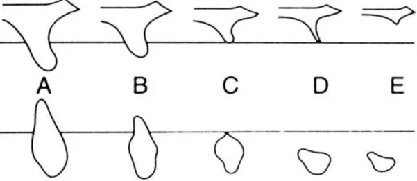
been debated if it affects implants outcomes. The current bone quality classification consists
on a scale of 1 to 4 based on the amount of cortical and trabecular bone present. Type 1 bone
is a homogenous dense cortical bone throughout the entirety of the implant site, while type 2
bone consists of a thick outer cortical layer surrounding a core of dense trabecular bone(104).
Type 3 bone consists of a thin layer of compact cortical bone surrounding a core of dense
trabecular bone, while type 4 bone is made of a thin layer of cortical bone surrounding
trabecular bone with low density(104). The differences in bone quality types as well as the
Several longitudinal studies on implant failure rates reports increased failure risk for
implants placed in type 4 bone locations(106, 107). Increased failures in type 4 bone are
important because patients affected with osteoporosis classically have type 4 bone due to loss
of bone mineral density(108). With controversy in the literature, histologic studies
demonstrated no difference in bone to implant contact (BIC) among healthy and osteoporotic
patients (109). However, a large retrospective study concluded that osteoporosis was a
significant variable for early implant failure(7).
Currently, the role of anti-resorptive medications is still unclear on their effects of
implant outcomes(7). The first reports of osteonecrosis of the jaw due to anti-resorptive
medications, (109, 110) specifically bisphosphonates, was in 2003(111). Monoclonal
antibodies gained popularity to aid in treating osteoporosis and other skeletal metabolic
and/or metastases as they were thought to have fewer side effects, however, the first report of
ONJ related to these types of medications was reported in 2010(112). Due to the increased
number of cases, and their effect on bone metabolism, there has been an increase in the
number of publications on bisphosphonates and other anti-resorptive drugs related to
osteonecrosis of the jaw(113, 114). Bisphosphonates are medications used to treat a variety
of skeletal disorders, ranging from metabolic bone conditions to metastatic bone diseases,
resulting in the maintenance of bone density and maintenance in serum calcium levels(62,
115). The prevalence of ONJ varies depending upon route of administration, condition being
treated, length of use, and population, ranging from <1% to as high as 28%(116, 117).
Because ONJ appears to be localized to the oral cavity, surgical dental interventions may put
there is little solid epidemiological evidence supporting compromised outcomes of implant
therapy in patients treated with anti-resorptive medications. Compromised outcomes in
dental implant therapy due to anti-resorptive mediations can have an extensive impact as
millions of patients, as both men and women with osteoporosis or other metabolic bone
disorder are taking these medications.
Overall opinion in the field is more research is needed to ascertain if a true correlation
exists between alveolar changes in the maxillofacial region and overall skeletal bone
mass(118). Also, additional research will be able to evaluate if medications used to treat
osteoporosis have effects on implant outcomes.
Section 1.10: Porous Tantalum Trabecular Metal: Production and Biologic Influence
Porous tantalum trabecular metal (PTTM) is made up of a rare transitional metal,
Tantalum (Ta), and is known for its resistance to corrosion. It is a member of the refractory
metal groups, allowing it to be incorporated in various alloys(119). Anders Gustav
Ekebereg, a Swedish chemist discovered Ta in 1802(120). Ta is predominantly mined in
western Australia and extracted from tantalite, but can be produced as a by product of tin
mining(121). Previously, Ta had restricted applications in the medical field due to its rarity
and the difficulty in manipulating solid Ta(119). Ta is overly reactive to oxygen, however,
due to this exaggerated reactivity, the formation of oxides on the surface is immediate when
exposed to oxygen, rendering the surface inert(119). Its use in the medical field includes
orthopedic implants, electrodes for pacemakers, and devices for nerve repair(122, 123). Ta
was incorporated in orthopedic implants to mimic the natural structure of trabecular bone.
PTTM has a structure similar to trabecular bone through the use of repeating dodecahedron
thermosetting foam polymer foam, which has undergone pyrolysis, thus creating a vitreous
carbon scaffold(125). Tantalum is then deposited on the surface of the scaffold through the
use of vapor deposition and infiltration(126). Due to the microarchitecture of PTTM it has a
low modulus of elasticity, which is similar to that of cancellous bone, leading to more ideal
dispersion of load and a reduced stress shielding phenomenon(124, 127).
The porous structure of PTTM has a biologic impact through the induction of rapid
angiogenesis by adhesion of serum proteins, leading to recruitment of osteoblastic precursor
cells and subsequent matrix formation(128). PTTM has a higher degree of porosity than
traditional titanium implants(124). Orthopedic surgeons have long utilized porous tantalum
trabecular metal (PTTM) for its ability to enhance peri-implant wound healing. The use of
PTTM in orthopedic implants over the years has shown excellent vascularization, bone
ingrowth, osteoconductivity and biocompatibility(125, 129). Tantalum has been shown to be
bioactive by forming a bone-like apatite layer when exposed to body fluid, and biologically
binds to bone(130, 131). The porous structure provides a biologically similar scaffold shape
to trabecular bone and allows for the bone in-growth and mechanical attachment to implant
surfaces(125, 132). The bone ingrowth due to the porous structure has lead to the concept
known as “osseoincorporation”(124, 125).
Summary
Throughout the history of implant therapy, the goal has been the formation and
maintenance of osseointegration, a direct connection between the hard tissues of the jaw
and the implant surface. With the evolution of macrostructure, micro- and eventually
there are local and systemic factors play a role in the formation and maintenance of this
structural and functional relationship between the implant surface and bone(7, 97). Once
implant success became more predictable, the focus then shifted to hastening the process
and inducing a more vigorous osseointegration response in healthy and patients
predisposed to implant failure. The introduction of porous tantalum trabecular metal
(PTTM) was utilized during orthopedic implants as an alternative to titanium due to its
biologic response and ability to be formed in porous three-dimensional open cell structures
to facilitate enhanced bone ingrowth(124, 132). The designed porosity allows for its
enhanced osteoconduction and angiogenesis, permitting bone to actually anchor onto the
outer surface and inside the interconnected pores of PTTM, characterized as
“osseoincorporation”(119, 130, 134, 135). PTTM has recently crossed over to use in the
oral cavity to replicate the hard tissue response seen in orthopedic implants(119, 136).
Tantalum was selected as an alternative to titanium due to its modulus of elasticity that is
similar to trabecular bone and its resistance to corrosion coupled with improved frictional
properties(121, 125, 126). However, it is similar to titanium with concern to the
biocompatibility, biochemical and biomechanical properties that support
osseointegration(125, 126). What continues to warrant further investigation is whether
PTTM implants are able to more robustly induce osseointegration in patients with risk
CHAPTER 2: OSTEOGENIC ACTIVITY ASSOCIATED WITH DENTAL IMPLANT PLACEMENT IN PATIENTS WITH OSTEOPENIA/OSTEOPOROSIS AS
COMPARED TO HEALTHY INDIVIDUALS
Introduction
The implementation of dental implants for the treatment of partial and fully
edentulous patients has evolved through the years and become common practice throughout
the world. Dental implant therapy is considered an effective, safe and reliable method of
treatment that is a viable alternative to conventional fixed and removable prostheses.
Regardless of the documented predictability, failures still occur and patients with certain
behavioral and systemic conditions are at increased risk of failure(8). The predictability of
dental implant therapy is predicated on the ability to achieve and maintain intimate contact
with the alveolar bone that is both a functional and structural relationship, known as
osseointegration(137, 138). The gold standard for determining the success and degree of
osseointegration is histology, however, this is not a viable option clinical practice(139).
Therefore, the success of osseointegration has been defined by a lack of increasing mobility
between the implant and the surrounding trabecular bone after implant placement(83).
Throughout the years, research has been done to investigate the complex pathways
involved in bone healing in vivo, however the minutia involved in these signaling
intricate interaction of pathways involved in osseointegration in vivo medically
compromised patients(22, 141).
With the introduction of new biomaterials, such as PTTM, their ability to aid in
more efficient osseointegration, not only in healthy but medically compromised patients is
currently being researched(142).
Section 2.1 Methods and Materials
Clinical Relevance
Porous tantalum trabecular metal (PTTM) may enhance initial implant healing in the oral
cavity as shown in orthopedic implant studies and therefore may be indicated for early
implant loading and restoration in healthy subjects. PTTM may also be useful for subjects
with compromised bone healing or density; however, future studies are needed for
compromised healing population. The primary aim of the study was to examine whether or
not the PTTM can improve bone ingrowth, thereby increasing the bone to implant contact
(BIC). It was theorized that the micro porous structure would induce earlier bone deposition
around implants when compared to conventional titanium (Ti) alloy, leading to more robust
osseointegration.
Participants
This study was approved by the Biomedical Institutional Review Board of the
University of North Carolina at Chapel Hill, IRB:11-2539. Written informed consent was
obtained from all study participants. The study population consisted of 13 adults in adequate
periodontal health that were eligible and treatment-planned to receive mandibular dental
symmetrical edentulous areas in the mandible, requiring placement of at least 4 implants,
were recruited through UNC and its healthcare system. The control group for the histologic
arm of the study consisted of 6 systemically healthy individuals, and the experimental group
consisted of 6 patients with osteoporosis, diagnosed previously by a physician. Major
exclusion criteria included: use of medications known to affect periodontal status within one
month prior to initial examination, systemic conditions that are known to affect periodontal
status, history of IV bisphosphonates, active infectious disease, pregnancy, current smokers
or history of smoking within the last two years, subjects with blood disorders and/or
anticoagulant therapy, chemotherapy, and radiotherapy. Specific inclusion and exclusion
The included subjects’ demographic information is shown in Table 2.1.
Demographics
Healthy
Osteopenia/
Osteoporosis
N=12
6
6
Age
64.3±6.0
66.38±5.15
Male
4
0
Female
2
6
Caucasian
5
5
African American
1
1
Cross Sectional
Processing
2
3
Longitudinal
Processing
An edentulous ridge area with sufficient space to place two test cylinders (each
approximately 2.9-3 x 5 mm) was confirmed radiographically. Patients had a CBCT taken
with a radiographic stent to confirm adequate alveolar dimension. For each patient, two
titanium test cylinders were placed on the mandible at the level of the crestal bone and
covered with a collagen membrane (BioMend, Zimmer Dental, Carlsbad, CA, USA) to
prevent soft tissue down growth, primary closure was obtained. Test cylinders were removed
at two and four weeks using a 5.0 mm diameter tissue punch and 4.5mm trephine drill and
sites received a screw vent implant (Zimmer Dental, Carlsbad, CA, USA), upon the removal
of the test cylinder.
Study design
Each subject was screened clinically and radiographically. Cone-beam computed tomography
(CBCT) scans were used to examine the pre-existing alveolar bone. The CBCT scans were
reviewed by one of the implant surgeon investigators (SB or TM). Simplant 16 (Dentsply,
York, PA) or 360dps implant planning software (360imaging, Atlanta, GA) was used to
determine if there was sufficient bone for future dental implant therapy. A minimal width of
7mm and height of 8mm for each dental implant was required for inclusion into the study.
The minimal bone volume was needed to ensure that the 3mm x 5mm diameter device could
be placed, preventing bone dehiscence or approximating any anatomical structures. At each
surgical visit, subjects were asked to rinse their mouth with 0.12% Chlorhexidine gluconate
prior to any procedure. All surgical procedures were completed under local anesthetics
involving bilateral inferior alveolar nerve blocks, long buccal nerve blocks and local
infiltration of the surgical sites. Full thickness flaps were raised in all sites. A split-mouth
CBCT scans, implant planning software, and a surgical guide. Two 3mm x 5mm Ti alloy
tapered screw and two 3mm x 5mm PTTM cylinder devices were placed in each edentulous
site, Ti test cylinders on one edentulous side of the jaw and PTTM test cylinders on the other
as seen in Figure 2.2 and 2.3.
Figure 2.3: Test cylinders immediately after insertion, right photo is PTTM test cylinders, left photo is Ti test cylinders in the same patient. (Photo courtesy of Dr. Thiago Morelli).
The top of each test cylinder was placed at the level of crestal bone level to ensure that the
device was completely surrounded by bone. Note that since the Ti device was a self-tapping
tapered screw, a 2.3 mm drill was used and the Ti device was self-tapped in place. Ti device
also has HA blasted surface treatment mimicking the clinically used and commercially
available dental implants (MTX, Zimmer Biomet, Palm Beach Garden, FL). However, since
the PTTM device was a straight cylinder, a 3 mm drill was used to place the device. The
PTTM device was press-fitted in place. A 5 mm diameter resorbable collagen membrane
(Biomend, Zimmer Biomet, Warsaw, IN) was placed on top of each device. A combination
of continuous interlocking and interrupted suturing techniques with 4.0 chromic gut sutures
were used to ensure the primary closure and hemostasis. The subjects were instructed to