ROS-INDUCED DNA ADDUCTS IN THE RODENTS AFTER EXPOSURE TO SUPERFUND HAZARDOUS CHEMICALS
Lina Gao
A dissertation submitted to the faculty of the University of North Carolina at Chapel Hill in partial fulfillment of the requirements for the degree of Doctor of Philosophy in the
Department of Environmental Sciences and Engineering
Chapel Hill 2011
© 2011 Lina Gao
ABSTRACT
LINA GAO: ROS-Induced DNA Damage in Rodents after Exposure to Superfund Hazardous Chemicals
(Under the direction of James A. Swenberg)
The accumulation of oxidative DNA damage has been hypothesized as a key event in chemical carcinogenesis. In this study, oxidative DNA damage was evaluated in the livers of rats exposed to vinyl chloride (VC), 2,3,7,8-tetrachlorodibenzo-p-dioxin (TCDD), 3,3΄,4,4΄,5-pentachlorobiphenyl (PCB 126), 2,3,4,7,8-pentachlorodibenzofuran (PeCDF) and 2,2΄,4,4΄,5,5΄-Hexachlorobiphenyl (PCB153). Eight oxidative DNA adducts were measured, 8-hydroxyl-2′-deoxyguanosine (8-OHdG), 1, N6 -etheno-2’-deoxyadenosine (εdA), N2, 3-εG, 1, N2-etheno-2’-deoxyguanosine (1, N2-εdG), 3-(2′-deoxy-β-d-erythro-pentofuranosyl) pyrimido[1,2-α]purin-10(3H) (M1dG), acrolein,
crotonaldehyde and HNE -derived dG adducts (assigned as AcrdG, CrdG, and 4-HNEdG respectively).
εdA is one of the promutagenic DNA adducts formed by VC, which can also be formed by lipid peroxidation. In this study, both adult and weanling Sprague-Dawley rats were exposed to 1100 ppm (13C2)-VC for 1 week (6 h/day, 5 days/week). The results
indicated that NA-εdA concentration did not show significant difference in the liver of adult and weanling rats after VC exposure. The distribution pattern of (13C2)-εdA in liver,
adult and weanling rats. ROS-induced DNA adducts were detected in the liver of female intact, ovariectomized (OVX) and male Sprague-Dawley rats, including 8-OHdG, 1, N6
-εdA, AcrdG, and CrdG. These animals were exposed to TCDD for 30 weeks after diethylnitrosamine (DEN) initiation. Induction of these adducts was consistently found in liver DNA of TCDD-treated intact female rats and 17β-estradiol (E2) supplemented OVX
female rats, but not detected in OVX rats without E2 supplement or male rats. These
results further confirmed that the induction of these adducts occurs via a sex-specific and estrogen-dependent mechanism reported previously. Oxidative DNA damage was measured in liver DNA of female Sprague-Dawley rats following 53-week exposure of PHAHs, including PCB153, PCB126, TCDD, and the ternary mixture of TCDD, PCB126 and PeCDF. Increases of 8-OHdG, N2, 3-εG and 1, N6-εdA were observed in PCB153 or PCB126 exposed animals. Significant increases of 1, N6-εdA were observed in all animals exposed to TCDD and the ternary mixture. Increases of 1, N2-εdG, CrdG, AcrdG, 4-HNEdG and M1dG were detected in animals exposed to the ternary mixture, but not the
ACKNOWLEDGEMENTS
TABLE OF CONTENTS
LIST OF TABLES ... xi
LIST OF FIGURES ... xii
LIST OF ABBREVIATIONS ... xv
CHAPTER I INTRODUCTION: OVERVIEW OF ROS-INDUCED DNA DAMAGE, VINYL CHLORIDE (VC) AND POLYHALOGENATED AROMATIC HYDROCARBONS (PHAHS) ... 1
A. ROS, DNA DAMAGE AND MUTATION ... 1
B. THE DETECTION OF ROS-INDUCED DNA ADDUCTS AND THEIR CONNECTION WITH DISEASE ... 3
C. DNA REPAIR PATHWAYS FOR ROS-INDUCED DNA ADDUCTS ... 4
D. VINYL CHLORIDE AND ETHENO DNA ADDUCTS ... 5
E. TCDD, OXIDATIVE STRESS AND ROS-INDUCED DNA DAMAGE ... 6
F. PHAHs, TOXIC EQUAVELENCY FACTOR, OXIDATIVE STRESS AND OXIDATIVE DNA DAMAGE ... 9
G. REFERENCES... 20
CHAPTERII DETECTION OF ROS-INDUCED DNA ADDUCTS BY LC-MS/MS AFTER HPLC PURIFICATION ... 28
A. INTRODUCTION ... 28
B. MATERIALS AND METHODS ... 31
Instrumentation ... 31
Animal exposure ... 32
DNA isolation ... 33
Standard synthesis ... 33
Enzymatic digestion and HPLC purification ... 35
LC-MS/MS detection ... 37
C. RESULTS... 39
Enzymatic hydrolysis and HPLC purification ... 39
LC-MS/MS detection and assay validation ... 40
E. DISCUSSION... 42
F. REFERENCES ... 71
CHAPTER III ENDOGENOUS AND EXOGENOUS 1, N6 -ETHENO-2´-DEOXYADENOSINE IN WEANLING AND ADULT SPRAGUE-DAWLEY RATS EXPOSED TO VINYL CHLORIDE BY INHALATION ... 81
A. INTRODUCTION ... 81
B. MATERIALS AND METHODS ... 84
Materials ... 84
Animal exposures... 85
DNA isolation ... 85
1, N6-εdA measurement in isolated DNA ... 86
Statistical analysis ... 87
C. RESULTS... 87
1, N6-εdA adducts in kidney and lung DNA of VC exposed weanling
and adult rats ... 89
D. DISCUSSION ... 90
E. REFERENCES ... 107
CHAPTER IV SPECTRUM OF DNA LESIONS INDUCED BY REACTIVE OXYGEN SPECIES IN SPRAGUE-DAWLEY RATS CHRONICALLY EXPOSED TO 2, 3, 7, 8-TETRACHLORODIBENZO-p-DIOXIN AFTER DIETHYLNITROSAMINE INITIATION ... 112
A. INTRODUCTION ... 112
B. MATERIALS AND METHODS ... 115
Matertials ... 115
Animal exposures... 116
DNA isolation ... 117
Enzymatic digestion and HPLC purification ... 118
LC-MS/MS detection ... 118
Statistical analysis ... 119
C. RESULTS... 120
D. DISCUSSION ... 122
F. REFERENCES ... 137
CHAPTER V OXIDATIVE DNA DAMAGE IN THE LIVER OF FEMALE SPARAGUE-DAWLEY RATS CHRONICALLY EXPOSED TO POLYHALOGENATED AROMATIC HYDROCARBONS (PHAHS) ... 144
A. INTRODUCTION ... 144
Chemicals ... 148
Animal exposures... 148
DNA isolation ... 149
Enzymatic digestion and HPLC purification ... 150
LC-MS/MS detection ... 151
N2, 3-εG assay ... 153
Statistical analysis ... 153
C. RESULTS... 154
D. DISCUSSION ... 157
E. REFERENCES ... 173
CHAPTER VI GENERAL DISCUSSION ... 180
A. SUMMARY OF FINDINGS ... 180
The detection of ROS-induced DNA adducts ... 182
The endogenous and exogenous 1, N6-εdA in weanling and adult Sprague-Dawley rats exposed to VC by inhalation ... 183
ROS-induced DNA adducts in the liver of Sprague-Dawley rats after DEN initiation and TCDD promotion ... 184
ROS-induced DNA adducts in the liver of Sprague-Dawley rats after chronic exposure of PHAHs and their mixtures ... 185
B. FUTURE DIRECTIONS... 187
Evaluate the sensitivity of different ROS-induced DNA adducts as biomarker of oxidative stress ... 187
Examine the length of exposure on ROS-induced DNA adducts ... 188
Compare the dose/time-response of ROS-induced DNA adducts with oxidative stress-related gene expression in the livers of
female Sprague-Dawley rats exposed to PHAHs ... 1910 Measure ROS-induced DNA adducts in the other target organs of
LIST OF TABLES
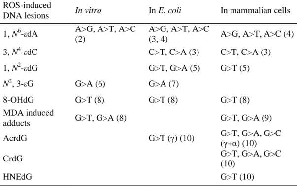
Table 1.1 Single-base changes induced by ROS related DNA adducts
in the site-directed mutagenesis experiments ... 17 Table 1.2 Reported endogenous amounts of ROS-induced DNA adducts ... 18 Table 1.3 Reported repair pathways for ROS-induced DNA adducts ... 19 Table 2.1 ROS-induced DNA adducts in control liver DNA of adult
LIST OF FIGURES
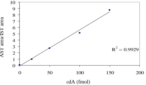
Figure 1.1 Major DNA adducts induced by reactive oxygen species (ROS)……….. … 12 Figure 1.2 Major propano-derived adducts induced by lipid peroxidation (LPO) … ... 13 Figure 1.3 Etheno DNA adduct formation in VC exposed animals ... 14 Figure 1.4 Oxidative DNA damage induced by dioxin-like compounds (DLCs) ... 15 Figure 1.5 Hypothetical scheme for the role of cumulative oxidative
stress in carcinogenesis ... 16 Figure 2.1 Mass spectrometry scheme for analysis of DNA adducts ... 55 Figure 2.2 1, N6-εdA level using the orginal method B (PD) and the
optimized method B (OPD) ... 56 Figure 2.3 The optimization of enzyme system for the measurement of
AcrdG and CrdG ... 57 Figure 2.4 The typical chromatograms for LC purification of CrdG,
4-HNEdG, AcrdG and 1, N2-εdG by UV detector ... 58 Figure 2.5 The chromatogram of LC purification for 1, N6-εdA and
8-OHdG by UV detector ... 59 Figure 2.6 Assay validations for εdA measurement……… ... 60 Figure 2.7 Instrument sensitivity comparison for 1, N6-εdA detection
labeled VC exposed adult rat liver DNA (UPLC vs nano) ... 61 Figure 2.8 Instrument sensitivity comparison for 1, N6-εdA detection
labeled VC exposed adult rat liver DNA (UPLC vs nano) ... 62 Figure 2.9A The typical chromatogram for ctDNA samples and
calibration curve for 8-OHdG by UPLC-MS/MS ... 63 Figure 2.9B The typical chromatogram for 1, N2-εdG in ctDNA and
Figure 2.9D The typical chromatogram for M1dG and AcrdG in
ctDNA by nanoUPLC-MS/MS………... ... 66 Figure 2.9E The typical chromatogram for CrdG and 4-HNEdG in
ctDNA by nanoUPLC-MS/MS……….. ... 67 Figure 2.10A 1, N2-εdG, M1dG and AcrdG in wild type C57BL/6J mice
exposed to CCl4 (1600 mg/kg in olive oil) or olive oil (10
ml/kg) by intraperitoneal injection for 13 days ... 69 Figure 2.10B1, N2-εdG, M1dG and AcrdG in XPA-/- C57BL/6J mice
exposed to CCl4 1600 mg/kg in olive oil by
intraperitoneal injection for 13 days ... 69 Figure 2.10C 1, N2-εdG, M1dG and AcrdG in the liver of Shasta strain
rainbow trout exposed to dibenzo[a,l]pyrene (DBP) for
29 days ... 70 Figure 2.10D CrdG and 4-HNEdG in the liver of Shasta strain rainbow
trout exposed to dibenzo[a,l]pyrene (DBP) for 29 days ... 70 Figure 3.1 The experimental design... 97 Figure 3.2 NA-1, N6-εdA amounts in (13C2)-VC exposed and control weanling rats ... 98
Figure 3.3 (13C2)-1, N6-εdA amounts in the (13C2)-VC exposed
weanling and adult liver DNA ... 99 Figure 3.4 NA-1, N6-εdA amounts in the NA-VC exposed and control weanling rats .. 100 Figure 3.6 Typical chromatogram for 1, N6-εdA detection in the liver of
(13C2)-VC treated adult rats ... 101
Figure 3.7 NA-1, N6-εdA amounts in the control and (13C2)-VC exposed adult liver ... 102
Figure 3.8 NA-1, N6-εdA amounts in the NA-VC exposed and control adult DNA ... 103 Figure 3.10 NA-1, N6-εdA amounts in the (13C2)-VC exposed and control adult rats ... 104
Figure 3.9 Total amount of 1, N6-εdA in adult rats after 1-week
exposure of (13C2)-VC by inhalation... 105
Figure 3.6 Total amount of 1, N6-εdA in weaning rats after 1-week
exposure of (13C2)-VC by inhalation... 106
Figure 4.2 8-OHdG in the liver of intact or OVX female Sprague-Dawley rats ... 132 Figure 4.3A 1, N6-εdA, AcrdG and CrdG in the liver of intact or OVX
female Sprague-Dawley rats ... 133 Figure 4.3B AcrdG and CrdG in the liver of intact or OVX female
Sprague-Dawley rats ... 134
Figure 4.4 8-OHdG and 1, N6-εdA in the hepatic DNA of male Sprague Dawley rats .. 135 Figure 4.5 The formation of ROS and bulky DNA adducts by estrogen ... 136 Figure 5.1 The structures of typical PHAHs in this study ... 166 Figure 5.2 N2, 3-εG, 1, N6-εdA and 8-OHdG in the liver of female
Sprague-Dawley rats exposed to PCB153 alone or PCB126
alone for 53 weeks ... 167 Figure 5.3 N2, 3-εG, 1, N6-εdA and 8-OHdG in the liver of
femaleSprague-Dawley rats exposed to the binary mixture of
PCB153 and PCB126 for 53 weeks ... 168 Figure 5.4 1, N6-εdA and 8-OHdG in the liver of female
Sprague-Dawley rats exposed to TCDD or the ternary mixture of
TCDD, PCB126 and PeCDF for 53 weeks ... 169 Figure 5.5 1, N2-εdG, M1dG, AcrdG, CrdG and 4-HNEdG in the liver
of female Sprague-Dawley rats exposed to TCDD or the ternary mixture of TCDD, PCB126 and PeCDF for 53
weeks ... 170 Figure 5.6 Tumor incidence (2 years) and enzyme induction in the liver
of female Sprague-Dawley rats exposed to PCB153 or
PCB126 for 53 weeks ... 171 Figure 5.7 Tumor incidence (2 years) and enzyme induction in the liver
of female Sprague-Dawley rats exposed to TCDD or the
LIST OF ABBREVIATIONS
AcrdG Acrolein-derived dG adducts
AP Alkaline Phosphatase
ARP Aldehyde Reactive Probe
BER Base Excision Repair
CE Collision energy
CrdG Crotonaldehyde-derived dG adducts
DBP Dibenzo[a, l]pyrene
DLCs Dioxin-like compounds
dA 2´-Deoxyadenosine dC 2´-Deoxycytidine
εdC 3, N4-Etheno-2’-deoxycytidine
DEN Diethylnitrosamine
dG 2´-Deoxyguanosine
dT Thymidine
E2 17β-Estradiol
ESCODD The European Standards Committee on Oxidative DNA Damage
ESI Electrospray ionization
GC-MS Gas chromatography-mass spectrometry
4-HNE trans-4-Hydroxy-2-Nonenal
4-HNEdG trans-4-hydroxy-2-nonenal-derived dG adducts IARC International Agency for Research on Cancer
LC-APCIMS/MS Liquid chromatography-atmosphere pressure chemical ionization- tandem mass spectrometry
LC-ECD Liquid chromatography-electrochemical detection
LC-ESIMS/MS Liquid chromatography-electrospray tandem mass spectrometry
LOD Limit of detection
LOQ Limit of quantitation
LPO Lipid peroxidation
MDA Malondialdehyde
M1dG 3-(2´-deoxy-β-D-erythropentofuranosyl) pyrimido[1,2-α
]purin-10(3H)-one
MRM Multiple reaction monitoring
NDLCs Non-dioxin-like compounds
NER Nucleotide excision repair pathway NICI Negative ion chemical ionization
NLM Neutral loss monitor
NP1 Nuclease P1
NTP National Toxicology Program
N2, 3εG N2, 3-Etheno-guanine
1, N6-εdA 1, N6-Etheno-2’-deoxyadenosine 1, N2-εdG 1, N2-Etheno-2’-deoxyguanosine
OVX Ovariectomized
8-OHdG 7, 8-Dihydro-8-oxo-2′-deoxyguanosine
PDE Phosphodiesterase I
PFB Pentafluorobenzylation
PCB126 3,3΄,4,4΄,5-Pentachlorobiphenyl PCB153 2,2΄,4,4΄,5,5΄-Hexachlorobiphenyl PCDDs Polychlorinated dibenzodioxins PCDFs Polychlorinated dibenzofurans PeCDF 2,3,4,7,8-Pentachlorodibenzofuran PHAHs Polyhalogenated aromatic hydrocarbons
ROS Reactive oxygen species
SIM Selected ion monitoring
SPE Solid phase extraction
TCDD 2,3,7,8-Tetrachlorodibenzo-p-dioxin
TEF Toxic Equivalency Factor
TEMPO 2,2’,6,6’-Tetramethylpiperidinyl-1-oxy
TEQ Toxic Equivalent
CHAPTER I
INTRODUCTION: OVERVIEW OF ROS-INDUCED DNA DAMAGE, VINYL CHLORIDE (VC) AND POLYHALOGENATED AROMATIC HYDROCARBONS
(PHAHS)
A. ROS, DNA DAMAGE AND MUTATION
ethenoguanine (N2, 3-εG), 3-(2-deoxy-β-D-erythropentofuranosyl) pyrimido[1,2-α ]purin-10(3H)-one (M1dG) and 1, N2-propano-2’-deoxyguanosine generated from acrolein,
crotonaldehyde and 4-hydroxy-2-nonenal (4-HNE). The specific chiral isomers for propano-derived DNA adducts are shown in figure 1.2.
The accumulation of ROS-induced DNA adducts was shown to potentially represent a hazard in vivo because of its association with mutagenesis (2-10). Site-directed mutagenicity studies found that many can induce specific transition and/or transversion point mutations in bacteria and/or mammalian cells, as shown in table 1.1. The promutagenicity of etheno DNA adducts was further demonstrated by studying their behavior in vinyl chloride (VC) exposed humans and animals (13, 14). Genetic changes similar to those induced by VC induced DNA adducts were demonstrated in p53 and H-ras genes in animal and human angiosarcomas. The presence of 8-OHdG during DNA replication can cause G to T transversions (8). Replication of MDA-modified single-stranded M13 genomes in E coli caused G→T, A→G, and C→T mutations (9, 15). M1dG
hamster lung cells and human lymphoblastoid cells with G→T transversions as the most prevalent base changes (22, 23).
B. THE DETECTION OF ROS-INDUCED DNA ADDUCTS AND THEIR CONNECTION WITH DISEASE
Many different methods have been developed to detect ROS-induced DNA adducts. For etheno adducts, the dominant detection technique is the 32P-postlabeling method, which has been successfully applied to measure εdA and εdC in rodent and human tissues (24). Persistent increases of εdA and εdC were found in premalignant target organs affected by various diseases, such as genetic metal storage disorders (Wilson’s disease and hemochromatosis), chronic pancreatitis, and chronic hepatitis (24). 8-OHdG was ever detected by LC-ECD (electrochemical detector), GC-MS, immunoassay and LC-MS/MS (25). Using different detection techniques, elevated 8-OHdG has been detected in patients with various malignancies, including acute leukemia, colorectal cancer, hepatic cancer, oral squamous cell carcinoma, and breast cancer (25-29). M1dG was measured in human
and animal tissues by 32P-postlabling, immunoslot blot, GC-MS and LC-MS/MS techniques (30-32). Higher M1dG was detected in smoking patients with lung cancer
importance of propano adducts in chemical carcinogenesis or chronic inflammatory diseases (24). Using different techniques, the endogenous levels of these DNA adducts in human and rodent tissues were obtained, as depicted in table 1.2.
C. DNA REPAIR PATHWAYS FOR ROS-INDUCED DNA ADDUCTS
8-OHdG:dA mismatches (41). Similar results were found for 1, N6-εdA (43), which could be repair by both BER and AlkB, a direct reversal repair pathway, as shown in table 1.3. At present, NER is regarded as the primary repair pathway for propano-adducts, but it is possible that other repair pathways are involved in their repair (10, 44).
D. VINYL CHLORIDE AND ETHENO DNA ADDUCTS
VC is a colorless organic gas used almost exclusively by the plastic industry to produce polyvinyl chloride (PVC) and copolymers. Regarded as carcinogenic to humans by IARC (45), VC can induce angiosarcoma of liver in animals and humans (14). After metabolic activation by CYP2E1 mainly in liver, VC is transformed into an active metabolite, chloroetheylene oxide, which can react with DNA directly and form several DNA adducts, 7-(2-oxoethyl)guanine (7OEG), and the minor adducts, 1, N6-εdA, N2,3-εG and 3,N4-εdC (46-49), as shown in figure 1.3. Because all these etheno-adducts are promutagenic, it is possible for them to cause base-pair substitution mutations in key cancer genes in vivo, if not repaired before cell replication. Several changes of oncogenes and tumor suppressor genes were reported in humans and animals exposed to VC, which would be consistent with the promutagenic properties of those DNA adducts (50-57). The accumulation of such mutations could further contribute to the formation of cancer.
role during this process, such as 4-HNE. Because of the existence of these background etheno adducts, it is impossible to differentiate adducts arising from VC and those formed endogenously. In this study, exposure of rats to 13C labeled VC will be utilized to distinguish endogenous NA- from exogenous (13C2)-1, N6-εdA. The distribution of
both NA- and (13C2)-1, N6-εdA and the persistence of (13C2)-1, N6-εdA will be
evaluated in the primary target (liver) and non-target organs of rats. By this study,
the reliability of nanoUPLC-MS/MS will be evaluated, which will applied to detect
other LPO-induced adducts in animal tissues exposed to other superfund chemicals.
E. TCDD, OXIDATIVE STRESS AND ROS-INDUCED DNA DAMAGE
contribute to the toxic effects of TCDD, since TCDD is not readily metabolized and can result in a prolonged and amplified response.
not ovariectomized female rats after diethylnitrosamine (DEN) initatiation. Wyde et al. (69) further found the induction of hepatic 8-OHdG by TCDD in rats after DEN initiation was female-specific, estrogen-dependant and was a chronic effect. Recently, Hassoun et al. (67) observed increased production of superoxide anion, lipid peroxidaiton and DNA single-strand breaks in hepatic tissues of rats after 30 weeks of exposure to TCDD. All these studies supported the importance of oxidative DNA damage in carcinogenesis of TCDD.
In summary, the contribution of ROS to the toxic effects of TCDD is still controversial. Further scientific studies are necessary to explore whether oxidative stress is important or just a hypothesis of scientists to explain the complicated mechanisms for the toxic effects of chemicals. Because of the high propensity for artifact formation during sample processing, 8-OHdG can be a problematic biomarker for ROS-induced DNA damage. Different DNA adducts have different formation and repair pathways, which may result in different toxicities/responses in treated animals. Therefore, in this
study, a battery of ROS-induced DNA adducts will be applied to further evaluate
ROS-induced DNA damage in a two-stage initiation-promotion rat liver model with
DEN as the initiator and TCDD as the promoter (69). This information will help us
better understand the mode of action of hepatocarcinogensis of TCDD in female rats.
F. PHAHs, TOXIC EQUAVELENCY FACTOR, OXIDATIVE STRESS AND OXIDATIVE DNA DAMAGE
in the environment, they accumulate in tissues. Humans and wildlife animals are exposed daily to complex mixtures of these chemicals, primarily via trace amounts present in food, resulting in chronic lifetime exposure which may evoke toxicity and carcinogenicity (74).
Depending on the location and type of halogenation, some PHAHs induce a similar spectrum of biochemical and toxic responses in experimental animals characterized by severe weight loss, thymic atrophy, hepatotoxicity, immunotoxicity and enzyme induction (58). These common biological effects are mediated through a similar mechanism of action as described for TCDD binding to AhR, as shown in figure 1.4. Therefore, these structurally related compounds are commonly referred to as dioxin-like compounds (DLCs) (58, 74). Due to similarity in toxicity and mechanisms of DLCs, the concept of the Toxic Equivalency Factor (TEF) has been used for the assessment of risk and regulatory control for these compounds. The TEF methodology is a relative potency scheme determined from the toxicity of each congener relative to that of TCDD on the basis of available in vivo and in vitro data. This allows for the estimation of the potential dioxin-like activity of a mixture of DLCs that are found in environment (74, 75), as shown in the following equation.
∑
∑
× +∑
× + ×= ni PCDDi TEFi n ni PCDFi TEFi n ni PCBi TEFi n
TEQ ( ) ( ) ( )
where i= the individual congener and its respective TEF, and n = all congeners within each class of DLCs.
found the production of superoxide anion, lipid peroxidation and DNA SSBs in liver DNA of rats after exposure to various mixtures of TCDD and two of its congeners, PeCDF and PCB 126 for 13 weeks (67). Other chronic animal studies demonstrated the involvement of ROS in the toxicity and carcinogenicity of PHAHs (77).
Recently, in an effort to evaluate the TEF methodology for the chronic toxicity and carcinogenicity of DLCs and structurally related polychlorinated biphenyls (PCBs), the National Toxicology Program (NTP) conducted a series of 2-year bioassays in female Harlan Sprague-Dawley rats exposed to DLCs (such as TCDD, PCB 126, and PeCDF and non-dioxin-like compounds (NDLCs) PCB153). The toxic effects of the ternary mixture of TCDD, PCB126 and PeCDF and the binary mixture of PCB126 and PCB153 were also evaluated in these studies. Besides inducing a high incidence of hepatic tumors in female rats, chronic inflammation in liver tissues was also observed in the exposed animals. Since the DNA damage caused by ROS has been implicated in a myriad of diseases, it is reasonable to hypothesize that ROS-induced DNA damage is an
important mode of action for carcinogensis of these PHAHs, which can be estimated
Figure 1.2 Major propano-derived adducts induced by lipid peroxidation (LPO)
O
H O O
OH N N N O N H N O H OH H H H H HO OH N N N O N H N O H OH H H H H HO OH N N N O N H N O H OH H H H H HO OH CH3 N N N O N H N O H OH H H H H HO OH CH3 Acrolein Crotonaldehyde Gamma-OH-PdG Alpha-OH-PdG (6R,8R)-crotonaldehyde adduct (6S,8S)-crotonaldehyde adduct 4-hydroxynonenal N N N O N H N O H OH H H H H HO OH
C5H11
OH N N N O N H N O H OH H H H H HO OH
C5H11
OH N N N O N H N O H OH H H H H HO OH
C5H11
OH N N N O N H N O H OH H H H H HO OH
C5H11
OH
(6S,8R,11S)-HNE adduct
(6R,8S,11R)-HNE adduct
(6S,8R,11R)-HNE adduct
Figure 1.3 Etheno DNA adduct formation in VC exposed animals
Vinyl chloride
Cl
P450 2E1
Chloroethylene epoxide
Cl O
7OEG dominant DNA adduct
Etheno DNA adducts minor DNA adduct Direct pathway
Oxidative stress /lipid peroxidation (LPO)
Reactive LPO products
Indirect pathway
Mutation ef f ects on key cancer genes
1, N6-εdA
N2, 3-εG
3, N4-εdC
Figure 1.5 Hypothetical schemes for the role of cumulative oxidative stress in
Table 1.1Single-base changes induced by ROS related DNA adducts in the site-directed mutagenesis experiments
ROS-induced
DNA lesions In vitro In E. coli In mammalian cells 1, N6-εdA A>G, A>T, A>C
(2)
A>G, A>T, A>C
(3, 4) A>G, A>T, A>C (4) 3, N4-εdC C>T, C>A (3) C>T, C>A (3)
1, N2-εdG G>T, G>A (5) G>T (5)
N2, 3-εG G>A (6) G>A (7)
8-OHdG G>T (8) G>T (8) G>T (8)
MDA induced
adducts G>T, G>A (8) G>T, G>A (9)
AcrdG G>T (γ) (10) G>T, G>A, G>C
(γ+α) (10)
CrdG G>T, G>A, G>C
(10)
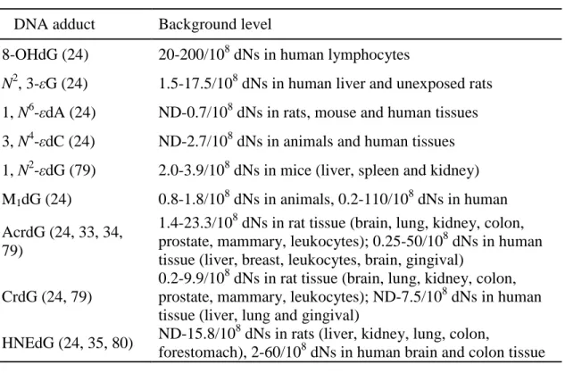
Table 1.2 Reported endogenous amounts of ROS-induced DNA adducts
DNA adduct Background level
8-OHdG (24) 20-200/108 dNs in human lymphocytes
N2, 3-εG (24) 1.5-17.5/108 dNs in human liver and unexposed rats 1, N6-εdA (24) ND-0.7/108 dNs in rats, mouse and human tissues 3, N4-εdC (24) ND-2.7/108 dNs in animals and human tissues 1, N2-εdG (79) 2.0-3.9/108 dNs in mice (liver, spleen and kidney) M1dG (24) 0.8-1.8/108 dNs in animals, 0.2-110/108 dNs in human
AcrdG (24, 33, 34, 79)
1.4-23.3/108 dNs in rat tissue (brain, lung, kidney, colon, prostate, mammary, leukocytes); 0.25-50/108 dNs in human tissue (liver, breast, leukocytes, brain, gingival)
CrdG (24, 79)
0.2-9.9/108 dNs in rat tissue (brain, lung, kidney, colon, prostate, mammary, leukocytes); ND-7.5/108 dNs in human tissue (liver, lung and gingival)
HNEdG (24, 35, 80) ND-15.8/10
8
dNs in rats (liver, kidney, lung, colon,
Table 1.3 Reported repair pathways for ROS-induced DNA adducts
DNA
lesions Bacterial enzyme/system Mammalian enzyme/system 1, N6-εdA AlkA (38), AlkB ANPG/Aag (39) (BER), ABH2
(43) (direct reversal pathway) 3, N4-εdC AlkB (81) mismatch-specific thymine-DNA
glycosylase (82) (BER)
1, N2-εdG ANPG (39) (BER)
N2, 3-εG E.Coli 3-methyladenine DNA glycosylase (39) (BER)
8-OHdG MYH, MMR
8-oxoguanine DNA glycosylase (42) (OGG1), hMYH, hMPG, hNEIL1 (BER), MMR, NER M1dG NER (10)
AcrdG NER (10)
CrdG NER (10)
HNEdG NER (44), HR (44) NER (10)
G. REFERENCES
(1) Lyrenas L., Zotova E., Ekström L., Morgenster R (2006) Oxidative Stress, Genetic Variation, and Disease. In Oxidative stress, disease and cancer (Keshav K Singh, Ed.); pp 371-460, Imperial College Press, Roswell Park Cancer Institute, Buffalo, New York.
(2) Litinski, V.; Chenna, A.; Sagi, J.; Singer, B. (1997) Sequence context is an important determinant in the mutagenic potential of 1, N6-ethenodeoxyadenosine (epsilonA): formation of epsilonA basepairs and elongation in defined templates. Carcinogenesis 18, 1609-1615.
(3) Basu, A. K.; Wood, M. L.; Niedernhofer, L. J.; Ramos, L. A.; Essigmann, J. M. (1993) Mutagenic and genotoxic effects of three vinyl chloride-induced DNA lesions: 1,N6-ethenoadenine, 3,N4-ethenocytosine, and 4-amino-5-(imidazol-2-yl)imidazole. Biochemistry 32, 12793-12801.
(4) Pandya, G. A.; Moriya, M. (1996) 1, N6-ethenodeoxyadenosine, a DNA adduct highly mutagenic in mammalian cells. Biochemistry 35, 11487-11492.
(5) Moriya, M.; Zhang, W.; Johnson, F.; Grollman, A. P. (1994) Mutagenic potency of exocyclic DNA adducts: marked differences between Escherichia coli and simian kidney cells. Proc. Natl. Acad. Sci. U. S. A. 91, 11899-11903.
(6) Singer, B.; Essigmann, J. M. (1991) Site-specific mutagenesis: retrospective and prospective. Carcinogenesis 12, 949-955.
(7) Cheng, K. C.; Preston, B. D.; Cahill, D. S.; Dosanjh, M. K.; Singer, B.; Loeb, L. A. (1991) The vinyl chloride DNA derivative N2,3-ethenoguanine produces G----A transitions in Escherichia coli. Proc. Natl. Acad. Sci. U. S. A. 88, 9974-9978. (8) Kamiya, H. (2003) Mutagenic potentials of damaged nucleic acids produced by
reactive oxygen/nitrogen species: approaches using synthetic oligonucleotides and nucleotides: survey and summary. Nucleic Acids Res. 31, 517-531.
(9) Niedernhofer, L. J.; Daniels, J. S.; Rouzer, C. A.; Greene, R. E.; Marnett, L. J. (2003) Malondialdehyde, a product of lipid peroxidation, is mutagenic in human cells. J. Biol. Chem. 278, 31426-31433.
(10) Minko, I. G.; Kozekov, I. D.; Harris, T. M.; Rizzo, C. J.; Lloyd, R. S.; Stone, M. P. (2009) Chemistry and biology of DNA containing 1,N(2)-deoxyguanosine adducts of the alpha,beta-unsaturated aldehydes acrolein, crotonaldehyde, and
(11) European Standards Committee on Oxidative DNA Damage (ESCODD) (2003) Measurement of DNA oxidation in human cells by chromatographic and enzymic methods. Free Radic. Biol. Med. 34, 1089-1099.
(12) McBrien, D. C. H.; Slater T. F. (1982) Free radicals and cancer. In Free Radicals, Lipid Peroxidation and Cancer (McBrien, D. C. H., Slater T. F., Eds.) pp 55-60, Academic Press, London, England.
(13) Barbin, A. (1999) Role of etheno DNA adducts in carcinogenesis induced by vinyl chloride in rats. In Exocyclic DNA Adducts in Mutagenesis and Carcinogensis (Singer, B., and Bartsch, H., Eds.) pp 303-313, IARC Scientific Publications, Lyon, France.
(14) Bolt, H. M. (2005) Vinyl chloride-a classical industrial toxicant of new interest. Crit. Rev. Toxicol. 35, 307-323.
(15) Benamira, M.; Johnson, K.; Chaudhary, A.; Bruner, K.; Tibbetts, C.; Marnett, L. J. (1995) Induction of mutations by replication of malondialdehyde-modified M13 DNA in Escherichia coli: determination of the extent of DNA modification, genetic requirements for mutagenesis, and types of mutations induced. Carcinogenesis 16, 93-99.
(16) Yang, I. Y.; Hossain, M.; Miller, H.; Khullar, S.; Johnson, F.; Grollman, A.; Moriya, M. (2001) Responses to the major acrolein-derived deoxyguanosine adduct in
Escherichia coli. J. Biol. Chem. 276, 9071-9076.
(17) VanderVeen, L. A.; Hashim, M. F.; Nechev, L. V.; Harris, T. M.; Harris, C. M.; Marnett, L. J. (2001) Evaluation of the mutagenic potential of the principal DNA adduct of acrolein. J. Biol. Chem. 276, 9066-9070.
(18) Kanuri, M.; Minko, I. G.; Nechev, L. V.; Harris, T. M.; Harris, C. M.; Lloyd, R. S. (2002) Error prone translesion synthesis past gamma-hydroxypropano
deoxyguanosine, the primary acrolein-derived adduct in mammalian cells. J. Biol. Chem. 277, 18257-18265.
(19) Yang, I. Y.; Chan, G.; Miller, H.; Huang, Y.; Torres, M. C.; Johnson, F.; Moriya, M. (2002) Mutagenesis by acrolein-derived propanodeoxyguanosine adducts in human cells. Biochemistry 41, 13826-13832.
(20) Sanchez, A. M.; Minko, I. G.; Kurtz, A. J.; Kanuri, M.; Moriya, M.; Lloyd, R. S. (2003) Comparative evaluation of the bioreactivity and mutagenic spectra of
(21) Fernandes, P. H.; Kanuri, M.; Nechev, L. V.; Harris, T. M.; Lloyd, R. S. (2005) Mammalian cell mutagenesis of the DNA adducts of vinyl chloride and
crotonaldehyde. Environ. Mol. Mutagen. 45, 455-459.
(22) Cajelli, E.; Ferraris, A.; Brambilla, G. (1987) Mutagenicity of 4-hydroxynonenal in V79 Chinese hamster cells. Mutat. Res. 190, 169-171.
(23) Hussain, S. P., et al. (2000) Increased p53 mutation load in nontumorous human liver of wilson disease and hemochromatosis: oxyradical overload diseases. Proc. Natl. Acad. Sci. U. S. A. 97, 12770-12775.
(24) Nair, U.; Bartsch, H.; Nair, J. (2007) Lipid peroxidation-induced DNA damage in cancer-prone inflammatory diseases: a review of published adduct types and levels in humans. Free Radic. Biol. Med. 43, 1109-1120.
(25) Valavanidis, A.; Vlachogianni, T.; Fiotakis, C. (2009) 8hydroxy2' -deoxyguanosine (8-OHdG): A critical biomarker of oxidative stress and
carcinogenesis. J. Environ. Sci. Health. C. Environ. Carcinog. Ecotoxicol. Rev. 27, 120-139.
(26) Isobe, C.; Abe, T.; Terayama, Y. (2010) Levels of reduced and oxidized coenzyme Q-10 and 8-hydroxy-2'-deoxyguanosine in the CSF of patients with Alzheimer's disease demonstrate that mitochondrial oxidative damage and/or oxidative DNA damage contributes to the neurodegenerative process. J. Neurol. 257, 399-404. (27) Olinski, R.; Gackowski, D.; Rozalski, R.; Foksinski, M.; Bialkowski, K. (2003)
Oxidative DNA damage in cancer patients: a cause or a consequence of the disease development? Mutat. Res. 531, 177-190.
(28) Nakabeppu, Y.; Tsuchimoto, D.; Yamaguchi, H.; Sakumi, K. (2007) Oxidative damage in nucleic acids and Parkinson's disease. J. Neurosci. Res. 85, 919-934. (29) Mercer, J.; Mahmoudi, M.; Bennett, M. (2007) DNA damage, p53, apoptosis and
vascular disease. Mutat. Res. 621, 75-86.
(30) Munnia, A.; Bonassi, S.; Verna, A.; Quaglia, R.; Pelucco, D.; Ceppi, M.; Neri, M.; Buratti, M.; Taioli, E.; Garte, S.; Peluso, M. (2006) Bronchial malondialdehyde DNA adducts, tobacco smoking, and lung cancer. Free Radic. Biol. Med. 41, 1499-1505.
(31) Marnett, L. J. (2002) Oxy radicals, lipid peroxidation and DNA damage. Toxicology 181-182, 219-222.
(33) Zhang, S.; Villalta, P. W.; Wang, M.; Hecht, S. S. (2007) Detection and quantitation of acrolein-derived 1, N2-propanodeoxyguanosine adducts in human lung by liquid chromatography-electrospray ionization-tandem mass spectrometry. Chem. Res. Toxicol. 20, 565-571.
(34) Liu, X.; Lovell, M. A.; Lynn, B. C. (2005) Development of a method for
quantification of acrolein-deoxyguanosine adducts in DNA using isotope dilution-capillary LC/MS/MS and its application to human brain tissue. Anal. Chem. 77, 5982-5989.
(35) Liu, X.; Lovell, M. A.; Lynn, B. C. (2006) Detection and quantification of
endogenous cyclic DNA adducts derived from trans-4-hydroxy-2-nonenal in human brain tissue by isotope dilution capillary liquid chromatography nanoelectrospray tandem mass spectrometry. Chem. Res. Toxicol. 19, 710-718.
(36) Maynard, S.; Schurman, S. H.; Harboe, C.; de Souza-Pinto, N. C.; Bohr, V. A. (2009) Base excision repair of oxidative DNA damage and association with cancer and aging. Carcinogenesis 30, 2-10.
(37) Baute, J.; Depicker, A. (2008) Base excision repair and its role in maintaining genome stability. Crit. Rev. Biochem. Mol. Biol. 43, 239-276.
(38) Saparbaev, M.; Kleibl, K.; Laval, J. (1995) Escherichia coli, Saccharomyces cerevisiae, rat and human 3-methyladenine DNA glycosylases repair 1,N6-ethenoadenine when present in DNA. Nucleic Acids Res. 23, 3750-3755. (39) Rydberg, B.; Dosanjh, M. K.; Singer, B. (1991) Human cells contain protein
specifically binding to a single 1, N6-ethenoadenine in a DNA fragment. Proc. Natl. Acad. Sci. U. S. A. 88, 6839-6842.
(40) Friedberg E. C. (2005) Base excision repair. In DNA Repair and Mutagenesis (Friedberg E. C. Ed.) pp135-181, American Society of Microbiology, Washington, D.C.
(41) Slupphaug, G.; Kavli, B.; Krokan, H. E. (2003) The interacting pathways for prevention and repair of oxidative DNA damage. Mutat. Res. 531, 231-251. (42) Nishioka, K.; Ohtsubo, T.; Oda, H.; Fujiwara, T.; Kang, D.; Sugimachi, K.;
Nakabeppu, Y. (1999) Expression and differential intracellular localization of two major forms of human 8-oxoguanine DNA glycosylase encoded by alternatively spliced OGG1 mRNAs. Mol. Biol. Cell 10, 1637-1652.
(44) Janowska, B.; Komisarski, M.; Prorok, P.; Sokolowska, B.; Kusmierek, J.; Janion, C.; Tudek, B. (2009) Nucleotide excision repair and recombination are engaged in repair of trans-4-hydroxy-2-nonenal adducts to DNA bases in Escherichia coli. Int. J. Biol. Sci. 5, 611-620.
(45) IARC monographs on the evaluation of carcinogenic risks to humans. International Agency for Research on Cancer (1999) IARC Monogr Eval.Carcinog. Risks Hum. 7, 291.
(46) Barbin, A.; Bresil, H.; Croisy, A.; Jacquignon, P.; Malaveille, C.; Montesano, R.; Bartsch, H. (1975) Liver-microsome-mediated formation of alkylating agents from vinyl bromide and vinyl chloride. Biochem. Biophys. Res. Commun. 67, 596-603. (47) Ottenwalder, H.; Laib, R. J.; Bolt, H. M. (1979) Alkylation of RNA by vinyl
bromide metabolites in vitro and in vivo. Arch. Toxicol. 41, 279-286.
(48) Green, T.; Hathway, D. E. (1978) Interactions of vinyl chloride with rat-liver DNA in vivo. Chem. Biol. Interact. 22, 211-224.
(49) Eberle, G.; Barbin, A.; Laib, R. J.; Ciroussel, F.; Thomale, J.; Bartsch, H.; Rajewsky, M. F. (1989) 1,N6-etheno-2'-deoxyadenosine and 3,N4-etheno-2'-deoxycytidine detected by monoclonal antibodies in lung and liver DNA of rats exposed to vinyl chloride. Carcinogenesis 10, 209-212.
(50) Barbin, A. (2000) Etheno-adduct-forming chemicals: from mutagenicity testing to tumor mutation spectra. Mutat. Res. 462, 55-69.
(51) Hollstein, M.; Marion, M. J.; Lehman, T.; Welsh, J.; Harris, C. C.; Martel-Planche, G.; Kusters, I.; Montesano, R. (1994) p53 mutations at A:T base pairs in
angiosarcomas of vinyl chloride-exposed factory workers. Carcinogenesis 15, 1-3. (52) Weihrauch, M.; Benick, M.; Lehner, G.; Wittekind, M.; Bader, M.; Wrbitzk, R.;
Tannapfel, A. (2001) High prevalence of K-ras-2 mutations in hepatocellular carcinomas in workers exposed to vinyl chloride. Int. Arch. Occup. Environ. Health 74, 405-410.
(53) Marion, M. J.; Froment, O.; Trepo, C. (1991) Activation of Ki-ras gene by point mutation in human liver angiosarcoma associated with vinyl chloride exposure. Mol. Carcinog. 4, 450-454.
(54) Smith, S. J.; Li, Y.; Whitley, R.; Marion, M. J.; Partilo, S.; Carney, W. P.; Brandt-Rauf, P. W. (1998) Molecular epidemiology of p53 protein mutations in workers exposed to vinyl chloride. Am. J. Epidemiol. 147, 302-308.
adjacent nonneoplastic liver tissue from patients occupationally exposed to vinyl chloride. Environ. Mol. Mutagen. 40, 36-40.
(56) Weihrauch, M.; Benicke, M.; Lehnert, G.; Wittekind, C.; Wrbitzky, R.; Tannapfel, A. (2001) Frequent k- ras -2 mutations and p16(INK4A)methylation in hepatocellular carcinomas in workers exposed to vinyl chloride. Br. J. Cancer 84, 982-989.
(57) Wiseman, R. W.; Stowers, S. J.; Miller, E. C.; Anderson, M. W.; Miller, J. A. (1986) Activating mutations of the c-Ha-ras protooncogene in chemically induced
hepatomas of the male B6C3 F1 mouse. Proc. Natl. Acad. Sci. U. S. A. 83, 5825-5829.
(58) Gilpin, R. K., Wagel, D. J., and Solch, J. G. (2003) Production, distribution, and fate of polychlorinated dibenzo-p-dioxins, dibenzofurans and related organohalogens in the environment. In Dioxins and Health (Schecter, A., and Gasiewicz, T. A., Eds.) pp 55-87, John Wiley & Sons, Inc., Hoboken, New Jersey.
(59) Wilson, C. L.; Safe, S. (1998) Mechanisms of ligand-induced aryl hydrocarbon receptor-mediated biochemical and toxic responses. Toxicol. Pathol. 26, 657-671. (60) Alsharif, N. Z.; Lawson, T.; Stohs, S. J. (1994) Oxidative stress induced by
2,3,7,8-tetrachlorodibenzo-p-dioxin is mediated by the aryl hydrocarbon (Ah) receptor complex. Toxicology 92, 39-51.
(61) Reichard, J. F.; Dalton, T. P.; Shertzer, H. G.; Puga, A. (2006) Induction of oxidative stress responses by dioxin and other ligands of the aryl hydrocarbon receptor. Dose Response 3, 306-331.
(62) Shertzer, H. G.; Nebert, D. W.; Puga, A.; Ary, M.; Sonntag, D.; Dixon, K.; Robinson, L. J.; Cianciolo, E.; Dalton, T. P. (1998) Dioxin causes a sustained oxidative stress response in the mouse. Biochem. Biophys. Res. Commun. 253, 44-48.
(63) Mohammadpour, H.; Murray, W. J.; Stohs, S. J. (1988) 2,3,7,8-Tetrachlorodibenzo-p-dioxin (TCDD)-induced lipid peroxidation in genetically responsive and non-responsive mice. Arch. Environ. Contam. Toxicol. 17, 645-650.
(64) Pantopoulos, K.; Hentze, M. W. (1995) Rapid responses to oxidative stress mediated by iron regulatory protein. EMBO J. 14, 2917-2924.
(65) Smith, A. G.; Clothier, B.; Robinson, S.; Scullion, M. J.; Carthew, P.; Edwards, R.; Luo, J.; Lim, C. K.; Toledano, M. (1998) Interaction between iron metabolism and 2,3,7,8-tetrachlorodibenzo-p-dioxin in mice with variants of the Ahr gene: a hepatic oxidative mechanism. Mol. Pharmacol. 53, 52-61.
following acute and subchronic exposure to 2,3,7,8-tetrachlorodibenzo-p-dioxin (TCDD). Toxicol. Sci. 54, 390-398.
(67) Hassoun, E. A.; Li, F.; Abushaban, A.; Stohs, S. J. (2001) Production of superoxide anion, lipid peroxidation and DNA damage in the hepatic and brain tissues of rats after subchronic exposure to mixtures of TCDD and its congeners. J. Appl. Toxicol. 21, 211-219.
(68) Tritscher, A. M.; Seacat, A. M.; Yager, J. D.; Groopman, J. D.; Miller, B. D.; Bell, D.; Sutter, T. R.; Lucier, G. W. (1996) Increased oxidative DNA damage in livers of 2,3,7,8-tetrachlorodibenzo-p-dioxin treated intact but not ovariectomized rats. Cancer Lett. 98, 219-225.
(69) Wyde, M. E.; Wong, V. A.; Kim, A. H.; Lucier, G. W.; Walker, N. J. (2001)
Induction of hepatic 8-oxo-deoxyguanosine adducts by 2,3,7,8-tetrachlorodibenzo-p-dioxin in Sprague-Dawley rats is female-specific and estrogen-dependent. Chem. Res. Toxicol. 14, 849-855.
(70) Thornton, A. S.; Oda, Y.; Stuart, G. R.; Glickman, B. W.; de Boer, J. G. (2001) Mutagenicity of TCDD in Big Blue transgenic rats. Mutat. Res. 478, 45-50. (71) Vezina, C. M.; Walker, N. J.; Olson, J. R. (2004) Subchronic exposure to TCDD,
PeCDF, PCB126, and PCB153: effect on hepatic gene expression. Environ. Health Perspect. 112, 1636-1644.
(72) National Toxicology Program NTP technical report on the toxicology and
carcinogenesis studies of a mixture of 2,3,7,8-tetrachlorodibenzo-p-dioxin (TCDD) (CAS No. 1746-01-6) in female Harlan Sprague-Dawley rats (Gavage studies) (2006) Natl. Toxicol. Program Tech. Rep. Ser. pp 4–232, U.S. Department of Health and Human Services, Public Health Service, National Toxicology Program, Washington, D.C.
(73) Robertson W. L., Hansen G. L. (2001) Origin of PCBs and characterization of exposures. In PCBs: Recent Advances in Environmental Toxicology and Health Effects (Larry W. R., Larry G. H., Ed.) pp 3-93, The University Press of Kentucky: Lexington, Kentucky.
(74) National Research Council (2006) TCDD, other dioxin and DLCs In Health Risks from Dioxin and Related Compounds: Evaluation of the EPA Reassessment pp 30-39, The National Academies Press, Washington, D.C.
(76) Brown, J. F.,Jr; Mayes, B. A.; Silkworth, J. B.; Hamilton, S. B. (2007) Polychlorinated biphenyls modulated tumorigenesis in Sprague Dawley rats: correlation with mixed function oxidase activities and superoxide (O2* ) formation potentials and implied mode of action. Toxicol. Sci. 98, 375-394.
(77) Jeong, Y. C.; Walker, N. J.; Burgin, D. E.; Kissling, G.; Gupta, M.; Kupper, L.; Birnbaum, L. S.; Swenberg, J. A. (2008) Accumulation of M1dG DNA adducts after chronic exposure to PCBs, but not from acute exposure to polychlorinated aromatic hydrocarbons. Free Radic. Biol. Med. 45, 585-591.
(78) Simha, D.; Palejwala, V. A.; Humayun, M. Z. (1991) Mechanisms of mutagenesis by exocyclic DNA adducts. Construction and in vitro template characteristics of an oligonucleotide bearing a single site-specific ethenocytosine. Biochemistry 30, 8727-8735.
(79) Nath, R. G.; Ocando, J. E.; Chung, F. L. (1996) Detection of 1,
N2-propanodeoxyguanosine adducts as potential endogenous DNA lesions in rodent and human tissues. Cancer Res. 56, 452-456.
(80) Wacker, M.; Wanek, P.; Eder, E. (2001) Detection of 1,N2-propanodeoxyguanosine adducts of trans-4-hydroxy-2-nonenal after gavage of trans-4-hydroxy-2-nonenal or induction of lipid peroxidation with carbon tetrachloride in F344 rats. Chem. Biol. Interact. 137, 269-283.
(81) Maciejewska, A. M.; Ruszel, K. P.; Nieminuszczy, J.; Lewicka, J.; Sokolowska, B.; Grzesiuk, E.; Kusmierek, J. T. (2010) Chloroacetaldehyde-induced mutagenesis in Escherichia coli: the role of AlkB protein in repair of 3,N(4)-ethenocytosine and 3,N(4)-alpha-hydroxyethanocytosine. Mutat. Res. 684, 24-34.
(82) Singer, B.; Hang, B. (1999) Mammalian enzymatic repair of etheno and para-benzoquinone exocyclic adducts derived from the carcinogens vinyl chloride and benzene. IARC Sci. Publ. (150), 233-247
CHAPTERII
DETECTION OF ROS-INDUCED DNA ADDUCTS BY LC-MS/MS
A. INTRODUCTION
Oxidative stress is a common state in pathophysiology, where the number of reactive radicals or molecules being formed exceeds those being detoxified. It is regarded as playing an important role in many diseases including cancer, neurodegeneration and aging (1). Reactive oxygen species induced by oxidative stress can react with DNA directly and form various DNA lesions. Among them, the most studied is 8-hydroxyl-2′-deoxyguanosine (8-OHdG). Persistent oxidative stress can also induce excessive lipid peroxidation products (LPO), especially unsaturated aldehydes, which are particularly reactive in forming exocyclic DNA adducts (2). The primary exocylic adducts are a five-membered ring (etheno adducts) or a six-five-membered ring (propano adducts) attached to DNA bases. The major etheno adducts include 1, N6-etheno-2’-deoxyadenosine (1, N6
-εdA), 3, N4-etheno-2’-deoxycytidine (3, N4-εdC), 1, N2-etheno-2’-deoxyguanosine (1, N2
biomarkers for assessing oxidative stress-derived DNA damage (2-3). With the development of sensitive and robust detection techniques, growing evidence supports that these DNA adducts are significantly induced in patients with various chronic inflammation diseases including cancer (2, 4-9).
especially valuable for epidemiology studies (14). At present, most propano adducts in animal tissues are detected by a HPLC-32P-postlabelling method (15, 16). With LC sample purification, this assay can differentiate the stereoisomers of propano adducts. In general, the amount of DNA required was more than 100 µg. Recently, several studies reported the detection of propano adducts in human samples by LC-MS/MS (19-22). Using a capillary LC-MS/MS system, higher sensitivity was obtained in these studies (19-22). Because the measurement of M1dG was affected by the competitive reaction of
pH-dependent ring-opened formation of N2-(3-oxo-1-propenyl)-deoxyguanosine anion (N2OPdG-), most studies chose M1G as the target molecule for assay development (23,
24). 32P-postlabeling was the first method employed for the detection and quantitation of M1G using 10 µg DNA (17), followed by GC/MS with electron capture negative
chemical ionization detection and LC-MS/MS with chemical derivatization (17, 25-29). However, most of these assays were time-consuming and required extensive sample manipulation by highly skilled personnel.
oxidative DNA damage in animals with chronic inflammatory diseases after chemical exposure in the future.
B. MATERIALS AND METHODS
Chemicals
Nucleic acid purification grade lysis buffer, protein precipitation solution and Proteinase K were purchased from Gentra Systems (Minneapolis, MN). TEMPO was obtained from Acros (Morris Plains, NJ). Other chemical reagents including 1, N6-εdA and 8-OHdG standards were from Sigma-Aldrich Chemical Company (St Louis, MO). HPLC grade water, methanol and other chemical solutions were purchased from Thermo Fisher Scientific Company (Raleigh, NC). [15N5]-8oxodG standard, [15N5]-dG, [15N3
]-2′-Deoxycytidine, and [13C10]-2´-deoxyguanosine was purchased from Cambridge Isotope
Laboratories (Andover, MA, USA).
Instrumentation
nanoUPLC-MS/MS system, comprised of a 10 kpsi nanoacquity Ultra performance LC system and a TSQ-Quantum ultra triple quad mass analyzer. An Agilent 1200 Series analytical fraction collector system was used for the LC sample purification of ROS-induced DNA adducts.
Animal exposure
The livers of Shasta strain rainbow trout (O. mykiss) exposed to DBP were from OSU Sinnhuber Aquatic Research Laboratory (SARL). The animal protocols were describled in detail previously (88). Trout fry (1.5g) were fed standardized OTD with different levels of DBP incorporated. On days 29, fish were sampled from tank for analysis of DNA adducts.
Male wide type and XPA null (XPA-/-) mice were dosed with olive oil (10ml/kg) or carbon tetrachloride (1600 mg/kg) by intraperitoneal injection for 13 days. After the treatment, these animals were sacrificed and livers were harvested.
The animal protocol for PHAHs treated female Sprague-Dawley rats (Harlan Sprague-Dawley Inc., Indianapolis, IN) was described in detail in the technical reports
DNA isolation
The DNA isolation protocol was slightly modified from Jeong’s study (90). Briefly, ~300 mg liver DNA was thawed and homogenized in ice-cold PBS solution. After centrifugation for 15 min at 1000g and washing by PBS buffer, the nuclear fraction was collected and reconsitituted in 6 ml lysis buffer. Protein was removed by incubation with proteinase K (400 U/ml, 60 µl) overnight at 4 ˚C and extracted twice with 6ml 70% phenol solution. The nuclear acid was precipated from the sample by adding 300 µl 3 M NaCl and 12 ml cold ethanol, followed by centrifugation and rinsing by 70% ethanol. After reconstitution in PBS buffer, the samples were incubated with RNase A (0.8 KeU/ml) and RNase T1 (3 mU/ml) for 1h at 37 ˚C. Finally, the DNA was precipated from the sample by the sequential addition of 100 µl 3 M NaCl and 4 ml cold ethanol, and rinsed with 70% ethenal after centrifugation. After the sample was reconstituted in HPLC water, the concentration and purity of the DNA was measured by UV.
Standard synthesis
15N5-1, N6-εdA standard was synthesized and characterized as described by Ham et
al. (30). 15N5-2΄-deoxyadenosine (15 mg) and ClCH2CHO (8 M) were added in a solution
followed by purification by HPLC on a C18 column. The purity of the final product was verified by UV, NMR and mass spectrometry.
1, N2-εdG and 13C10-1, N2-εdG were synthesized based on the report of Kusmierek
et al. (31). Chloroacetaldehyde (57 µmol) was added into the stirred solution of N, N′-dimethylformamide (166 µl) with 10 mg [13C10]-2′-deoxyguanosine or
2’-deoxyguanosine (34 µmol) and 7 mg K2CO3 (50 µmol). After reacting for 3 h at room
temperature, K2CO3 (33.3 µmol) and chloroacetaldehyde (33 µmol) were added and
stirred for an additional 12 h. The product was purified by LC (C18 column, 250 × 4.6 mm; 4µ). Solvent A was methanol; solvent B was 0.5 M NH4HCO3 in water. The flow
rate was 1000 µl/min. The elution time of 1, N2-εdG and 13C10-1, N2-εdG was verified by
UV and the purity was determined by mass spectrometry and NMR.
Malondialdehyde (MDA) modified 15N- or natural abundance (NA-) DNA was made based on Jeong’s study (28). Briefly, MDA was prepared from tetramethoxypropane in 1 M HCl by hydrolysis at 45 ˚C for 45 min. 15N-DNA was prepared using E. coli cultured in minimum salt medium containing (15NH4)2SO4 as a
single nitrogen source for bacterial growth. 3 mg 15N- or NA-DNA was incubated for 2 h in cold room (4 ˚C) with 6 ml of an aqueous solution containing 50 mM MDA as IS or AS DNA. Excess MDA was removed by precipitating DNA using ethanol and water.
15
N5 or NA-M1dG in DNA was determined by measuring M1G concentration using
The standards for AcrdG, CrdG and 4-HNEdG and their 15N5 labeled internal
standards were synthesized based on previous studies (21, 32, 33). In brief, 1 ml of 400 mM acrolein, crotonaldehyde or 4-HNE were allowed to react with dG or 15N5-dG (50mg)
in 10 ml of 0.02 M phosphate buffer (pH = 7) at 37 ˚C overnight. Then 200 µl of 4 M acrolein, crotonaldehyde or 4-HNE was added into the sample and incubated at 37 ˚C overnight followed by two extractions with 10 ml chloroform and solvent evaporation using argon gas. The products were further purified on C18 column (150 mm × 4.6 mm, 5 µm) by HPLC using PDA detector with separate collection of the stereoisomer. The product peak was verified by UV spectrometry and mass spectrometry.
Enzymatic digestion and HPLC purification
Three different enzymatic systems were evaluated for 1, N6-εdA detection (30, 34, 35). Method A (30): 100 µg DNA was incubated in Tris buffer (pH 7.0, 80 mM Tris-Hcl, 20 mM MgCl2) with DNaseI (200 U) for 10 min at 37 ˚C. Alkaline phosphatase I (AP, 10
After DNA hydrolysis, the samples were purified on an Atlantis T3 column (4.5×150 mm, 5 µ). Solvent A was 10 mM ammonium acetate in water; B was 100% methanol. The initial condition was 5% B at a flow rate of 1 ml/min for 10 min. Then solvent B increased to 10% in the next 5 min, 20% in 10 min and 80% in 13 min and return to the initial condition 5% in the last 5 min.
50 µg DNA was processed with TEMPO (5µl, 1.5mM) added at the beginning of 8-OHdG assay. The same enzymatic hydrolysis as 1, N6-εdA was applied to digest DNA. A short LC purification method was developed especially for 8-OHdG using the same LC columns and mobile phases as 1, N6-εdA. The gradient program was as follows: solvent B was 15% for the initial 5 min, increased to to 30% in 20 min and 80% in 10 min before returning to 15% in 5 min. The LC flow rate was 0.6 ml/min.
65% in 5 min. For simultaneous purification of AcrdG, M1dG and 1, N2-εdG, the mobile
phases were 5 mM ammonium formate and 0.1% formic acid in water (pH 3.5) as solvent A and methanol as solvent B. The flow rate was 0.6 µl/min. Solvent B was 10% for the initial 8 min, increased to 30% in 12 min and kept stable for 10 min before returning to 10% in 5 min.
LC-MS/MS detection
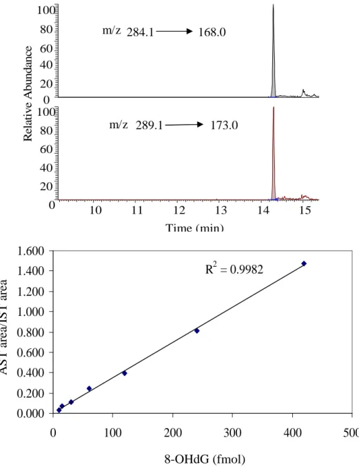
Three LC-MS/MS systems were evaluated for 1, N6-εdA detection, UPLC-MS/MS, capillary LC-MS/MS and nanoUPLC-MS/MS. In nano UPLC-MS/MS, a nanoacquity symmetry trap column (180µm×20mm, 5 µm), was connected with a nanoUPLC BEH C18 analytical column (1.0mm×100mm, 1.7µm) through a control valve. UPLC and capillary LC-MS/MS systems consisted of an Aquity UPLC system or Agilent 1200 capillary system and a TSQ triple quadrupole mass spectrometer. In the UPLC system, 0.1% formic acid in water was solvent A and 0.1% formic acid in methanol was solvent B. The flow rate was 200µl/min. A HSS T3 UPLC (1.8 µm, 2.1×100 mm) column was used to purify the samples before MS detection. In capillary system, the separation was performed on a ZORBAX SB-C18 column (100mm×0.5mm, 3 µm) with the flow rate of 7µl/min. Solvent A and B were 5mM ammonium formate in water and methanol respectively. The same collision energy and transitions were applied in all the three systems. 1, N6-εdA was detected under multiple reaction monitoring (MRM) in the positive ESI mode, m/z 276.0 → 160.0 for 1, N6-εdA and m/z 281.0 → 165.0 for 15N5-1,
16 eV. The sheath and auxillary gas pressure was 35 and 30 Torr respectively in UPLC systems, but none of them was used in capillary or nano system.
8-OHdG was detected by UPLC-MS/MS on a T3 HSS column (2.1×100mm, 1.7 µ) with 0.1% acetic acid in water as solvent A and methanol as solvent B at flow rate of 200µl/min. The gradient program was as follows: solvent B was increased from 1% to 5% in 2 min, held at 5% for 10 min, and increased to 50% in 2 min. m/z 284.1 → 168.0 and m/z 289.1 → 173.0 were monitored for 8-OHdG and [15N5]8-OHdG respectively with
CE 12 eV. The spray voltage was 3000 V. The vaporizer temperature was 250 ˚C. The sheath and auxillary gas pressure was 35 and 30 Torr respectively. The capillary temperature was 285 ˚C.
NanoLC-MS/MS was used to detect all LPO-induced DNA adducts. For the detection of 1, N2-εdG, M1dG, CrdG, 4-HNEdG and AcrdG, solvent A and B were 0.1%
formic acid in water and acetonitrile respectively. The sample was first loaded on the trap column by 99% A for 3 min with flow rate of 5µl/min. For 1, N2-εdG, M1dG, and AcrdG
detection, solvent B was 5% for the initial 1 min, followed by a increase to 30% in 10 min before returning to 5% in 15 min. For CrdG detection, solvent B was 10% for the initial 5 min, increased to 50% in 10 min and kept stable for 5 min. For 4-HNEdG detection, solvent B was 15% for the initial 2 min, increased to 70% in 10 min and returning to 15% in 10 min. The flow rate was 1.0 µl/min for the detection of all above adducts. Similar MS parameters were used to detect 1, N2-εdG, M1dG, AcrdG, CrdG and
was set as 285 ˚C. The spray voltage was 1500 V. Neither nebulizing nor drying gas was used in this system. Two transitions were monitored for the detection of 1, N2-εdG, M1dG
and 4-HNEdG respectively with CE 16 eV, m/z 304.0 → 188.0 and 308.0 → 193.0 for M1dG and [15N5]-M1dG, m/z 292 → 176.0 and 302 → 181.0 for 1, N2-εdG and [13C10]-1,
N2-εdG, and m/z 424.0 → 308 and 429.0 → 313.0 for 4-HNEdG and [15N5]-4-HNEdG.
Three and four transitions were monitored for CrdG and AcrdG detection respectively, m/z 338 → 222 with CE 12 eV and m/z 338 → 178 with CE 32 eV for CrdG, 343 → 227
with CE 12 eV for [15N5]-CrdG, m/z 324 → 208 with CE 12 eV and 324 → 164 with CE
32 eV for AcrdG, 329 → 213 with CE 12 eV and m/z 329 → 190 with CE 32 eV for [15N5]-AcrdG.
C. RESULTS
Enzymatic hydrolysis and HPLC purification
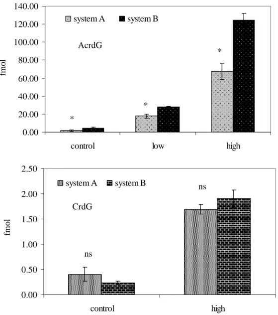
Higher amounts of AcrdG were observed in the acrolein treated ctDNA samples digested by system B than A and C, although no difference was found for the amount of CrdG in the crotaldehyde treated samples, as shown in figure 2.3. Background noise appeared for the detection of 4-HNEdG and M1dG in control and 4-HNE or MDA treated
ctDNA by system A. Thus, system B was selected for the assay development of propano adducts, 1, N2-εdG and M1dG. 200 U DNase I was added into system B at the beginning
of digestion, because some studies mentioned that it was necessary for the detection of bulky DNA adducts (36). 2΄-Deoxyadenosine (dA) co-eluted with AcrdG on C18 columns when 5 mM ammonium formate in water was used as solvent A and methanol as B. With 0.1% formic acid added in 5mM ammonium formate aqueous solution as solvent A, complete separation of dA and AcrdG was achieved, as shown in figure 2.4. The peak of M1dG appeared after that of 1, N2-εdG determined by LC-MS/MS using MDA-treated
DNA. The concentration of M1dG in MDA-treated ctDNA was below the LOD of the
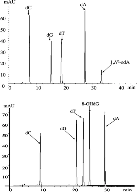
UV detector of the fraction collector. The typical chromatograms for LC purification of 1, N2-εdG, AcrdG, 4-HNEdG and CrdG are shown in figure 2.4. The fractions containing 1, N6-εdA and 8-OHdG were collected based on retention time of standards on fraction collector, as shown in figure 2.5.
LC-MS/MS detection and assay validation
dynamic curve), as shown in figure 2.7. The sensitivity of nanoUPLC system was more than 20 times higher than the UPLC system, followed by the capillary system which had 3 to 4 times higher sensitivity than the UPLC system. After LC purification, no significant matrix effects were observed with the nanoUPLC system, as indicated by figure 2.6. Better results were also obtained for the detection of 1, N6-εdA using the nanoUPLC than the UPLC system when evaluating rat liver DNA exposed to (13C2)-vinyl
chloride (VC), as depicted in figure 2.8. Transitions m/z 276 → 160, 278 → 162 and 281 → 165 were used to monitor endogenous NA-1, N6-εdA (lipid peroxidation induced 1, N6-εdA), exogenous [13C2]- 1, N6-εdA (VC induced 1, N6-εdA) and internal standard
[15N5]- 1, N6-εdA respectively.
The typical chromatograms of 8-OHdG, 1, N2-εdG and propano adducts in ctDNA and their calibration curves are shown in figure 2.9 A~E. Well-shaped peaks appeared in each monitored transition of these adducts. With R2 ≥ 0.98, all of these calibration curves indicated a good linear dynamic range (from sub-femto mole to several hundred femto mole) for the detection of these adducts by nanoUPLC-MS/MS system. The LOQ for propano adducts was 50 atmol on-column, which was determined by injecting standards with S/N higher than 10. Using this battery of assays, the background concentrations of these adducts in control liver DNA of female Sprague-Dawley rats (61 weeks old) were determined (table 2.1). 8-OHdG had the highest concentration in these samples, followed subsequently by AcrdG, M1dG, 1, N2-εdG, 1, N6-εdA, 4-HNEdG and CrdG. Besides,
M1dG, 1, N2-εdG and propano adducts were also measured in liver DNA from mice
DNA exposed by dibenzo[α,l]pyrene (DBP). The results are shown in figure 2.10A~D. After i.p. exposure to carbon tetrachloride for 13 days, significant increases of M1dG, 1,
N2-dG and AcrdG were observed in the liver DNA of wild type mice (2 to 3 fold increase in the exposed compared with the control), as shown in figure 2.10A. With the same chemical treatment, M1dG, 1, N2-εdG and AcrdG were significantly induced in liver
DNA of exposed XPA-/- mice compared with the control, as depicted in figure 2.10B. Three- to four-fold increases of M1dG, 1, N2-εdG and AcrdG were present in the liver
DNA from XPA-/- mice exposed to carbon tetrachloride, compared with exposed wide type mice, suggesting that NER is a dominant repair pathway for these DNA adducts. After 28 days exposure, no significant increases of 1, N2-εdG, M1dG, AcrdG, 4-HNEdG
and CrdG were observed in the liver of trout treated by 80 ppm DBP, compared with the 0.45 ppm treated group, as indicated in figure 2.10C and 2.10D.
D. DISCUSSION
With background adducts usually around 1/106 guanine, 8-OHdG has been detected by many different techniques, including immunoaffinity assay, LC-ECD, LC-MS/MS,
32
purification techniques for 8-OHdG detection by LC-MS/MS (40-51). With LC purification, decent peaks of 8-OHdG were observed by LC-MS/MS and the LOQ was reach several fmol on-column in the present and other published studies (10, 34, 48, 49). However, comprehensive comparison between LC-MS/MS assay with LC-ECD with sample purification before injection is still necessary. Besides the reported artifact formation during DNA isolation by ESCODD (37), Chao et al. found the production of artifacts during vacuum drying, which was supported by other reports (44, 49-51). Their studies indicated that as a concentration process, both vacuum and freeze drying for intact or hydrolyzed DNA could introduce artifact formation. With SPE purification before column switching LC-MS/MS detection, higher 8-OHdG was observed than the one without SPE, which indicated that excessive sample processing also contributed to the formation of artifact. In our studies, the further oxidation of [15N5]8-OHdG in samples
was observed when offline LC sample purification was applied before LC-MS/MS detection, which made the quantification of 8-OHdG even more complicated. Besides, background 8-OHdG levels, similar to other reports (48, 49) were detected in this study. These results suggest that online column switching may work well for the detection of 8-OHdG.
column purification, 32P-postlabelling procedure and thin-layer chromatographic separation. The mean 1, N6-εdA and 3, N4-εdC levels per 108 parent nucleotides in normal human liver were determined to be 1.9 ± 0.5 and 2.8 ± 1.0 respectively by this assay (53). Although it has excellent sensitivity, this assay also has shortcomings. The accuracy of the measurements was not well defined due to the lack of an authentic internal standard and no chemical structural information is provided. Compared with 32P-postlabelling method, mass spectrometry is a powerful technique with the particular advantage in providing the information on chemical structure of adducts. Using isotopically labeled internal standards, the entire sample processing scheme can be monitored to better assess the accuracy of the assay. Using various sample purification techniques, GC-MS/MS or LC-MS/MS has been applied to detect etheno adducts, including 1, N6-εdA or εA, 3, N4
Background 1, N6-εdA and 1, N2-εdG can be consistently detected. Because of the instability of the glycosyl bond in N2, 3-εdG, the direct detection of this adduct by LC-MS/MS was impeded (34, 49, 64).
M1G has been measured by MS, 32P-postlabeling or immunochemical techniques
(25-29, 67). Each technique offers advantages and disadvantages based on a combination of sensitivity and specificity. 32P-Postlabeling was first employed for the detection and quantitation of M1G (17). Increased M1G was reported in the kidneys of Syrian hamster
treated with estradiol, in white blood cells from women fed a diet high in sunflower oil and in normal breast tissues of women with breast cancer using this technique (68-70). Marnett’s laboratory employed GC-MS with electron capture negative chemical ionization detection for quantitation of M1G after immunoaffinity column purification
and derivatization with pentafluorobenzyl bromide (17). The LOD of the assay was approximately 1 M1G per 108 nucleotides with 1mg of DNA. Using 500 ug MDA-treated
ctDNA, Hakala evaluated the application of LC-ESIMS/MS for the measurement of M1G.
Besides, several studies developed assays to measure M1dG indirectly and directly (26-29,
67). Jeong et al. developed a novel method for analysis of M1dG by aldehyde reactive
probe (ARP) labeling and LC-MS/MS with SPE purification (28). Both ring-open and ring-closed forms of M1dG were conjugated with a stable ARP group before detection,
with an LOD of 2 M1G adducts/108 guanines. Both APCIMS/MS and
LC-ESIMS/MS were evaluated to measure the 5, 6-dihydro derivative of M1dG in urine after