Edited by: Philippe Saas, INSERM UMR1098 Interactions Hôte-Greffon-Tumeur & Ingénierie Cellulaire et Génique, France
Reviewed by: Joanne E. Konkel, University of Manchester, United Kingdom Julien Diana, Institut National de la Santé et de la Recherche Médicale, France Thomas J. Hawke, McMaster University, Canada Sylvaine You, Institut National de la Santé et de la Recherche Médicale, France
*Correspondence: Roland M. Tisch [email protected]
†These authors have contributed
equally to this work.
Specialty section: This article was submitted to Inflammation, a section of the journal Frontiers in Immunology
Received: 20 October 2017 Accepted: 12 December 2017 Published: 22 December 2017 Citation: Clark M, Kroger CJ and Tisch RM (2017) Type 1 Diabetes: A Chronic Anti-Self-Inflammatory Response. Front. Immunol. 8:1898. doi: 10.3389/fimmu.2017.01898
Type 1 Diabetes: A Chronic Anti-
Self-inflammatory Response
Matthew Clark
1†, Charles J. Kroger
1†and Roland M. Tisch
1,2*
1 Department of Microbiology and Immunology, University of North Carolina at Chapel Hill, Chapel Hill, NC, United States, 2 Lineberger Comprehensive Cancer Center, University of North Carolina at Chapel Hill, Chapel Hill, NC, United States
Inflammation is typically induced in response to a microbial infection. The release of
proinflammatory cytokines enhances the stimulatory capacity of antigen-presenting
cells, as well as recruits adaptive and innate immune effectors to the site of infection.
Once the microbe is cleared, inflammation is resolved by various mechanisms to avoid
unnecessary tissue damage. Autoimmunity arises when aberrant immune responses
target self-tissues causing inflammation. In type 1 diabetes (T1D), T cells attack the
insu-lin producing
β
cells in the pancreatic islets. Genetic and environmental factors increase
T1D risk by in part altering central and peripheral tolerance inducing events. This results
in the development and expansion of
β
cell-specific effector T cells (Teff) which mediate
islet inflammation. Unlike protective immunity where inflammation is terminated,
auto-immunity is sustained by chronic inflammation. In this review, we will highlight the key
events which initiate and sustain T cell-driven pancreatic islet inflammation in nonobese
diabetic mice and in human T1D. Specifically, we will discuss: (i) dysregulation of thymic
selection events, (ii) the role of intrinsic and extrinsic factors that enhance the expansion
and pathogenicity of Teff, (iii) defects which impair homeostasis and suppressor activity
of FoxP3-expressing regulatory T cells, and (iv) properties of
β
cells which contribute to
islet inflammation.
Keywords: autoimmunity, type 1 diabetes, immunoregulation, inflammation, T cells
iNTRODUCTiON
Type 1 diabetes (T1D) is an autoimmune disease characterized by the chronic inflammation of
the pancreatic islets of Langerhans (
1
–
4
). Islet inflammation is typically marked by infiltrating
adaptive and innate immune effectors. Insulitis progresses over time and when a sufficient amount
of
β
cell mass has been rendered nonfunctional and/or destroyed, hyperglycemic blood levels are
achieved, and clinical diabetes established. The immune mechanisms mediating
β
cell
autoim-munity are heterogeneous, as reflected by the nature of the islet infiltrate and the age of clinical
onset. Nevertheless, T1D is generally viewed as a T cell-driven autoimmune disease, particularly
for the more prevalent and aggressive type of T1D that develops in children and adolescents versus
adults (
5
–
17
). A T cell-independent subtype of T1D, however, may also exist that is thought to
be largely mediated by innate immune effectors (
18
,
19
). The events leading to the loss of
β
cell-specific tolerance and chronic islet inflammation are complex, and influenced by both genetic and
environmental factors (
20
–
22
).
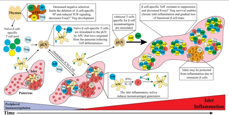
FigURe 1 | Dysregulated thymic and peripheral events culminate in chronic islet inflammation. In general, overt diabetes results from the gradual loss of functional insulin producing β cells due to the inflammatory environment driven by infiltrating self-reactive T cells and antigen-presenting cell (APC). Although β cell-specific T cell clones are detected in both healthy and type 1 diabetes (T1D) susceptible individuals, a number of factors promote T1D development in the latter population. Decreased efficiency of negative selection in the thymus, either due to altered tissue-specific antigen expression or due to T cell receptor (TCR) signaling, allows for the increased escape of β cell-specific T cell clones into the periphery. In addition, β cell-specific Foxp3+Treg development may also be suboptimal due to dysregulation of TCR signaling. In the periphery, β cell-specific T cells are stimulated in the pancreatic lymph nodes (pLN) by APC derived from the islets, leading to effector T cell (Teff) differentiation. These pathogenic Teff then infiltrate the islets and drive inflammation leading to reduced β cell function and/or survival. Not all islets are infiltrated potentially due to an immature phenotype and reduced autoantigen expression by β cells. Ongoing islet inflammation also leads to the generation of neoautoantigens either directly in β cells or during antigen processing by APC. The presentation of neoautoantigens within the pLN promotes the activation and expansion of additional Teff pools. These events amplify and drive a chronic state of islet inflammation leading to impaired functional β cell mass and clinical onset of T1D.
class I and II haplotypes, consistent with a key role for T cells
in T1D (
29
,
30
). A number of genes regulating T, B, and innate
cell immunobiology are also linked with T1D, as are genetic
vari-ants intrinsic to
β
cells, which deleteriously affect
β
cell function
and/or responses to inflammation (
31
–
37
).
The identity and role of environmental factors in T1D are
poorly understood. The most common hypothesis is that
micro-bial infections initiate and/or exacerbate islet inflammation in
genetically susceptible individuals (
38
,
39
). For instance, T1D is
associated with enteroviruses such as coxsackievirus B1 (
40
–
44
).
Viral infection of
β
cells may result in direct cytolysis and/or
elicit local inflammation that initiates and/or drives
autoimmun-ity (
45
–
47
). The gut microbiota also has a profound regulatory
effect on
β
cell autoimmunity (
48
,
49
). In the nonobese diabetic
(NOD) mouse, a spontaneous model of T1D,
β
cell destruction
can be either promoted or prevented by changes in the
composi-tion of the gut microbiota (
50
,
51
). Here bacterial components
and metabolites are thought to impact the activation and/or
differentiation status of innate and adaptive immune effectors.
Longitudinal studies of at risk subjects also indicate a role for gut
microbiota in human T1D (
52
–
54
).
The T cell-related events that drive chronic islet
inflamma-tion in T1D stem from dysregulainflamma-tion of central and peripheral
tolerance, alterations in self-antigen processing, and modified
β
cell responses (Figure 1). Here, we discuss how events critical
for initiating and amplifying the development of T cell-mediated
islet inflammation are regulated in NOD mice and human T1D.
THYMiC ORigiNS OF DiABeTOgeNiC
T CeLLS: SeTTiNg THe STAge FOR
iSLeT iNFLAMMATiON
The generation of an autoreactive T cell receptor (TCR)
reper toire in the periphery is established in part by inefficient
negative selection of anti-self-single positive thymocytes (SP)
in the thymus (
55
,
56
). Early in ontogeny negative selection is
lax, resulting in increased escape of anti-self-SP (
57
–
59
). This
temporal decrease in negative selection and elevated survival of
β
cell-specific clonotypes may help explain the predominance of
T1D onset in childhood. With time, changes in thymic structural
organization and maturation of thymic antigen-presenting cells
(APC) leads to more efficient negative selection and increased
death of autoreactive SP (
57
).
express and present several tissue-specific antigens (TSA)
(
60
–
63
). Recognition of MHC-self-peptide complexes with
increasing avidity/affinity results in elevated TCR signaling and
SP apoptosis. Dysregulation of negative selection generates a
peripheral pool of anti-self-T cells displaying increased avidity/
affinity, and likely an enhanced “pathogenic potential.”
Parameters influencing the efficiency of negative selection
both intrinsic and extrinsic to thymocytes have been linked to
the development of
β
cell-specific T cells and T1D. Thymocyte
intrinsic properties reported in NOD mice have included
reduced SP sensitivity to apoptosis and altered double positive
thymocyte differentiation to SP (
64
–
67
). In humans, TCR
signal-ing needed to drive apoptosis of
β
cell-specific SP may be limited
by a T1D-associated variant of the protein tyrosine phosphatase
non-receptor 22 (
PTPN22
) gene (
31
,
32
). PTPN22 is a negative
regulator of TCR signaling, and elevated phosphatase activity
by PTPN22 is predicted to reduce TCR signaling strength and
diminish apoptosis induction in SP (
68
). An increase in PTPN22
activity may also limit thymic development of
β
cell-specific
FoxP3-expressing regulatory CD4
+T cells (FOXP3
+Treg),
which is dependent on high(er) avidity/affinity recognition of
self-peptide.
Thymocyte extrinsic factors that impact negative
selec-tion include aberrant expression of TSA in the medulla. The
importance of thymic expression and presentation of TSA
is readily evident in mice and humans deficient of the
tran-scription factor autoimmune regulator (AIRE) (
60
,
69
). Lack
of AIRE, which drives expression of select TSA by mTEC,
results in inefficient thymic negative selection and reduced
development of tissue-specific Foxp3
+Treg, leading to
multio-rgan autoimmunity in mice (
70
–
74
). Similarly, aberrant AIRE
expression and function in humans results in the development
of autoimmune polyendorinology candidiasis and ectodermal
dysplasia (APECD) in which a variety of organs are targeted
by T cells; notably a subset of APCED patients develop T1D
(
75
,
76
). Reduced AIRE expression has been reported in NOD
mice, which reflects not only T1D development but also
T cell-mediated inflammation of other tissues such as the thyroid,
salivary, and lacrimal glands (
77
).
In human T1D, a strong genetic association is linked to the
insulin encoding gene
INS2
found in
IDDM2
(
78
). Insulin is
believed to be a key autoantigen driving human T1D, which is
supported by studies in NOD mice (
79
–
81
).
INS2
is preceded
by a variable number of tandem repeats (VNTRs). Individuals
that have 26–63 VNTRs, associated with decreased thymic
INS2
expression, have an increased risk of developing T1D. In contrast,
INS2
expression is increased with VNTRs ranging between 140
and 210, which in turn is associated with a protective phenotype
(
82
,
83
). Reduced thymic insulin expression is expected to both
limit negative selection and development of insulin-specific
SP and FOXP3
+Treg, respectively. Future studies are needed
to directly demonstrate that thymic selection is dysregulated,
and contributes to an expanded
β
cell-specific peripheral T cell
pool in human T1D. Whether defects in thymic selection and
development of
β
cell-specific T cells are necessary only early on
or required throughout the disease process is another issue that
needs to be tackled.
It is noteworthy that
β
cell-specific T cells are detected in
the blood of healthy individuals, likely reflecting in part the
reduced efficiency of thymic negative selection early in
ontog-eny. However, the phenotype of circulating
β
cell-specific T cells
is distinct in T1D patients versus healthy subjects (
84
–
89
).
The former exhibit mostly an effector/memory phenotype
and expression of proinflammatory cytokines consistent with
ongoing
β
cell autoimmunity (
84
–
88
). These findings indicate
that in addition to the TCR repertoire, other factors contribute
to the differentiation and expansion of diabetogenic effector
T cells (Teff). For instance, the extent of tissue destruction and
lethality of AIRE deficiency in mice is influenced by genotype
with AIRE-deficient NOD versus C57BL/6 mice exhibiting more
severe systemic autoimmunity (
90
,
91
). Additionally, distinct
TCR repertoires have been found in NOD mice in contrast to
MHC matched C57BL/6 mice (
92
). Overall, dysregulation of
thymic selection events in NOD mice acts as a precursor for islet
inflammation.
eXTRiNSiC AND iNTRiNSiC FACTORS
PROMOTe PATHOgeNiC eFFeCTOR
T CeLLS iN T1D
The initiation of islet inflammation in NOD mice and humans
is ill-defined. In NOD mice pancreatic remodeling shortly after
birth is thought to play a key role starting the diabetogenic
response (
93
,
94
). Remodeling of the pancreas results in a wave
of
β
cell apoptosis and release of antigens which are endocytosed
by resident macrophages and DC (
95
). These APC then traffick
to the draining pancreatic lymph nodes (pLN) to prime
β
cell-specific T cells and promote Teff differentiation (
96
,
97
). Once
established Teff migrate into the islets and mediate inflammation
(
97
–
99
).
Both CD4
+and CD8
+T cells are required for efficient
β
cell
destruction in NOD mice (
105
). Islet CD8
+T cells primarily
mediate
β
cell destruction by a cognate interaction involving
perforin and granzyme B-, and Fas-Fas ligand-mediated killing
(
106
,
107
). On the other hand, islet CD4
+T cells drive
β
cell
destruction in a bystander manner
via
secretion of
proinflamma-tory cytokines. CD4
+and CD8
+T cells are also detected in the
islets of diabetic subjects, with CD8
+T cells often predominating
(
6
,
106
). Several
β
cell autoantigens are recognized by the islet
infiltrating T cells, and a number of these are similarly targeted
in both the NOD and human diabetogenic responses including
glutamic acid decarboxylase 65, proinsulin, insulin B chain, islet
antigen-2, and islet-specific glucose-6-phosphatase catalytic
subunit-related protein (
108
).
The majority of CD4
+and CD8
+T cells infiltrating the islets
of NOD mice and T1D subjects exhibit a T helper 1 (Th1)
effector phenotype, marked by IFN
γ
secretion (
109
). Increased
Th17 cells are seen in the islets of NOD mice and the pLN of
T1D subjects (
109
–
111
). The role of Th17 cells in mediating
islet inflammation, however, is ill-defined. Elevated local levels
of IFN
γ
are believed to establish a feed-forward loop that drives
islet pathology. Based on NOD mouse studies, IFN
γ
secreted
by islet CD4
+(and CD8
+) Teff results in local upregulation of
chemotactic cues that induce additional T, B, and innate cells
to migrate into the islets, as well as promote islet retention of
these effectors (
109
,
112
). IFN
γ
also activates islet resident
APC and stromal cells to elevate production of additional
inflammatory mediators, such reactive oxygen species, which
impair function and mediate
β
cell necrosis (
107
,
113
,
114
).
Furthermore, IFN
γ
in the context of IL-1
β
and TNF
α
induces
β
cell apoptosis (
113
,
114
).
Another proinflammatory cytokine thought to contribute
to islet inflammation is IL-21 which is elevated in T1D patient
serum (
115
). Notably, the murine IL-21 gene is located in the
Idd3
locus and IL-21 receptor (R) deficiency prevents T1D
in NOD mice (
116
). CD4
+T follicular helper cells, which are
increased in the pLN of NOD mice, are the primary source of
IL-21 (
112
,
117
–
119
). IL-21 has a critical role in supporting B cell
development and antibody production. B cells, serving as APC,
are required for efficient
β
cell destruction in NOD mice and
likely in human T1D (
117
,
118
,
120
,
121
). IL-21 also enhances
maintenance of CD8
+Teff by preventing exhaustion during
chronic inflammation (
122
,
123
). Interestingly, the pathogenicity
of
β
cell-specific CD8
+T cells is dependent on IL-21R expression
(
124
,
125
).
Defects intrinsic to Teff are also thought to facilitate chronic
islet inflammation. Variants of the
CTLA4
gene are linked to
T1D susceptibility in both NOD mice (
Idd5.1
) and human T1D
(
IDDM12
) (
33
,
126
). CTLA-4 which binds to the costimulatory
molecules CD80 and CD86 expressed on APC, is a negative
regu-lator of T cell activation and proliferation (
34
). Polymorphisms
in the human
CTLA4
gene region are associated with reduced
mRNA levels and a decrease in expression of the soluble (s)
CTLA-4 isoform (
33
,
34
,
126
). sCTLA-4 also negatively regulates
TCR signaling (
33
,
34
,
126
). Reduced expression of CTLA-4 and
sCTLA-4 is expected to facilitate expansion of
β
cell-specific
T cells. This scenario is consistent with the exacerbated
β
cell
autoimmunity seen in NOD mice expressing a diabetogenic TCR
transgene and lacking CTLA-4 expression (
127
). Noteworthy is
that both NOD-derived and human T1D Teff also exhibit reduced
sensitivity to Foxp3
+Treg-mediated suppression (
128
,
129
).
In sum, the culmination of a variety of extrinsic and intrinsic
factors enables Teff to expand, persist, and in turn amplify islet
inflammation.
DeFeCTS iN THe Foxp3
+Treg POOL
CONTRiBUTe TO T1D
In addition to Teff that are resistant to regulatory mechanisms
that limit expansion and function, evidence indicates that the
Foxp3
+Treg pool is compromised in T1D (
130
,
131
). Here,
dysregulation of Foxp3
+Treg homeostasis is thought to permit
preferential differentiation and expansion of pathogenic
β
cell-specific Teff. Foxp3
+Treg have an essential role in regulating
immune homeostasis and reactivity to self (
132
–
135
). The lack of
thymic development of Foxp3
+Treg due to deficient expression
or function of the FoxP3 transcription factor, results in systemic
autoimmunity in both mice and humans. Foxp3
+Treg mediate
suppression of T cells and other immune effectors
via
multiple
mechanisms including cell-contact dependent suppression, and
secretion of anti-inflammatory cytokines and mediators such
as IL-10, TGF
β
1, and IL-35, and adenosine, respectively (
136
).
Foxp3
+Treg also function as an “IL-2 depot” to deprive Teff of
IL-2 needed for expansion (
136
). The latter is mediated by
con-stitutive expression of CD25, the
α
subunit of the IL-2R (
136
).
Therefore, Foxp3
+Treg, expressing the high affinity IL-2R, are
able to out compete Teff for IL-2, which transiently express high
affinity IL-2R.
IL-2 is essential for Foxp3
+Treg homeostasis, expansion, and
function (
136
). Unlike conventional T cells, Foxp3
+Treg do not
produce IL-2 due to FoxP3-mediated negative regulation of
Il2
transcription. Therefore, Foxp3
+Treg are dependent on T cells
and DC as IL-2 sources (
136
). This dependency is thought to
enable Foxp3
+Treg to more readily sense and respond to ongoing
inflammation. Accordingly, defects in the IL-2/IL-2R axis have
been described in the NOD model and human T1D (
137
–
142
).
In NOD mice, an
Il2
variant located in
Idd3
results in reduced
levels of IL-2 expression by Teff, and impaired survival and
func-tion of islet resident Foxp3
+Treg (
130
,
143
). Increased levels of
proinflammatory cytokines, such as IFN
γ
and IL-6 that
down-regulate FoxP3 expression may also promote dedifferentiation of
islet Foxp3
+Treg into a Teff-like subsets (
144
,
145
). These events
lead to a progressive loss of islet Foxp3
+Treg suppression, thereby
“releasing the brakes” and favoring pathogenic Teff expansion.
The frequency of FOXP3
+Treg found in blood is largely
unaffected in T1D subjects (
140
,
141
,
146
–
148
). However,
FOXP3
+Treg from T1D subjects exhibit reduced suppressor
function measured
in vitro
(
128
,
129
). This aberrant activity is
linked to T1D risk variants of
IL2RA
(CD25) and
PTPN2
, a
phos-phatase involved in IL-2R signaling (
149
). Notably, FOXP3
+Treg
in T1D subjects (
149
,
151
). This approach has been effective in
preventing and/or reversing diabetes in NOD mice by increasing
the number and function of islet Foxp3
+Treg (
149
,
152
). One
key question not addressed is the specificity of FOXP3
+Treg in
T1D subjects. Reduced thymic development of
β
cell-specific
FOXP3
+Treg, as discussed above, would be expected to limit the
“anti-diabetogenic” effects of the peripheral FOXP3
+Treg pool.
AMPLiFYiNg THe PATHOgeNiC
eFFeCTOR T CeLL ReSPONSe
VIA
NeOAUTOANTigeNS
Recent findings have demonstrated that the proinflammatory
milieu
of the islets promotes processing of “neoautoantigens”
(
153
). Importantly, these neoautoantigens are only found in the
periphery so that corresponding T cell clonotypes, not deleted
in the thymus and possibly expressing high affinity TCR, can
be recruited into the inflammatory response. Neoautoantigens
are generated
via
posttranslational modifications (PTM), such
as deamidation by tissue transglutaminase (tTG) (
153
). PTM
can occur during APC antigen processing or directly in
β
cells
(
154
). tTG-dependent deamidation of a proinsulin C-peptide
for instance is detected in both human DC and islets under
inflammatory conditions (
154
). Notably the resulting modified
peptide is recognized by CD4
+T cells derived from T1D subjects.
The MHC binding and in turn T cell stimulatory properties of
peptides can also be enhanced by deamidation (
155
).
Neoautoantigens consisting of hybrid peptides have recently
been identified. In NOD mice hybrid insulin peptides are
gener-ated
via
covalent crosslinking of a proinsulin C peptide with
peptides derived from naturally occurring cleavage products
produced in the
β
cell secretory granules (
156
). In addition to
ongoing inflammation, PTM occurs
via
endoplasmic reticulum
stress, which can be induced in
β
cells by the normal
physi-ological demands associated with high levels of insulin secretion
(
157
). Therefore, it is possible that
β
cell neoautoantigens in
addition to amplifying inflammation, play a role in initiating the
diabetogenic response. Neoautoantigens are also generated at a
transcriptional level. A mutation in the open reading frame of
insulin mRNA generates a neoautoantigen that stimulates CD8
+T cells from T1D subjects causing
β
cells lysis
in vitro
(
158
).
Alternative RNA splicing may be another mechanism leading
to neoautoantigen expression, particularly since ~30% of genes
in inflamed
β
cells undergo aberrant alternative splicing (
159
).
In sum,
β
cell neoautoantigens serve as
bona fide
targets of
patho-genic CD4
+and CD8
+T cells. The breadth of the peptidome of
neoautoantigens produced and presented, and the properties of
neoautoantigen-specific T cells, in terms of frequency, avidity/
affinity, subset phenotype (e.g., pathogenic versus regulatory),
and overall contribution to islet inflammation require further
investigation.
β
CeLL-iNTRiNSiC PROPeRTieS THAT
RegULATe iSLeT iNFLAMMATiON
Studies have demonstrated that intrinsic properties of
β
cells also
influence islet inflammation. For instance, CXCL10 is produced by
β
cells although the role of this chemokine in disease is controversial
(
160
). CXCL10 regulates migration of CXCR3 expressing Teff
and Foxp3
+Treg into the islets (
97
–
99
,
161
). Overexpression of
CXCL10 in
β
cells accelerates T1D progression, and antibody
blockade of CXCL10 prevents Teff migration into the islets of
NOD mice (
97
–
99
,
161
). On the other hand,
Cxcr3
deficiency
accelerates T1D by reducing islet resident Foxp3
+Treg (
162
,
163
).
Therefore, depending on the context,
β
cells may affect
inflamma-tory and immunoregulainflamma-tory events
via
CXCL10 production. The
chemokine CCL2 is also secreted by
β
cells, and over-expression
of ectopic CCL2 recruits tolerogenic CCR2-expressing DC and
blocks T1D progression in NOD mice (
164
). Interestingly, NOD
APC shows defective migration in response to CCL2, and human
T1D patients have reduced serum levels of CCL2 (
164
–
166
).
Additionally,
β
cells produce CXCL1 and CXCL2 that recruit
CXCR2-expressing neutrophils to the islets, which contribute to
stimulating
β
cell-specific T cell reactivity (
167
,
168
). Overall,
β
cell produced chemotactic cues regulate the progression of the
diabetogenic response.
Along with chemokines,
β
cells secrete the cytokine IL-1
β
,
which at low levels promotes
β
cell proliferation, and enhances
production of CCL2, CXCL1, CXCL2, and insulin (
169
,
170
).
However, IL-1
β
also primes leukocyte effector-mediated
inflammation, and as noted above, IL-1
β
in the context of TNF
α
,
and/or IFN-
γ
induces
β
cell apoptosis
in vitro
(
113
,
114
).
Notably, glucagon-secreting
α
cells also produce IL-1
β
,
indicat-ing that other islet resident endocrine cells may also contribute
to local inflammation (
171
,
172
). Islet inflammation also
induces upregulation of MHC class I and II on
β
cells to further
increase
β
cell immunogenicity (
173
). Interestingly, a
subpopu-lation of
β
cells have been identified which under inflammatory
conditions acquires resistance to immune-mediated
destruc-tion in NOD mice (
174
). The latter is associated with a more
immature
β
cell phenotype coupled with reduced expression
of autoantigens and upregulation of immunomodulatory
molecules such as PD-L1, an inducer of T cell exhaustion.
A similar phenotype is seen for human
β
cells (
174
). Therefore,
β
cells not only contribute to islet inflammation but also adapt
under the inflammatory conditions in order to persist. A
bet-ter understanding of the events regulating this dichotomy has
important implications for the treatment of T1D patients
via
β
cell replacement strategies for instance.
SUMMARY
negative selection (
57
–
59
). This is coupled with
β
cell-specific
Teff that are insensitive to peripheral tolerance inducing events,
β
cell-specific FOXP3
+Treg with impaired suppressor activity,
and
β
cells which readily promote islet inflammation (
128
,
129
,
147
,
169
,
170
). Robust inflammation leads to increased
β
cell
neoautoantigen production further amplifying the kinetics
and overall inflammatory response (
153
,
154
,
156
,
158
). Under
these “ideal” conditions, early onset T1D develops. On the other
hand, in individuals with only a partial complement of these
key “disease components,” islet inflammation is less robust and
the kinetics of T1D onset proportionately delayed. Defining
the events driving early versus late(r) T1D onset is critical for a
better understanding of how islet inflammation is regulated in
humans. The latter is also important for developing rational and
effective immunotherapies to prevent and/or treat T1D. Devising
strategies to enhance thymic negative selection early in ontogeny
for instance, would be expected to purge the diabetogenic TCR
repertoire to prevent T1D. Indeed, approaches are currently
being studied to manipulate thymic negative selection in the
context of cancer treatment by expanding the T cell repertoire
specific for self-tumor antigens (
175
–
177
). Altering the gut
microbiome early in life may also prove to be an effective strategy
to limit expansion of the anti-self-T cell repertoire and establish
robust immunoregulation in the periphery. Administration of
β
cell neoautoantigens may augment the efficacy of antigen-based
immunotherapies to block disease progression at later stages of
T1D (
157
). Depending on the mode of administration,
β
cell
neoautoantigens can be used to target the corresponding
clono-types by tolerizing pathogenic Teff and/or inducing/expanding
FOXP3
+Treg. In view of the heterogeneity in the
immunopathol-ogy of T1D, it is very likely these approaches and others currently
being studied will need to be combined to effectively suppress the
chronic islet inflammation and
β
cell autoimmunity long term.
AUTHOR CONTRiBUTiONS
MC, CJK, and RMT contributed to the preparation of the review
article.
FUNDiNg
This work was supported by National Institutes of Health grants
R01DK100256 and R01DK1035486 (RMT) and T32AI007273
(MC).
ReFeReNCeS
1. Bach JF. Insulin-dependent diabetes mellitus as an autoimmune disease. Endocr Rev (1994) 15(4):516–42. doi:10.1210/edrv-15-4-516
2. Tisch R, McDevitt H. Insulin-dependent diabetes mellitus. Cell (1996) 85(3):291–7. doi:10.1016/S0092-8674(00)81106-X
3. Eisenbarth GS. Type 1 diabetes: molecular, cellular and clinical immunology. Adv Exp Med Biol (2004) 552:306–10.
4. Anderson MS, Bluestone JA. The NOD mouse: a model of immune
dys-regulation. Annu Rev Immunol (2005) 23:447–85. doi:10.1146/annurev.
immunol.23.021704.115643
5. Pugliese A. Autoreactive T cells in type 1 diabetes. J Clin Invest (2017) 127(8):2881–91. doi:10.1172/JCI94549
6. Coppieters KT, Dotta F, Amirian N, Campbell PD, Kay TW, Atkinson MA, et al. Demonstration of islet-autoreactive CD8 T cells in insulitic lesions from recent onset and long-term type 1 diabetes patients. J Exp Med (2012) 209(1):51–60. doi:10.1084/jem.20111187
7. Atkinson MA, von Herrath M, Powers AC, Clare-Salzler M. Current con-cepts on the pathogenesis of type 1 diabetes – considerations for attempts to prevent and reverse the disease. Diabetes Care (2015) 38(6):979–88. doi:10.2337/dc15-0144
8. Arif S, Gibson VB, Nguyen V, Bingley PJ, Todd JA, Guy C, et al. Beta-cell specific T-lymphocyte response has a distinct inflammatory phenotype in children with Type 1 diabetes compared with adults. Diabet Med (2017) 34(3):419–25. doi:10.1111/dme.13153
9. Heninger AK, Eugster A, Kuehn D, Buettner F, Kuhn M, Lindner A, et al. A divergent population of autoantigen-responsive CD4+ T cells in infants prior to beta cell autoimmunity. Sci Transl Med (2017) 9(378):eaaf8848. doi:10.1126/scitranslmed.aaf8848
10. Willcox A, Richardson SJ, Bone AJ, Foulis AK, Morgan NG. Analysis of islet inflammation in human type 1 diabetes. Clin Exp Immunol (2009) 155(2):173–81. doi:10.1111/j.1365-2249.2008.03860.x
11. Atkinson MA, Gianani R. The pancreas in human type 1 diabetes: providing new answers to age-old questions. Curr Opin Endocrinol Diabetes Obes (2009) 16(4):279–85. doi:10.1097/MED.0b013e32832e06ba
12. Coppieters KT, Roep BO, von Herrath MG. Beta cells under attack: toward a better understanding of type 1 diabetes immunopathology. Semin Immunopathol (2011) 33(1):1–7. doi:10.1007/s00281-010-0236-6 13. Richardson SJ, Willcox A, Bone AJ, Morgan NG, Foulis AK. Immunopathology
of the human pancreas in type-I diabetes. Semin Immunopathol (2011) 33(1):9–21. doi:10.1007/s00281-010-0205-0
14. Campbell-Thompson M. Organ donor specimens: what can they tell us about type 1 diabetes? Pediatr Diabetes (2015) 16(5):320–30. doi:10.1111/ pedi.12286
15. Campbell-Thompson M, Fu A, Kaddis JS, Wasserfall C, Schatz DA, Pugliese A, et al. Insulitis and beta-cell mass in the natural history of type 1 diabetes. Diabetes (2016) 65(3):719–31. doi:10.2337/db15-0779
16. Babon JA, DeNicola ME, Blodgett DM, Crevecoeur I, Buttrick TS, Maehr R, et al. Analysis of self-antigen specificity of islet-infiltrating T cells from human donors with type 1 diabetes. Nat Med (2016) 22(12):1482–7. doi:10.1038/nm.4203
17. Michels AW, Landry LG, McDaniel KA, Yu L, Campbell-Thompson M, Kwok WW, et al. Islet-derived CD4 T cells targeting proinsulin in human autoimmune diabetes. Diabetes (2017) 66(3):722–34. doi:10.2337/db16-1025 18. In’t Veld P. Insulitis in human type 1 diabetes: the quest for an elusive lesion.
Islets (2011) 3(4):131–8. doi:10.4161/isl.3.4.15728
19. Skog O, Korsgren S, Melhus A, Korsgren O. Revisiting the notion of type 1 diabetes being a T-cell-mediated autoimmune disease. Curr Opin Endocrinol Diabetes Obes (2013) 20(2):118–23. doi:10.1097/MED. 0b013e32835edb89
20. van Belle TL, Coppieters KT, von Herrath MG. Type 1 diabetes: etiology, immunology, and therapeutic strategies. Physiol Rev (2011) 91(1):79–118. doi:10.1152/physrev.00003.2010
21. Atkinson MA. The pathogenesis and natural history of type 1 diabetes. Cold Spring Harb Perspect Med (2012) 2(11):a007641. doi:10.1101/cshperspect. a007641
22. Katsarou A, Gudbjornsdottir S, Rawshani A, Dabelea D, Bonifacio E, Anderson BJ, et al. Type 1 diabetes mellitus. Nat Rev Dis Primers (2017) 3:17016. doi:10.1038/nrdp.2017.16
23. Barrett JC, Clayton DG, Concannon P, Akolkar B, Cooper JD, Erlich HA, et al. Genome-wide association study and meta-analysis find that over 40 loci affect risk of type 1 diabetes. Nat Genet (2009) 41(6):703–7. doi:10.1038/ ng.381
24. Concannon P, Erlich HA, Julier C, Morahan G, Nerup J, Pociot F, et al. Type 1 diabetes: evidence for susceptibility loci from four genome-wide linkage scans in 1,435 multiplex families. Diabetes (2005) 54(10):2995–3001. doi:10.2337/diabetes.54.10.2995
25. Pociot F, Akolkar B, Concannon P, Erlich HA, Julier C, Morahan G, et al. Genetics of type 1 diabetes: what’s next? Diabetes (2010) 59(7):1561–71. doi:10.2337/db10-0076
27. Noble JA, Erlich HA. Genetics of type 1 diabetes. Cold Spring Harb Perspect Med (2012) 2(1):a007732. doi:10.1101/cshperspect.a007732
28. Onengut-Gumuscu S, Chen WM, Burren O, Cooper NJ, Quinlan AR, Mychaleckyj JC, et al. Fine mapping of type 1 diabetes susceptibility loci and evidence for colocalization of causal variants with lymphoid gene enhancers. Nat Genet (2015) 47(4):381–6. doi:10.1038/ng.3245
29. Singal DP, Blajchman MA. Histocompatibility (HL-A) antigens, lymphocy-totoxic antibodies and tissue antibodies in patients with diabetes mellitus. Diabetes (1973) 22(6):429–32. doi:10.2337/diab.22.6.429
30. Nerup J, Platz P, Andersen OO, Christy M, Lyngsoe J, Poulsen JE, et al. HL-A antigens and diabetes mellitus. Lancet (1974) 2(7885):864–6. doi:10.1016/S0140-6736(74)91201-X
31. Bottini N, Musumeci L, Alonso A, Rahmouni S, Nika K, Rostamkhani M, et al. A functional variant of lymphoid tyrosine phosphatase is associated with type I diabetes. Nat Genet (2004) 36(4):337–8. doi:10.1038/ng1323 32. Smyth D, Cooper JD, Collins JE, Heward JM, Franklyn JA, Howson JM,
et al. Replication of an association between the lymphoid tyrosine phos-phatase locus (LYP/PTPN22) with type 1 diabetes, and evidence for its role as a general autoimmunity locus. Diabetes (2004) 53(11):3020–3. doi:10.2337/diabetes.53.11.3020
33. Nistico L, Buzzetti R, Pritchard LE, Van der Auwera B, Giovannini C, Bosi E, et al. The CTLA-4 gene region of chromosome 2q33 is linked to, and associated with, type 1 diabetes. Belgian Diabetes Registry. Hum Mol Genet (1996) 5(7):1075–80. doi:10.1093/hmg/5.7.1075
34. Ueda H, Howson JM, Esposito L, Heward J, Snook H, Chamberlain G, et al. Association of the T-cell regulatory gene CTLA4 with susceptibility to autoimmune disease. Nature (2003) 423(6939):506–11. doi:10.1038/ nature01621
35. Field LL, Larsen Z, Pociot F, Nerup J, Tobias R, Bonnevie-Nielsen V. Evidence for a locus (IDDM16) in the immunoglobulin heavy chain region on chromosome 14q32.3 producing susceptibility to type 1 diabetes. Genes Immun (2002) 3(6):338–44. doi:10.1038/sj.gene.6363857
36. Morahan G, Huang D, Ymer SI, Cancilla MR, Stephen K, Dabadghao P, et al. Linkage disequilibrium of a type 1 diabetes susceptibility locus with a regulatory IL12B allele. Nat Genet (2001) 27(2):218–21. doi:10.1038/ 84872
37. Dooley J, Tian L, Schonefeldt S, Delghingaro-Augusto V, Garcia-Perez JE, Pasciuto E, et al. Genetic predisposition for beta cell fragility underlies type 1 and type 2 diabetes. Nat Genet (2016) 48(5):519–27. doi:10.1038/ ng.3531
38. Filippi CM, von Herrath MG. Viral trigger for type 1 diabetes: pros and cons. Diabetes (2008) 57(11):2863–71. doi:10.2337/db07-1023
39. Op de Beeck A, Eizirik DL. Viral infections in type 1 diabetes mellitus – why the beta cells? Nat Rev Endocrinol (2016) 12(5):263–73. doi:10.1038/ nrendo.2016.30
40. Schulte BM, Bakkers J, Lanke KH, Melchers WJ, Westerlaken C, Allebes W, et al. Detection of enterovirus RNA in peripheral blood mononuclear cells of type 1 diabetic patients beyond the stage of acute infection. Viral Immunol (2010) 23(1):99–104. doi:10.1089/vim.2009.0072
41. Laitinen OH, Honkanen H, Pakkanen O, Oikarinen S, Hankaniemi MM, Huhtala H, et al. Coxsackievirus B1 is associated with induction of beta-cell autoimmunity that portends type 1 diabetes. Diabetes (2014) 63(2):446–55. doi:10.2337/db13-0619
42. Oikarinen S, Tauriainen S, Hober D, Lucas B, Vazeou A, Sioofy-Khojine A, et al. Virus antibody survey in different European populations indicates risk association between coxsackievirus B1 and type 1 diabetes. Diabetes (2014) 63(2):655–62. doi:10.2337/db13-0620
43. Richardson SJ, Leete P, Dhayal S, Russell MA, Oikarinen M, Laiho JE, et al. Detection of enterovirus in the islet cells of patients with type 1 dia-betes: what do we learn from immunohistochemistry? Reply to Hansson SF, Korsgren S, Ponten F et al [letter]. Diabetologia (2014) 57(3):647–9. doi:10.1007/s00125-014-3167-2
44. Lin HC, Wang CH, Tsai FJ, Hwang KP, Chen W, Lin CC, et al. Enterovirus infection is associated with an increased risk of childhood type 1 diabetes in Taiwan: a nationwide population-based cohort study. Diabetologia (2015) 58(1):79–86. doi:10.1007/s00125-014-3400-z
45. Ylipaasto P, Klingel K, Lindberg AM, Otonkoski T, Kandolf R, Hovi T, et al. Enterovirus infection in human pancreatic islet cells, islet tropism in vivo
and receptor involvement in cultured islet beta cells. Diabetologia (2004) 47(2):225–39. doi:10.1007/s00125-003-1297-z
46. Dotta F, Censini S, van Halteren AG, Marselli L, Masini M, Dionisi S, et al. Coxsackie B4 virus infection of beta cells and natural killer cell insulitis in recent-onset type 1 diabetic patients. Proc Natl Acad Sci U S A (2007) 104(12):5115–20. doi:10.1073/pnas.0700442104
47. Krogvold L, Edwin B, Buanes T, Frisk G, Skog O, Anagandula M, et al. Detection of a low-grade enteroviral infection in the islets of Langerhans of living patients newly diagnosed with type 1 diabetes. Diabetes (2015) 64(5):1682–7. doi:10.2337/db14-1370
48. Knip M, Siljander H. The role of the intestinal microbiota in type 1 diabetes mellitus. Nat Rev Endocrinol (2016) 12(3):154–67. doi:10.1038/ nrendo.2015.218
49. Vaarala O, Atkinson MA, Neu J. The “perfect storm” for type 1 diabetes: the complex interplay between intestinal microbiota, gut permeability, and mucosal immunity. Diabetes (2008) 57(10):2555–62. doi:10.2337/ db08-0331
50. Wen L, Ley RE, Volchkov PY, Stranges PB, Avanesyan L, Stonebraker AC, et al. Innate immunity and intestinal microbiota in the development of type 1 diabetes. Nature (2008) 455(7216):1109–13. doi:10.1038/nature07336 51. Markle JG, Frank DN, Mortin-Toth S, Robertson CE, Feazel LM,
Rolle-Kampczyk U, et al. Sex differences in the gut microbiome drive hormone- dependent regulation of autoimmunity. Science (2013) 339(6123):1084–8. doi:10.1126/science.1233521
52. de Goffau MC, Luopajarvi K, Knip M, Ilonen J, Ruohtula T, Harkonen T, et al. Fecal microbiota composition differs between children with beta-cell autoimmunity and those without. Diabetes (2013) 62(4):1238–44. doi:10.2337/db12-0526
53. Endesfelder D, Zu Castell W, Ardissone A, Davis-Richardson AG, Achenbach P, Hagen M, et al. Compromised gut microbiota networks in children with anti-islet cell autoimmunity. Diabetes (2014) 63(6):2006–14. doi:10.2337/ db13-1676
54. Kostic AD, Gevers D, Siljander H, Vatanen T, Hyotylainen T, Hamalainen AM, et al. The dynamics of the human infant gut microbiome in development and in progression toward type 1 diabetes. Cell Host Microbe (2015) 17(2):260–73. doi:10.1016/j.chom.2015.01.001
55. Takaba H, Takayanagi H. The mechanisms of T cell selection in the thymus. Trends Immunol (2017) 38(11):805–16. doi:10.1016/j.it.2017.07.010 56. Klein L, Kyewski B, Allen PM, Hogquist KA. Positive and negative
selec-tion of the T cell repertoire: what thymocytes see (and don’t see). Nat Rev Immunol (2014) 14(6):377–91. doi:10.1038/nri3667
57. He Q, Morillon YM II, Spidale NA, Kroger CJ, Liu B, Sartor RB, et al. Thymic development of autoreactive T cells in NOD mice is regulated in an age-dependent manner. J Immunol (2013) 191(12):5858–66. doi:10.4049/ jimmunol.1302273
58. Kroger CJ, Wang B, Tisch R. Temporal increase in thymocyte negative
selec-tion parallels enhanced thymic SIRPalpha+ DC function. Eur J Immunol
(2016) 46(10):2352–62. doi:10.1002/eji.201646354
59. Guerau-de-Arellano M, Martinic M, Benoist C, Mathis D. Neonatal tolerance revisited: a perinatal window for Aire control of autoimmunity. J Exp Med (2009) 206(6):1245–52. doi:10.1084/jem.20090300
60. Anderson MS, Su MA. AIRE expands: new roles in immune tolerance and beyond. Nat Rev Immunol (2016) 16(4):247–58. doi:10.1038/nri.2016.9 61. Anderson MS, Venanzi ES, Klein L, Chen Z, Berzins SP, Turley SJ, et al.
Projection of an immunological self shadow within the thymus by the aire protein. Science (2002) 298(5597):1395–401. doi:10.1126/science.1075958 62. Derbinski J, Schulte A, Kyewski B, Klein L. Promiscuous gene expression in
medullary thymic epithelial cells mirrors the peripheral self. Nat Immunol (2001) 2(11):1032–9. doi:10.1038/ni723
63. Liston A, Lesage S, Wilson J, Peltonen L, Goodnow CC. Aire regulates negative selection of organ-specific T cells. Nat Immunol (2003) 4(4):350–4. doi:10.1038/ni906
64. Liston A, Lesage S, Gray DH, O’Reilly LA, Strasser A, Fahrer AM, et al. Generalized resistance to thymic deletion in the NOD mouse; a poly-genic trait characterized by defective induction of Bim. Immunity (2004) 21(6):817–30. doi:10.1016/j.immuni.2004.10.014
66. Mingueneau M, Jiang W, Feuerer M, Mathis D, Benoist C. Thymic negative selection is functional in NOD mice. J Exp Med (2012) 209(3):623–37. doi:10.1084/jem.20112593
67. Zucchelli S, Holler P, Yamagata T, Roy M, Benoist C, Mathis D. Defective central tolerance induction in NOD mice: genomics and genetics. Immunity (2005) 22(3):385–96. doi:10.1016/j.immuni.2005.01.015
68. Vang T, Congia M, Macis MD, Musumeci L, Orru V, Zavattari P, et al. Autoimmune-associated lymphoid tyrosine phosphatase is a gain-of- function variant. Nat Genet (2005) 37(12):1317–9. doi:10.1038/ng1673 69. Abramson J, Husebye ES. Autoimmune regulator and self-tolerance –
molecular and clinical aspects. Immunol Rev (2016) 271(1):127–40. doi:10.1111/imr.12419
70. Ramsey C, Winqvist O, Puhakka L, Halonen M, Moro A, Kampe O, et al. Aire deficient mice develop multiple features of APECED phenotype and show altered immune response. Hum Mol Genet (2002) 11(4):397–409. doi:10.1093/hmg/11.4.397
71. Zuklys S, Balciunaite G, Agarwal A, Fasler-Kan E, Palmer E, Hollander GA. Normal thymic architecture and negative selection are associated with Aire expression, the gene defective in the autoimmune-polyendocrinopathy- candidiasis-ectodermal dystrophy (APECED). J Immunol (2000) 165(4): 1976–83. doi:10.4049/jimmunol.165.4.1976
72. Yang S, Fujikado N, Kolodin D, Benoist C, Mathis D. Immune tolerance. Regulatory T cells generated early in life play a distinct role in maintaining
self-tolerance. Science (2015) 348(6234):589–94. doi:10.1126/science.
aaa7017
73. Aricha R, Feferman T, Scott HS, Souroujon MC, Berrih-Aknin S, Fuchs S. The susceptibility of Aire(-/-) mice to experimental myasthenia gravis involves alterations in regulatory T cells. J Autoimmun (2011) 36(1):16–24. doi:10.1016/j.jaut.2010.09.007
74. Lei Y, Ripen AM, Ishimaru N, Ohigashi I, Nagasawa T, Jeker LT, et al. Aire-dependent production of XCL1 mediates medullary accumulation of thymic dendritic cells and contributes to regulatory T cell development. J Exp Med (2011) 208(2):383–94. doi:10.1084/jem.20102327
75. Nagamine K, Peterson P, Scott HS, Kudoh J, Minoshima S, Heino M, et al. Positional cloning of the APECED gene. Nat Genet (1997) 17(4):393–8. doi:10.1038/ng1297-393
76. Finnish-German AC. An autoimmune disease, APECED, caused by muta-tions in a novel gene featuring two PHD-type zinc-finger domains. Nat Genet (1997) 17(4):399–403. doi:10.1038/ng1297-399
77. Fornari TA, Donate PB, Macedo C, Marques MM, Magalhaes DA, Passos GA. Age-related deregulation of Aire and peripheral tissue antigen genes in the thymic stroma of non-obese diabetic (NOD) mice is associated with autoimmune type 1 diabetes mellitus (DM-1). Mol Cell Biochem (2010) 342(1–2):21–8. doi:10.1007/s11010-010-0464-z
78. Bell GI, Horita S, Karam JH. A polymorphic locus near the human insulin gene is associated with insulin-dependent diabetes mellitus. Diabetes (1984) 33(2):176–83. doi:10.2337/diabetes.33.2.176
79. Unanue ER, Ferris ST, Carrero JA. The role of islet antigen presenting cells and the presentation of insulin in the initiation of autoimmune diabetes in the NOD mouse. Immunol Rev (2016) 272(1):183–201. doi:10.1111/ imr.12430
80. Zhang L, Nakayama M, Eisenbarth GS. Insulin as an autoantigen in NOD/ human diabetes. Curr Opin Immunol (2008) 20(1):111–8. doi:10.1016/j. coi.2007.11.005
81. Krishnamurthy B, Dudek NL, McKenzie MD, Purcell AW, Brooks AG, Gellert S, et al. Responses against islet antigens in NOD mice are prevented by tolerance to proinsulin but not IGRP. J Clin Invest (2006) 116(12):3258–65. doi:10.1172/JCI29602
82. Pugliese A, Zeller M, Fernandez A Jr, Zalcberg LJ, Bartlett RJ, Ricordi C, et al. The insulin gene is transcribed in the human thymus and transcription levels correlated with allelic variation at the INS VNTR-IDDM2 suscepti-bility locus for type 1 diabetes. Nat Genet (1997) 15(3):293–7. doi:10.1038/ ng0397-293
83. Vafiadis P, Bennett ST, Todd JA, Nadeau J, Grabs R, Goodyer CG, et al. Insulin expression in human thymus is modulated by INS VNTR alleles at the IDDM2 locus. Nat Genet (1997) 15(3):289–92. doi:10.1038/ng0397-289 84. Monti P, Scirpoli M, Rigamonti A, Mayr A, Jaeger A, Bonfanti R, et al.
Evidence for in vivo primed and expanded autoreactive T cells as a specific
feature of patients with type 1 diabetes. J Immunol (2007) 179(9):5785–92. doi:10.4049/jimmunol.179.9.5785
85. Skowera A, Ladell K, McLaren JE, Dolton G, Matthews KK, Gostick E, et al. beta-cell-specific CD8 T cell phenotype in type 1 diabetes reflects chronic autoantigen exposure. Diabetes (2015) 64(3):916–25. doi:10.2337/ db14-0332
86. Viglietta V, Kent SC, Orban T, Hafler DA. GAD65-reactive T cells are activated in patients with autoimmune type 1a diabetes. J Clin Invest (2002) 109(7):895–903. doi:10.1172/JCI14114
87. Arif S, Tree TI, Astill TP, Tremble JM, Bishop AJ, Dayan CM, et al. Autoreactive T cell responses show proinflammatory polarization in diabe-tes but a regulatory phenotype in health. J Clin Invest (2004) 113(3):451–63. doi:10.1172/JCI19585
88. Luce S, Lemonnier F, Briand JP, Coste J, Lahlou N, Muller S, et al. Single insulin-specific CD8+ T cells show characteristic gene expression pro-files in human type 1 diabetes. Diabetes (2011) 60(12):3289–99. doi:10.2337/ db11-0270
89. Velthuis JH, Unger WW, Abreu JR, Duinkerken G, Franken K, Peakman M,
et al. Simultaneous detection of circulating autoreactive CD8+ T-cells
specific for different islet cell-associated epitopes using combinatorial MHC multimers. Diabetes (2010) 59(7):1721–30. doi:10.2337/db09-1486 90. Chen Z, Benoist C, Mathis D. How defects in central tolerance impinge
on a deficiency in regulatory T cells. Proc Natl Acad Sci U S A (2005) 102(41):14735–40. doi:10.1073/pnas.0507014102
91. Jiang W, Anderson MS, Bronson R, Mathis D, Benoist C. Modifier loci condition autoimmunity provoked by Aire deficiency. J Exp Med (2005) 202(6):805–15. doi:10.1084/jem.20050693
92. Ferreira C, Singh Y, Furmanski AL, Wong FS, Garden OA, Dyson J. Non-obese diabetic mice select a low-diversity repertoire of natural regulatory
T cells. Proc Natl Acad Sci U S A (2009) 106(20):8320–5. doi:10.1073/
pnas.0808493106
93. St-Onge L, Wehr R, Gruss P. Pancreas development and diabetes. Curr Opin Genet Dev (1999) 9(3):295–300. doi:10.1016/S0959-437X(99)80044-6 94. Finegood DT, Scaglia L, Bonner-Weir S. Dynamics of beta-cell mass in
the growing rat pancreas. Estimation with a simple mathematical model. Diabetes (1995) 44(3):249–56. doi:10.2337/diabetes.44.3.249
95. Gagnerault MC, Luan JJ, Lotton C, Lepault F. Pancreatic lymph nodes are required for priming of beta cell reactive T cells in NOD mice. J Exp Med (2002) 196(3):369–77. doi:10.1084/jem.20011353
96. Turley S, Poirot L, Hattori M, Benoist C, Mathis D. Physiological beta cell death triggers priming of self-reactive T cells by dendritic cells in a type-1 diabetes model. J Exp Med (2003) 198(10):1527–37. doi:10.1084/ jem.20030966
97. Rotondi M, Chiovato L, Romagnani S, Serio M, Romagnani P. Role of chemo-kines in endocrine autoimmune diseases. Endocr Rev (2007) 28(5):492–520. doi:10.1210/er.2006-0044
98. Sarkar SA, Lee CE, Victorino F, Nguyen TT, Walters JA, Burrack A, et al. Expression and regulation of chemokines in murine and human type 1 diabetes. Diabetes (2012) 61(2):436–46. doi:10.2337/db11-0853
99. Rhode A, Pauza ME, Barral AM, Rodrigo E, Oldstone MB, von Herrath MG, et al. Islet-specific expression of CXCL10 causes spontaneous islet infiltration and accelerates diabetes development. J Immunol (2005) 175(6):3516–24. doi:10.4049/jimmunol.175.6.3516
100. Yurkovetskiy L, Burrows M, Khan AA, Graham L, Volchkov P, Becker L, et al. Gender bias in autoimmunity is influenced by microbiota. Immunity (2013) 39(2):400–12. doi:10.1016/j.immuni.2013.08.013
101. Kawabe T, Jankovic D, Kawabe S, Huang Y, Lee PH, Yamane H,
et al. Memory-phenotype CD4+ T cells spontaneously generated under
steady-state conditions exert innate TH1-like effector function. Sci Immunol (2017) 2(12):eaam9304. doi:10.1126/sciimmunol.aam9304
102. Zhang X, Zhivaki D, Lo-Man R. Unique aspects of the perinatal immune system. Nat Rev Immunol (2017) 17(8):495–507. doi:10.1038/nri.2017.54 103. Jameson SC, Lee YJ, Hogquist KA. Innate memory T cells. Adv Immunol
(2015) 126:173–213. doi:10.1016/bs.ai.2014.12.001
105. Christianson SW, Shultz LD, Leiter EH. Adoptive transfer of diabetes into immunodeficient NOD-scid/scid mice. Relative contributions
of CD4+ and CD8+ T-cells from diabetic versus prediabetic NOD.
NON-Thy-1a donors. Diabetes (1993) 42(1):44–55. doi:10.2337/ diab.42.1.44
106. Roep BO. The role of T-cells in the pathogenesis of Type 1 diabetes: from cause to cure. Diabetologia (2003) 46(3):305–21. doi:10.1007/s00125- 003-1089-5
107. Pang S, Zhang L, Wang H, Yi Z, Li L, Gao L, et al. CD8(+) T cells specific for beta cells encounter their cognate antigens in the islets of NOD mice. Eur J Immunol (2009) 39(10):2716–24. doi:10.1002/eji.200939408 108. Mallone R, Brezar V, Boitard C. T cell recognition of autoantigens in
human type 1 diabetes: clinical perspectives. Clin Dev Immunol (2011) 2011:513210. doi:10.1155/2011/513210
109. Walker LS, von Herrath M. CD4 T cell differentiation in type 1 diabetes. Clin Exp Immunol (2016) 183(1):16–29. doi:10.1111/cei.12672
110. Kuriya G, Uchida T, Akazawa S, Kobayashi M, Nakamura K, Satoh T, et al. Double deficiency in IL-17 and IFN-gamma signalling significantly suppresses the development of diabetes in the NOD mouse. Diabetologia (2013) 56(8):1773–80. doi:10.1007/s00125-013-2935-8
111. Ferraro A, Socci C, Stabilini A, Valle A, Monti P, Piemonti L, et al. Expansion of Th17 cells and functional defects in T regulatory cells are key features of the pancreatic lymph nodes in patients with type 1 diabetes. Diabetes (2011) 60(11):2903–13. doi:10.2337/db11-0090
112. Martin AJ, Clark M, Gojanovich G, Manzoor F, Miller K, Kline DE, et al. Anti-coreceptor therapy drives selective T cell egress by suppressing inflammation-dependent chemotactic cues. JCI Insight (2016) 1(17):e87636. doi:10.1172/jci.insight.87636
113. Cnop M, Welsh N, Jonas JC, Jorns A, Lenzen S, Eizirik DL. Mechanisms of pancreatic beta-cell death in type 1 and type 2 diabetes: many differences, few similarities. Diabetes (2005) 54(Suppl 2):S97–107. doi:10.2337/diabe-tes.54.suppl_2.S97
114. Yoon JW, Jun HS. Autoimmune destruction of pancreatic beta cells. Am J Ther (2005) 12(6):580–91. doi:10.1097/01.mjt.0000178767.67857.63 115. Ferreira RC, Simons HZ, Thompson WS, Cutler AJ, Dopico XC, Smyth DJ,
et al. IL-21 production by CD4+ effector T cells and frequency of circulating follicular helper T cells are increased in type 1 diabetes patients. Diabetologia (2015) 58(4):781–90. doi:10.1007/s00125-015-3509-8
116. Sutherland AP, Van Belle T, Wurster AL, Suto A, Michaud M, Zhang D, et al. Interleukin-21 is required for the development of type 1 diabetes in NOD mice. Diabetes (2009) 58(5):1144–55. doi:10.2337/db08-0882 117. Vinuesa CG, Tangye SG, Moser B, Mackay CR. Follicular B helper T cells
in antibody responses and autoimmunity. Nat Rev Immunol (2005) 5(11): 853–65. doi:10.1038/nri1714
118. Crotty S. Follicular helper CD4 T cells (TFH). Annu Rev Immunol (2011) 29:621–63. doi:10.1146/annurev-immunol-031210-101400
119. Kenefeck R, Wang CJ, Kapadi T, Wardzinski L, Attridge K, Clough LE, et al. Follicular helper T cell signature in type 1 diabetes. J Clin Invest (2015) 125(1):292–303. doi:10.1172/JCI76238
120. Serreze DV, Chapman HD, Varnum DS, Hanson MS, Reifsnyder PC, Richard SD, et al. B lymphocytes are essential for the initiation of T cell- mediated autoimmune diabetes: analysis of a new “speed congenic” stock of NOD.Ig mu null mice. J Exp Med (1996) 184(5):2049–53. doi:10.1084/ jem.184.5.2049
121. Noorchashm H, Lieu YK, Noorchashm N, Rostami SY, Greeley SA, Schlachterman A, et al. I-Ag7-mediated antigen presentation by B lympho-cytes is critical in overcoming a checkpoint in T cell tolerance to islet beta cells of nonobese diabetic mice. J Immunol (1999) 163(2):743–50. 122. Frohlich A, Kisielow J, Schmitz I, Freigang S, Shamshiev AT, Weber J,
et al. IL-21R on T cells is critical for sustained functionality and control of chronic viral infection. Science (2009) 324(5934):1576–80. doi:10.1126/ science.1172815
123. Yi JS, Du M, Zajac AJ. A vital role for interleukin-21 in the control of a chronic viral infection. Science (2009) 324(5934):1572–6. doi:10.1126/ science.1175194
124. McGuire HM, Vogelzang A, Ma CS, Hughes WE, Silveira PA, Tangye SG, et al. A subset of interleukin-21+ chemokine receptor CCR9+ T helper cells target accessory organs of the digestive system in autoimmunity. Immunity (2011) 34(4):602–15. doi:10.1016/j.immuni.2011.01.021
125. Sutherland AP, Joller N, Michaud M, Liu SM, Kuchroo VK, Grusby MJ.
IL-21 promotes CD8+ CTL activity via the transcription factor T-bet.
J Immunol (2013) 190(8):3977–84. doi:10.4049/jimmunol.1201730 126. Anjos S, Nguyen A, Ounissi-Benkalha H, Tessier MC, Polychronakos C.
A common autoimmunity predisposing signal peptide variant of the cytotoxic T-lymphocyte antigen 4 results in inefficient glycosylation of the susceptibility allele. J Biol Chem (2002) 277(48):46478–86. doi:10.1074/jbc. M206894200
127. Luhder F, Chambers C, Allison JP, Benoist C, Mathis D. Pinpointing when T cell costimulatory receptor CTLA-4 must be engaged to dampen diabeto-genic T cells. Proc Natl Acad Sci U S A (2000) 97(22):12204–9. doi:10.1073/ pnas.200348397
128. Schneider A, Rieck M, Sanda S, Pihoker C, Greenbaum C, Buckner JH. The effector T cells of diabetic subjects are resistant to regulation via CD4+
FOXP3+ regulatory T cells. J Immunol (2008) 181(10):7350–5. doi:10.4049/ jimmunol.181.10.7350
129. D’Alise AM, Auyeung V, Feuerer M, Nishio J, Fontenot J, Benoist C, et al. The defect in T-cell regulation in NOD mice is an effect on the T-cell effectors. Proc Natl Acad Sci U S A (2008) 105(50):19857–62. doi:10.1073/ pnas.0810713105
130. Tang Q, Adams JY, Penaranda C, Melli K, Piaggio E, Sgouroudis E, et al. Central role of defective interleukin-2 production in the triggering of islet autoimmune destruction. Immunity (2008) 28(5):687–97. doi:10.1016/j. immuni.2008.03.016
131. Xufre C, Costa M, Roura-Mir C, Codina-Busqueta E, Usero L, Pizarro E, et al. Low frequency of GITR+ T cells in ex vivo and in vitro expanded Treg cells from type 1 diabetic patients. Int Immunol (2013) 25(10):563–74. doi:10.1093/intimm/dxt020
132. Brunkow ME, Jeffery EW, Hjerrild KA, Paeper B, Clark LB, Yasayko SA, et al. Disruption of a new forkhead/winged-helix protein, scurfin, results in the fatal lymphoproliferative disorder of the scurfy mouse. Nat Genet (2001) 27(1):68–73. doi:10.1038/83784
133. Wildin RS, Smyk-Pearson S, Filipovich AH. Clinical and molecular features of the immunodysregulation, polyendocrinopathy, enteropathy, X linked (IPEX) syndrome. J Med Genet (2002) 39(8):537–45. doi:10.1136/ jmg.39.8.537
134. Fontenot JD, Gavin MA, Rudensky AY. Foxp3 programs the development
and function of CD4+CD25+ regulatory T cells. Nat Immunol (2003)
4(4):330–6. doi:10.1038/ni904
135. Collison LW, Chaturvedi V, Henderson AL, Giacomin PR, Guy C, Bankoti J, et al. IL-35-mediated induction of a potent regulatory T cell population. Nat Immunol (2010) 11(12):1093–101. doi:10.1038/ni.1952 136. Vignali DA, Collison LW, Workman CJ. How regulatory T cells work. Nat
Rev Immunol (2008) 8(7):523–32. doi:10.1038/nri2343
137. Lowe CE, Cooper JD, Brusko T, Walker NM, Smyth DJ, Bailey R, et al. Large-scale genetic fine mapping and genotype-phenotype associations implicate polymorphism in the IL2RA region in type 1 diabetes. Nat Genet (2007) 39(9):1074–82. doi:10.1038/ng2102
138. Maier LM, Anderson DE, Severson CA, Baecher-Allan C, Healy B, Liu DV, et al. Soluble IL-2RA levels in multiple sclerosis subjects and the effect of soluble IL-2RA on immune responses. J Immunol (2009) 182(3):1541–7. doi:10.4049/jimmunol.182.3.1541
139. Giordano C, Panto F, Caruso C, Modica MA, Zambito AM, Sapienza N, et al. Interleukin 2 and soluble interleukin 2-receptor secretion defect in vitro in newly diagnosed type I diabetic patients. Diabetes (1989) 38(3):310–5. doi:10.2337/diab.38.3.310
140. Brusko T, Wasserfall C, McGrail K, Schatz R, Viener HL, Schatz D, et al.
No alterations in the frequency of FOXP3+ regulatory T-cells in type 1
diabetes. Diabetes (2007) 56(3):604–12. doi:10.2337/db06-1248
141. Brusko TM, Wasserfall CH, Clare-Salzler MJ, Schatz DA, Atkinson MA. Functional defects and the influence of age on the frequency of CD4+ CD25+
T-cells in type 1 diabetes. Diabetes (2005) 54(5):1407–14. doi:10.2337/ diabetes.54.5.1407
142. Qu HQ, Verlaan DJ, Ge B, Lu Y, Lam KC, Grabs R, et al. A cis-acting regulatory variant in the IL2RA locus. J Immunol (2009) 183(8):5158–62. doi:10.4049/jimmunol.0901337