An Immunocompetent Mouse
Model of Zika Virus Infection
Matthew J. Gorman,1,15Elizabeth A. Caine,2,15Konstantin Zaitsev,1,14Matthew C. Begley,6,7James Weger-Lucarelli,8 Melissa B. Uccellini,9,11Shashank Tripathi,9,11Juliet Morrison,9Boyd L. Yount,6,7Kenneth H. Dinnon III,6,7
Claudia R€uckert,8Michael C. Young,8Zhe Zhu,12Shelly J. Robertson,13Kristin L. McNally,13Jing Ye,2Bin Cao,4 Indira U. Mysorekar,1,4Gregory D. Ebel,8Ralph S. Baric,6,7Sonja M. Best,13Maxim N. Artyomov,1,5
Adolfo Garcia-Sastre,9,10,11and Michael S. Diamond1,2,3,5,16,*
1Department of Pathology and Immunology, Washington University School of Medicine, St. Louis, MO, USA 2Department of Medicine, Washington University School of Medicine, St. Louis, MO, USA
3Department of Molecular Microbiology, Washington University School of Medicine, St. Louis, MO, USA 4Department of Obstetrics and Gynecology, Washington University School of Medicine, St. Louis, MO, USA
5The Center of Human Immunology and Immunotherapy Programs, Washington University School of Medicine, St. Louis, MO, USA 6Department of Microbiology and Immunology, University of North Carolina at Chapel Hill, Chapel Hill, NC, USA
7Department of Epidemiology, University of North Caroline at Chapel Hill, Chapel Hill, NC, USA
8Department of Microbiology, Immunology, and Pathology, Colorado State University, Fort Collins, CO, USA 9Department of Microbiology, Icahn School of Medicine at Mount Sinai, New York, NY, USA
10Department of Medicine, Division of Infectious Diseases, Icahn School of Medicine at Mount Sinai, New York, NY, USA 11Global Health and Emerging Pathogens Institute, Icahn School of Medicine at Mount Sinai, New York, NY, USA
12Department of Medicine, Division of Regenerative Medicine, University of California, San Diego, School of Medicine, La Jolla, CA, USA
13Laboratory of Virology, Rocky Mountain Laboratories, National Institute of Allergy and Infectious Diseases, NIH, Hamilton, MT, USA
14Computer Technologies Department, ITMO University, St. Petersburg, Russia 15These authors contributed equally
16Lead Contact
*Correspondence:[email protected] https://doi.org/10.1016/j.chom.2018.04.003
SUMMARY
Progress toward understanding Zika virus (ZIKV)
pathogenesis is hindered by lack of
immunocom-petent small animal models, in part because ZIKV
fails to effectively antagonize Stat2-dependent
interferon (IFN) responses in mice. To address
this limitation, we first passaged an African ZIKV
strain (ZIKV-Dak-41525) through
Rag1
/mice to
obtain a mouse-adapted virus (ZIKV-Dak-MA) that
was more virulent than ZIKV-Dak-41525 in mice
treated with an anti-Ifnar1 antibody. A G18R
substi-tution in NS4B was the genetic basis for the
increased replication, and resulted in decreased
IFN-
b
production, diminished IFN-stimulated gene
expression,
and
the
greater
brain
infection
observed with ZIKV-Dak-MA. To generate a fully
immunocompetent mouse model of ZIKV infection,
human
STAT2
was introduced into the mouse
Stat2 locus (hSTAT2 KI). Subcutaneous inoculation
of pregnant hSTAT2 KI mice with ZIKV-Dak-MA
resulted in spread to the placenta and fetal brain.
An immunocompetent mouse model of ZIKV
infec-tion may prove valuable for evaluating
countermea-sures to limit disease.
INTRODUCTION
Zika virus (ZIKV) is a mosquito, sexually, and vertically trans-mitted flavivirus in the Flaviviridae family and was isolated origi-nally in 1947 from a febrile sentinel rhesus monkey in Uganda. Historically, ZIKV caused a self-limiting febrile illness in about 20% of adults, with the remainder of infections being subclinical (Weaver et al., 2016), and was understudied until an outbreak in 2013–2014 in French Polynesia and an epidemic in Brazil in 2015–2016 (Cao-Lormeau et al., 2014; Heymann et al., 2016; Marrs et al., 2016). The recent epidemics have been associated with ZIKV congenital syndrome and microcephaly in fetuses of infected mothers and Guillain-Barre´ syndrome in adults.
The type I interferon (IFN) signaling pathway is a primary response to RNA virus infections in mammals and is triggered by RIG-I-like and Toll-like recognition receptors. The resultant induction of type I (e.g., IFN-a and IFN-b) and type III (e.g., IFN-l) IFNs and engagement of their respective heterodimeric receptors (IFNAR1/IFNAR2 and IFNLR1/IL10Rb) result in Janus (Jak) and tyrosine (Tyk) kinase phosphorylation, dimerization, and nuclear translocation of STAT1 and STAT2, and the produc-tion of hundreds of antiviral and immunomodulatory IFN-stimu-lated genes (ISGs). Although the ZIKV NS5 protein evades the human IFN signaling response by binding to and degrading STAT2, it is unable to bind efficiently to mouse Stat2 and thus lacks the ability to antagonize the mouse IFN response (Bowen et al., 2017; Grant et al., 2016; Kumar et al., 2016).
While existing mouse models of ZIKV infection in adult animals have provided insight into pathogenesis and disease outcome, they have been performed principally in mice with genetic or acquired deficiencies of IFN signaling (reviewed in Morrison and Diamond, 2017). It has been challenging to infect adult immunocompetent mice productively through a natural periph-eral route; viral replication is readily aborted by the innate immune response before systemic dissemination (Aliota et al., 2016; Lazear et al., 2016). An immunocompetent mouse model of ZIKV infection that more closely reflects the human disease could improve our understanding of the pathogenesis of ZIKV and the ensuing host immune response, and have greater utility for evaluating vaccines and therapeutics. Here, we first gener-ated a mouse-adapted strain of ZIKV with a key mutation in the viral NS4B gene, which facilitated less induction of IFN-b and greater replication in NSCs and the brains of mice. By combining this adapted virus with a transgenic mouse that replaces mouse Stat2 with human STAT2 and thus allows evasion of IFN signaling cascades in infected cells, we created an immunocompetent mouse model of ZIKV infection that al-lowed for trans-placental transmission and fetal infection.
RESULTS
A Mouse-Adapted ZIKV Strain
Adult immunocompetent wild-type (WT) mice are resistant to ZIKV infection (Lazear et al., 2016) in part because ZIKV cannot efficiently antagonize mouse Stat2 and type I IFN signaling (Grant et al., 2016; Kumar et al., 2016). In an attempt to overcome this restriction, we passaged an African strain of ZIKV (Dakar 41525, Senegal, 1984; GenBank: KU955591.1) in C57BL/6
Rag1 / mice with an intact type I IFN response. As these mice lack mature B and T cell responses, we hypothesized that the sustained pressure of an innate immune response might facilitate evolution of viral escape mutants against IFN. We selected an African strain of ZIKV (ZIKV-Dak) because of its greater pathogenicity in WT neonatal mice than Asian strains (Lazear et al., 2016). We initially tested 4- to 6-week-oldRag1 /
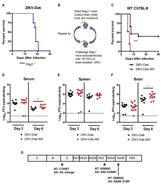
mice for susceptibility to ZIKV-Dak, and all mice succumbed within 31 days of infection (Figure 1A). To adapt ZIKV-Dak, virus-containing brain homogenates isolated from moribund
Rag1 / mice were used for subcutaneous infection of naive
Rag1 / mice (Figure 1B). After four passages in Rag1 / mice, we inoculated this mouse-adapted virus (ZIKV-Dak-MA) into 4- to 5-week-old WT mice after treatment with 2 mg
anti-Ifnar1 monoclonal antibody (mAb) and compared it with the parental virus. Anti-Ifnar1-treated WT mice were more vulnerable to infection with ZIKV-Dak-MA than ZIKV-Dak with a shorter time to death (Figure 1C). We investigated the basis for increased lethality by comparing viral RNA levels in serum, spleen, and the brain at 3 and 6 days after subcutaneous inoculation with 105 focus-forming units (FFU) of ZIKV-Dak or ZIKV-Dak-MA. In serum, no difference was observed at day 3 between the strains, whereas a slightly higher level was measured at day 6 in ZIKV-Dak-MA-infected animals (Figure 1D). No differ-ence in ZIKV-Dak and ZIKV-Dak-MA levels was detected in the spleen at days 3 or 6 after infection (Figure 1E). However, we observed higher levels of infection in the brains of ZIKV-Dak-MA than ZIKV-Dak-infected mice on day 6 (Figure 1F). Thus, ZIKV-Dak-MA replicated to moderately higher levels in the brain of anti-Ifnar1-treated mice, and this phenotype was associated with an increase in lethality.
To determine the genetic basis for the increased pathogenicity of ZIKV-Dak-MA in mice, we performed next-generation sequencing of the parental and adapted viruses. This analysis revealed three mutations in greater than 95% of the ZIKV-Dak-MA population (Figure 1G): a synonymous C to T change at nucleotide 1,006, a non-synonymous A to G change at nucleo-tide 5,808 resulting in a conservative lysine to arginine substitu-tion at amino acid posisubstitu-tion 399 in NS3, and a non-synonymous G to A change at nucleotide 6,965 resulting in a non-conservative glycine to arginine substitution at amino acid position 18 in NS4B. Analysis of the viral sequences obtained from the earlier sequential passages (P2 and P3) inRag1 / mice revealed that the mutations in NS4B arose during the second virus passage, with the NS3 mutation arising during the fourth passage (Table S1). These mutations in NS3 and NS4B in ZIKV-Dak-MA were not present in any ZIKV sequences available in the VIPR (Virus Pathogen Database and Analysis Resource) databases, but were present in some related mosquito- and tick-transmitted fla-viviruses (Figures S1A and S1B).
ZIKV-Dak-MA Replicates More Efficiently in the Brain and in NSCs
The increased infectivity of ZIKV-Dak-MA in brain tissue could reflect an enhanced ability to cross the blood-brain barrier or an intrinsic ability to replicate more efficiently in neuronal cell targets. To evaluate these possibilities, we inoculated 4- to 5-week-old WT C57BL/6 mice with 104FFU Dak or ZIKV-Dak-MA via an intracranial route (no anti-Ifnar1 mAb treatment) and compared lethality and viral burden. Remarkably, the ani-mals did not succumb to infection with either virus, suggesting that the changes in ZIKV-Dak-MA were not sufficient to result in lethal infection in immunocompetent mice (Figure 2A). Although both strains replicated in different regions of the brains of WT mice, ZIKV-Dak-MA accumulated to greater levels (Fig-ures 2B–2E). At day 4, the midbrain of ZIKV-Dak-MA-infected mice had higher viral RNA burden (Figure 2D) than ZIKV-Dak-challenged mice. By day 6, multiple regions of the brain had higher levels of ZIKV-Dak-MA than ZIKV-Dak RNA (brain stem, cerebellum, midbrain, and cortex) (Figures 2B–2E). In contrast, the olfactory bulb showed no differences (Figure 2F).
ZIKV-Dak-MA replicated to higher levels than ZIKV-Dak at multi-ple time points (Figures 2G and 2H). The increased yield of ZIKV-Dak-MA was not due to greater cell survival, as ZIKV-ZIKV-Dak-MA resulted in more, not less, cell death than ZIKV-Dak (Figure 2I). Figure 1. Generation of an Adapted ZIKV with Increased Lethality and CNS Viral Burden in Mice
(A) Four- to 6-week-oldRag1 /
mice were inoculated subcutaneously with ZIKV-Dakar 41525 (ZIKV-Dak) (three experiments, n = 8). (B) Scheme ofin vivopassaging of ZIKV-Dak. Brain homogenates (106
plaque-forming units [PFU]) fromRag1 /
mice that succumbed to infection were inoculated subcutaneously into naiveRag1 /
mice.
(C) Four- to 5-week-old WT C57BL/6 mice were treated with anti-Ifnar1 mAb 1 day prior to subcutaneous inoculation with 105
FFU ZIKV-Dak or ZIKV-Dak-MA (three experiments, n = 15, log rank test; ***p < 0.001).
(D–F) ZIKV RNA levels in serum (D), spleen (E), and brain (F) of WT mice after treatment with anti-Ifnar1 mAb and subcutaneous inoculation of ZIKV-Dak or ZIKV-Dak-MA (three experiments, n = 8–12, Mann-Whitney test; *p < 0.05). Solid lines are median values, and dotted lines denote the limit of detection of the assay.
(G) RNA was isolated from ZIKV-Dak-MA and subjected to next-generation sequencing. Mutations present in over 95% of genomes are shown. See alsoTable S1andFigure S1.
AsZIKVreplicatesefficientlyinNSCs(Lietal.,2016b;McGrath
et al.,2017), we isolated these cellsfrom the subventricular
Figure 2. Enhanced Infectivity in the CNS and NSCs of ZIKV-Dak-Ma
(A) Four- to 5-week-old WT C57BL/6 mice (without Ifnar1 blockade) were inoculated via intracranial route with ZIKV-Dak or ZIKV-Dak-MA and moni-tored for survival (three experiments, n = 7). (B–F) ZIKV RNA levels in (B) brainstem, (C) cere-bellum, (D) midbrain, (E) cortex, and (F) olfactory bulb in WT mice at days 4 or 6 after intracranial challenge with ZIKV-Dak or ZIKV-Dak-MA (three experiments, n = 6–9, Mann-Whitney test; **p < 0.01). Solid lines are median values, and dotted lines denote the limit of detection of the assay. (G–I) Adult NSCs were inoculated at an MOI of 1 (H and I) or 0.01 (G) with ZIKV-Dak or ZIKV-Dak-MA. Infectious titers were determined by focus-forming assay (FFA). Viability of NSCs (I) was normalized to uninfected control cells (three experiments per-formed in duplicate). Mean values ± SEM. A two-way and one-two-way ANOVA with multiple comparison correction were for viral titers and cell viability, respectively (*p < 0.05; **p < 0.01; ***p < 0.001). (J–N) Viral RNA levels in the (J) brainstem, (K) cere-bellum, (L) midbrain, (M) cortex, and (N) olfactory bulb in congenic Ifnar1 /
Figure 3. The NS4B G18R Mutation Is Required for Enhanced Mouse Lethality and Increased NSC Infection by ZIKV
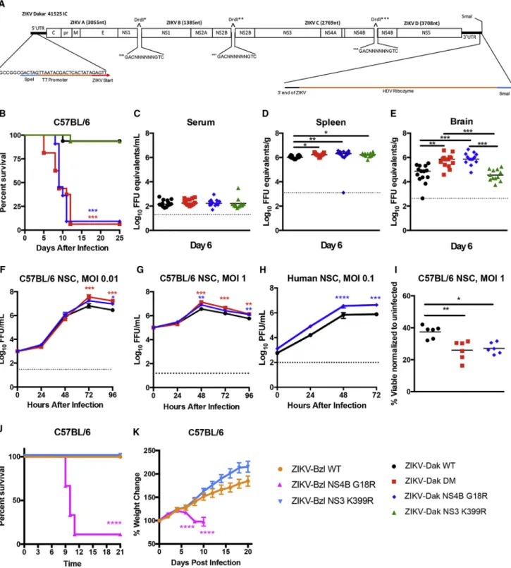
(A) ZIKV-Dakar 41525 infectious cDNA clone.
(B) Four- to 5-week-old WT C57BL/6 mice were treated with anti-Ifnar1 mAb 1 day prior to subcutaneous inoculation with infectious clone derived WT ZIKV-Dakar 41525 (ZIKV-Dak-WT), ZIKV-Dak NS3 K399R, ZIKV-Dak NS4B G18R, or ZIKV-Dak NS3 K399R + NS4B G18R (ZIKV-Dak DM) (three to four experiments, n = 15–16, log rank test compared with ZIKV-Dak-WT; ZIKV-Dak DM and ZIKV-Dak NS4B G18R, p < 0.001; ZIKV-Dak NS3 K399R, p > 0.9).
(C–E) Viral burden in serum (C), spleen (D), and brain (E) at day 6 post-subcutaneous inoculation with ZIKV-Dak-WT, ZIKV-Dak-DM, ZIKV-Dak NS3 K399R, or ZIKV-Dak NS4B G18R of 4- to 5-week-old WT mice after treatment with anti-Ifnar1 mAb (3 experiments, n = 12–14, one-way ANOVA with multiple comparisons correction; *p < 0.05; **p < 0.01; ***p < 0.001).
We hypothesized that, compared with the parental virus, ZIKV-Dak-MA may have acquired an ability to evade a type I IFN response in the brain. To test this hypothesis, we compared infectivity of ZIKV-Dak and ZIKV-Dak-MA in different brain regions after intracranial inoculation ofIfnar1 / mice. No differ-ences in infection were observed between ZIKV-Dak and ZIKV-Dak-MA in different brain regions at day 2 after infection (Figures 2J–2N). By day 4 after infection, minimal differences in viral RNA levels (<2-fold) were observed between ZIKV-Dak and ZIKV-ZIKV-Dak-MA in different brain regions. Because ZIKV-Dak-MA infection resulted in up to 9-fold greater viral titers than ZIKV-Dak in different brain regions of WT mice (Figures 2B– 2E), in aggregate, these data suggest that the majority of the repli-cation disparity between ZIKV-Dak and ZIKV-Dak-MA in the brain is likely due to a differential ability to evade type I IFN responses. However, there is a small difference in relative infectivity that could reflect effects on type I IFN-independent host defenses.
The NS4B G18R Mutation Determines the Increased Pathogenicity of ZIKV-Dak-MA
ZIKV-Dak-MA had three mutations present in over 95% of the viral population, two of which (NS3 K399R and NS4B G18R) were non-synonymous changes. To evaluate which mutations were responsible for enhanced infection and disease, we engi-neered isogenic viruses from a quadripartite infectious clone of ZIKV-Dakar 41525 (Figure 3A;STAR Methods). We produced WT and isogenic variants with mutations in NS3 K399R, NS4B G18R, or NS3 K399R and NS4B G18R (DM). Four- to 5-week-old WT mice were pretreated with anti-Ifnar1 mAb and chal-lenged with 105FFU of the isogenic viruses (Figure 3B). Approx-imately 95% of mice succumbed to ZIKV-Dak NS4B G18R or ZIKV-Dak DM, whereas only 5% of animals died after inoculation with ZIKV-Dak WT or ZIKV-Dak NS3 K399R (Figure 3B). Thus, the NS4B G18R mutation was necessary and sufficient for enhanced virulence. Viral burden analysis at day 6 after inocula-tion corroborated these findings. Although no differences were observed in serum (Figure 3C), ZIKV-Dak NS4B G18R and ZIKV-Dak DM replicated to slightly higher titers in the spleen than ZIKV-Dak WT (Figure 3D). Moreover, in the brain, at day 6, ZIKV-Dak NS4B G18R and ZIKV-Dak DM accumulated to higher levels than ZIKV-Dak WT (Figure 3E). Consistent with these results, ZIKV-Dak NS4B G18R and ZIKV-Dak DM pro-duced higher viral yields in the supernatants of mouse and human NSCs (Figures 3F–3H) and induced greater cell death than ZIKV-Dak WT at 96 hr post-infection (Figure 3I).
To assess whether the adaptive mutations could enhance the pathogenicity of other ZIKV strains, we generated isogenic viruses using an infectious clone of a Brazilian ZIKV strain (BeH819015) (Widman et al., 2017). Three-week-old WT C57BL/6 mice were pretreated with 2 mg anti-Ifnar1 mAb and
inoculated subcutaneously with 105 FFU of the WT clone-derived ZIKV from Brazil (ZIKV-Bzl WT), a single NS3 K399R mutant (ZIKV-Bzl NS3 K399R), or a single NS4B G18R mutant (ZIKV-Bzl NS4B G18R). A majority of animals inoculated with ZIKV-Bzl NS4B G18R succumbed to infection, whereas those in-fected with ZIKV-Bzl WT or ZIKV-Bzl NS3 K399R did not (Fig-ure 3J). Moreover, ZIKV-Bzl NS4B G18R-infected animals lost weight, whereas ZIKV-Bzl WT or ZIKV-Bzl NS3 K399R-infected animals did not (Figure 3K).
Single-Cell RNA Sequencing of Infected and Uninfected Murine NSCs
To begin to determine how the adaptive NS4B mutation in ZIKV affected IFN responses, we first performed single-cell RNA sequencing (RNA-seq) analysis on uninfected and ZIKV-Dak WT-infected murine NSCs at 24 and 48 hr time points (Figure 4A). We detected large transcriptional changes in the ZIKV-infected NSCs compared with the uninfected cells, particularly by 48 hr (Figures 4B andS2). As expected, many cells expressed NSC markers, including Id3, Rpl32, Cdk4, Nes, and Sox2, which confirmed their cellular identity (Figure 4C) (Dulken et al., 2017). Clustering analysis partitioned transcriptionally similar cells from all uninfected and infected samples into six groups (Figure 4D). The clusters were then manually labeled based on which RNAs were highly differential between clusters (e.g., cell cycle, ISGs, and levels of ZIKV RNA) (Figure 4E). ZIKV-infected cells were defined as having greater than 0.03% of viral transcripts relative to total cellular transcripts (STAR Methods), and increased from none (uninfected), to a minority of cells at 24 hr (9%), to a majority of cells (67%) at 48 hr after infection (Figure 4F). ISGs were increased in cells exposed to ZIKV (Figure S2), with some (e.g.,
Ifnb1) correlating with viral RNA and infection, and others (e.g.,
Isg15, Rsad2, and Cxcl10) expressed in both infected (ZIKV RNA+) and uninfected (ZIKV RNA ) NSCs (Figures 4E–4G). By 48 hr, most cells responded to type I IFN signals based on expres-sion ofRsad2andIsg15, but only a small fraction accumulated a high level ofIfnb1transcripts and these were among the most in-fected ones. The highly ZIKV-inin-fected cells were the sole pro-ducers of pro-inflammatory chemokines, including Ccl5 and
Cxcl10. The overall IFN effect also manifested in cell cycle arrest, as the majority of the cells at 48 hr were in G0/G1 phase, whereas at 0 and 24 hr cells were equally distributed across G0/G1, G2/M, and S cell-cycle phases (Figure 4D). This analysis illustrates the utility of single-cell RNA-seq in determining the transcriptional changes in uninfected and virus-infected cell populations.
ZIKV-Dak NS4B G18R Induces Less of a Type I IFN Response
We next used our single-cell RNA-seq analysis to define tran-scriptional differences between ZIKV-Dak WT and ZIKV-Dak
(F–I) Mouse NSCs were inoculated at an MOI of 0.01 (F) or 1 (G and I) with ZIKV-Dak WT, ZIKV-Dak DM, or ZIKV-Dak NS4B G18R and analyzed by FFA (F and G) or cell viability assay (I). Human NSCs were inoculated with ZIKV-Dak WT or ZIKV-Dak NS4B G18R (MOI of 0.1) and analyzed by plaque assay (H) (three experiments performed in duplicate). For (F)–(H), mean ± SEM is shown. Viral titer and cell viability were analyzed (ANOVA with multiple comparisons corrections; *p < 0.05; **p < 0.01; ***p < 0.001; ****p<0.0001).
NS4B G18R-infected murine NSCs (Figure 5A). Large transcrip-tional differences were found in cells infected with either ZIKV strain at the 48 hr time point compared with uninfected cells (Figures 5B andS2). ZIKV RNA was present in NSCs inoculated with either ZIKV-Dak WT or ZIKV-Dak NS4B G18R, and the tran-scriptional signatures clustered similarly (Figure 5C). Although more ZIKV-Dak NS4B G18R RNA was detected at 24 and 48 hr after infection (Figure 5D), many ISGs (e.g.,Ifit1,Ifitm3, and Isg15) were induced at lower levels in ZIKV-Dak NS4B G18R than ZIKV-Dak WT-infected NSCs (Figures 5E andS3A). Single-cell RNA-seq allowed separate transcriptional interroga-tion of the uninfected and ZIKV-infected NSC populainterroga-tions. At 24 and 48 hr, ISGs were induced at a higher level in uninfected cells from the ZIKV-Dak WT group than the ZIKV-Dak NS4B G18R group (Figures 5E and S3A); this suggested that the ZIKV-Dak NS4B G18R might antagonize induction of the type I IFN response more efficiently than the parental virus, which would have its greatest effect in cells responding to, but not infected with, ZIKV. At 24 and 48 hr, ISG levels also were higher in the infected cell subset from the ZIKV-Dak WT compared with ZIKV-Dak NS4B G18R group, although the differences were less than in the uninfected cells (Figure S3B).
To begin to address whether ZIKV-Dak NS4B G18R antago-nized type I IFN responses and induction of ISGs, we first as-sessed the levels of IFN-bin the supernatant of ZIKV-Dak WT and ZIKV-Dak NS4B G18R-infected murine NSCs. Despite higher levels of infection (Figure 3G), less IFN-bwas detected in the supernatant of ZIKV-Dak NS4B G18R than ZIKV-Dak WT-infected cells at 48 hr (Figure 5F). However, both RNA-seq and qRT-PCR data failed to show differences in IFN-bmRNA in-duction after ZIKV-Dak WT and ZIKV-Dak NS4B G18R infection of NSCs (Figures 5E and 5G). The lack of significant changes in the IFN-bmRNA levels in the context of decreased IFN-bprotein in the supernatant of ZIKV-Dak NS4B G18R-infected cells sug-gests that the NS4B G18R mutation might contribute to an IFN-btranslational block. Inhibition of IFN-bprotein translation has been described as a ZIKV immune evasion mechanism in human dendritic cells (Bowen et al., 2017), although the mecha-nism currently remains unknown.
To assess whether adapted ZIKV modulates type I IFN signaling responses, murine NSCs that were pretreated with anti-Ifnar1 antibody or exogenous IFN-bwere inoculated with ZIKV-Dak WT or ZIKV-Dak NS4B G18R. The greater infectivity phenotype seen with ZIKV-Dak NS4B G18R by viral yield assays or antigen staining (Figures 5H and 5I) was lost after anti-Ifnar1 mAb treatment, with both viruses replicating equivalently in NSCs lacking type I IFN signaling (Figure 5J); these data suggest that differences in replication in NSCs between ZIKV-Dak WT
and ZIKV-Dak NS4B G18R are dependent on type I IFN signaling. However, pretreatment of NSCs with IFN-bresulted in equivalent restriction of ZIKV-Dak WT and ZIKV-Dak NS4B G18R (Figure 5K). As the WT and adapted ZIKV strains produced different amounts of IFN-bprotein, showed infection differences that were abolished by anti-Ifnar1 mAb treatment, yet were equally sensitive to exogenous IFN-bpretreatment, the NS4B G18R mutation in ZIKV likely acts through its ability to diminish IFN-blevels that initiate the antiviral IFN signaling pathway in infected NSCs.
An Immunocompetent Mouse Model of ZIKV Infection Our experiments suggested that ZIKV-Dak-MA acquired an abil-ity to minimize induction of type I IFN yet still remained sensitive to the antiviral effects of exogenous or paracrine-derived IFN, likely because of its inability to bind to and degrade mouse Stat2 (Bowen et al., 2017; Grant et al., 2016; Kumar et al., 2016; Tripathi et al., 2017). To overcome this limitation and create a fully immunocompetent mouse model of ZIKV patho-genesis, we inserted by homologous recombination a human
STAT2allele into the mouseStat2locus but retained the mouse Stat2 promoter: exon 1 and part of exon 2 were derived from the mouse gene (promoter sequences), and the remainder of exon 2 and all of exons 3 through 24 were from the human gene (coding sequences) (Figure 6A). Southern blotting and PCR corroborated the correct integration (Figures 6B and 6C), and western blotting of splenocytes from mice treated with poly(I:C) confirmed the loss of mouse Stat2 and the expression of human STAT2 (Figure 6D). ISG induction (e.g.,Oas1,Irf7, andIfnb) in hSTAT2 knockin (KI) mice downstream of IFN signaling was es-tablished by administration of poly(I:C) via intranasal route and interrogation of lungs 6 hr later, and appeared equivalent to that observed in WT mice (Figure 6E).
We investigated the pathogenic potential of ZIKV-Dak-MA in hSTAT2 KI mice without IFN blockade. Three-week-old hSTAT2 KI mice were inoculated subcutaneously with ZIKV-Dak or ZIKV-Dak-MA and monitored. Thirty percent of hSTAT2 KI mice succumbed to lethal ZIKV-Dak-MA infection, whereas none died after inoculation with parental ZIKV-Dak (Figure 6F). Viral burden analysis revealed higher levels of ZIKV-Dak-MA than ZIKV-Dak in the spleen and brain, but not in serum, at day 9 (Figures 6G–6I). We next tested if pregnant female hSTAT2 KI mice could transmit ZIKV efficiently to developing fetuses. Eleven-week-old WT or hSTAT2 KI dams that had been mated to WT or hSTAT2 KI sires, respectively (fetuses are homozygous WT or STAT2 KI), were inoculated with Dak or ZIKV-Dak-MA via subcutaneous route on embryonic day (E)6.5, and maternal and fetal tissues were collected on E13.5. Higher levels
Figure 4. Single-Cell RNA-Seq of NSCs after Infection with ZIKV-Dak
(A) Scheme for single-cell RNA-seq experiment: 100,000 NSCs (uninfected or 24 or 48 hr after ZIKV-Dak WT infection) were collected from three technical replicates and pooled, and subsequently 17,500 cells were subjected to microfluidic-based single-cell RNA-seq library generation and sequencing. (B) T-distributed stochastic neighbor embedding (tSNE) plots comparing uninfected and ZIKV-Dak WT-infected cells.
(C) Violin plots showing expression of NSC-specific markers and RLR-related genes in NSCs from all three conditions.
(D) tSNE plots of all conditions with cell type clusters identified with graph-based clustering. Clusters were manually named (e.g., cell cycle and IFN-responding) after their most highly differential genes.
(E) Expression of a subset of ISGs and cell-cycle markers used to determine clusters of (D) overlaid on the tSNE analysis from (B). (F) Expression of ZIKV RNA overlaid on the tSNE analysis from (B).
Figure 5. ZIKV-Dak NS4B G18R Induces Less IFN-band Induction of ISGs in Infected NSCs
(A) Scheme of experiment: 100,000 NSCs (uninfected, 24 or 48 hr after ZIKV-Dak WT infection, or 24 or 48 hr after ZIKV-Dak NS4B G18R) were collected from three technical replicates and pooled, and then 17,500 cells were subjected to microfluidic-based single-cell RNA-seq library generation and sequencing. (B) tSNE plots comparing uninfected and ZIKV-Dak WT and ZIKV-Dak NS4B G18R-infected cells.
(C) Expression of ZIKV RNA overlaid on the tSNE plots from (B).
(D) Violin plots showing the expression of ZIKV viral RNA in all populations. Wilcoxon rank-sum test with continuity correction was performed (**p < 1010 ).
of ZIKV-Dak-MA RNA were present in maternal serum and spleen in hSTAT2 KI compared with WT dams (Figures 6J and 6K). ZIKV-Dak-MA infected the placenta and fetal head to significantly higher levels in hSTAT2 KI compared with WT mice (Figures 6L and 6M). Indeed, ZIKV RNA was detected in the junctional layer of the placenta and uterine lining from hSTAT2-infected dams (Figure S4). In comparison, hSTAT2 KI dams infected with parental ZIKV-Dak had lower viral RNA levels in maternal and fetal tissues (Figures S5A–S5E) than ZIKV-Dak-MA-infected hSTAT2 KI dams (Figures 6J–6M). Expression of the human STAT2 allele for transmission was important for the dam, as placentas and fetuses from hSTAT2 KI female mice mated to WT males (fetuses are heterozygous for hSTAT2 allele) also accumulated high levels of ZIKV (Figures S5F–S5J); these data suggest that evasion of IFN signaling by ZIKV is necessary for efficient dissemination of ZIKV to the maternal-fetal interface but is largely dispensable for infection of the placenta and fetus. Thus, by combining the adapted ZIKV strain with hSTAT2 KI mice (Figure S5K), we generated lethality and placental trans-mission models in immunocompetent mice after subcutaneous inoculation.
DISCUSSION
ZIKV has emerged as an important global health concern due to its teratogenic potential. With no currently approved vaccine or treatment, there is an urgent need for the development of animal models that recapitulate key features of human disease to un-derstand the biology of this unique flavivirus and facilitate the development of countermeasures. Here, we generated an adapted ZIKV strain through passage in Rag1 / mice. This strain had one critical mutation in the NS4B gene, which allowed it to replicate to higher titers in NSCs and in the brains of mice, and to some degree evade type I IFN responses. The NS4B G18R substitution was necessary and sufficient to enhance the pathogenicity of both African and Brazilian strains of ZIKV in mice. Single-cell RNA-seq analysis of Dak- and ZIKV-Dak-MA-infected NSCs revealed transcriptional changes be-tween infected and uninfected cells and confirmed a reduced type I IFN response in ZIKV-Dak-MA-infected cells. We com-bined the pathogenic features of this adapted strain with hSTAT2 KI mice, which overcome the inherent inability of ZIKV strains to bind and degrade mouse Stat2 (Bowen et al., 2017; Grant et al., 2016; Kumar et al., 2016), to generate an immunocompetent small animal model of ZIKV pathogenesis.
RNA viruses rapidly mutate to overcome selective immune or drug pressures, gain host tropism, or acquire immune evasive
functions. Several recent studies have identified mutations in ZIKV that correlate with its epidemic emergence. One report used phylogenetic analysis to predict positively selected muta-tions in the N terminus (residue 26) and second ER loop (residues 87 and 88) of NS4B (Sironi et al., 2016), regions that inhibit type I IFN responses and STING activity and also bind NS1 (Mun˜oz-Jorda´n et al., 2005; Youn et al., 2012). A second study used an evolutionary approach to identify two amino acid substitutions in NS4B (V180I and L182S) present in epidemic strains (Zhu et al., 2016). Neither of these studies identified the NS4B G18 residue as under strong positive selection, and indeed G18R substitution is absent from all historical and circulating ZIKV strains. Several explanations for this are possible, including that residue 18 of NS4B is under strong purifying selection due to compromising effects on virulence in the epidemic or enzootic human, non-human primate, or mosquito hosts. Alternatively, the substitution could be specific for certain hosts, which might explain why some mosquito- and tick-borne flaviviruses with other natural reservoirs (e.g., rodent or marsupial) have an arginine at this position (Figure S1), and there is no selection pressure to retain it in human/non-human primate/mosquito transmission cycles.
The flavivirus NS4B protein contributes to remodeling of the endoplasmic reticulum, which facilitates viral replication (Kaufusi et al., 2014; Zmurko et al., 2015) and modulation of host defense responses. Dengue virus NS4B inhibits IFN-aand -binduction by altering mitochondrial morphology and RIG-I activation or by inhibiting TBK1 and IRF3 activation (Chatel-Chaix et al., 2016; Dalrymple et al., 2015). ZIKV NS4B also has been reported to diminish innate immune responses by preventing or altering activation of TBK1 (Onorati et al., 2016; Wu et al., 2017). Our studies with ZIKV-Dak and ZIKV-Dak NS4B G18R inIfnar1 /
mice, anti-Ifnar1 mAb-treated NSCs, and IFN-b-pretreated NSCs suggest that the adaptive substitution in NS4B does not affect sensitivity to type I IFN or ISGs but rather affects IFN-b induction. Our single-cell RNA-seq data in murine NSCs showed decreased ISG induction in ZIKV-Dak NS4B G18R-infected cells at 24 and 48 hr despite higher levels of intracellular viral RNA and extracellular virus compared with ZIKV-Dak WT. Because we distinguished transcriptional signatures of uninfected and ZIKV-infected cells, we were able to observe that higher levels of ISGs were induced in uninfected NSCs from the ZIKV-Dak WT group compared with the ZIKV-Dak NS4B G18R group, which likely rendered the uninfected cells in the ZIKV-Dak WT group less permissive (Figure S3B). Our data are consistent with a recent study in human dendritic cells showing that ZIKV evades type I IFN immunity in part by diminishing translation of
(E) Violin plots were generated showing expression of selected RLRs (Ddx58,Ifih1, andMavs), type I IFNs (Ifna2andIfnb1), and ISGs (Eif2ak2,Ifit1,Ifit3,Ifitm2, Ifitm3,Isg15,Oasl1,Oasl2, andRsad2) in uninfected (ZIKV RNA ) and infected (ZIKV RNA+
) cells in the culture at 24 hr after inoculation with ZIKV-Dak WT or ZIKV-Dak NS4B G18R. MAST test was used to determine the statistical difference in gene expression between different conditions (**p adjusted < 0.01). (F) NSCs were inoculated with ZIKV-Dak WT and ZIKV-Dak NS4B G18R at an MOI of 1. Supernatant was collected and IFN-bwas quantified by ELISA (three experiments in duplicate, mean ± SEM, Student’s t test; **p < 0.01).
(G) NSCs were inoculated with ZIKV-Dak WT and ZIKV-Dak NS4B G18R (MOI of 1). Cells were lysed, RNA was isolated, and18S,Ifnb1, andIsg15mRNA was quantified by qRT-PCR. Expression was compared with uninfected cells (three experiments in duplicate, Student’s t test forDDCt data).
(H–K) NSCs were untreated (no mAb) (H and I) or pretreated with anti-Ifnar1 blocking mAb (J) or 3 U/mL IFN-b(K) for 16 hr and then inoculated with ZIKV-Dak WT or ZIKV-Dak NS4B G18R (MOI of 0.01). Infectious titers were determined by FFA. In (I), NSCs were inoculated with ZIKV-Dak or ZIKV-Dak NS4B G18R. At 96 hr, cells were stained for ZIKV E protein and processed by flow cytometry.
Figure 6. hSTAT2 KI Mice Are Susceptible to ZIKV-Dak-MA
(A) Diagram of targeting construct for hSTAT2 KI mice.
(B and C) Southern blot (B) and PCR (C) confirmation of integration of hSTAT2 allele. (D) Western blot of hSTAT2 KI (h/h), WT (m/m), andStat2 /
( / ) splenocytes from poly(I:C)-treated mice.
(E) ISG (Oas1,Irf7, andIfnb) induction in WT and hSTAT2 KI mice after administration of poly(I:C) via an intranasal route and interrogation of lungs 6 hr later.
(F) Three-week-old hSTAT2 KI mice were inoculated subcutaneously with ZIKV-Dak or ZIKV-Dak-MA (three experiments; n = 12–26; log rank test, p = 0.06). (G–I) Viral burden in serum (G), spleen (H), and brain (I) of 3-week-old hSTAT2 KI at day 9 after inoculation with ZIKV-Dak or ZIKV-Dak-MA (two experiments, n = 9, Mann-Whitney test; **p < 0.01; ***p < 0.001). Solid lines are median values, and dotted lines denote the limit of detection of the assay.
IFN-bprotein (Bowen et al., 2017). Given that IFN-bmRNA levels were similar in ZIKV-Dak-MA and ZIKV-Dak NS4B G18R-infected cells, we speculate that the NS4B G18R mutation may enhance the efficiency of a ZIKV-induced IFN-b transla-tional block. The precise means by which changes in NS4B sequence differentially affect IFN-btranslation remain undeter-mined, as the mechanism by which ZIKV diminishes translation of IFN-bprotein (Bowen et al., 2017) is unknown.
One caveat to our experiments with Dak and ZIKV-Dak-MA is the relative disparity of phenotype between peripheral organs and the brain after subcutaneous virus inoculation. As many of these studies were performed in anti-Ifnar1 mAb-treated mice, the differences in viral titers in peripheral organs (serum and spleen) are small, as expected, between the WT and adapt-ed ZIKV virus, analogous to our NSC experiments (Figure 5J). In comparison, because anti-Ifnar1 mAb does not appreciably cross the blood-brain barrier, the phenotype of the mutant ZIKV-Dak-MA is more prominent in the brain. Notwithstanding these data, ZIKV-Dak-MA still replicated slightly more effectively than ZIKV-Dak in the CNS ofIfnar1 / mice after direct intracra-nial inoculation, suggesting there may be an additional pathway, independent of type I IFN signaling, by which the adaptive muta-tion funcmuta-tions; NS4B G18R could antagonize or co-opt other host defense pathways, including type II or III IFN signaling or autophagy in cells of the brain. Indeed, ZIKV NS4B reportedly modulates Akt-mTOR signaling and autophagy induction (Liang et al., 2016), and autophagy confers a proviral advantage for ZIKV (Cao et al., 2017).
Most adult mouse models of ZIKV infection require eliminating or inhibiting the mouse type I IFN response to achieve infection and pathogenesis (reviewed inMorrison and Diamond, 2017). Nonetheless, several immunocompetent mouse models of ZIKV infection have been described, although each has limita-tions. These include (1) peripheral infection of neonatal (<1-week-old) WT mice (Lazear et al., 2016; Manangeeswaran et al., 2016), which have immature immune responses; (2) direct intrauterine injection of WT pregnant dams (Vermillion et al., 2017), which bypasses trans-placental transmission; (3) direct injection of fetal brains during pregnancy (Wu et al., 2016), which bypasses the placenta entirely; and (4) administration of high-dose ZIKV via an intravenous route to pregnant dams (Cugola et al., 2016), which bypasses peripheral immune responses. To overcome the lack of Stat2 antagonism, which limits ZIKV repli-cation in the peripheral tissues likely due to sustained type I and III IFN responses, we engineered a transgenic mouse that replaced most of mouseStat2with humanSTAT2. This mouse showed normal immune responses to poly(I:C), a stimulus that activates IFN induction. ZIKV, especially the NS4B G18R adapt-ed strain, was able to replicate, spread to the brain, cause lethality in juvenile (3-week-old) hSTAT2 KI mice, and result in trans-placental transmission and fetal infection in pregnant dams. Of note, the pregnant dams themselves, despite vertically transmitting ZIKV, did not sustain infection in the brain, which is
characteristic of human infection. Thus, ZIKV infection in adult hSTAT2 KI mice also did not recapitulate the extreme vulnera-bility and lethality ofIfnar1 / (Lazear et al., 2016) orStat2 / mice (Tripathi et al., 2017); this could reflect cell-extrinsic anti-viral effects of IFN signaling in uninfected cells (as seen in our single-cell RNA-seq data) or the lack of additional species-spe-cific evasion mechanisms that are not accounted for by the NS4B mutation (e.g., DENV antagonizes human, but not mouse, STING;Aguirre et al., 2012). Alternatively, the absence of brain infection in adult hSTAT2 KI mice could reflect the age-depen-dent maturation of the blood-brain barrier and the tightening effects of type I and III IFNs, both of which signal through Stat2 (Daniels et al., 2014, 2017), which would be absent inIfnar1 /
andStat2 / mice.
In summary, we identified an adaptive mutation in ZIKV NS4B that confers enhanced virulence in mice and NSCs for African and American ZIKV strains. We applied single-cell RNA-seq analysis in uninfected and ZIKV-infected NSCs to define unique cell-intrinsic transcriptional signatures and used this information to establish mechanistically how the adaptive NS4B mutation enhanced infectivity and virulence. By combining the adapted virus with transgenic mice that exchanged human and mouse STAT2, which restricts ZIKV infection in a species-dependent manner, we developed an immunocompetent model of ZIKV infection and pathogenesis after peripheral inoculation that more closely recapitulates features of human infection. This immunocompetent small animal model of ZIKV infection may prove a resource for determining mechanisms of pathogenesis, defining correlates of innate and adaptive immune protection, and evaluating countermeasures to limit disease.
STAR+METHODS
Detailed methods are provided in the online version of this paper and include the following:
d KEY RESOURCES TABLE
d CONTACT FOR REAGENT AND RESOURCE SHARING
d EXPERIMENTAL MODEL AND SUBJECT DETAILS
B Animals and Ethics B Cell Lines
B Primary Cell Cultures B Viruses
d METHOD DETAILS
B Mouse Experiments
B hSTAT2 KI Mouse Generation B IFN Responses in Mice B Viral Burden Measurements B NSC Isolation and Infection
B IFN-bProduction in NSCs during ZIKV Infection B Cell Viability Assay
B Next-Generation Sequencing of ZIKV B ZIKV-Dak and ZIKV-Bzl Infectious Clones
(J–M) Eleven-week-old hSTAT2 KI or WT dams mated to hSTAT2 KI and WT sires, respectively, were infected with ZIKV-Dak-MA on E6.5 and tissues were harvested on E13.5. Viral burden in maternal serum (J), maternal spleen (H), placenta (L), or fetal heads (M) is shown (two experiments, n = 9 WT and 10 hSTAT2 KI dams, n = 36 WT and 32 hSTAT2 KI placentas and fetal heads, Mann-Whitney test; **p < 0.01; ****p < 0.0001). Solid lines are median values and dotted lines denote the limit of detection of the assay.
B Single Cell RNA-Seq
B Alignment, Barcode Assignment and Unique Molecular Identifier (UMI) Counting
B Preprocessing Analysis with Seurat Package B Dimensionality Reduction and Clustering
B Identification of Cluster-Specific Genes and Marker-Based Classification
B Single Cell RNA-Seq Differential Expression B qRT-PCR of IFN-bmRNA
B Sequence Alignment for ZIKV and Related Flaviviruses B Sanger Sequencing of Passaged ZIKV Isolates B Viral RNAIn SituHybridization
d QUANTIFICATION AND STATISTICAL ANALYSIS
d DATA AND SOFTWARE AVAILABILITY
(2017). Zika virus antagonizes type I interferon responses during infection of
human dendritic cells. PLoS Pathog.13, e1006164.
Butler, A., and Satija, R. (2017). Integrated analysis of single cell transcriptomic
data across conditions, technologies, and species. bioRxiv21, 627–636.
Cao, B., Parnell, L.A., Diamond, M.S., and Mysorekar, I.U. (2017). Inhibition of autophagy limits vertical transmission of Zika virus in pregnant mice. J. Exp.
Med.214, 2303–2313.
Cao-Lormeau, V.-M., Roche, C., Teissier, A., Robin, E., Berry, A.-L., Mallet, H.-P., Sall, A.A., and Musso, D. (2014). Zika virus, French Polynesia, South Pacific, 2013. Emerg. Infect. Dis.20, 1085–1086.
Chatel-Chaix, L., Cortese, M., Romero-Brey, I., Bender, S., Neufeldt, C.J., Fischl, W., Scaturro, P., Schieber, N., Schwab, Y., Fischer, B., et al. (2016). Dengue virus perturbs mitochondrial morphodynamics to dampen innate
immune responses. Cell Host Microbe20, 342–356.
Chavali, P.L., Stojic, L., Meredith, L.W., Joseph, N., Nahorski, M.S., Sanford, T.J., Sweeney, T.R., Krishna, B.A., Hosmillo, M., Firth, A.E., et al. (2017). Neurodevelopmental protein Musashi-1 interacts with the Zika genome and promotes viral replication. Science357, 83–88.
Cugola, F.R., Fernandes, I.R., Russo, F.B., Freitas, B.C., Dias, J.L.M., Guimara˜es, K.P., Benazzato, C., Almeida, N., Pignatari, G.C., Romero, S., et al. (2016). The Brazilian Zika virus strain causes birth defects in experimental
models. Nature534, 267–271.
Dalrymple, N.A., Cimica, V., and Mackow, E.R. (2015). Dengue virus NS pro-teins inhibit RIG-I/MAVS signaling by blocking TBK1/IRF3 phosphorylation: dengue virus serotype 1 NS4A is a unique interferon-regulating virulence
determinant. MBio6, e00553–15.
Daniels, B.P., Holman, D.W., Cruz-Orengo, L., Jujjavarapu, H., Durrant, D.M., and Klein, R.S. (2014). Viral pathogen-associated molecular patterns regulate
blood-brain barrier integrity via competing innate cytokine signals. MBio5,
e01476–14.
Daniels, B.P., Jujjavarapu, H., Durrant, D.M., Williams, J.L., Green, R.R., White, J.P., Lazear, H.M., Gale, M., Diamond, M.S., and Klein, R.S. (2017). Regional astrocyte IFN signaling restricts pathogenesis during neurotropic viral infec-tion. J. Clin. Invest.127, 843–856.
Dulken, B.W., Leeman, D.S., Boutet, S.C., Hebestreit, K., and Brunet, A. (2017). Single-cell transcriptomic analysis defines heterogeneity and
tran-scriptional dynamics in the adult neural stem cell lineage. Cell Rep.18,
777–790.
Finak, G., McDavid, A., Yajima, M., Deng, J., Gersuk, V., Shalek, A.K., Slichter, C.K., Miller, H.W., McElrath, M.J., Prlic, M., et al. (2015). MAST: a flexible statistical framework for assessing transcriptional changes and characterizing
heterogeneity in single-cell RNA sequencing data. Genome Biol.16, 278.
Govero, J., Esakky, P., Scheaffer, S.M., Fernandez, E., Drury, A., Platt, D.J., Gorman, M.J., Richner, J.M., Caine, E.A., Salazar, V., et al. (2016). Zika virus infection damages the testes in mice. Nature540, 438–442.
Grant, A., Ponia, S.S., Tripathi, S., Balasubramaniam, V., Miorin, L., Sourisseau, M., Schwarz, M.C., Sa´nchez-Seco, M.P., Evans, M.J., Best, S.M., et al. (2016). Zika virus targets human STAT2 to inhibit type I interferon signaling. Cell Host Microbe19, 882–890.
Heymann, D.L., Hodgson, A., Sall, A.A., Freedman, D.O., Staples, J.E., Althabe, F., Baruah, K., Mahmud, G., Kandun, N., Vasconcelos, P.F., et al. (2016). Zika virus and microcephaly: why is this situation a PHEIC? Lancet 387, 719–721.
Joguet, G., Mansuy, J., Matusali, G., Hamdi, S., Walschaerts, M., Pavili, L., Guyomard, S., Prisant, N., Lamarre, P., Dejucq-rainsford, N., et al. (2017). Effect of acute Zika virus infection on sperm and virus clearance in body fluids: a prospective observational study. Lancet Infect. Dis.17, 1200–1208.
Kaufusi, P.H., Kelley, J.F., Yanagihara, R., and Nerurkar, V.R. (2014). Induction of endoplasmic reticulum-derived replication-competent membrane struc-tures by West Nile virus non-structural protein 4B. PLoS One9, e84040.
Kearse, M., Moir, R., Wilson, A., Stones-Havas, S., Cheung, M., Sturrock, S., Buxton, S., Cooper, A., Markowitz, S., Duran, C., et al. (2012). Geneious Basic: an integrated and extendable desktop software platform for the organization
and analysis of sequence data. Bioinformatics28, 1647–1649.
SUPPLEMENTALINFORMATION
SupplementalInformationincludesfivefiguresandtwotablesandcanbe foundwiththisarticleonlineathttps://doi.org/10.1016/j.chom.2018.04.003.
ACKNOWLEDGMENTS
WeacknowledgetheGenomeTechnologyAccessCenteratWashington Uni-versityforhelpwithgenomicanalysis.ThecenterispartiallysupportedbyNCI CancerCenterSupportGrantP30CA91842andbyICTS/CTSAGrantUL1 TR000448.This work also wassupported by grants from theNIH (R01 AI073755,R01AI104972,andU19AI083019toM.S.D.;R01HD091218to I.U.M.andM.S.D.;U19AI118610andR21AI129486toA.G.-S;andR01 AI100625andR01AI107810 toR.S.B.)andby theDivisionofIntramural Research,NationalInstituteofAllergyandInfectious Diseases,NIH.K.Z. wassupportedbyGovernmentofRussianFederationgrant074-U01.
AUTHORCONTRIBUTIONS
M.J.G.,E.A.C.,K.Z.,J.W.-L.,M.B.U.,S.T.,G.D.E.,S.M.B.,M.N.A.,andM.S.D. designedtheexperiments.M.J.G.,E.A.C.,J.W.-L.,C.R.,M.C.Y.,B.C.,S.J.R., K.L.M.,M.B.U.,andS.T.performedtheexperiments.J.M.contributedtothe generationofthehSTAT2KImice.M.C.B.,K.H.D.,B.L.Y.,Z.Z.,J.Y.,R.S.B., and A.G.-S. contributed key reagents, including recombinant viruses. M.J.G.,E.A.C.,K.Z.,M.B.U.,B.C.,I.U.M.,S.T.,J.W.-L.,C.R.,M.N.A.,and G.D.E.analyzed the data. M.J.G. and E.A.C. wrote the first draft, with M.S.D.providingmajoreditorialcomments.Allauthorsparticipatedinediting thefinalversionofthemanuscript.
DECLARATIONOFINTERESTS
M.S.D.isaconsultantforInbios,Aviana,andSanofi-Pasteur,andisonthe Sci-entificAdvisoryBoardofModerna.
Received:January9,2018 Revised:March16,2018 Accepted:April10,2018 Published:May9,2018
REFERENCES
Aguirre,S.,Maestre,A.M.,Pagni,S.,Patel,J.R.,Savage,T.,Gutman,D.,
Maringer,K.,Bernal-Rubio, D.,Shabman, R.S.,Simon, V., etal. (2012).
DENVinhibitstype IIFN productionininfectedcells bycleavinghuman
STING.PLoSPathog.8,e1002934.
Aliota,M.T.,Caine,E.A.,Walker,E.C.,Larkin,K.E.,Camacho,E.,andOsorio, J.E.(2016).CharacterizationoflethalZikavirusinfectioninAG129mice.PLoS Negl.Trop.Dis.10,e0004682.
Bowen,J.R.,Quicke,K.M., Maddur,M.S.,O’Neal, J.T.,McDonald,C.E.,
Kowalczyk, M.S., Tirosh, I., Heckl, D., Rao, T.N., Dixit, A., Haas, B.J., Schneider, R.K., Wagers, A.J., Ebert, B.L., and Regev, A. (2015). Single-cell RNA-seq reveals changes in cell cycle and differentiation programs upon
ag-ing of hematopoietic stem cells. Genome Res.25, 1860–1872.
Kumar, A., Hou, S., Airo, A.M., Limonta, D., Mancinelli, V., Branton, W., Power, C., and Hobman, T.C. (2016). Zika virus inhibits type-I interferon production
and downstream signaling. EMBO Rep.17, 1766–1775.
Lazear, H.M., Govero, J., Smith, A.M., Platt, D.J., Fernandez, E., Miner, J.J., Diamond, M.S., Fernandez, E., Miner, J.J., and Diamond, M.S. (2016). A
mouse model of Zika virus pathogenesis. Cell Host Microbe19, 720–730.
Li, C., Xu, D., Ye, Q., Hong, S., Jiang, Y., Liu, X., Zhang, N., Shi, L., Qin, C., and Xu, Z. (2016a). Zika virus disrupts neural progenitor development and leads to microcephaly in mice. Cell Stem Cell19, 120–126.
Li, H., Saucedo-Cuevas, L., Regla-Nava, J.A., Chai, G., Sheets, N., Tang, W., Terskikh, A.V., Shresta, S., and Gleeson, J.G. (2016b). Zika virus infects neural progenitors in the adult mouse brain and alters proliferation. Cell Stem Cell19,
593–598.
Liang, Q., Luo, Z., Zeng, J., Chen, W., Foo, S.-S., Lee, S.-A., Ge, J., Wang, S., Goldman, S.A., Zlokovic, B.V., et al. (2016). Zika virus NS4A and NS4B pro-teins deregulate Akt-mTOR signaling in human fetal neural stem cells to inhibit
neurogenesis and induce autophagy. Cell Stem Cell19, 663–671.
Ma, W., Li, S., Ma, S., Jia, L., Zhang, F., Zhang, Y., Zhang, J., Wong, G., Zhang, S., Lu, X., et al. (2016). Zika virus causes testis damage and leads to male infer-tility in mice. Cell167, 1511–1518.e10.
Manangeeswaran, M., Ireland, D.D.C., and Verthelyi, D. (2016). Zika (PRVABC59) infection is associated with T cell infiltration and
neurodegenera-tion in CNS of immunocompetent neonatal C57Bl/6 mice. PLoS Pathog.12,
e1006004.
Marrs, C., Olson, G., Saade, G., Hankins, G., Wen, T., Patel, J., and Weaver, S. (2016). Zika virus and pregnancy: a review of the literature and clinical consid-erations. Am. J. Perinatol.33, 625–639.
McGrath, E.L., Rossi, S.L., Gao, J., Widen, S.G., Grant, A.C., Dunn, T.J., Azar, S.R., Roundy, C.M., Xiong, Y., Prusak, D.J., et al. (2017). Differential responses of human fetal brain neural stem cells to Zika virus infection. Stem Cell Reports 8, 715–727.
Meertens, L., Labeau, A., Dejarnac, O., Cipriani, S., Sinigaglia, L., Bonnet-Madin, L., Le Charpentier, T., Hafirassou, M.L., Zamborlini, A., Cao-Lormeau, V.M., et al. (2017). Axl mediates ZIKA virus entry in human glial cells
and modulates innate immune responses. Cell Rep.18, 324–333.
Messer, W.B., Yount, B., Hacker, K.E., Donaldson, E.F., Huynh, J.P., de Silva, A.M., and Baric, R.S. (2012). Development and characterization of a reverse genetic system for studying dengue virus serotype 3 strain variation and neutralization. PLoS Negl. Trop. Dis.6, e1486.
Miner, J.J., Sene, A., Richner, J.M., Smith, A.M., Santeford, A., Ban, N., Weger-Lucarelli, J., Manzella, F., R€uckert, C., Govero, J., et al. (2016). Zika vi-rus infection in mice causes panuveitis with shedding of vivi-rus in tears. Cell Rep. 16, 3208–3218.
Morrison, T.E., and Diamond, M.S. (2017). Animal models of Zika virus infec-tion, pathogenesis, and immunity. J. Virol.91, e00009–17.
Mun˜oz-Jorda´n, J.L., Laurent-Rolle, M., Ashour, J., Martı´nez-Sobrido, L., Ashok, M., Lipkin, W.I., and Garcı´a-Sastre, A. (2005). Inhibition of alpha/beta interferon signaling by the NS4B protein of flaviviruses. J. Virol.79, 8004–8013.
Oliphant, T., Engle, M., Nybakken, G.E., Doane, C., Johnson, S., Huang, L., Gorlatov, S., Mehlhop, E., Marri, A., Chung, K.M., et al. (2005). Development of a humanized monoclonal antibody with therapeutic potential against West Nile virus. Nat. Med.11, 522–530.
Onorati, M., Li, Z., Liu, F., Sousa, A.M.M., Nakagawa, N., Li, M., Dell’Anno, M.T., Gulden, F.O., Pochareddy, S., Tebbenkamp, A.T.N., et al. (2016). Zika virus disrupts phospho-TBK1 localization and mitosis in human neuroepithelial stem cells and radial glia. Cell Rep.16, 2576–2592.
Pal, P., Dowd, K.A., Brien, J.D., Edeling, M.A., Gorlatov, S., Johnson, S., Lee, I., Akahata, W., Nabel, G.J., Richter, M.K., et al. (2013). Development of a
highly protective combination monoclonal antibody therapy against
Chikungunya virus. PLoS Pathog.9, e1003312.
Park, C., Li, S., Cha, E., and Schindler, C. (2000). Immune response in Stat2
knockout mice. Immunity13, 795–804.
Pickett, B.E., Sadat, E.L., Zhang, Y., Noronha, J.M., Squires, R.B., Hunt, V., Liu, M., Kumar, S., Zaremba, S., Gu, Z., et al. (2012). ViPR: an open bioinfor-matics database and analysis resource for virology research. Nucleic Acids
Res.40, D593–D598.
Richard, A.S., Shim, B.-S., Kwon, Y.-C., Zhang, R., Otsuka, Y., Schmitt, K., Berri, F., Diamond, M.S., and Choe, H. (2017). AXL-dependent infection of human fetal endothelial cells distinguishes Zika virus from other pathogenic flaviviruses. Proc. Natl. Acad. Sci. USA114, 2024–2029.
Schaft, J., Ashery-Padan, R., van der Hoeven, F., Gruss, P., and Stewart, A.F. (2001). Efficient FLP recombination in mouse ES cells and oocytes. Genesis 31, 6–10.
Sironi, M., Forni, D., Clerici, M., and Cagliani, R. (2016). Nonstructural proteins are preferential positive selection targets in Zika virus and related flaviviruses.
PLoS Negl. Trop. Dis.10, e0004978.
Tripathi, S., Balasubramaniam, V.R., Brown, J.A., Mena, I., Grant, A., Bardina, S.V., Maringer, K., Schwarz, M.C., Maestre, A.M., Sourisseau, M., et al. (2017). A novel Zika virus mouse model reveals strain specific differences in virus
pathogenesis and host inflammatory immune responses. PLoS Pathog.13,
e1006258.
Vermillion, M.S., Lei, J., Shabi, Y., Baxter, V.K., Crilly, N.P., McLane, M., Griffin, D.E., Pekosz, A., Klein, S.L., and Burd, I. (2017). Intrauterine Zika virus infection of pregnant immunocompetent mice models transplacental transmission and
adverse perinatal outcomes. Nat. Commun.8, 14575.
Weaver, S.C., Costa, F., Garcia-Blanco, M.A., Ko, A.I., Ribeiro, G.S., Saade, G., Shi, P.-Y., and Vasilakis, N. (2016). Zika virus: history, emergence, biology, and prospects for control. Antiviral Res.130, 69–80.
Widman, D.G., Young, E., Yount, B.L., Plante, K.S., Gallichotte, E.N., Carbaugh, D.L., Peck, K.M., Plante, J., Swanstrom, J., Heise, M.T., et al. (2017). A reverse genetics platform that spans the Zika virus family tree. MBio8, 1–15.
Wilm, A., Aw, P.P.K., Bertrand, D., Yeo, G.H.T., Ong, S.H., Wong, C.H., Khor, C.C., Petric, R., Hibberd, M.L., and Nagarajan, N. (2012). LoFreq: a sequence-quality aware, ultra-sensitive variant caller for uncovering cell-population
het-erogeneity from high-throughput sequencing datasets. Nucleic Acids Res.40,
11189–11201.
Wu, K.-Y., Zuo, G.-L., Li, X., Ye, Q., Deng, Y.-Q., Huang, X.-Y., Cao, W.-C., Qin, C.-F., and Luo, Z.-G. (2016). Vertical transmission of Zika virus targeting the radial glial cells affects cortex development of offspring mice. Cell Res.26,
645–654.
Wu, Y., Liu, Q., Zhou, J., Xie, W., Chen, C., Wang, Z., Yang, H., and Cui, J. (2017). Zika virus evades interferon-mediated antiviral response through the co-operation of multiple nonstructural proteins in vitro. Cell Discov.3, 17006.
Youn, S., Li, T., McCune, B.T., Edeling, M.A., Fremont, D.H., Cristea, I.M., and Diamond, M.S. (2012). Evidence for a genetic and physical interaction be-tween nonstructural proteins NS1 and NS4B that modulates replication of West Nile virus. J. Virol.86, 7360–7371.
Zhao, H., Fernandez, E., Dowd, K.A., Speer, S.D., Platt, D.J., Gorman, M.J., Govero, J., Nelson, C.A., Pierson, T.C., Diamond, M.S., et al. (2016). Structural basis of Zika virus-specific antibody protection. Cell166, 1016–1027.
Zhu, Z., Chan, J.F., Tee, K., Choi, G.K., Lau, S.K., Woo, P.C., Tse, H., and Yuen, K. (2016). Comparative genomic analysis of pre-epidemic and epidemic Zika virus strains for virological factors potentially associated with the rapidly
expanding epidemic. Emerg. Microbes Infect.5, e22.
STAR+METHODS
KEY RESOURCES TABLE
REAGENT or RESOURCE SOURCE IDENTIFIER
Antibodies
E60 antibody Diamond Lab Oliphant et al., 2005
Polyclonal anti mouse HRP Sigma A8924
Human STAT2 Santa Cruz Sc-476
Stat2 Cell signaling 4597
Anti-mouse Ifnar antibody Leinco Technologies MAR1-5A3
Anti-human CD119 antibody Leinco Technologies GIR-208 Goat anti-mouse IgG Alexa Fluor 647 Thermo Fisher Scientific A-21235
ZIKV-2 Diamond Lab Zhao et al., 2016
CHK-152 Diamond Lab Pal et al., 2013
ZIKV-13 Diamond Lab Zhao et al., 2016
Bacterial and Virus Strains
ZIKV-Dakar 41525 World Reference center for Emerging Viruses and Arboviruses
GenBank: KU955591
ZIKV-Dakar 41525 Parental This paper N/A
ZIKV-Dakar 41525 Mouse Adapted This paper N/A
ZIKV-Dakar WT clone derived This paper N/A
ZIKV-Dakar NS4B G18R clone derived This paper N/A
ZIKV-Dakar NS3 K399R clone derived This paper N/A ZIKV-Dakar NS4B G18R/NS3 K399R clone derived This paper N/A
ZIKV-Brazil WT (BeH819015) clone derived Widman et al., 2017 N/A ZIKV-Brazil NS4B G18R clone derived This paper N/A
ZIKV Brazil NS3 K399R clone derived This paper N/A
Chemicals, Peptides, and Recombinant Proteins
Phusion High Fidelity PCR MasterMix New England Biolabs M0531S
RNase-Free DNase set Qiagen 79254
Matrigel Corning 08-774-552
Q5 DNA polymerase New England Biolabs M0491S
dsDNA fragmentase New England Biolabs M0348S
RT-PCR-Superscript III Reverse Transcriptase Invitrogen S1230S
Mouse IFN-b PBL assay science 12400-1
RNA ISH ZIKV RNA probe Advanced Cell Diagnostics 467771
Critical Commercial Assays
Verikine IFNbELISA PBL Assay Science 42400-1
RNeasy mini kit Qiagen 74104
QIAamp viral RNA Mini kit Qiagen 52906
NEBnexy ultraII DNA library prep kit New England Biolabs E7645S Superscript III first strand synthesis supermix Thermo Fisher Scientific 18080400
Taqman RNA-to-CT1-step kit Applied Biosystem 4392938 FoxP3/Transcription Factor Staining Buffer Set Thermo Fisher Scientific 00-5523-00 T7 Kit – Ambion mMESSAGE mMACHINE T7
Transcription Kit
Thermo Fisher Scientific AM1344
CONTACT FOR REAGENT AND RESOURCE SHARING
Further information and requests for resources and reagents should be directed to and will be fulfilled by the Lead Contact, Michael S. Diamond ([email protected]).
EXPERIMENTAL MODEL AND SUBJECT DETAILS
Animals and Ethics
Wild-type (WT) C57BL/6 (000664) mice were purchased from Jackson Laboratory. CongenicRag1-/-,Ifnar1-/-,Stat2-/-(Park et al., 2000), and hSTAT2 KI C57BL/6 mice were bred in a specific-pathogen-free facility at Washington University or the Icahn School of Medicine at Mount Sinai. 4 to 6 week-old mice, or 11 week old pregnant female mice were used. Littermates of the same sex were randomly assigned to experimental groups. This study was carried out in accordance with the recommendations in the Guide for the Care and Use of Laboratory Animals of the National Institutes of Health. The protocols were approved by the Continued
REAGENT or RESOURCE SOURCE IDENTIFIER
Deposited Data
ZIKV-Dak MA RNAseq This paper SRA: PRJNA413540
Single cell RNAseq experiment This paper GEO: GSE112711
ZIKV-Dak WT consensus sequence This paper Genbank: MG758785 ZIKV-Dak MA consensus sequence This paper Genbank: MG758786
Experimental Models: Cell Lines
Mouse adult Neuronal Stem cells from C57BL/6 mice This paper N/A
African green monkey kidney (Vero) cells WHO Reference cell bank WHO Vero cells
Experimental Models: Organisms/Strains
Mouse: C57BL/6J The Jackson Laboratory 000664
Mouse:Ifnar1-/- Michel Aguet N/A
Mouse: hSTAT2 KI This paper N/A
Mouse:Rag1-/- The Jackson Laboratory 002216
Mouse:Stat2-/- N/A Park et al., 2000
Oligonucleotides
ZIKV-Dakar titering primers This paper SeeTable S2
hSTAT2 KI genotyping primers This paper SeeTable S2
Sequencing ZIKV primers This paper SeeTable S2
ZIKV amplicon primers This paper SeeTable S2
Eukaryotic 18S RNA Endogenous control Thermo Fisher Scientific 4319413E
PrimeTime Predesigned PCR AssayIfnb1 IDT Mm.PT.58.30132453.g
Recombinant DNA
pUC57 BioBasic http://www.snapgene.com/
resources/plasmid_files/basic_ cloning_vectors/pUC57/
Software and Algorithms
Prism GraphPad Version 6.0e
Geneius Geneius V 11.0.4
DNAstar Megalign DNAstar V14.0
Seurat R package Butler and Satija, 2017 https://github.com/satijalab/ seurat/; Version 2.1
Cell Ranger Single Cell Software Suite 10x Genomics https://github.com/10XGenomics/ cellranger/; Version 2.0.2
InstitutionalAnimalCareandUseCommitteeattheWashingtonUniversitySchoolofMedicine(AssurancenumberA3381-01).Animal studiesalsowereapproved bytheInstitutional Animal Careand Use Committeeof IcahnSchool ofMedicineatMount Sinai (AssurancenumberA311-01).Virusinoculationswereperformedunderanaesthesiathatwasinducedandmaintainedwithketamine hydrochlorideandxylazine,andalleffortsweremadetominimizeanimalsuffering.
CellLines
Verocellswereculturedat37CinDulbecco’sModifiedEagleMedium(DMEM)supplementedwith5%fetalbovineserum(FBS)and 25mMHEPES.
PrimaryCellCultures
NSCswereisolatedfrom4to6week-oldWTC57BL/6mice.NSCswereculturedinNeurobasalmedia(ThermoFisher)without phenolred,supplementedwithB27withoutvitaminA(ThermoFisher)andN-2supplement(ThermoFisher)at37C in 5% CO2. NSCswereplatedat105cellsin2mlofNeurobasalmediaperwellin6wellplatestreatedwithmatrigel(Corning,08-774-552).
Viruses
ZIKVstrainDakar41525(Senegal,1984,GenBank:KU955591)wasprovidedbytheWorldReferencecenterforEmergingViruses andArboviruses(R.Tesh,UniversityofTexasMedicalBranch).ZIKVstockswerepropagatedinVerocells,andcellsupernatants wereharvested66-72hpost inoculation.Virus stocks weretitrated byfocus formingassay(FFA) onVero cellsaspreviously described(Lazearetal.,2016)andstoredaliquotedat-80C.
METHODDETAILS
MouseExperiments
Insomeexperiments,miceweretreatedwith2mgofanIfnar1-blockingantibody(MAR1-5A3,LeincoTechnologies)by intraperito-nealinjectionone-daybeforevirusinfection.MicewereinoculatedwithZIKVbyasubcutaneous(viafootpad)routewith104to106 FFUPFUofZIKVinavolumeof50mlPBSorbyanintracranialroutewith104FFUofZIKVinavolumeof10mlPBS.Forsurvival studies,miceweremonitoredfor21-45days.Forenhancedsafety,allmouseinfectionexperimentswithZIKVwereperformedunder A-BSL3conditions.
hSTAT2KIMouseGeneration
ThehSTAT2KImouselinewasgeneratedcommerciallybyTaconicArtemisGmbh.Thetargetingvectorwasconstructedusing bac-terialartificialchromosomeclonesC57BL/6JRPCIB-731andhumanRPCIB-753,transfectedintotheTaconicArtemisC57BL/6N TacEScellline,andselectedwithneomycinandpuromycin.TheintegrationinEScellswasconfirmedbySouthernblottingand PCR.GermlinepupswerebredtoC57BL/6-Tg(CAG-Flpe)2Arte(Schaftetal.,2001)forremovaloftheneomycinandpuromycin cassettes,andtheFlpetransgenewasbredawaybybackcrossing.MiceweregenotypedwithprimersCTGAGGTAGAATCACTTT GACTTCC-5079-42andaGATGGCTCAGAGGTTAAGAGC-5079-43,orGGCAAAGCCAAGACATAAACC-5080-46andACAGGTTC CAGGCCATCAAG-5080-47andPCRconditions:94C30sec,60C30sec,72C1min.WesternblottingforhumanSTAT2(Santa Cruzsc-476)ormouseStat2(Cellsignaling4597)wasperformedonsplenocytesisolated24hafterintravenousinjectionof100mg of poly(I:C).
IFNResponsesinMice
WT,hSTAT2KI,andStat2-/-micewereadministered50mgofpoly(I:C)(Invivogen,tlrl-pic)viaanintranasalroutein50mlofPBS,and lungswereharvested6hlater.LungswerehomogenizedinRLTbuffer(QiagenRNAeasykit),andtotalRNAwasisolatedfrom200ml oftissuehomogenate.RT-PCRwasperformedasdescribed(Tripathietal.,2017).ISGsweredetectedwiththefollowingprimersets: mouseOas1aForward5’-ATGGAGCACGGACTCAGGA-3’mouseOas1aRev5’-TCACACACGACATTGACGGC-3’;mouse Ifnb1
Forward 5’-CAGCTCCAAGAAAGGACGAAC-3’ mouse Ifnb1 Reverse 5’-GGCAGTGTAACTCTTCTGCAT-3’; mouse Irf7
Forward5’-GAGACTGGCTATTGGGGGAG-3’mouseIrf7Reverse5’-GACCGAAATGCTTCCAGGG-3’;mouse18srRNAForward 5’-GTAACCCGTTGAACCCCATT-3’mouse18srRNAReverse5’-CCATCCAATCGGTAGTAGCG-3’
ViralBurdenMeasurements
by bead dissociation using a MagNA Lyzer (Roche) in 1 ml of DMEM containing 2% FBS. Samples were clarified by centrifugation (2,000xgat 4C for 10 min), diluted serially, and then added to Vero cell monolayers in 6 well plates. Cells were overlaid with 2% low melting point agarose (Lonza) and 4 days later were fixed with 10% formaldehyde and stained with crystal violet for manual counting on a lightbox.
NSC Isolation and Infection
NSCs were isolated from 4 to 6 week-old WT C57BL/6 mice. The subventricular zone of the brain was digested in 0.05% trypsin (Invitrogen) for 30 min at 37C. Cells were resuspended after pipetting up and down twenty times. NSCs were cultured in Neurobasal media (Thermo Fisher) without phenol red, supplemented with B27 without vitamin A (Thermo Fisher) and N-2 supplement (Thermo Fisher) at 37C in 5% CO2. NSCs were plated at 105cells in 2 ml of Neurobasal media per well in 6 well plates treated with matrigel (Corning, 08-774-552) and inoculated with ZIKV at an MOI of 0.01 or 1. For some experiments, NSCs were pretreated with 25mg/mL of Ifanr1 blocking antibody (MAR-1 5A3), 3 or 10 U/mL of IFN-b(PBL Assay science, 12400-1) for 16 h before infection. Supernatants were harvested and medium was replaced at indicated time points. Viral burden in the supernatant was determined by focus-forming assay (FFA). NSCs were harvested at 96 h after infection, fixed in Foxp3/transcription factor staining buffer (eBioscience), and stained with a combination of anti-ZIKV murine mAbs (ZIKV-2 and ZIKV-13) or only CHK-152 as an isotype control using 200 ng of mAb per 50ml. Cells were then incubated with goat anti-mouse Alexa Fluor 647 antibody (Thermo Fisher Scientific) and analyzed on a MACsQuant8 flow cytometer (Miltenyi Biotec).
IFN-bProduction in NSCs during ZIKV Infection
NSCs were cultured in Neurobasal media without phenol red, supplemented with B27 without vitamin A and N-2 supplement at 37C in 5% CO2. NSCs were plated at 105cells in 2 mL of Neurobasal media per well in 6 well plates treated with matrigel and inoculated with ZIKV at an MOI of 1. After 4 h, cells were washed three times in PBS, and media was replaced with 2 mL of fresh Neurobasal media. At the indicated time points, supernatant was collected and Ifnbin the supernatant was measured using VeriKine Mouse Ifnb ELISA (PBL Assay Science, 42400-1) according to the manufacturer’s protocol. In brief, 100 ul of supernatant was added to precoated wells and incubated for 1 h at room temperature. Detection, development, and washing steps were performed as described by the manufacturer’s protocol. OD450 was recorded using a Tristar LB 941 reader (Berthold Technologies). IFN-b con-centration was determined by comparing OD450 values to a standard curve. Limit of detection (LOD) was determined by calculating the mean and standard deviation (SD) of blank controls, and setting the LOD at three SDs above the mean of the blank controls.
Cell Viability Assay
Cell viability was measured using CellTiter-Glo reagents (Promega) according to the manufacturer’s protocol. Briefly, NSCs were plated at 105cells in 2 mL of Neurobasal media in 6 well plates treated with matrigel. Cells were inoculated with ZIKV at an MOI of 1. Ninety-six hours after infection, 1.7 ml of media was removed and 300ml of CellTiter-Glo reagent was added to each well. Plates were incubated for 15 min at room temperature and luminescence was recorded using a Synergy H1 Hybrid Reader (Bio-tek).
Next-Generation Sequencing of ZIKV
Preparation of sequencing libraries was performed as described (Miner et al., 2016). Briefly, RNA was extracted from the input or passaged virus, reverse-transcribed into cDNA, and then amplified in eight segments using Q5 DNA polymerase (New England Biolabs). DNA was fragmented using dsDNA fragmentase (New England Biolabs), and adapters and indexes were added to prepare Illumina compatible libraries using the NEBNext Ultra II DNA library prep kit (New England Biolabs). Libraries were sequenced on an Illumina Nextseq 500. The demultiplexed fastq files for the input were aligned to the ZIKV genome (Strain ArD_41519, Accession HQ234501) to create a consensus sequence. The input consensus sequence was used as a reference file to assess mutations pre-sent in the passaged virus. Variant calling was performed with LoFreq (Wilm et al., 2012) and consensus sequences were generated with Geneious software. All sequences have been uploaded and will be published upon publication of a finalized manuscript.
ZIKV-Dak and ZIKV-Bzl Infectious Clones
We developed an infectious clone utilizing a published reverse genetics platform (Widman et al., 2017). We applied a unidirectional quadripartite assembly method using asymmetric, nonpalindromic, naturally encoded restriction enzyme sites located near known centers of toxicity for bacterial expression (Messer et al., 2012; Widman et al., 2017). Cleavage sites were located at nucleotide positions 3064, 4451, and 7220 of the ZIKV-Dak (strain 41525) genome and resulted in four fragments for expression in bacterial plasmids. Non-native modifications added to the consensus sequence included hepatitis D virus ribozyme following the native 3’ untranslated region (UTR) as well as a T7 promotor upstream of the 5’-UTR forin vitro transcription. Site-directed mutagenesis overlap PCR was performed to generate mutations at positions NS3 K339R and/or NS4B G18R in ZIKV-Dak WT and ZIKV-Bzl WT clones (Widman et al., 2017). Screening was performed via Sanger sequencing to confirm mutagenesis as well as genetic integrity of the rest of the fragment.