Diagnostic Accuracy of Transvaginal Ultrasound in Women with
Endometriosis at Deeply Infiltrating, Ovarian, and Superficial Peritoneal
Sites
By
Rebecca Turner PA-S
A Capstone Paper submitted to the faculty of the University of North Carolina at Chapel Hill
in partial fulfillment of the requirements for the degree of Master of Health Sciences
in the Physician Assistant Program
Chapel Hill
December 2019
Keturah R Faurot PhD, MPH, PA
11/14/2019
Introduction
Endometriosis is a multifaceted disease that is associated with dysmenorrhea, noncyclic
pelvic pain, dyspareunia, and infertility 1. It affects approximately 1 in 10 women, up to 200
million worldwide, and can disrupt every aspect of a woman’s life, including sexual
relationships, appetite, sleep, exercise, work productivity, and emotional well-being 2. When
extrapolated, endometriosis is a disease that has a high cost to both patients and society as a
whole, as patients incur personal charges related to pain control and infertility management,
while absenteeism and loss of productivity contribute to lost earnings worldwide 3. Despite the
disease’s prevalence and devastating effects on a woman’s life, the average time from symptom
presentation to endometriosis diagnosis is 8-12 years, a conservative estimate when considering
most women with the disease will never be diagnosed 3. This delay in diagnosis and treatment
has been shown to cause an increase in long term disease sequelae, significant degradation in the
patient-provider relationship, and further progression of the disease 2,4.
The cause of this unfortunate delay in diagnosis is multifaceted. A large narrative review evaluating the social and psychological impact of endometriosis on women’s lives found that the average time between a woman’s initial symptoms and initial presentation for evaluation was 3.7
years. Many causative social factors were cited, including difficulty perceiving the difference
between normal and pathologic symptoms, viewing menstruation as shameful or needing to be
hidden, and reinforcement of symptom concealment from male and female peers 4. The average
delay from initial clinical presentation to diagnosis was between 3.7 and 5.7 years. Factors
contributing to medical delay of diagnosis included referral delay, misdiagnosis, and lack of
provider knowledge, all of which contributed to women feeling ignored or dismissed by
providers4. A significant number of women reported feeling relieved, legitimized, and often
angry at the delay after receiving an endometriosis diagnosis, all of which have been shown to
contribute to a degradation in the patient-provider relationship. After diagnosis, women report
negative impacts to many aspects of their lives, including intimate relationships, work and
productivity, social lives, family planning, fertility, sleep quality, and mental health 4.
At this time, laparoscopy with histologic sampling is the only tool available for definitive
diagnosis. Due to its cost and inherent risks, this tool is out of reach for many patients, especially
those that do not have access to a tertiary gynecologic referral center. A less invasive diagnostic
allow the diagnosis to be made in the primary care setting 3. Transvaginal ultrasound is a
low-cost, highly available tool that has shown significant utility in diagnosing many causes of pelvic
pain 5. This narrative review will seek to evaluate the current research on the diagnostic
capability of transvaginal ultrasound (TVUS) for diagnosing endometriotic lesions at ovarian,
superficial peritoneal, and deeply infiltrating sites. If accuracy is high enough, TVUS as a less
invasive, less expensive tool for diagnosis could allow providers to evaluate patients for
endometriosis earlier in their disease, increase quality of patient care, and decrease associated
costs incurred by both the patient and the health care system.
This narrative review will begin with an overview of the natural history of endometriosis,
including epidemiology, pathophysiology, clinical manifestations, current available treatments,
and diagnostics. This will be followed by an evaluation and discussion of the current research on
the diagnostic accuracy of TVUS in the diagnosis of endometriosis. The primary outcome will be
to analyze whether TVUS can accurately diagnose ovarian, superficial peritoneal, and deeply
infiltrating endometriosis when compared to laparoscopy with histologic examination.
Background
Epidemiology
Many patients with endometriosis will experience either no symptoms or mild symptoms
that may be considered within the normal realm of menstrual-related symptoms. This makes
identifying the prevalence of endometriosis quite difficult 3. Various studies that have sought to
determine the general population prevalence have produced highly variable results ranging from
1% to 15%, with approximately 10% being the accepted value 2. For women with symptomatic
presentation, prevalence has been reported up to 70% when the presentation includes pelvic pain
and 50% when the presentation includes infertility 6. There appears to be a genetic association, as
women with first degree female relatives affected by the disease have a higher likelihood of
being diagnosed with endometriosis 7. Factors that increase a woman’s risk for endometriosis
include being nulliparous, early menarche, menstrual cycles shorter than 27 days, menorrhagia,
height over 68 inches, and low BMI 8. Conversely, protective factors include cycles longer than
Pathophysiology
By definition, endometriosis is the presence of endometrial tissue outside of the uterus.
The pathogenesis of the disease likely includes genetic factors, autoimmune dysregulation, and
abnormal endocrine signaling, in addition to the presence of ectopic endometrial tissue 9. The
ectopic endometrial tissue implants, grows, and causes an inflammatory reaction which can lead
to scarring and anatomic dysmorphism 8. The exact cause of these ectopic implants is poorly
understood. A 2014 meta-analysis of 8 papers analyzed genetic data from a total of 11506
endometriosis cases showed that six genetic loci were significantly associated with
endometriosis 7. However, analyses have failed to identify a genetic marker consistently
associated with the disease.
The leading theory for the pathogenesis of ectopic endometrial tissue implantation is
retrograde menstruation, in which endometrial tissue flows backward through the fallopian tubes
and into the abdomen during menstruation 8. However, there is uncertainty surrounding this
theory, as up to 90% of women have been shown to have retrograde menstruation and
endometriosis has been diagnosed in prepubescent girls 7.
After implantation, the endometriotic implants cause pain through inflammatory and
neurologic responses. A variety of inflammatory markers are involved in the inflammatory
process, with prostaglandins being the primary culprit 9. The implants have also been shown to
cause surrounding changes to sympathetic and sensory nerve fibers, with some studies showing
endometriosis patients to have a higher density of nerve endings within and around implants 9.
This suggests another possible genetic link to endometriosis symptoms, as a predisposition to
higher inflammatory markers or increased nerve endings could increase pain signaling and
symptoms 7.
The pathogenesis for infertility or subfertility caused by endometriosis is thought to
correlate with the stage of the disease, with a greater burden of disease associated with more
inflammatory markers and more scar tissue 9. This increase in inflammatory markers can lead to
ovarian or endometrial hormonal dysfunction, which is thought to cause a subprime environment
for ovulation, fertilization, and implantation of a zygote. This dysfunction can occur at any stage
of endometriosis, including early, minimally-invasive disease, although the process remains
significant scarring, adhesions, and subsequent pelvic anatomic dysmorphism. This is also
thought to lead to a hostile and non-ideal environment for fertility 8.
Clinical Manifestations
Endometriotic lesions can implant in a variety of locations, both pelvic and non-pelvic.
Figure 1 demonstrates common locations of pelvic endometriosis, but does not depict
endometriosis outside of the pelvis 10. These locations most commonly include the ovaries,
anterior or posterior uterine cul-de-sac, any of the uterine ligaments, the uterus itself, the
fallopian tubes, and the sigmoidal bowel 9. A majority of patients present with lesions in more
than one location, with the ovaries being the most common. In rare cases, endometriotic lesions
have been diagnosed in locations including the breast, thorax, lung, central nervous system, and
abdominal organs. This can represent diagnostic challenges, as the symptoms associated with
lesions in rare locations can be
nonspecific and intermittent 3.
When a lesion is located within the
pelvis, it is classified into one of
three categories: ovarian,
superficial peritoneal, or deeply
infiltrating. Ovarian lesions occur
on the ovaries, superficial
peritoneal lesions are less than
5mm into the peritoneum, and
deeply infiltrating lesions are 5mm
or more deep into the peritoneum 8.
The most common
presenting complaints of women with endometriosis are pelvic pain, infertility, and ovarian mass
3. The majority of patients present with some complaint of pain, which can include
dysmenorrhea, noncyclic pelvic pain, dyspareunia [pain with sex], dyschezia [pain with
defecation], and dysuria. There is generally a direct association between the location of
endometriotic lesions and the symptoms experienced. However, a patient’s symptom burden is
not necessarily correlated with disease burden 3. For example, one patient may present with
severe pain but only have two to three superficial lesions, while another patient may present with
mild pain but have 10+ deeply infiltrating and superficial lesions with severe scarring 2. This
represents another diagnostic challenge, and demonstrates the importance of practitioners
maintaining a high degree of suspicion in patients presenting with any degree of chronic pelvic
pain.
Current Treatment Options
There is no cure for endometriosis; rather, the current treatment options rely on symptom
management. Treatment plans typically involve a two-fold approach utilizing both medical and
surgical modalities. The first line medical therapy for endometriosis-related pain are
non-steroidal anti-inflammatory drugs (NSAIDs).11 These medications are particularly well-suited to
combat the prostaglandin mediated pain caused by endometriosis because of their
anti-prostaglandin properties. NSAIDs are also generally very safe and well-tolerated, making them a
great option for most women 11. Combined oral hormonal contraceptive (COCs) medications are
another option for the management of endometriosis-related pain, particularly the pain associated with menstrual cycles. By regulating the menstrual cycle and making a patient’s periods lighter,
shorter, and more regular, COCs can decrease the pain associated with menstruation11. COCs are
also generally safe and well-tolerated by most populations. If NSAIDs and COCs are ineffective
at managing pain, medications such as GnRH agonists, Danazol, and aromatase inhibitors are
further options. These medications work in different ways to reduce the effects of estrogen in a woman’s body, including suppressing menstruation and associated pain, but they generally have
more side effects and are less well tolerated than first line treatments11.
The medical treatment of endometriosis-related infertility is similar to the approach to
infertility treatment in patients without endometriosis. The mainstay of medical treatment is
clomiphene citrate (Clomid), a medication that stimulates follicle growth and ovulation12. The
addition of gonadotropins and aromatase inhibitors can also be used to enhance follicle
stimulation. The next step in medical assistance in infertility is the use of assisted reproduction
technology (ART). This includes In-Vitro Fertilization (IVF) and Intrauterine Insemination (IUI)
12. Despite these methods, a diagnosis of endometriosis significantly increases a woman’s
probability of treatment failure compared to women without endometriosis 12. Progression of the
disease is shown to be directly related to the probability of failure, with greater severity of
The surgical management of endometriosis related pain and infertility are similar.
Surgical management for infertility focuses on correcting distorted pelvic anatomy in women
with moderate to severe disease 12. Unfortunately, this approach has not been validated in RCTs
and the fertility benefit from surgery is unclear. However, laparoscopic removal or ablation of
lesions has been shown to significantly reduce pain symptoms in women with mild and moderate
symptoms 11. While there is a high return to surgery rate (over 50% at the seven year mark),
multiple RCTs have demonstrated a benefit to removing lesions at the time of laparoscopic
diagnosis. This is a major strength of laparoscopy, in that it can be both diagnostic and
therapeutic. Other surgical options for pain management include removal of ovarian
endometriomas, neurectomy, and hysterectomy with bilateral salpingo-oophorectomy 11. The
latter is viewed as a last-resort option for women with debilitating symptoms who have failed
other therapies and do not desire child-bearing. Even after hysterectomy, disease recurrence is
still possible and many patients do not achieve complete pain relief. 13
Diagnosis
For definitive diagnosis of endometriosis, surgical biopsy with histologic review remains
the gold standard 14. This has been the case since the early 20th century. The current standard
surgical technique is laparoscopy 14. One benefit from this technique is that it can be both a
diagnostic and therapeutic procedure. This can be helpful if a patient desires surgical
management for their disease. However, despite years of research, there is no noninvasive
diagnostic test available 2. Some experienced providers may be comfortable managing the
disease symptomatically without a surgical diagnosis, but many primary care providers do not
have sufficient gynecology training to feel comfortable with this 2. This is a significant reason
why most women will go years without a definitive diagnosis for their endometriosis symptoms.
At least part of this diagnostic and symptomatic burden could be relieved with a noninvasive
diagnostic test 2. Research is currently being conductive in a variety of outlets, including
transvaginal ultrasound (TVUS), transrectal ultrasound (TRUS), and serum biomarkers. TVUS
currently has the most promise and the most research available as a potential diagnostic test for
the most common endometriosis locations, and will be the focus of this narrative review.
Methods
AND deep,’ ‘endometriosis AND ultrasound AND ovarian,’ and ‘endometriosis AND ultrasound
AND superficial’ were used. To be included in this review, studies needed to be systematic
reviews, published since January 1st 2009, evaluate human subjects, include no author overlap
with other studies, and compare TVUS at ovarian, deeply infiltrating, and/or superficial
peritoneal endometriosis against laparoscopy with biopsy. Reviews including pregnant patients
were excluded. For superficial peritoneal endometriosis, the criteria were broadened to include
primary research due to a lack of systematic reviews evaluating superficial endometriosis. From
this broadened criteria, 51 studies resulted. To prevent overlap of patient data within reviews,
only the most recent, relevant paper for each lead author was evaluated. A list of excluded
studies and the reasons for exclusion can be found in Appendix 1. The four final studies selected
for this paper were chosen based on relevancy to the clinical question of this paper, methodologic quality, and assessment by the “A MeaSurement Tool to Assess systematic Reviews 2” (AMSTAR 2) criteria and the “Quality Assessment of Diagnostic Accuracy Studies
2 (QUADAS 2) tool. A table of the AMSTAR 2 and QUADAS 2 evaluations is found in
Appendix 2 and Appendix 3.
Results
Out of the search and evaluation, two systematic reviews and two diagnostic accuracy
studies were identified as eligible, relevant, and of good methodological quality in the evaluation
of TVUS diagnostic accuracy of endometriosis at deeply infiltrating (DIE), ovarian, and
superficial peritoneal sites. Details on each paper’s features, risk of bias, and methodologic
quality is available in Table 1. Statistical results from each paper were identified and are listed in
Table 2, along with the endometriosis locations that were evaluated.
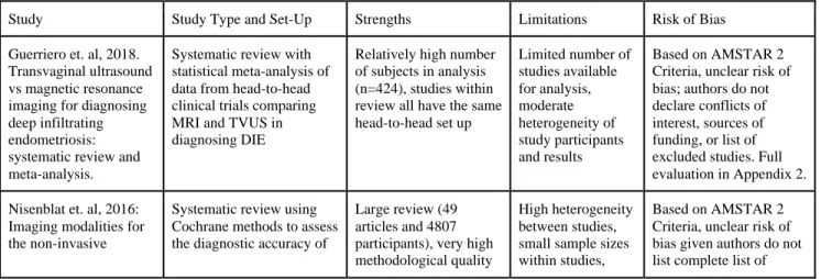
Table 1. Strengths, limitations, and risk of bias for each paper
Study Study Type and Set-Up Strengths Limitations Risk of Bias
Guerriero et. al, 2018. Transvaginal ultrasound vs magnetic resonance imaging for diagnosing deep infiltrating endometriosis: systematic review and meta-analysis.
Systematic review with statistical meta-analysis of data from head-to-head clinical trials comparing MRI and TVUS in diagnosing DIE
Relatively high number of subjects in analysis (n=424), studies within review all have the same head-to-head set up
Limited number of studies available for analysis, moderate heterogeneity of study participants and results
Based on AMSTAR 2 Criteria, unclear risk of bias; authors do not declare conflicts of interest, sources of funding, or list of excluded studies. Full evaluation in Appendix 2.
Nisenblat et. al, 2016: Imaging modalities for the non-invasive
Systematic review using Cochrane methods to assess the diagnostic accuracy of
Large review (49 articles and 4807 participants), very high methodological quality
High heterogeneity between studies, small sample sizes within studies,
diagnosis of endometriosis.
imaging tools in the diagnosis of endometriosis
(extensive details for methods, results, and discussion sections), extensive statistical analysis
high/unclear risk of bias for each study
excluded studies; however, in setting of high volume of studies evaluated and the authors listing reasons for exclusion, would consider a low risk of overall bias given comprehensive and detailed review otherwise. Full
evaluation in Appendix 2.
Reid et. al, 2019. The association between ultrasound-based “soft markers” and endometriosis type/location: A prospective observational study. Multicenter prospective observational study. Participants with chronic pelvic pain were recruited from tertiary gynecologic referral centers, then underwent TVUS, history, and laparoscopy. Primary results included correlation between test results and accuracy of TVUS findings in predicting location of endometriosis
Findings are consistent with previous studies with similar objectives, highly detailed description TVUS “soft-markers” involved in the study
Small sample size, high potential for referral bias, non-standardized format for history taking, potential for subjective reporting from sonographers, unclear if surgeons were blind to TVUS results
Based on the QUADAS-2 tool, there is a low risk of bias in patient selection, index test, and flow and timing. There is an unclear risk of bias in the reference standard. There is low concern regarding applicability. Full evaluation in Appendix 3.
Chowdary et. al, 2018. Multicentre
retrospective study to assess diagnostic accuracy of ultrasound for superficial endometriosis-Are we any closer?
Retrospective analysis of women who received TVUS and laparoscopy in their work-up for endometriosis. Women found to have isolated superficial endometriosis were included in analysis, with primary objective to determine accuracy of TVUS in detecting superficial endometriosis.
Single sonographer for TVUS assessment, highly detailed description of imaging protocol
Small sample size, retrospective analysis, limited statistical analysis, subjective assessment by sonographer could limit reproducibility
Based on the QUADAS-2 tool, there is a low risk of bias in patient selection, index test, reference standard, and flow and timing. There is low concern regarding applicability. Full evaluation in Appendix 3.
Quality Assessment of Diagnostic Accuracy Studies-2 Tool: 15, A MeaSurement Tool to Assess systematic Reviews-2 Criteria: 16
Deeply Infiltrating Endometriosis
Guerriero et. al is a systematic review with meta-analysis that examines and compares the
accuracy of transvaginal ultrasound (TVUS) and MRI in the diagnosis of deeply infiltrating
endometriosis (DIE) 17. A total of six studies (n=424) were considered eligible, in that all of the
study's participants received TVUS, MRI, and laparoscopy (the criterion standard) in the
evaluation for endometriosis. For the purposes of this narrative review, only the TVUS results
were examined. The authors broke down their evaluation into specific locations for DIE.
Specifically, they examined the rectosigmoid, rectovaginal septum, and uterosacral ligaments,
three of the most common locations for DIE. For the rectosigmoid, the pooled sensitivity and
pooled sensitivity and specificity of TVUS was found to be 0.59 and 0.97 respectively. For the
uterosacral ligaments, pooled sensitivity and specificity of TVUS was found to be 0.67 and 0.86
respectively. The results are detailed further in Table 2, including confidence intervals. For all locations, heterogeneity was found to be moderate to high through the Cochran's Q‐statistic and
the I2 index. Meta-regression was performed on sample size, prevalence, median patient age,
number of observers (single/multiple), index test description and reference standard description.
The authors were unable to find an explanation for the heterogeneity. The authors also did not
provide a total sensitivity or specificity value for all locations of DIE. Overall, the authors
concluded that TVUS has valuable diagnostic capability and should be a first line technique for
evaluating DIE.
Nisenblat et. al conducted a systematic review evaluating and comparing the diagnostic
capabilities of a variety of noninvasive tests for diagnosing endometriosis 8 with the laparoscopic
and histologic sampling standard. This included TVUS, TRUS (trans-rectal ultrasound), MRI,
and biomarkers. For the purposes of this narrative review, only data involving TVUS was evaluated. The authors’ criteria for a test to be considered a replacement diagnostic test for
laparoscopy is sensitivity 94% or above and specificity 79% or above. For a test to be considered a study specified “SpPin” rule-in triage test, sensitivity needed to be 50% or above and
specificity 95% or above. For a test to be considered a study specified “SnNout” rule-out triage
test, sensitivity needed to be 95% or above and specificity 50% or above. Through a
meta-analysis of 49 studies including 4807 women, the authors found that TVUS met criteria as a
SpPin triage test for evaluating DIE at the uterosacral ligaments, rectosigmoid, rectovaginal
septum, vaginal wall, and the Pouch of Douglas. It failed to meet criteria as a replacement
diagnostic test. The authors note significant heterogeneity between papers for most of the results.
The results are detailed further in Table 2, including confidence intervals.This was assessed
through visual examination of forest plots and co-variate testing when more than 10 studies were
available for a specific diagnostic test. In these cases, the authors were unable to identify the
cause of heterogeneity.
Ovarian Endometriosis
Nisenblat et. al included 10 studies that specifically evaluated the diagnostic capability of
noninvasive tests in the evaluation of ovarian endometriosis 8. Data from these 10 studies plus
meta-analysis for this evaluation. Using the same criteria as described in the deeply infiltrating
endometriosis subsection of the results section, the authors found that TVUS met criteria as a
SpPin triage test (Sn 50% or above, Sp 95% or above) for ovarian endometriosis, in that a
positive test can rule-in the presence of endometriosis at that location. It failed to meet criteria as
a replacement diagnostic test for ovarian endometriosis. The results are detailed further in Table
2, including confidence intervals. The authors note significant heterogeneity between papers for
most of the results. This was assessed through visual examination of forest plots and co-variate
testing when more than 10 studies were available for a specific diagnostic test. In these cases, the
authors were unable to identify the cause of heterogeneity.
Superficial Peritoneal Endometriosis
Reid et. al evaluated 189 women in a multicenter prospective diagnostic accuracy study 5.
Each woman suffered from chronic pelvic pain, underwent TVUS evaluation, and laparoscopic confirmation for endometriosis. The study used a specific TVUS technique to look for “soft-markers” such as ovarian immobility, Pouch of Douglas obliteration, and site-specific
tenderness, then correlated the results with findings from laparoscopy to assess for diagnostic
capabilities. For right ovary immobility, sensitivity and specificity for ipsilateral pelvic sidewall
superficial endometriosis was 7.0% and 94% respectively. For left ovary immobility, sensitivity
and specificity was 16% and 87% respectively. Confidence intervals were not provided.
Additionally, site-specific tenderness to the left adnexa in the absence of ovarian immobility,
Pouch of Douglas obliteration, and DIE was shown to be significantly correlated with left pelvic
sidewall superficial endometriosis (p=0.024), although the sensitivity and specificity values were
not provided and only 112 women met the criteria for this analysis.
Chowdary et. al conducted a retrospective diagnostic accuracy study to look specifically
at pre-surgical factors that could be correlated with superficial endometriosis, including
symptoms and TVUS characteristics 18. Fifty-three women were identified as eligible for
analysis in that they were receiving surgical evaluation of chronic pelvic pain or endometriosis,
received TVUS as part of their preoperative work-up, and were not found to have DIE, ovarian
endometriosis, or adenomyosis. One sonographer performed all the ultrasounds and was called “an experienced sonologist who has specialised in endometriosis” by the authors. Seventy-nine
percent (42/53) of patients were found to have laparoscopic findings that matched TVUS
have a sensitivity and specificity of 0.62 and 0.73 respectively. Overall sensitivity and specificity
values for any positive findings on TVUS were not provided.
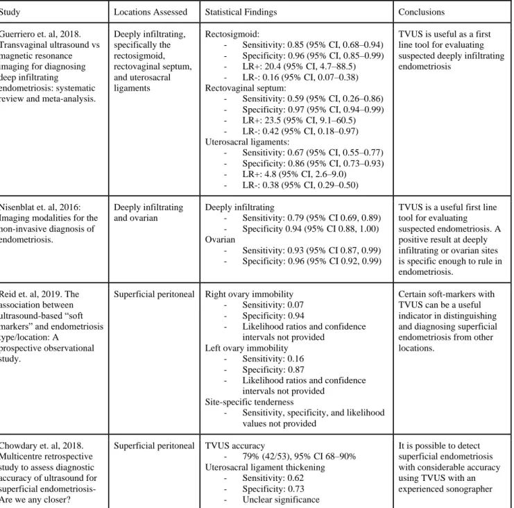
Table 2. Sensitivity and specificity results from the included papers.
Study Locations Assessed Statistical Findings Conclusions
Guerriero et. al, 2018. Transvaginal ultrasound vs magnetic resonance imaging for diagnosing deep infiltrating
endometriosis: systematic review and meta-analysis.
Deeply infiltrating, specifically the rectosigmoid, rectovaginal septum, and uterosacral ligaments Rectosigmoid:
- Sensitivity: 0.85 (95% CI, 0.68–0.94) - Specificity: 0.96 (95% CI, 0.85–0.99)
- LR+: 20.4 (95% CI, 4.7–88.5)
- LR-: 0.16 (95% CI, 0.07–0.38) Rectovaginal septum:
- Sensitivity: 0.59 (95% CI, 0.26–0.86) - Specificity: 0.97 (95% CI, 0.94–0.99)
- LR+: 23.5 (95% CI, 9.1–60.5)
- LR-: 0.42 (95% CI, 0.18–0.97) Uterosacral ligaments:
- Sensitivity: 0.67 (95% CI, 0.55–0.77) - Specificity: 0.86 (95% CI, 0.73–0.93)
- LR+: 4.8 (95% CI, 2.6–9.0)
- LR-: 0.38 (95% CI, 0.29–0.50)
TVUS is useful as a first line tool for evaluating suspected deeply infiltrating endometriosis
Nisenblat et. al, 2016: Imaging modalities for the non-invasive diagnosis of endometriosis.
Deeply infiltrating and ovarian
Deeply infiltrating
- Sensitivity: 0.79 (95% CI 0.69, 0.89) - Specificity 0.94 (95% CI 0.88, 1.00) Ovarian
- Sensitivity: 0.93 (95% CI 0.87, 0.99) - Specificity: 0.96 (95% CI 0.92, 0.99)
TVUS is a useful first line tool for evaluating suspected endometriosis. A positive result at deeply infiltrating or ovarian sites is specific enough to rule in endometriosis.
Reid et. al, 2019. The association between ultrasound-based “soft markers” and endometriosis type/location: A
prospective observational study.
Superficial peritoneal Right ovary immobility - Sensitivity: 0.07 - Specificity: 0.94
- Likelihood ratios and confidence intervals not provided
Left ovary immobility - Sensitivity: 0.16 - Specificity: 0.87
- Likelihood ratios and confidence intervals not provided
Site-specific tenderness
- Sensitivity, specificity, and likelihood values not provided
Certain soft-markers with TVUS can be a useful indicator in distinguishing and diagnosing superficial endometriosis from other locations.
Chowdary et. al, 2018. Multicentre retrospective study to assess diagnostic accuracy of ultrasound for superficial endometriosis-Are we any closer?
Superficial peritoneal TVUS accuracy
- 79% (42/53), 95% CI 68–90%
Uterosacral ligament thickening - Sensitivity: 0.62 - Specificity: 0.73 - Unclear significance
It is possible to detect superficial endometriosis with considerable accuracy using TVUS with an experienced sonographer
Discussion
The primary objective of this narrative review is to analyze the diagnostic accuracy of
TVUS compared to the traditional laparoscopic technique in diagnosing endometriosis at deeply
enough to make it a valuable “rule-in” tool for evaluating deeply infiltrating and ovarian
endometriosis, it lacks the sensitivity necessary to replace laparoscopy as a definitive diagnostic
tool for these locations. Guerriero et. al concluded that TVUS performed well enough to be
considered a first-line tool for evaluating a woman for DIE 17. Nisenblat et. al concluded that
TVUS meets criteria to be a useful tool to “rule-in” DIE and ovarian endometriosis, in that a
positive TVUS from a well-trained sonographer can reliably diagnose endometriosis in those
locations and, thus, should be considered a first line tool for evaluating women with suspected
endometriosis 8. For superficial peritoneal endometriosis, the data indicate that TVUS has
potential to be a useful tool in assessing a woman with endometriosis, but Reid et. al and
Chowdary et. al both concluded that more research is needed to be convincing 5,18.
While the generally low sensitivity values prohibit TVUS from being used as a
replacement for laparoscopic diagnosis, the specificity values are impressively high across the
board. Altogether, the research shows diagnostic utility in a positive TVUS, in that a patient can
be diagnosed with endometriosis with reasonable certainty if they have a positive TVUS.
However, a negative TVUS does not have the same utility. If a woman has a negative TVUS, the
data here suggest further investigation is warranted before a provider can rule out endometriosis
with reasonable certainty. This finding holds true for ovarian and deeply infiltrating sites.
Despite only one of the reviews evaluating ovarian sites, the quality and completeness of the
review is high enough to consider the findings reliable. The data is less convincing for superficial
peritoneal sites given the lack of systematic reviews and overall limited data, but the foundation
has been laid for future research at this location.
The quality of data available remains a major limitation of this narrative review. There
are a limited number of studies evaluating the accuracy of TVUS in endometriosis diagnosis at
deeply infiltrating and ovarian sites, and there are far fewer that analyze superficial peritoneal
sites. As such, the quality of data available is stronger for deeply infiltrating and ovarian sites
than superficial peritoneal sites. Of the studies that are available, they are limited by small
sample sizes and poor methodological quality. This includes the diagnostic accuracy studies
from Reid et. al and Chowdary et. al 5,18. The challenge of small sample sizes can be partially
alleviated with systematic reviews that include meta-analysis, although this produces the
nature of the research question makes it difficult to include healthy controls, as it would be
unethical to perform surgery on an otherwise healthy subject.
The inclusion of high-quality systematic reviews with extensive statistical analysis is the
major strength of this narrative review. Nisenblat et. al in particular was impressively done, with
data from over 4800 participants and methods that resulted in a low risk of bias 8. Guerriero et. al
included over 400 participants and was shown to have an unclear risk of bias, but included an
extensive and very strong meta-analysis 17. Strengths of this review are otherwise limited due to
the reasons stated above.
The results of this narrative review reveal many opportunities for future research. In
regards to ovarian and deeply infiltrating endometriosis, it is reasonable to conclude that TVUS
is a useful tool in evaluating endometriosis in these locations when used by an experienced
sonographer. Research evaluating the diagnostic accuracy of TVUS in the hands of a
sonographer that does not specialize in endometriosis would serve to improve the usefulness of
the tool in a setting outside of a tertiary gynecologic referral center. One study evaluating the
learning curve for sonography students found that a two week course in endometriosis markers can improve a sonographer’s accuracy to above 90% for most DIE locations 19. This represents
another opportunity for future research, as the validation of a sonography curriculum for
endometriosis can greatly increase the standardization and access of endometriosis trained
sonographers. In regards to superficial peritoneal endometriosis, larger studies evaluating TVUS
as a tool for diagnosis are greatly needed. Future research opportunities should include larger
studies specifically evaluating this location, inclusion of this location in systematic reviews and
meta-analyses, and validation of positive TVUS markers for this location.
Conclusions
Endometriosis is a complicated disease process that results in ectopic implantation of
endometrial tissue. Most implants can be classified as deeply-infiltrating, ovarian, or superficial
peritoneal based on their locations within the pelvis. Women who have endometriosis can suffer
from physical and non-physical sequelae, including dysmenorrhea, dyspareunia, noncyclic pelvic
pain, dyschezia, dysuria, depression, anxiety, lost income, and mistrust of healthcare
professionals. The average time to diagnosis is 8-12 years, a delay that is in part due to the
This narrative review sought to understand the current research available for evaluating
TVUS as a diagnostic tool for deeply infiltrating, ovarian, and superficial peritoneal
endometriosis. The results reveal a high specificity and limited sensitivity for TVUS at DIE and
ovarian sites. More research is needed to further validate and standardize evaluation at these
sites, but the data here is strong enough to consider TVUS a useful first-line tool in the
evaluation of endometriosis at deeply infiltrating and ovarian sites. In fact, these results suggest a
positive TVUS can reliably establish the diagnosis of endometriosis at ovarian and deeply
infiltrating sites. However, a negative TVUS cannot rule out the diagnosis of endometriosis, and
a provider should pursue further testing and maintain a high degree of suspicion for the disease.
The data available for superficial peritoneal sites is currently too limited to draw conclusions and
significant more research is needed.
Overall, this review reveals that TVUS is a useful, noninvasive, low-cost tool in
evaluating a woman for endometriosis and has the potential to reduce the time to diagnosis and
treatment, thereby greatly reducing the burden of disease for a woman. Providers should be
confident and empowered to use TVUS as a first line in evaluating a woman with a clinical
picture consistent with endometriosis. This tool can be an important component of improving a
patient-provider relationship, in that this is a relatively easy, low-cost way for a provider to try
and reach some answers for a patient. However, the consequences of the poor sensitivity values
should be discussed in detail with patients. Patients and providers should be aware that a
negative TVUS does not rule out endometriosis, and providers will need to maintain a high
Appendix 1: Table of excluded studies
Study [Year] Reason for Exclusion
Aloisi [2018] 20 Does not evaluate TVUS (only evaluates laparoscopic narrow band imaging)
Anaf [2009] 21 Does not evaluate TVUS (only evaluates barium enema)
Audebert [2015] 22 Not related to primary outcome
Barra [2018] 23 Not related to primary outcome
Borsellino [1993] 24 Published prior to 2009
Casasayas-Carles [2014] 25
Not related to primary outcome
Daraï [2014] 26 Not related to primary outcome
Deffieux [2004] 27 Published prior to 2009
Fancellu [2013] 28 Case report and not related to primary outcome
Fastrez [2017] 29 Does not evaluate TVUS (only evaluates specific type of PET-CT scan)
Fernandez [2003] 30 Published prior to 2009
Gabriel [2011] 31 Not related to primary outcome
Gonçalves [2016] 32 Not related to primary outcome
Guerriero [2015] 33 More recent study published from the same lead author
Guerriero [2016] 34 More recent study published from the same lead author
Hernández [2005] 35 Published prior to 2009
Hudelist [2011] 36 Results are specific to bowel endometriosis and are not suitable for comparison with diagnosing DIE
as a whole
Jaramillo-Cardoso [2018] 37
Not a systematic review and does not evaluate superficial endometriosis (only evaluates abdominal-wall endometriosis)
Keckstein [2000] 38 Published prior to 2009
Khan [2018] 39 Does not evaluate TVUS (only evaluates MRI)
Kiesel [2019] 3 Not a systematic review and does not evaluate superficial endometriosis
Kruse [2012] 40 Not a systematic review and does not evaluate superficial endometriosis
Leone [2016] 41 Only evaluated women during active pregnancy
Levy [2013] 42 Not related to primary outcome
Ma [2019] 43 Not related to primary outcome
McCausland [1996] 45 Published prior to 2009
McCausland [1998] 46 Published prior to 2009
Moawad [2013] 47 Not a systematic review and does not evaluate superficial endometriosis
Moore [2002] 48 Published prior to 2009
Muzii [2016] 49 Not related to primary outcome
Nisenblat [2016] 50 Only evaluated TVUS diagnostic potential when combined with other tests; derivative of included
systematic review (Nisenblat, 2016)
Noventa [2015] 51 Included diagnostic data from non-TVUS techniques
O’Callaghan [2006] 52 Published prior to 2009
Parazzini [2018] 53 Not related to primary outcome
Pickhardt [2007] 54 Published prior to 2009
Piessens [2019] 55 Does not include laparoscopy and histology as reference value
Ribeiro [2006] 56 Published prior to 2009
Rimondi [2018] 57 Not related to primary outcome
Salvat [2001] 58 Published prior to 2009
Scardapane [2013] 59 Does not evaluate TVUS (MRI-only review)
Shoji [2016] 60 Case report, not related to primary outcome
Silveira [2018] 61 Does not evaluate TVUS; animal study
Streuli [2017] 62 Not related to primary outcome
Valentini [2014] 63 Does not evaluate TVUS (only evaluates MRI)
Wozniak [2015] 64 Not related to primary outcome
Appendix 2: AMSTAR 2 Risk of Bias tool for systematic reviews
AMSTAR 2 Criteria Nisenblat et. al, 2016 Guerriero et. al, 2018
1. Did the research questions and inclusion criteria for the review include the components of PICO?
Yes Yes
2. Did the report of the review contain an explicit statement that the review methods were established prior to the conduct of the review and did the report justify any significant deviations from the protocol?
Yes Yes
3. Did the review authors explain their selection of the study designs for inclusion in the review?
Yes Yes
4. Did the review authors use a comprehensive literature search strategy?
Yes Yes
5. Did the review authors perform study selection in duplicate?
Yes Yes
6. Did the review authors perform data extraction in duplicate?
Yes Yes
7. Did the review authors provide a list of excluded studies and justify the exclusions?
No; they did provide a list of reasons why studies were excluded, but not a list of the specific studies
No; they did provide a list of reasons why studies were excluded, but not a list of the specific studies
8. Did the review authors describe the included studies in adequate detail?
Yes Yes
9. Did the review authors use a satisfactory technique for assessing the risk of bias (RoB) in individual studies that were included in the review?
Yes, QUADAS-2 tool Yes, QUADAS-2 tool
10. Did the review authors report on the sources of funding for the studies included in the review?
Yes, within the QUADAS-2 tool Yes, within the QUADAS-2 tool
11. If meta-analysis was performed, did the review authors use appropriate methods for statistical combination of results?
Yes Yes
12. If meta-analysis was performed, did the review authors assess the potential impact of RoB in individual studies on the results of the meta-analysis or other evidence synthesis?
Yes Yes
13. Did the review authors account for RoB in primary studies when
interpreting/discussing the results of the review?
14. Did the review authors provide a satisfactory explanation for, and discussion of, any heterogeneity observed in the results of the review?
Yes Yes
15. If they performed quantitative synthesis did the review authors carry out an adequate investigation of publication bias (small study bias) and discuss its likely impact on the results of the review?
Yes Yes
16. Did the review authors report any potential sources of conflict of interest, including any funding they received for conducting the review?
Yes No, there is no explicit statement
Appendix 3: QUADAS-2 tool for primary literature
QUADAS-2 Criteria Chowdary et. al Reid et. al
Review Question Do ultrasound findings of
superficial endometriosis correlate with laparoscopic findings?
Are ultrasound ‘soft markers’ associated with endometriosis type and location based on laparoscopic findings?
Index Test Transvaginal ultrasound Transvaginal ultrasound
Reference Test Laparoscopy with histologic
sampling
Laparoscopy with histologic sampling
Patient Selection: Risk of Bias
1. Was a consecutive or random sample of patients enrolled?
2. Was a case-control design avoided?
3. Did the study avoid inappropriate exclusions? 4. Could the selection of patients have introduced
bias?
1. Yes, consecutive 2. Yes, all cases 3. Yes, only excluded
incomplete patients
4. LOW RISK
1. Yes, consecutive 2. Yes, all cases 3. Yes, only excluded
women who did not receive laparoscopy
4. LOW RISK
Patient Selection: Applicability
1. Is there concern that the patients do not match the review question?
1. LOW RISK 1. LOW RISK
Index Test: Risk of Bias
1. Were the index test results interpreted without knowledge of the results of the reference standard?
2. If a threshold was used, was it pre-specified? 3. Could the conduct or interpretation of the index
test have introduced bias?
1. Yes, performed
prior
2. Yes, predefined and only one technician
3. LOW RISK
1. Yes, performed prior 2. Yes, positive test was
pre-defined and given to technicians
3. LOW RISK
Index Test: Applicability
1. Is there concern that the index test, its conduct, or interpretation differ from the review question?
1. LOW RISK 1. LOW RISK
Reference Standard: Risk of Bias
1. Is the reference standard likely to correctly classify the target condition?
2. Were the reference standard results interpreted without knowledge of the results of the index test? 3. Could the reference standard, its conduct, or its
interpretation have introduced bias?
1. Yes, gold standard
2. Yes, no knowledge
of index test results
3. LOW RISK
1. Yes, gold standard 2. Unclear, does not state
if surgeons knew TVUS results
3. UNCLEAR RISK
Reference Standard: Applicability
1. Is there concern that the target condition as defined by the reference standard does not match the review question?
1. LOW RISK 1. LOW RISK
Flow and Timing: Risk of Bias
1. Was there an appropriate interval between index test(s) and reference standard?
2. Did all patients receive a reference standard? 3. Did patients receive the same reference standard? 4. Were all patients included in the analysis? 5. Could the patient flow have introduced bias?
1. Yes, reference test after index
2. Yes
3. Yes
4. Yes, 30 histories incomplete
5. LOW RISK
1. Yes, reference test after index test
2. Yes
3. Yes
4. Yes, 31 excluded for not receiving reference
References Bibliography
1. Brown J, Farquhar C. Endometriosis: an overview of Cochrane Reviews. Cochrane Database Syst Rev. 2014;(3):CD009590. doi:10.1002/14651858.CD009590.pub2
2. As-Sanie S, Black R, Giudice LC, et al. Assessing research gaps and unmet needs in endometriosis.
Am J Obstet Gynecol. 2019;221(2):86-94. doi:10.1016/j.ajog.2019.02.033
3. Kiesel L, Sourouni M. Diagnosis of endometriosis in the 21st century. Climacteric. 2019;22(3):1-7. doi:10.1080/13697137.2019.1578743
4. Culley L, Law C, Hudson N, et al. The social and psychological impact of endometriosis on women’s lives: a critical narrative review. Hum Reprod Update. 2013;19(6):625-639. doi:10.1093/humupd/dmt027
5. Reid S, Leonardi M, Lu C, Condous G. The association between ultrasound-based “soft markers” and endometriosis type/location: A prospective observational study. Eur J Obstet Gynecol Reprod Biol. 2019;234:171-178. doi:10.1016/j.ejogrb.2019.01.018
6. Reid R, Steel A, Wardle J, et al. The prevalence of self-reported diagnosed endometriosis in the Australian population: results from a nationally-representative survey. BMC Res Notes.
2019;12(1):88. doi:10.1186/s13104-019-4114-6
7. Rahmioglu N, Nyholt DR, Morris AP, Missmer SA, Montgomery GW, Zondervan KT. Genetic variants underlying risk of endometriosis: insights from meta-analysis of eight genome-wide association and replication datasets. Hum Reprod Update. 2014;20(5):702-716.
doi:10.1093/humupd/dmu015
8. Nisenblat V, Bossuyt PMM, Farquhar C, Johnson N, Hull ML. Imaging modalities for the non-invasive diagnosis of endometriosis. Cochrane Database Syst Rev. 2016;2:CD009591.
doi:10.1002/14651858.CD009591.pub2
9. Morotti M, Vincent K, Becker CM. Mechanisms of pain in endometriosis. Eur J Obstet Gynecol Reprod Biol. 2017;209:8-13. doi:10.1016/j.ejogrb.2016.07.497
10. UNC Center for Endometriosis - UNC Department of Obstetrics & Gynecology.
https://www.med.unc.edu/obgyn/migs/our-services/unc-center-for-endometriosis/. Accessed August 30, 2019.
11. Practice Committee of the American Society for Reproductive Medicine. Treatment of pelvic pain associated with endometriosis: a committee opinion. Fertil Steril. 2014;101(4):927-935.
doi:10.1016/j.fertnstert.2014.02.012
12. Tanbo T, Fedorcsak P. Endometriosis-associated infertility: aspects of pathophysiological mechanisms and treatment options. Acta Obstet Gynecol Scand. 2017;96(6):659-667. doi:10.1111/aogs.13082
13. Vercellini P, Viganò P, Somigliana E, Fedele L. Endometriosis: pathogenesis and treatment. Nat Rev Endocrinol. 2014;10(5):261-275. doi:10.1038/nrendo.2013.255
2012;98(6 Suppl):S1-62. doi:10.1016/j.fertnstert.2012.08.001
15. Whiting PF, Rutjes AWS, Westwood ME, et al. QUADAS-2: a revised tool for the quality assessment of diagnostic accuracy studies. Ann Intern Med. 2011;155(8):529-536.
doi:10.7326/0003-4819-155-8-201110180-00009
16. Shea BJ, Reeves BC, Wells G, et al. AMSTAR 2: a critical appraisal tool for systematic reviews that include randomised or non-randomised studies of healthcare interventions, or both. BMJ. 2017;358:j4008. doi:10.1136/bmj.j4008
17. Guerriero S, Saba L, Pascual MA, et al. Transvaginal ultrasound vs magnetic resonance imaging for diagnosing deep infiltrating endometriosis: systematic review and meta-analysis. Ultrasound Obstet Gynecol. 2018;51(5):586-595. doi:10.1002/uog.18961
18. Chowdary P, Stone K, Ma T, et al. Multicentre retrospective study to assess diagnostic accuracy of ultrasound for superficial endometriosis-Are we any closer? Aust N Z J Obstet Gynaecol.
2018;59(2):279-284. doi:10.1111/ajo.12911
19. Guerriero S, Pascual MA, Ajossa S, et al. Learning curve for ultrasonographic diagnosis of deep infiltrating endometriosis using structured offline training program. Ultrasound Obstet Gynecol. 2019;54(2):262-269. doi:10.1002/uog.20176
20. Aloisi A, Sonoda Y, Gardner GJ, et al. Prospective comparative study of laparoscopic narrow band imaging (NBI) versus standard imaging in gynecologic oncology. Ann Surg Oncol.
2018;25(4):984-990. doi:10.1245/s10434-017-6314-4
21. Anaf V, El Nakadi I, De Moor V, Coppens E, Zalcman M, Noel J-C. Anatomic significance of a positive barium enema in deep infiltrating endometriosis of the large bowel. World J Surg. 2009;33(4):822-827. doi:10.1007/s00268-008-9903-3
22. Audebert A, Lecointre L, Afors K, Koch A, Wattiez A, Akladios C. Adolescent Endometriosis: Report of a Series of 55 Cases With a Focus on Clinical Presentation and Long-Term Issues. J Minim Invasive Gynecol. 2015;22(5):834-840. doi:10.1016/j.jmig.2015.04.001
23. Barra F, Scala C, Biscaldi E, et al. Ureteral endometriosis: a systematic review of epidemiology, pathogenesis, diagnosis, treatment, risk of malignant transformation and fertility. Hum Reprod Update. 2018;24(6):710-730. doi:10.1093/humupd/dmy027
24. Borsellino G, Buonaguidi A, Veneziano S, Borsellino V, Mariscalco G, Minnici G. [Endometriosis of the large intestine. A report of 2 clinical cases]. Minerva Ginecol. 1993;45(9):443-447.
25. Casasayas-Carles P, Fuentes-Marquez I, Tarrasa-Sagristá F, Gutiérrez Sanz-Gadea C.
Müllerianosis of the urinary bladder: report of three new cases. Arch Esp Urol. 2014;67(9):771-775.
26. Daraï E, Bazot M, Ballester M, Belghiti J. [Endometriosis]. Rev Prat. 2014;64(4):545-550. 27. Deffieux X, Fernandez H. [Physiopathologic, diagnostic and therapeutic evolution in the
management of adenomyosis: review of the literature]. J Gynecol Obstet Biol Reprod (Paris). 2004;33(8):703-712.
doi:10.1016/j.ijscr.2013.11.001
29. Fastrez M, Artigas C, Sirtaine N, et al. Value of the 68Ga-DOTATATE PET-CT in the diagnosis of endometriosis. A pilot study. Eur J Obstet Gynecol Reprod Biol. 2017;212:69-74.
doi:10.1016/j.ejogrb.2017.03.022
30. Fernandez H. [New concepts on pathophysiology, diagnosis and treatment of adenomyosis]. J Gynecol Obstet Biol Reprod (Paris). 2003;32(8 Pt 2):S23-7.
31. Gabriel B, Nassif J, Trompoukis P, Barata S, Wattiez A. Prevalence and management of urinary tract endometriosis: a clinical case series. Urology. 2011;78(6):1269-1274.
doi:10.1016/j.urology.2011.07.1403
32. Gonçalves FC, Andres MP, Passman LJ, Gonçalves MOC, Podgaec S. A systematic review of ultrasonography-guided transvaginal aspiration of recurrent ovarian endometrioma. Int J Gynaecol Obstet. 2016;134(1):3-7. doi:10.1016/j.ijgo.2015.10.021
33. Guerriero S, Ajossa S, Minguez JA, et al. Accuracy of transvaginal ultrasound for diagnosis of deep endometriosis in uterosacral ligaments, rectovaginal septum, vagina and bladder: systematic review and meta-analysis. Ultrasound Obstet Gynecol. 2015;46(5):534-545.
doi:10.1002/uog.15667
34. Guerriero S, Ajossa S, Orozco R, et al. Accuracy of transvaginal ultrasound for diagnosis of deep endometriosis in the rectosigmoid: systematic review and meta-analysis. Ultrasound Obstet Gynecol. 2016;47(3):281-289. doi:10.1002/uog.15662
35. Hernández Valencia M, Zárate A, Hernández Quijano T, Landero Montes de Oca ME, Escamilla Godínez G. [Endometriosis in delayed scarring of postpartum eutocic episiorrhaphy. Integral aspects and a case report]. Rev Med Inst Mex Seguro Soc. 2005;43(3):237-242.
36. Hudelist G, English J, Thomas AE, Tinelli A, Singer CF, Keckstein J. Diagnostic accuracy of transvaginal ultrasound for non-invasive diagnosis of bowel endometriosis: systematic review and meta-analysis. Ultrasound Obstet Gynecol. 2011;37(3):257-263. doi:10.1002/uog.8858
37. Jaramillo-Cardoso A, Balcacer P, Garces-Descovich A, et al. Multimodality imaging and
clinicopathologic assessment of abdominal wall endometriosis: knocking down the enigma. Abdom Radiol (NY). July 2018. doi:10.1007/s00261-018-1666-1
38. Keckstein J. Hysteroscopy and adenomyosis. Contrib Gynecol Obstet. 2000;20:41-50.
39. Khan KS, Tryposkiadis K, Tirlapur SA, et al. MRI versus laparoscopy to diagnose the main causes of chronic pelvic pain in women: a test-accuracy study and economic evaluation. Health Technol Assess. 2018;22(40):1-92. doi:10.3310/hta22400
40. Kruse C, Seyer-Hansen M, Forman A. Diagnosis and treatment of rectovaginal endometriosis: an overview. Acta Obstet Gynecol Scand. 2012;91(6):648-657.
doi:10.1111/j.1600-0412.2012.01367.x
41. Leone Roberti Maggiore U, Ferrero S, Mangili G, et al. A systematic review on endometriosis during pregnancy: diagnosis, misdiagnosis, complications and outcomes. Hum Reprod Update. 2016;22(1):70-103. doi:10.1093/humupd/dmv045
2013;94(1):3-25. doi:10.1016/j.diii.2012.10.012
43. Ma T, Chowdary P, Eskander A, et al. Can narrowband imaging improve the laparoscopic identification of superficial endometriosis? A prospective cohort trial. J Minim Invasive Gynecol. 2019;26(3):427-433. doi:10.1016/j.jmig.2018.05.007
44. Maignien C, Santulli P, Gayet V, et al. Prognostic factors for assisted reproductive technology in women with endometriosis-related infertility. Am J Obstet Gynecol. 2017;216(3):280.e1-280.e9. doi:10.1016/j.ajog.2016.11.1042
45. McCausland AM, McCausland VM. Depth of endometrial penetration in adenomyosis helps determine outcome of rollerball ablation. Am J Obstet Gynecol. 1996;174(6):1786-1793; 1793. doi:10.1016/s0002-9378(96)70211-9
46. McCausland V, McCausland A. The response of adenomyosis to endometrial ablation/resection.
Hum Reprod Update. 1998;4(4):350-359. doi:10.1093/humupd/4.4.350
47. Moawad NS, Caplin A. Diagnosis, management, and long-term outcomes of rectovaginal endometriosis. Int J Womens Health. 2013;5:753-763. doi:10.2147/IJWH.S37846
48. Moore J, Copley S, Morris J, Lindsell D, Golding S, Kennedy S. A systematic review of the accuracy of ultrasound in the diagnosis of endometriosis. Ultrasound Obstet Gynecol. 2002;20(6):630-634. doi:10.1046/j.1469-0705.2002.00862.x
49. Muzii L, Di Tucci C, Achilli C, et al. Continuous versus cyclic oral contraceptives after laparoscopic excision of ovarian endometriomas: a systematic review and metaanalysis. Am J Obstet Gynecol. 2016;214(2):203-211. doi:10.1016/j.ajog.2015.08.074
50. Nisenblat V, Prentice L, Bossuyt PMM, Farquhar C, Hull ML, Johnson N. Combination of the non-invasive tests for the diagnosis of endometriosis. Cochrane Database Syst Rev.
2016;7:CD012281. doi:10.1002/14651858.CD012281
51. Noventa M, Saccardi C, Litta P, et al. Ultrasound techniques in the diagnosis of deep pelvic endometriosis: algorithm based on a systematic review and meta-analysis. Fertil Steril. 2015;104(2):366-83.e2. doi:10.1016/j.fertnstert.2015.05.002
52. O’Callaghan D. Endometriosis--an update. Aust Fam Physician. 2006;35(11):864-867. 53. Parazzini F, Frattaruolo MP, Chiaffarino F, Dridi D, Roncella E, Vercellini P. The limited
oncogenic potential of unilocular adnexal cysts: A systematic review and meta-analysis. Eur J Obstet Gynecol Reprod Biol. 2018;225:101-109. doi:10.1016/j.ejogrb.2018.04.019
54. Pickhardt PJ, Kim DH, Menias CO, Gopal DV, Arluk GM, Heise CP. Evaluation of submucosal lesions of the large intestine: part 2. Nonneoplastic causes. Radiographics. 2007;27(6):1693-1703. doi:10.1148/rg.276075028
55. Piessens S, Edwards A. Sonographic evaluation for endometriosis in routine pelvic ultrasound. J Minim Invasive Gynecol. September 2019. doi:10.1016/j.jmig.2019.08.027
56. Ribeiro PAA, Rodrigues FC, Kehdi IPA, et al. Laparoscopic resection of intestinal endometriosis: a 5-year experience. J Minim Invasive Gynecol. 2006;13(5):442-446.
doi:10.1016/j.jmig.2006.05.010
hemangiomas. Radiol Med. 2018;123(7):538-544. doi:10.1007/s11547-018-0862-y
58. Salvat J. [Diagnosis and follow-up of endometriosis during consultation: changes]. Gynecol Obstet Fertil. 2001;29(9):616-623.
59. Scardapane A, Lorusso F, Bettocchi S, et al. Deep pelvic endometriosis: accuracy of pelvic MRI completed by MR colonography. Radiol Med. 2013;118(2):323-338. doi:10.1007/s11547-012-0850-6
60. Shoji T, Takatori E, Murakami K, et al. A case of ovarian adenosquamous carcinoma arising from endometrioid adenocarcinoma: a case report and systematic review. J Ovarian Res. 2016;9(1):48. doi:10.1186/s13048-016-0255-6
61. Silveira MB, Rodrigues DM, Araújo MR, et al. 18F-Fluorocholine Uptake and Positron Emission Tomography Imaging in Rat Peritoneal Endometriosis. Reprod Sci. 2018;25(1):19-25.
doi:10.1177/1933719117728799
62. Streuli I, Santulli P, Chouzenoux S, Chapron C, Batteux F. Serum osteopontin levels are decreased in focal adenomyosis. Reprod Sci. 2017;24(5):773-782. doi:10.1177/1933719116669054
63. Valentini AL, Gui B, Miccò M, et al. How to improve MRI accuracy in detecting deep infiltrating colorectal endometriosis: MRI findings vs. laparoscopy and histopathology. Radiol Med.
2014;119(5):291-297. doi:10.1007/s11547-013-0336-1
64. Wozniak S, Czuczwar P, Szkodziak P, et al. Elastography Improves the Accuracy of Ultrasound in the Preoperative Assessment of abdominal wall endometriosis. Ultraschall Med. 2015;36(6):623-629. doi:10.1055/s-0034-1398834