https://doi.org/10.1124/pr.119.017863
ASSOCIATE EDITOR: ERIC L. BARKER
Harnessing Ion-Binding Sites for GPCR Pharmacology
Barbara Zarzycka, Saheem A. Zaidi, Bryan L. Roth, and Vsevolod Katritch
Departments of Biological Sciences (B.Z., S.A.Z., V.K.) and Chemistry (V.K.), Bridge Institute, Michelson Center for Convergent Bioscience, University of Southern California, Los Angeles, California; and Department of Pharmacology (B.L.R.) and Division of Chemical Biology and Medicinal Chemistry, Eshelman School of Pharmacy (B.L.R.), University of North Carolina at Chapel Hill, Chapel Hill, North Carolina
Abstract. . . 572
Significance Statement . . . 572
I. Historical Overview . . . 572
II. Structural Data for Conserved and Nonconserved Ion Binding Sites in G-Protein-Coupled Receptors . . . 576
A. Conserved Sodium Binding Site in Class A G-Protein-Coupled Receptors . . . 576
1. High-Resolution Structures of Sodium in the Conserved Site . . . 576
2. Sodium Ion Detection Criteria . . . 577
3. Lower Resolution Inactive State Structures of Class A Compatible with Sodium Ion Binding . . . 577
4. Structures of Active State G-Protein-Coupled Receptors Are Incompatible with Sodium Binding . . . 578
5. Allosteric Ligands can Block Sodium Binding . . . 579
6. Mutations Abolishing Sodium Binding . . . 579
B. G-Protein-Coupled Receptors that Lack the Conserved Sodium Site . . . 580
1. Some Class A G-Protein-Coupled Receptors Lack Specific Sodium Site. . . 580
2. Non-Class A G-Protein-Coupled Receptors Lack Conserved Sodium Sites in 7-Transmembrane Domains . . . 581
C. Nonconserved Ion Binding Sites in G-Protein-Coupled Receptors. . . 581
D. Nonspecific Ion Binding in Crystal Structures . . . 582
III. Functional Role—Why is Sodium So Special for Class A G-Protein-Coupled Receptors? . . . 582
A. Allosteric Effects of Sodium on Agonist Binding . . . 582
1. “Classical”Allosteric Effect of Sodium Ion on Agonist Binding . . . 582
2. Binding of Sodium Not Always Detected by a“Classical”Allosteric Effect. . . 584
B. Evidence for the Functional Importance of the Sodium Ion Site . . . 584
1. Direct Functional Effects of Sodium Ion Presence . . . 584
2. Mutations in Sodium Ion Pocket Reduce or Abolish Receptor Stimulation by Agonists . . . 585
3. A Gain of Function by Introducing Acidic Residues in Sodium Ion Pocket . . . 586
4. Disease-Associated Mutations in the Sodium Pocket. . . 586
C. Mechanism of Sodium Ion Functional Involvement . . . 586
1. Sodium as an Allosteric Cofactor of Class A G-Protein-Coupled Receptor Signaling . . . . 586
D. Other Potential Functional Effects of the Conserved Sodium Ion Binding . . . 587
1. Voltage Sensing . . . 587
2. pH Dependence . . . 587
IV. Ion Binding Sites as Ligand Targets—New Approaches to Design Functional Properties . . . 587
Address correspondence to:Dr. Vsevolod Katritch, University of Southern California, 1002 West Childs Way, MCB-317, Los Angeles, CA 90089. E-mail: [email protected]; or Dr. Bryan L. Roth, University of North Carolina at Chapel Hill, 120 Mason Farm Rd., CB # 7365 Chapel Hill, NC 27599-7365. E-mail: [email protected]
The research was supported by National Institutes of Health National Institute on Drug Abuse Grants [DA038858, P01DA035764, R37DA035764]; National Institute of Mental Health Grant [R01MH112205]; the National Institute of Mental Health Psychoactive Drug Screening Program; and the Michael Hooker Distinguished Professorship to B.L.R. and The Netherlands Association for Scientific Research Rubicon fellowship [019.161LW.035].
A. Targeting Nonconserved Ion Binding Sites for Subtype Selectivity . . . 588
B. Allosteric Ligand Binding in the Conserved Sodium Ion Site. . . 589
C. Targeting Sodium Ion with Bitopic Ligands . . . 589
1. Concept of Bitopic Ligands . . . 589
2. Structure-Based Design of Bitopic Ligands for the Sodium Ion Site . . . 589
V. Biophysical and Computational Approaches for Studying Allosteric Ions . . . 590
A. Nuclear Magnetic Resonance Spectroscopy . . . 590
1. Study of G-Protein-Coupled Receptor Dynamics with and without Sodium Ion Site. . . 590
2. Direct Nuclear Magnetic Resonance Assessment of Allosteric Sodium Ion Binding Kinetics . . . 590
3. Binding and Effect of Divalent Ions in A2A Adrenergic Receptor is Potentially Nonspecific . . . 590
B. Molecular Dynamic Simulations . . . 590
1. Sodium Access and Universality of Sodium Ion Binding in Class A G-Protein-Coupled Receptors. . . 590
2. Molecular Dynamic Simulations Corroborate New Functional Hypotheses . . . 591
C. Phylogenetic Analysis. . . 592
VI. Unanswered Questions and Future Perspectives . . . 592
Acknowledgments . . . 593
References . . . 593
Abstract——Endogenous ions play important roles in the function and pharmacology of G-protein coupled receptors (GPCRs). Historically the evidence for ionic modulation of GPCR function dates to 1973 with studies of opioid receptors, where it was demonstrated that physiologic concentrations of sodium allosterically attenuated agonist binding. This Na1-selective effect was distinct from effects of other monovalent and divalent cations, with the latter usually counteracting sodium’s negative allosteric modulation of binding. Since then, numerous studies documenting the effects of mono- and divalent ions on GPCR function have been published. While ions can act selectively and nonselectively at many sites in different receptors, the discovery of the conserved sodium ion site in class A GPCR structures in 2012 revealed the unique nature of Na1site, which has emerged as a near-universal site for allosteric modulation of class A GPCR structure and function. In this review, we synthesize and highlight
recent advances in the functional, biophysical, and structural characterization of ions bound to GPCRs. Taken together, these findings provide a molecular understanding of the unique roles of Na1 and other ions as GPCR allosteric modulators. We will also discuss how this knowledge can be applied to the redesign of receptors and ligand probes for desired functional and pharmacological profiles.
Significance Statement——The function and pharma-cology of GPCRs strongly depend on the presence of mono and divalent ions in experimental assays and in living organisms. Recent insights into the molecular mechanism of this ion-dependent allosterism from structural, biophysical, biochemical, and computational studies provide quantitative understandings of the pharmacological effects of drugs in vitro and in vivo and open new avenues for the rational design of chemical probes and drug candidates with improved properties.
I. Historical Overview
Endogenous ions are involved in all aspects of human biology, including their key roles in the function and pharmacology of GPCRs, which comprise the largest family of clinically relevant protein targets (Lagerström and Schiöth, 2008; Katritch et al., 2013; Hauser et al., 2017). GPCRs signal both at the plasma membrane and
in intracellular membranes, including endosomes and golgi (Calebiro et al., 2010; Irannejad et al., 2013; Vilardaga et al., 2014; Godbole et al., 2017; Eichel and von Zastrow, 2018), and are likely exposed to large spatiotemporal variations in ionic and pH conditions that may affect their function. Thus, for instance, extra-cellular Na1 is normally maintained in 135–145 mM range, while its intracellular levels are about 10 times
ABBREVIATIONS:A2AR, A2Aadenosine receptor; BLT1, leukotriene B4 receptor 1; cAMP, cyclic adenosine monophosphate; CaSR, calcium
sensing receptor; CB1, cannabinoid receptor type 1; CCR2, C-C chemokine receptor type 2; CLTR1, cysteinyl leukotriene receptor 1; CPMG, Carr Purcell Meiboom and Gill NMR; DOR, delta opioid receptor; DRD2, dopamine receptor D2; DRD3, dopamine receptor D3; DRD4, dopamine receptor D4; EC, extracellular; ECL, extracellular loop; ECL2, extracellular loop 2; EM, electron microscopy; EP3, prostaglandin E receptor subtype EP3; ETA, endothelin ETA receptor; ETB1, endothelin ETB1 receptor; GPCR, G-protein-coupled receptors; HMA,
lower in most cells (Lodish et al., 2000); intracellular sodium levels rapidly increase during depolarization in neurons. Also, some GPCRs are directly (Wingler et al., 2019) and selectively modulated by inorganic ions as a part of their physiologic function, e.g., CaSR by Ca21 (Silve et al., 2005) and GPR39 by Zn1 (Sato et al., 2016). Other GPCRs are proton sensing, in-cluding GPR68, GPR4, TDAG8, and G2A (Ludwig et al., 2003; Radu et al., 2005; Yang et al., 2007; Liu et al., 2010; Huang et al., 2015b). In this review though, we will mostly focus on the function of endogenous ligands, and therapeutic drugs, being allosterically modulated by ions interacting with GPCRs.
Historically, the first evidence for ionic modulation of GPCRs dates well before they were recognized as a large family of receptors sharing a common seven-transmembrane (7TM) architecture. In 1973, studies of opioid receptors showed that agonist binding is negatively modulated by monovalent cations like Na1 (Pert et al., 1973; Pert and Snyder, 1974), while being positively modulated by divalent cations (Pasternak et al., 1975). Several subsequent studies provided biochemical data suggesting that these effects were mediated by an allosteric mechanism (Simon and Groth, 1975; Horstman et al., 1990). A similar negative allosteric modulation of agonist binding affinity was soon discovered for many other class A GPCRs including adrenergic (Tsai and Lefkowitz, 1978), dopaminergic (Neve, 1991;
Neve et al., 1991) and somatostatin (Kong et al., 1993) receptors. Since then, hundreds of papers have appeared documenting the actions of sodium, as well as other cations and anions on the function of many GPCRs [see Katritch et al. (2014) and (Strasser et al., 2015) for review].
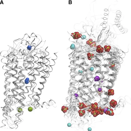
Moreover, high-resolution structural information for GPCRs and their complexes, which has emerged in the past few years (Liu et al., 2012b; Fenalti et al., 2014; Miller-Gallacher et al., 2014; Wang et al., 2017) has made it possible to identify a variety of ion binding sites in GPCRs (Fig. 1; Table 1). While some of the ions, like the multiple Zn21 and Hg21ions in rhodopsin struc-tures were introduced to assist crystallization and/or anomalous diffraction phasing (Teller et al., 2001), many other ion binding sites may be relevant for endogenous ligand binding at specific receptors. For example, the crystallographically observed PO432site in H1 histamine receptor (Shimamura et al., 2011) or Na1 binding in the extracellular loop in theb1AR adrenergic receptor (Miller-Gallacher et al., 2014).
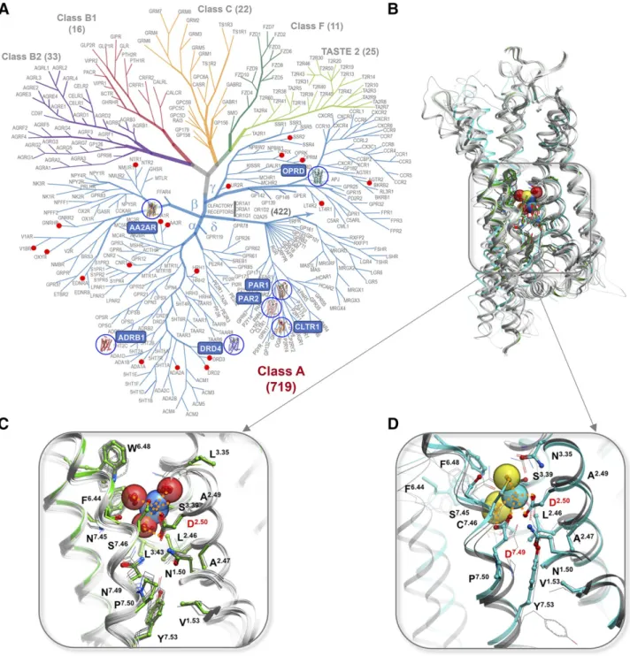
Only the sodium site, however, stands out as highly conserved among most class A GPCRs (Katritch et al., 2014) and potentially tracing its origin to other distant 7TM relatives like prokaryotic channel rhodopsins (Shalaeva et al., 2015). This site binds allosteric sodium in the middle of the 7TM helical bundle of class A GPCRs (Fig. 2, A and B), anchored at the most
conserved aspartate residue D2.50 (superscript shows generic numbering of GPCR residues as described in Isberg et al. (2015). Analysis of the structures and sequences of class A GPCRs revealed the highly con-served nature of the sodium pocket, 15 residues of which are conserved exactly in 45 diverse receptors and with minor variations in a vast majority of class A GPCRs families (Fig. 2, C and D) (Katritch et al., 2014). Moreover, those class A receptors that lack the key residues of the sodium site naturally, or via introduced
mutations, have their ligand-induced signaling dramat-ically reduced or completely abolished (Katritch et al., 2014; Massink et al., 2015; White et al., 2018). While our understanding of ion binding sites and their bio-chemical and physiologic effects on GPCR signaling has greatly expanded in the last few years, we are only beginning to understand the new possibilities for harnessing this knowledge for the discovery of safer and more efficient drugs that have improved subtype and/or functional selectivity (Roth, 2019). Fig. 2. Conserved Na1sites in GPCRs. (A) GPCR superfamily tree with blue circles highlighting receptor structures with Na1resolved in the conserved pocket and red dots marking GPCR with established allosteric effect of Na1. (B) Overview of the Na1position in 7TM helical bundle. (C) Type I Na1site, first discovered in A2AR (green cartoon and sticks) and conserved in majority of Class A GPCRs structures (gray).
(D) Distinct coordination of Na1by two acidic residues D2.50and D7.49in Type II sodium site as resolved in PAR1 (cyan), PAR2 and CLTR1
II. Structural Data for Conserved and Nonconserved Ion Binding Sites in
G-Protein-Coupled Receptors
A. Conserved Sodium Binding Site in Class A G-Protein-Coupled Receptors
1. High-Resolution Structures of Sodium in the Conserved Site. Sodium ion in the conserved sodium pocket was first crystallographically identified in the A2A adenosine receptor (Liu et al., 2012b), quickly followed by PAR1 thrombin (Zhang et al., 2012),
b1AR adrenergic (Miller-Gallacher et al., 2014),
d-OR opioid (Fenalti et al., 2014), and D4 dopamine receptors (Wang et al., 2017). Remarkably, despite these structures representing receptors in different major branches of class A GPCRs and having low sequence identity between them (20%–35%), the sodium-binding positions in the structures were found to be almost identical (within 0.5–1.5 Å) with all of them anchored at the negatively charged D2.50 side chain. Moreover, the sequence of all 16 residues lining the sodium binding pocket and their conformations in receptor structures are remarkably conserved either in the whole class A GPCR or its individual branches (Table 2). The unprecedented level of conservation of the Na1 pocket as a structural feature was also emphasized by the fact that the positions of up to 10 water molecules in the pocket comprising Na1/water cluster were found conserved between such distant receptors as A2A,b1AR, andd-OR. Such a high level of sequence and structural conservation implied a critical functional role of this Na1site in class A GPCRs (Liu et al., 2012b; Katritch et al., 2014). It is important to note, also, that the sodium pocket lies in close proximity and, in most structures of class A GPCRs, is directly connected to the orthosteric pocket making
it potentially accessible to ligand design, as discussed below insection IV.
A number of high-resolution structures have been more recently obtained (see Table 1), shedding light on the Na1pocket and revealing new features of the Na1 binding site. Thus, more than 20 additional structures of antagonist-bound complexes for each of the A2A adenosine and b1 adrenergic receptors show that the Na1/water cluster can be reliably resolved in GPCR structures at up to about 2.3 Å resolution. Among them, the X-ray free-electron laser crystal structure of A2AR (1.96 Å resolution) is especially important (Batyuk et al., 2016), because it was determined at room temperature. This structure demonstrated that exis-tence of a well-defined conformation of Na1/water in the pocket detected in crystal structures of many GPCRs was not an artifact of cryo-freezing, but rather a result of the unique stability of the cluster itself.
Key insights were obtained from the antagonist-bond dopamine receptor D4 (DRD4) high-resolution struc-ture (Wang et al., 2017), which was determined both with and without sodium. Importantly, an electron density for sodium was observed only when Na1 (;200 mM) was added during crystallization, thus providing the most direct structural evidence for Na1 in its binding site. Remarkably, even though another, the sodium-free structure of DRD4, was slightly higher resolution, the electron densities for water molecules forming the Na1/water cluster disappeared, showing that Na1is critical for the stability of the whole cluster. Indeed, water clusters in polar pockets can usually dynamically form many combinations of their hydrogen bonding network, which compromise detection of in-dividual water molecules. In contrast, the presence of the Na1 with a strong ionic bridge to D2.50 creates a specific configuration of the whole cluster, characterized
TABLE 2
Residue conservation in the sodium pocket for different branches of Class A GPCRs.
by well-defined electron densities, as observed previously in A2A,b1AR, and d-OR high-resolution structures. It is also worth noting that there was no significant difference in the receptor conformation itself between Na1-bound and sodium-free structures of DRD4, even in the sodium-coordinating pocket residues, suggesting that the presence of sodium ion does not“induce”any specific conformational macrostate of the receptor. Instead, the observed stabilizing role of Na1is manifested in shift-ing equilibrium toward the same inactive state confor-mation as observed without sodium.
The recently solved structures of the PAR2 proteinase-activated receptor (Cheng et al., 2017a) and of the cysteinyl leukotriene receptor CLTR1 (Luginina et al., 2019) further confirm the presence of sodium in the
d-branch of GPCRs as was identified previously for PAR1 (Zhang et al., 2012). These receptors provide a distinct structure of the Na1pocket (Fig. 2D; Table 2), where sodium is coordinated by two acidic residues, D2.50and D7.49, instead of only D2.50in most other class A receptors, which have N7.49. This double salt bridge coordination shifts the sodium position about 1.5 Å
“down”along the polar channel and changes the overall Na1 coordination and conservation pattern compared with“classical”sodium pocket in a- andg-branches of GPCRs.
2. Sodium Ion Detection Criteria. While divalent ions in crystal structures are often detected by their anomalous diffraction, monovalent ions including Na1 lack such anomalous diffraction. Reliable detection of monovalent ions like Na1 in the protein crystal struc-tures is based on high resolution (usually,2.3 Å), the strong electron density in a potential Na1ion position and the unambiguous detection of at least five oxygen (or potentially nitrogen) atoms that comprise the Na1 coordination shell. The sodium ion can be identified then by its 5-atom coordination geometry and short characteristic distances to the coordinating atoms (2.3–2.5 Å), as discussed in Liu et al. (2012b). These criteria help to differentiate Na1from four-atom tetra-hedral coordination of water molecules and character-istic water interaction distances (2.8–3.1 Å). At even higher resolution, e.g., ,2.0 Å, the accuracy of mea-surement may also be sufficient (Cheng et al., 2017a) to differentiate Na1 coordination distances from those of other monovalent ions, e.g., longer distances for K1 (2.6–2.8 Å) and shorter for Li1 (1.9–2.1 Å) (Kuppuraj et al., 2009), thus specifically detecting Na1.
It is important to note that the high structural stability of the protein and of the Na1/water cluster is as important for detection of Na1as the resolution of the structures. Thus, it was possible to reliably resolve Na1 in PAR2 even at somewhat lower 2.8 Å resolution, because Na1 was coordinated by five oxygen atoms of the protein side chains, including two from the charged carboxy groups of D2.50and D7.49. At the same time, in some higher resolution structures, for example OX2R
(PDB: 5WQC, resolution 1.96 Å), allosteric sodium was apparently absent from the conserved pocket (Suno et al., 2018a). Though some waters of the sodium pocket were resolved, the density for sodium and a neighboring position interacting with D2.50 were not well defined, precluding Na1 detection. This is not surprising, as the Na1 concentration used for this crystallization was 120 mM, which is in the range close to EC50 of Na1in some receptors (e.g.,;100 mM for DRD4), and thus may not allow the full saturation required for Na1/water cluster stability and detection. Therefore, the lack of sodium density in this structure does not necessarily mean that the fully conserved Na1 pocket in OX2R does not bind sodium, but rather that it could not be detected crystallographically under the condi-tions used. Further studies of Na1in OX2R, including validation of the classic Na1effect on agonist binding, may be needed to answer this question more definitively (Suno et al., 2018a).
All structures with bound Na1so far were resolved by crystallography, although it is possible that structural information about sodium pocket may also come in the future from cryo-EM studies. The best GPCR structures by cryo-EM have been solved at;3.0 Å resolution (Zhao et al., 2019), but the rapid progress in the cryo-EM field and detection of soluble protein structures at resolu-tions as high as 1.8 Å (Merk et al., 2016) suggests that this crystal-free technology may ultimately allow deci-phering the sodium cluster and other ion binding details as well.
3. Lower Resolution Inactive State Structures of Class A Compatible with Sodium Ion Binding. In many other crystal structures of diverse class A GPCRs where the modest resolution (2.4–2.9 Å) was insuffi-cient reliably to resolve sodium ion, the conserved pocket is still fully compatible with sodium presence in the structure.
• In the a-branch of class A GPCRs, this includes A1adenosine (Cheng et al., 2017b) and most of the aminergic GPCR structures, for example,
b2AR (2RH1), D3R (3PBL) that have closely related subtypes with Na1 explicitly determined crystallographically.
• In theg-branch, where sodium was only resolved in high-resolution structure of delta opioid receptor (DOR) so far (Fenalti et al., 2014), the pockets are fully conserved and compatible with Na1binding in structures of all other opioid receptors including mu (MOR) (Manglik et al., 2012), kappa (KOR) (Wu et al., 2012) and nociceptin (NOP) (Thompson et al., 2012) opioid receptors.
represented a partially active-like state. An inter-esting observation was made recently for another
b-branch GPCR, the ETB endothelin receptor (Shihoya et al., 2017). Though a weak allosteric sodium effect was detected in the ETBreceptor, it was above the physiologic sodium concentration (.1 M), probably owing it to the fact that one of the key residues of the otherwise conserved sodium pocket, Y7.53, was replaced by L7.53 in the ETB receptor. Correspondingly, the crystal structure of ETB solved at relatively high resolution 2.2 Å had the cavity filled with electron densities that were more compatible with water molecules than with sodium. Intriguingly, the closely related ETA has much stronger Na1 binding at EC50 5 245 mM, and a high-resolution ETA structure might provide new insights for this family.
4. Structures of Active State G-Protein-Coupled Receptors Are Incompatible with Sodium Binding.
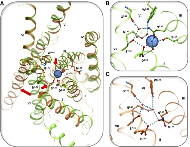
Comparison of the sodium pocket conformation in inactive- and active-state structures of A2A, b1AR, muscarinic, 5-HT, and opioid receptors (Fig. 3) reveals that the sodium pocket shape, conformation, and in-teraction network change dramatically upon activation,
resulting in a partially collapsed pocket that is not compatible with high-affinity binding of sodium (Katritch et al., 2014). In general, active state conformations are characterized by an inward movement of the TM7 backbone, which directly clashes with the sodium site and rearranges sodium-coordinating side chains so that they form direct hydrogen bonds instead of Na1 medi-ated (e.g., between D2.50and S3.39) that preclude sodium coordination (Fig. 3C). Other major activation related changes, like the outward movement of TM6, also change the shape of the Na1 pocket and disrupt the so-called hydrophobic layer (Yuan et al., 2014). Disrup-tion of the hydrophobic layer, comprising residues 1.53, 2.46, 3.43, 7.53 at the bottom of the Na1pocket, opens the floodgate for water and sodium ion egress toward the intracellular side.
Such conformational changes in active-like states were described recently for NTSR1 (White et al., 2012; Krumm et al., 2015) and AT2R (Zhang et al., 2015b, 2017) in structures of complexes with agonists, as well as the structures of fully active MOR (PDB: 5C1M) (Huang et al., 2015a) and KOR (PDB: 6B73) (Che et al., 2018) bond to both agonists and nanobodies. The MOR structure is especially important in this respect because
it was solved at a high 2.1 Å resolution. A detailed examination of the pocket structure reveals no electron density suitable for Na1, and in general, the active state conformation is incompatible with Na1binding (Huang et al., 2015a).
It should be noted, that while the described above general conformational rearrangements are common to active state conformations, the details of newly formed interactions in the pocket can differ between the struc-tures quite dramatically. This contrasts with the very conserved conformation of the residues in the structures of inactive GPCRs. This loss of uniformity can be explained by natural differences in the pocket between receptors, but also by different activation states (intermediate activated to fully active) and a range crystallographic resolution (from 2.1 to 3.5 Å) (Katritch et al., 2014).
5. Allosteric Ligands can Block Sodium Binding.
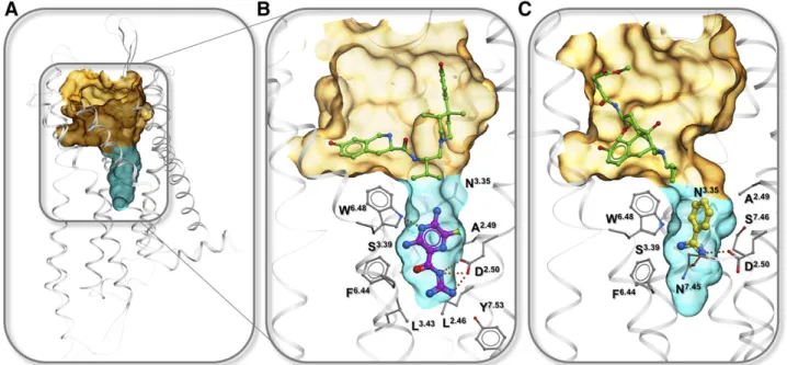
Although the Na1pocket is small,;200 Å3as estimated in A2A (Liu et al., 2012b), it can bind small molecules like amiloride and its analogs, which have a common positively charged moiety connected to an aromatic ring (Fig. 4) (Liu et al., 2012b; Katritch et al., 2014). The allosteric binding of amiloride has been shown biochemi-cally for several receptors, revealing direct competition with Na1 binding and a strong dependence on muta-tions in D2.50and other pocket residues (Howard et al., 1987; Gao and Ijzerman, 2000; Gao et al., 2003a,b; Heitman et al., 2008; Gutiérrez-de-Terán et al., 2013; Massink et al., 2015) (Fig. 4B). Crystallographic obser-vation of ligand binding in the Na1 pocket has been elusive, until recently the structure of leukotriene B4 receptor BLT1 was solved in complex with a ligand
reaching into Na1pocket (Hori et al., 2018). The bitopic ligand BIIL260, spanning the orthosteric pocket and reaching all the way to the sodium binding anchor D2.50, was characterized as an inverse agonist. This is an expected functional effect, as the ligand blocks the sodium site and precludes conformational rearrange-ments in the pocket, which are required for activa-tion. The bitopic ligand comprised an orthosteric BTL1 selective moiety and a positively charged benzamidine group that forms a salt bridge to D2.50 in a manner similar to amiloride. Interestingly, the study also shows the benzamidine itself has a negative allosteric effect of on BLT1 activation withKB;500mM, which is much weaker thanKBvalues reported for amilorides (Fig. 4B). This allosteric effect of benzamidine was also confirmed in the b2AR, suggesting its potential effect in many other class A GPCRs with a similar Na1 pocket struc-ture. A combination of orthosteric selectivity with a con-trolled allosteric sodium pocket functionality in bitopic ligands was suggested as a beneficial path for drug discovery, as discussed insection IVbelow.
6. Mutations Abolishing Sodium Binding. A central role of the Na1site in activation-related conformational changes suggests that mutation in this site can modu-late the stability of specific functional states. Moreover, by removing Na1 as a key “gear”in the transmission mechanism, the conformational space sampled by the receptor along the activation path is modified and can improve the thermostability of the receptor (Katritch et al., 2014). Indeed, several structures of GPCRs have been recently obtained with mutations in the sodium pocket that improved receptor thermostability.
Specifically, a mutation in the Na1 anchor residue D7.49N helped to crystallize and solve structures of P2Y1 receptor in complex with antagonists (PDB: 4XNV) (Zhang et al., 2015a), as well as P2Y12 complex with agonist (PDB: 4PXZ) (Zhang et al., 2014). Some of the established sodium-disrupting mutations (D2.50N, S3.39A, and D7.49N) were included as knowledge-based transferrable mutations in the GPCR thermostabiliza-tion algorithm (Popov et al., 2018, 2019). Other muta-tions in the pocket show promise, e.g., introducing Arg in 3.39 position was theoretically predicted as stabiliz-ing mutation (Yasuda et al., 2017) expected to block the Na1pocket. This mutation recently helped to solve new structures of muscarinic acetylcholine receptor 2 (M2R) (Suno et al., 2018b) and EP4 prostaglandin receptor (Toyoda et al., 2019) in the inactive state.
Because stabilization by sodium pocket destruc-tion usually comes at the expense of losing funcdestruc-tion, i.e., signaling response to agonists, we specifically studied the structural consequences of some of these mutations in a well-established system such as A2A adenosine receptor (AA2AR) (White et al., 2018). Muta-tions in sodium-coordinating posiMuta-tions D2.50N and S3.39A were introduced in A2AR and assessed both functionally and structurally. This study demonstrated robust improvement in thermostability for the D2.50N mutation in the apo-, agonist-, and antagonist-bound receptor, supporting its broad importance. Although D2.50N resulted in complete disruption of G-protein signaling mechanism, it retained a full affinity for antagonists, while even improving binding of agonist, as expected for GPCRs with decoupling mutations. Importantly, the crystal structures of A2AR D2.50N and S3.39A mutants in complex with agonist UK432097 were conformationally undistinguishable from the wild-type receptor in the same complex, with only minor local variations in the two to three residues directly interact-ing with the mutation site. This structural resilience to stabilizing mutations in the sodium pocket again sug-gests that such mutants can be used to facilitate GPCR crystallization (or improved cryo-EM resolution) with minimal disturbance to the resulting overall structure of the receptor.
B. G-Protein-Coupled Receptors that Lack the Conserved Sodium Site
1. Some Class A G-Protein-Coupled Receptors Lack Specific Sodium Site. A limited number of class A GPCRs lack the key polar residues in the sodium pocket and are apparently not suitable for the selective high-affinity binding of sodium. We estimated about 10%–30% of class A GPCRs lack specific Na1binding in the conserved pocket, depending on the Na1affinity cutoff. The most obvious 36 exceptions are listed in Supplemental Table S1 of our previous review (Katritch et al., 2014), including 1) visual rhodopsin and other opsins that lack conservation in the polar pocket,
2) GPCRs lacking D2.50anchor that are known to lack ligand-induced signaling, some constitutively activated and some acting via dimerization with signaling sub-type, 3) some orphan and“putative”GPCR lacking D2.50 anchor where ligand signaling has not been established, or 4) receptors where lack of D2.50 anchor may be compensated by acidic Asp and Glu in positions 7.49 or 3.39 of the sodium pocket.
Several other interesting cases of receptors with rare deviations in the pocket, which result in dramatically reduced or abolished Na1binding affinity have been studied more recently. Thus, in the NK1 neurokinin receptor (NK1R), the rare E2.50 carboxylic acid side chain, which is longer than the common D2.50, was predicted to occupy the sodium position in this site and make direct interactions with conserved S3.39, T7.46, and N7.49 (Valentin-Hansen et al., 2015). This modeling prediction was recently confirmed by a 2.2 Å resolution structure of NK1R (Schöppe et al., 2019), which also shows that while E2.50replaces Na1in the site, the structure of water molecules in the pocket is remarkably conserved as in Na1/water cluster resolved in other GPCRs. It was hypothesized that by replacing mobile Na1with direct and immobile carboxy side chain interactions of E2.50, the receptor more tightly controls its basal signaling; indeed NK1R lacks appreciable basal activity. Intriguingly, while NK1R lacks Na1 binding and allosteric effects, both can be restored in the NK1R receptor by“reintroducing”D2.50, as it is in other two NK receptors, NK2 and NK3 (Schöppe et al., 2019). The E2.50D and other mutations in the Na1 pocket of NK1R also dramatically change the constitutive and biased signaling profile of NK1R, suggesting that evolution uses deviations from the canonical Na1site as a way to modulate the functional properties of receptors.
Interestingly, another rare substitution of a small to larger side chain in 7.46 position of the pocket, e.g., S(T,A)7.46N, is found in only two class A GPCRs including angiotensin AT1 receptor. Assessment of the sodium pocket structure of AT1R, including in-active (Zhang et al., 2015b, 2017) and in-active-like state (Wingler et al., 2019) structures, suggests that N7.46 side chain and its hydrogen bond interactions with N3.35may interfere with sodium binding, replac-ing sodium as a conformational stabilizer and makreplac-ing the receptor insensitive to sodium concentration.
that compromise Na1binding appear to be common to a group of other inflammatory chemokine receptors, suggesting that the switch in Na1pocket played a key evolutionary role in differentiating the chemokine re-ceptor family into homeostatic (CXCR4-like) and in-flammatory (CCR5-like) (Taddese et al., 2018) (see more discussion in section V.C). In general, establishing an accurate structure-activity relationship for the Na1 pockets of all class A GPCRs is far from finished and will require a combination of computational modeling and experimental efforts.
2. Non-Class A G-Protein-Coupled Receptors Lack Conserved Sodium Sites in 7-Transmembrane Domains.
Potential ion binding sites have been identified or proposed for non-class A GPCRs, both in their 7TM domains and soluble extracellular domains; however, they are structurally distinct from the conserved class A GPCR Na1site and appear to have different functional and evolutionary roles. In the class B 7TM domain, an allosteric Na1 site was proposed by MD simulations in the glucagon receptor, with the ion coordinated by residues Glu3626.53b, Asn2383.43b, Tyr2393.44b, and Tyr4007.53b, although the predicted ion residence time in the binding site was very short (Selvam et al., 2018). Moreover, the site is conserved only in four class B receptors that have the key acidic Glu residue in 6.53b position, and the biologic significance of ion binding to class B GPCRs remains unclear. None of the class B structures have ions detected in their crystal structures that bind their 7TM or extracellular domains, even though many of the extracellular structures were solved at sub 2.0 Å resolution.
In Class C GPCRs, the internal cavity extends deep in the 7TM bundle reaching approximately the location of the class A Na1 site. There are polar residues like Y6593.40c, T7816.44c, and S8097.45cin this region that create a hydrophilic subpocket, and indeed a water molecule has been resolved in this position in the mGLuR5 metabotropic glutamate receptor structure (mGluR5) solved at 2.2 Å resolution (PDB: 6FFI) (Christopher et al., 2019). This polar triad is con-served in seven of eight mGLuR receptors (but not other class C), suggesting some role for a water binding site. However, the subpocket lacks any acidic residues compatible with specific binding of cations like Na1. None of the currently available class C structures have an ion resolved crystallographically, even at 2.2 Å resolution.
Similarly, in class F GPCRs, exemplified by the smoothened receptor structures (Wang et al., 2013) and the recently solved structure of apo FZD4 re-ceptor (Yang et al., 2018), there is an extended polar channel in the 7TM domain, with water molecules coordinated by the conserved in class F residues Y2622.52a, S3173.40a, and Y4446.41a. Again, none of the residues in the core of the 7TM has an acidic side chain, precluding specific ion binding in this region.
Some nonspecific binding ions have been detected in smoothened receptor (SMO) structures, though they are not likely to play a substantial functional role in these receptors.
C. Nonconserved Ion Binding Sites in G-Protein-Coupled Receptors
Sodium ions, as well as other single and polyatomic ions have been found in many crystal structures of GPCRs with sufficient resolution, as listed in Table 1. Thus, for the b1AR and b2AR, in addition to the conserved sodium site, a second Na1 ion was also identified in the ECL2, coordinated by three carboxy groups of the protein backbone and 2 water molecules. This tightly bound Na1, along with a disulfide bond formed by cysteines of the loop, apparently helps to stabilize thea-helix-loop structural motif in the ECL2 of these receptors, and thus apparently serves a struc-tural role. This particular sequence and strucstruc-tural motif, however, can be found in only three receptors of
b-AR subfamily and is not conserved in other GPCRs. Other notable nonconserved sites include phosphate ion PO432in the ECL region of the H1 histamine (H1R) receptor (Shimamura et al., 2011). As described in this H1R structural paper, the PO432 ion plays an impor-tant role in binding and selectivity of some of the ligands (see more discussion in section IV.C), but this site is unique for H1R.
D. Nonspecific Ion Binding in Crystal Structures
Multiple ions have been found crystallographically in the intracellular region of the receptor (Fig. 1; Table 1); however, most of them have loose interactions with the receptor, suggesting nonspecific binding. As the intra-cellular region is enriched with positively charged Arg and Lys residues, it is not surprising that all of the ions identified in this region are anions, including Cl2, PO432, and SO422. In most cases, the ions are coordinated by one or two positively charged side chains, although in general, the binding is rather loose and the ions remain highly exposed to solvent. Note also that these intracellular ion binding sites are not reproduced between receptors and, in most cases, not even between different subunits and different structures of the same receptor, so this binding is probably nonspecific and only identified due to the high concentration of these ions in crystallization conditions.
Some divalent cations like Zn21and Hg21have been also detected bound at the lipid interface of the 7TM bundle. Interestingly, in almost all these cases these metal cations have been found in structures of rhodop-sin, reflecting specific crystallization conditions that used a high concentration of the ions. One other crystal structure with Hg21 is a M2 muscarinic receptor structure (Suno et al., 2018b), where the ions are bound on the lipid interface of the 7TM domain and do not make any ionic or even substantial polar contacts, making their binding apparently nonspecific.
Many other ions present in high concentrations in crystallographic conditions are likely to be loosely and nonspecifically bound in GPCRs, but were not identified in crystal structures because they lack well-defined electron density. Their physiologic role is usually limited to nonspecific ionic strength effects at high concentrations. For example, some studies report a com-ponent of the Na1 allosteric effects that are indepen-dent of D2.50mutation in H1R (Hishinuma et al., 2017). This is not surprising, given numerous charged residues in GPCRs, often in the orthosteric ligand binding pockets, for example, negative anchor residues in all aminergic opioid and positive phosphate binding residues in P2Y purinergic receptors. Nonspecific ion binding in ICL3 region may also directly modulate downstream effector binding and activation. Although most of these effects, including the overall ionic strength of the solvent, can manifest themselves only at high concentration of ions and unlikely involved in GPCR function, they need to be accounted in experiments by using appropriate controls.
III. Functional Role—Why is Sodium So Special for Class A G-Protein-Coupled Receptors?
Sodium is one of the most abundant ions in the human body, essential for cell energetics, homeostasis, neural function, and many other physiologic functions.
However, our understanding of Na1and its role in the physiologic processes involving GPCR signaling is only now starting to unfold.
A. Allosteric Effects of Sodium on Agonist Binding
1. “Classical” Allosteric Effect of Sodium Ion on Agonist Binding. As mentioned above, the selective sodium effect was originally discovered in opioid recep-tors as a negative allosteric modulation of (NAM) of agonist binding upon increasing sodium concentration (Pert et al., 1973; Pert and Snyder, 1974; Simon and Groth, 1975; Roth et al., 1981). This NAM effect in the
m-opioid receptor (m-OR) correlated well with ligand efficacy, and for some time was the primary method for differentiating agonists from antagonists; for the latter, the effect was usually neutral or reversed (Pert and Snyder, 1974). This allosteric effect was observed for sodium within the physiologic concentration range of ;140 mM, and, in some cases, titration of the effect with sodium concentration allowed measurement of KB or EC50 values as a proxy for Na1 binding affinity. Importantly, the described above NAM effect on ago-nists was found specific for Na1, while showing much less magnitude for Li1 and lacking for K1 and larger monovalent cations (Pert and Snyder, 1974). The di-valent cations like Mn21, Mg21, and Ca21displayed the opposite effect on agonist binding, although the effect was not specific (Pasternak et al., 1975). Since then, studies for numerous other class A GPCRs have dem-onstrated similar sodium allosteric effect on ligand binding, many of these studies also showing the spec-ificity of Na1 binding by mutations in the pocket and corresponding effects on signaling (for historical data see Table 1 in Katritch et al., 2014).
In the years since the structural detection of Na1in the highly conserved GPCR site, a resurgence of interest led to further validation and more detailed biochemical characterization of the sodium allosteric effects in these and many other class A GPCRs (Table 3). Thus, for adenosine A2Areceptor titration of the NAM effect of Na1on agonist NECA allowed estimation of its IC50 value at 44 6 6 mM (Massink et al., 2015; White et al., 2018), while confirming the positive allosteric modulation (PAM) effect on antagonist ZM241385 (4- (2-(7-amino-2-(furan-2-yl)-[1,2,4]triazolo[1,5-a][1,3,5]triazin-5-ylamino)ethyl)phenol). The NAM effect was drastically reduced by S3.39A and W6.48A mutations and completely abolished by D2.50A, N7.45A, and N7.49A mutations in the Na1pocket.
withKBas high as 7.3 mM for MOR, 24.3 mM for DOR, and 32.4 mM for A2A. As expected (see functional effect inSection B.1), constitutive activity of the D4 receptor significantly increased at low concentrations of Na1, the effect of which can be blocked by the addition of antagonists (Wang et al., 2017).
For the BLT1 leukotriene receptor, structural studies (Hori et al., 2018) were complemented by biochemical assays that revealed a pronounced negative allosteric effect of Na1 on agonist leukotriene B4 binding. In-terestingly, the study also described similar effects for an allosteric small molecule benzamide that competes with Na1for the sodium pocket binding.
For V1b vasopressin receptors in cell-based assays, a recent study shows that reducing the concentration of external Na1 to below 50 mM dramatically increased cell surface binding of radiolabeled agonist [3H]arginine vasopressin (Koshimizu et al., 2016). Interestingly, though agonist binding was increased, the receptor signaling and internalization were reduced in low Na1concentrations. This is an important observation, suggesting that the functional effects of Na1 are not limited to NAM effect on agonist binding. Again, the biochemical and functional effects were selective for Na1compared with Cs1or NH41.
For the oxytocin receptor, while the endogenous agonist oxytocin was positively modulated by divalent ions like Mg21, specific NAM effect of Na1on oxytocin binding was detected at physiologic Na1concentration (Schiffmann and Gimpl, 2018). Thus, the increase of Na1concentration from 0 to 300 mM reduced oxytocin affinity ;15-fold, while no significant effect was ob-served for K1or other monovalent ions.
For the H1 histamine receptor (Hishinuma et al., 2017), all three agonists studied showed expected NAM effect at 100 mM concentration of Na1. While a maximal NAM effect of Na1 was observed for histamine and other two agonists, a set of diverse antagonists showed a whole range of effects from NAM to PAM, with the most pronounced PAM found for the most efficacious second-generation antagonists (antihistamines). While these effects were largely abolished by D2.50N mutation, residual D2.50-independent effects were observed for some of the antagonists like fexofenadine, depending on their physicochemical properties. This observa-tion emphasizes that the observed Na1 effects are often a combination of specific D2.50 pocket Na1 binding and nonspecific effects due to multiple low-affinity binding sites and charge screening effect in ligands and receptors.
For the muscarinic receptor subfamily, early studies suggested a classic Na1effect in M2 muscarinic recep-tors (Rosenberger et al., 1980), recently corroborated by mutation studies (Suga and Ehlert, 2013) and molecu-lar dynamics simulations in M3 muscarinic receptors (Miao et al., 2015). However, a strong nonspecific effect of ionic strength (Birdsall et al., 1979) and ionic
interactions in the orthosteric pocket may interfere with an accurate assessment of Na1selective binding in this subfamily.
In general, both the magnitude and affinity (e.g.,KB) of the sodium effect can vary dramatically between receptor and between ligands in the same receptor. As the systematic study for six different receptors shows (Wang et al., 2017), the magnitude of agonist potency change by Na1 can exceed 100-fold for some receptors (e.g., MOR), while in other receptors is barely detect-able at less than fivefold (DRD4). This may somewhat correlate with the KB of the sodium effect, which is shown to be much higher for MOR than for DRD4.
Interestingly, a strong sensitivity to allosteric Na1 was characterized recently for ligands that are alloste-ric modulators themselves. At the D2 dopamine re-ceptor, the allosteric ligand SB269652 completely loses its modulatory effect in the absence of Na1ion (Draper-Joyce et al., 2018). Similarly, allosteric ligands effect in MOR was found to be controlled by Na1 presence (Livingston and Traynor, 2014). The effect has been recently observed in other opioid receptors (Livingston et al., 2018), and the authors conclude that disruption of the Na1 ion binding site may represent a common mechanism for allosteric modulation of class A GPCRs.
2. Binding of Sodium Not Always Detected by a “Classical” Allosteric Effect. While the negative allosteric modulation of agonist binding has been long considered a hallmark effect of Na1 in some GPCRs, this effect may be much less pronounced and can easily go undetected in some cases, even when sodium is known to bind in their conserved pocket. Thus, sodium ion anchored by D2.50 has been resolved in high-resolution
b1AR structure, revealing the same Na1/water cluster as in A2AR (Miller-Gallacher et al., 2014). But in contrast to A2AR, any attempts to detect this NAM effect inb1AR have failed, suggesting that the b1AR adrenergic receptor lacks any observable dependence of agonist binding on Na1concentration. In the closely relatedb2AR (65% sequence identity), mutations D2.50A or D2.50N disrupting Na1 site also failed to detect classic Na1 NAM effect on agonist binding (Strader et al., 1988).
The lack of“classical”NAM sodium effects on GPCRs that actually bind Na1 can arise from combination of several factors, such as 1) weak coupling between allosteric and orthosteric pocket conformations, which reduce the magnitude of the allosteric effect and 2) presence of nonspecific binding effects that can at least partially mask/compensate the specific NAM effect. The detection of the“classical”sodium effect can be further complicated in those cases where affinity of Na1 bind-ing in the specific pocket is low (KB.100 mM), which makes it harder to differentiate from nonspecific ionic strength effect. Corroborating the first factor above, bothb1- andb2-adrenergic receptors are well known to have very weak coupling between extracellular ago-nist binding and intracellular conformational changes,
reflected in their high basal activity (;20%–40%) and incomplete activation by endogenous ligands (Yao et al., 2009). This weak coupling is reflected also in structural studies, showing the agonist binding per se does not convertb1AR andb2AR to active (R*) state (Rosenbaum et al., 2011; Warne et al., 2011), which needs G protein or arrestin binding for stabilization (Rasmussen et al., 2011). Importantly, this carefully documented case of absence of NAM effect of Na1 on agonist binding in some GPCRs suggest that this“classical”effect, though most easily measurable in vitro, is not, in fact, essential to the functional role of sodium. The NAM effect on agonist binding is only part of the story and probably not the most biologically important part.
B. Evidence for the Functional Importance of the Sodium Ion Site
Although the most commonly documented effect of sodium presence in class A GPCRs is NAM, i.e., reduction of agonist binding and reduction in constitutive activity (Quitterer et al., 1996; Seifert and Wenzel-Seifert, 2001; Wang et al., 2017) there is a substantial evidence that physiologic sodium is actually required for efficient stimulation of the receptors in response to agonists. Indeed, as early as 1982, Cooper et al. (1982) noted the amplification effect of Na1 on agonist-induced cAMP modulation in rat striatal plasma membrane. More recent studies are further corroborating this hypothesis, including both direct dependence of signal-ing on Na1 concentrations and its displacement by an agonist, as well as indirect effects of mutations in Na1 coordinating residues.
1. Direct Functional Effects of Sodium Ion Presence.
Because of the potential interference of changing Na1 concentration with signal transduction downstream from GPCRs, direct measurements of the Na1 concen-tration effect on GPCR signaling are challenging. Nevertheless, several studies demonstrate that such dependencies can be detected in well-controlled assays. Thus in 1982, Cooper et al. (1982) were the first to note the amplification effect of Na1 on agonist signaling via opioid receptors in rat striatal plasma membrane. Opioid receptors generally signal via theGipathway, which inhibits the production of cAMP. The authors found that the presence of Na1 at 80 mM concen-trations results in a dramatic increase of the cAMP inhibition effect of the morphine agonist, especially at high GTP concentrations.
Selley et al. (2000) studied the Na1 effect on [35 S]-GTPgS binding in CHO cells stably transfected with MOR (mMOR-CHO cells) and in rat thalamus. In both systems, an increase of sodium concentration to phys-iologic levels (;140 mM) had a dual effect of 1) reduced basal G-protein signaling and 2) increased receptor stimulation by full agonists, but not partial agonists. In other words, while in the absence of Na1, stimulation by full and partial agonists was almost indistinguish-able, increasing Na1concentrations“magnified relative efficacy differences among agonists”(Fig. 5B).
In the recent study on DRD4 (Wang et al., 2017), which combines structural, biochemical, and functional assessment of the receptor, the authors showed that basal (constitutive) activity dependence on Na1can be accurately measured. Thus, the study found that con-stitutive Gai/oactivity at D4 receptor was dramatically (twofold) reduced at physiologic Na1 concentrations. The potentiation of DRD4 constitutive activity in low Na1concentration can be abolished by selective DRD4 antagonist nemonapride, showing that the effect is entirely D4 receptor-mediated (Fig. 5C).
The aforementioned study of V1b vasopressin recep-tors (Koshimizu et al., 2016) also shows that agonist binding at high Na1 concentrations was reduced. However, IP3 production assays showed that Na1 in the external buffer was required for signaling. Thus, in the NaCl-containing buffer, the agonist increased the IP3 level from basal 6.661.2 to 13.260.7 nM in the stimulated receptor. In contrast, in the buffer without NaCl, the agonist-stimulated IP3 levels were below the detection level (,1 nM). These biochemical and functional effects were selective for Na1compared with Cs1or NH41.
In general, all these results suggest that presence of sodium at physiologic concentrations both reduces the basal activity of receptors and enhances the stimu-lated response to agonist, thus selectively enhancing the overall efficacy of full agonists. The effects were selec-tive to Na1, as shown in K1replacement experiments. Importantly, when the studies were able to accurately titrate sodium allosteric effects on signaling, whether basal or ligand induced (Costa et al., 1990; Selley et al., 2000; Wang et al., 2017), the Na1response curves show
KBor EC50values in the range of;10–200 mM, approx-imately corresponding to the affinity of Na1 in the conserved sodium pocket of these receptors.
2. Mutations in Sodium Ion Pocket Reduce or Abolish Receptor Stimulation by Agonists. A substantial body of evidence for the functional role of Na1 comes from indirect studies showing the dramatic impact of muta-tions in the sodium coordinating residue’s classic allo-steric binding effect and on signaling function of at least 20 class A GPCRs, as presented in Table 1 of our previous review (Katritch et al., 2014). Several recent studies corroborate these observations, suggesting that removal of Na1 site via mutations often has similar consequences as removal of Na1 itself from solution. Thus Massink et al. (2015) show that while in A2A adenosine receptor mutations in sodium-coordinating residues S3.39 and N7.45 reduce or abolish classic de-pendence of agonist binding on Na1, they also increased basal activity and reduced the maximal activity (Emax) of the agonist-induced signal. These mutations result in about a fivefold reduction of total cAMP response to ligand compared with the wild-type signal. Importantly, the mutations did not reduce but even slightly improved EC50values of agonists in these assays, which is similar to improved agonist affinities in lieu of Na1. In the case of the D2.50N(A) mutations (Massink et al., 2015; White et al., 2018) in adenosine A2AAR, however, any cAMP activity (basal or induced) of the mutants was disrup-ted, suggesting that in addition to the Na1 anchoring role, D2.50 has other roles in the receptor activation, which may also be related to dynamic change in its protonation state (Vickery et al., 2018). A similar effect was recently observed for GPR3, where D2.50A mutation in a recently characterized sodium site completely abolished signaling (Capaldi et al., 2018).
all resulted in constitutive activation in thyrotropin receptor (TSHR), but also inb2AR, luteinizing hormone (LHR), and follitropin (FSHR) receptors (Tao et al., 2000). Apparently, mutations breaking the hydrophobic layer facilitate Na1/water cluster disruption and egress into the cytoplasm, activating receptors even without agonist.
3. A Gain of Function by Introducing Acidic Residues in Sodium Ion Pocket. The importance of allosteric Na1 binding itself is further corroborated by gain-of-function effects, when acidic residues in the pocket other than D2.50were found to restore, at least partially, signaling function of the receptor lacking D2.50. Such gain of function studies performed for the 5-HT2A serotonin receptor and m-opioid receptor show that whereas the D2.50N mutant abrogated receptor coupling to G-protein, double mutant D2.50N/N7.49D with another Asp in position 7.49 restored Na1binding and regained most of the functional activity (Sealfon et al., 1995; Xu et al., 1999). Similarly, some GPCRs, for example the sodium-dependent GnRHR, have these residues natu-rally reversed as N2.50and D7.49in the wild-type protein (Flanagan et al., 1999). Another Na1 coordinating position of the pocket, 3.39, also bears Glu in a few olfactory receptors that lack D2.50, which likely helps them to retain their Na1 binding properties and signaling.
4. Disease-Associated Mutations in the Sodium Pocket. Because GPCRs play a critical role in many biologic and pathologic pathways, missense mutations modifying their signaling response underlie many monogenic disorders in retinal, endocrine, metabolic, developmental, and other systems (Spiegel and Weinstein, 2004; Insel et al., 2007; Vassart and Costagliola, 2011). Some of the critical mutations occur in the sodium pocket residues, impacting their functional profile. Thus, a dis-ease-relevant SNP in CLTR2 cysteinyl leukotriene re-ceptor residue L1293.43has been associated with uveal melanoma and blue nevi (Moore et al., 2016; Moller et al., 2017). Like other mutations in this position described above, the L1293.43N mutant of CLTR2 re-ceptor constitutively activates endogenous Gaq and is unresponsive to stimulation by leukotriene (Moore et al., 2016).
As predicted recently by Hauser et al. (2018), many more GPCR point mutations documented in the Exome Aggregation Consortium database may be pathologi-cally and therapeutipathologi-cally relevant and many of them are located in the sodium pocket. More than 220 potential disease-associated mutations have been suggested in the sodium pocket of more than 80 different clinical targets of class A GPCRs (Hauser et al., 2018). Of these, mutations at D2.50 position were predicted to be deleterious in 24 different class A GPCRs, S3.39 in 14, N7.45 in 13, S7.46 in 15, and Y7.53 in 15 GPCRs. Most of these SNPs are exceedingly rare (rate,1024) or unique, making their disease association hard to detect
and statistically validate. Thus, understanding of their functional role can facilitate full biochemical and in vivo characterization of the mutants, leading to new diag-nostics tools for range diseases. Importantly, as the effect of mutations can vary from elevated basal activity to reduced or completely abolished signaling, the same receptor may have several different disease associations.
C. Mechanism of Sodium Ion Functional Involvement
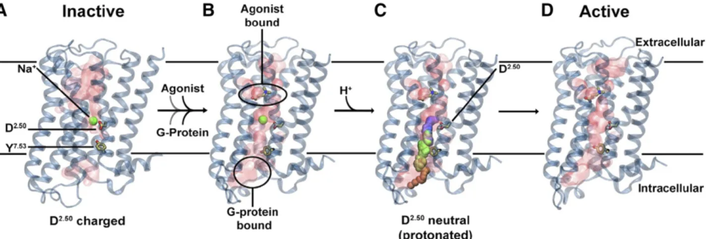
1. Sodium as an Allosteric Cofactor of Class A G-Protein-Coupled Receptor Signaling. The above ev-idence suggests that along with selective NAM effects on agonist binding, physiologic concentrations of Na1 can reduce the basal activity of receptors and, overall, enhance the magnitude of their stimulation by full agonists. These opposing effects of Na1 on agonist binding and signaling response at GPCRs have been characterized with EC50 or KB values in the same 20–100 mM concentration range, and the effects can be abolished by D2.50N or other mutations in the sodium pocket, suggesting a common functional mechanism that involves Na1binding in the conserved pocket. In 2014, we (Katritch et al., 2014) proposed a dynamic mechanism of Na1 as an allosteric cofactor in class A GPCR ligand induced signal transduction. It involves Na1 entrance into the conserved pocket from the extracellular side and along the hydrated channel, which is opened in most class A GPCRs. The extra-cellular entrance of Na1, also observed in all MD simulations (see section V.B below) is also corrobo-rated by the fact that the intracellular side of the pocket of class A GPCRs in an inactive state is sealed by the“hydrophobic layer”right beneath the sodium. Moreover, the intracellular entrance of Na1 is hin-dered by a major electrostatic barrier due to excess of positive charges (as high as 10–15) found at the cytoplasmic side of receptors. It is well established that the presence of the sodium/water cluster in the conserved pocket stabilizes the receptor in the in-active state, reducing its basal activity and reducing the availability of high-affinity binding sites for ago-nists (Chung et al., 2011). Unlike agoago-nists, binding of most antagonists is compatible with Na1 binding and therefore a synergistic stabilization of inactive state by Na1can enhance the affinity of antagonists and inverse agonists.
with electrostatic potential on the plasma membrane, and the reverse transfer against the electrochemical gradient is very unlikely. It was estimated that the transmembrane transfer of Na1 along with gradient would result in;3 kcal gain in energy, and this transfer can be coupled with signal amplification in class A GPCRs observed in presence of Na1.
One of the more recent studies also pointed to possible protonation of D2.50 upon activation, where increased mobility of Na1 in the pocket results in higher pKa of this acidic side chain (Vickery et al., 2018). Such protonation would result in the total disappearance of the barrier for sodium intracellular egress and thus facilitate activation (Fig. 6).
D. Other Potential Functional Effects of the Conserved Sodium Ion Binding
1. Voltage Sensing. Selective transfer of Na1 posi-tive charge through the GPCR transmembrane bundle and coupling of this transfer with receptor activation is likely to make GPCRs sensitive to both sodium concen-tration gradient and the electrostatic potential on the membrane (Ben-Chaim et al., 2006). Several recent studies, indeed, showed that membrane voltage in-creased the sensitivity of the a2Aadrenoreceptor to norepinephrine (Rinne et al., 2013). Activation of another adrenergic receptor, the b1AR, by catechol-amine agonists was also shown to be positively modulated by membrane voltage, while depolariza-tion of membrane dramatically reduced signaling (Birk et al., 2015). Similarly, voltage sensitivity of muscarinic acetylcholine receptors to their full ago-nists was shown for M2, M3, and M5 subtypes (Navarro-Polanco et al., 2011; Rinne et al., 2015). Several studies, including MD-simulations in M2 and the d-opioid receptor (Vickery et al., 2018), suggested that Na1 binding in the sodium pocket may explain
such voltage sensitivity. Limited experimental data from live cell assays, however, have not been conclusive so far. While D2.50 mutations to Ala (Navarro-Polanco et al., 2011) or Asn (Barchad-Avitzur et al., 2016) eliminated gating currents in M2R, voltage sensitivities for agonist binding and conformational changes of the receptor were still present in the mutant (Barchad-Avitzur et al., 2016). This suggests the presence of multiple voltage sensors in muscarinic receptors (Hoppe et al., 2018) and calls for similar assessments of voltage sensitivity in other class A GPCRs, where the effect may be more well defined.
2. pH Dependence. Protonation of D2.50 has been proposed as a facilitator of Na1 egress from class A GPCRs, thus shifting the conformational equilibrium toward their active state and facilitating signaling (Vickery et al., 2018; Hu et al., 2019). This mecha-nism is consistent with in vitro observations that lower pH increases both basal and ligand-induced activation, for example in theb2AR (Ghanouni et al., 2000). This pH dependence may have important phys-iologic consequences because, in addition to classic cell membrane signaling, GPCR have been shown to be signaling for an extended period of time from endo-somes, where pH is dramatically shifted toward an acidic environment (Calebiro et al., 2010; Irannejad et al., 2013; Vilardaga et al., 2014; Godbole et al., 2017; Eichel and von Zastrow, 2018). The conserved Na1site protonation would establish a common mechanism for pH dependence for the majority of class A GPCRs; however, more data and further details of the proton transfer need first to be established.
IV. Ion Binding Sites as Ligand Targets—New Approaches to Design Functional Properties
Beyond their physiologic importance, can the ion binding sites in GPCRs be directly exploited for the
Fig. 6. Updated version of the Na1involvement in GPR activation mechanism. (A) Inactive receptor conformation has an Na1ion bound to D2.50in
a pocket, which is sealed from the cytosol by a hydrophobic layer around Y7.53. (B) G-protein and agonist bind to the receptor, leading to the formation of
a continuous water channel across the GPCR. The increased mobility of the Na1ion results in a pKa shift and subsequent protonation of D2.50. (C)
Neutralization of D2.50and the presence of the hydrated pathway facilitate transfer of Na1to the intracellular side, driven by the TM Na1gradient and
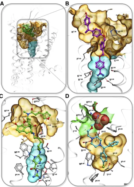
discovery of new ligands with potentially therapeuti-cally relevant properties? Indeed, structure-based anal-ysis of known GPCRs suggests that ion binding sites can be critical for designing both subtype and functionally selective ligands (Fig. 7).
A. Targeting Nonconserved Ion Binding Sites for Subtype Selectivity
Targeting selective ionic interactions can often serve as a beneficial strategy for creating subtype selectivity within closely related subfamily members, with some of such cases being characterized pharmacologically and structurally. One of the examples is the develop-ment of highly selective drugs for the H1 histamine receptor (H1R) (Fig. 7D). While the first generation of antihistamines, including doxepin, were not subtype selective, the crystal structure of the H1R-doxepin complex revealed a phosphate ion tightly bound in the
extracellular loop (ECL) region and coordinated by nonconserved basic side chains K179ELC2and K1915.39 (Shimamura et al., 2011). Docking of the second gener-ation antihistamines like acrivastine, levocetirizine, and fexofenadine showed that H1R selectivity and thus improved safety and pharmacological profile of these drugs can be explained by their acidic carboxy groups mimicking the interactions of the PO432ions.
Interactions with ion binding sites must be taken into consideration in other cases of design of GPCR ligands. The GPR39A, recently identified as Zn21 modulated receptor, presents a good example of ligand identification for an ion-binding receptor. Ligands were discovered by medium-throughput screening assays, which detected selective modulation of Zn21activity on this GPCR39A (Sato et al., 2016). Another interesting example is proton-sensing receptor GPR68, where the proton site has been detected via a combination of molecular modeling and
mutagenesis (Huang et al., 2015b). After initial de-tection of lorazepam as a selective positive allosteric modulator of the proton activation in GPR68, homology modeling and ligand guided optimization approaches were used to develop a model for virtual screening of ;3M available compounds. The screening yielded sev-eral new selective PAMs for GPR68, some of them showing in vivo activity (Huang et al., 2015b). Rapidly improving availability of receptor structures and more relevant templates for structural homology modeling makes such approaches more and more practical in application to other ion-binding GPCRs.
B. Allosteric Ligand Binding in the Conserved Sodium Ion Site
The importance of the conserved Na1 site for the function of class A GPCRs suggests that targeting this site with allosteric or bitopic ligands may be a viable general strategy for modulation of these receptor signal-ing. The volume of the pocket is usually about 150–250 Å3, thus permitting binding of small fragment-like molecules. Indeed, the Na1pocket has been character-ized as a binding site of an antidiuretic drug, sodium channel blocker amiloride, and its derivatives (Liu et al., 2012b). Amilorides are known as negative allosteric modulators (NAMs) of many class A GPCRs, including adenosine (Howard et al., 1987; Leppik et al., 2000; Gao et al., 2003a,b, 2011; Gutiérrez-de-Terán et al., 2013), dopamine (Neve, 1991; Hoare et al., 2000), muscarinic (Dehaye and Verhasselt, 1995), 5-HT (Pauwels, 1997), GnRHR (Heitman et al., 2008), and potentially many more receptors, as summarized in Katritch et al. (2014). Various affinity estimates for amiloride derivatives show KB raging from ;1 to 50 mM in their negative allosteric modulation of orthosteric ligand binding. Docking of amiloride and a bulkier derivative HMA (5-(N,N-hexamethylene)amiloride) (Gutiérrez-de-Terán et al., 2013) shows that the positively charged guanidine moiety of the ligand forms a salt bridge to the D2.50 carboxyl, while the bulkier N5 substituents point toward the orthosteric site. The induced docking and confor-mational modeling also suggested that the fitting of amilorides, especially HMA into the pocket, requires substantial expansion of the pocket, which manifested in adjustments in the N7.45, N7.49, and especially W6.48 side chains. Accordingly, mutations of these residues to alanine in this study only improved affinity of amiloride and HMA severalfold, suggesting that amiloride might not be the optimal chemotype for targeting the pocket. On the chemistry side, a number of additional amiloride derivatives with longer 5N substitutes were character-ized in a recent study (Massink et al., 2016), showing that extension of the allosteric ligand into the orthos-teric pocket is possible without major reduction of the binding affinity.
Another small molecule characterized recently as an allosteric Na1 pocket binder in the BLT1 receptor is
benzamidine (Hori et al., 2018), though its affinity (KB) was estimated much lower than amiloride at;500mM, making it only ;10 times more potent than Na1 ion itself (based on Fig. 4A in Hori et al., 2018). The study revealed binding of benzamidine and its NAM effect on G-protein activity in two very different receptors, the BLT1 receptor andb1AR, suggesting that it likely binds at the sodium ion binding site in other class A GPCRs as well.
Intriguingly, because Li1 can compete with Na1 in the conserved pocket (Pert et al., 1973), some studies hypothesized that effects of Li1on functional properties of GPCRs can be implicated in physiologic and psycho-active effects of the Li1 (Dudev et al., 2018). The Li1 effect as a competitor to Na1 binding is especially intriguing because lithium is widely used in treatment of bipolar disorders; however, more evidence is needed to establish the GPCR mode of action of Li1, as this ion can also impart a central nervous system effect via ion channel modulation.
C. Targeting Sodium Ion with Bitopic Ligands
1. Concept of Bitopic Ligands. The highly conserved nature and small size of the sodium pocket itself limit its selectivity, and therefore, the practical utility of small ligands like amilorides and benzamidines as allosteric modulators. On the other hand, a combination of high affinity selective orthosteric moieties with the unique functional properties of the Na1 site allosteric binders could make bitopic ligands an attractive target for ligand design. One recently characterized example of such a bitopic ligand is benzamidine-containing ligand BIIL260 found in the BTL1 receptor structure (Hori et al., 2018). By reaching into the Na1site and forming a salt bridge with D2.50carboxyl, as well as hydrogen bonds to S3.39and S7.45, the positively charged benza-midine moiety is expected to block activation related changes. In agreement with this prediction, BIIL260 was characterized as an inverse agonist, completely blocking the basal activity of the receptor. There are several other benzamidine-containing compounds for BLT1 predicted to bind in a similar manner (Hori et al., 2018).