Cover Page
The handle http://hdl.handle.net/1887/66265 holds various files of this Leiden University dissertation.
Author: Gao, Y.
Design and Application of Dextran Based
Cross-linked Networks
Proefschrift
ter verkrijging van
de graad van Doctor aan de Universiteit Leiden,
op gezag van Rector Magnificus Prof. mr. C.J.J.M. Stolker,
volgens besluit van het College voor Promoties
te verdedigen op donderdag 18 oktober 2018
klokke 10:00 uur
door
Yue Gao
Promotiecommissie:
Promoter:
Prof. dr Alexander Kros
Co-promotor:
Dr. Roxanne Kieltyka
Overige leden:
Prof. Dr. H.S. Overkleeft (voorzitter)
Prof. Dr. J. Brouwer (secretaris)
Prof. Dr. J. Bouwstra
Prof. Dr. J van der Gucht, Wageningen Universiteit
TABLE
OF
CONTENTS
CHAPTER 1 General introduction 5
CHAPTER 2 Thiolated human serum albumin crosslinked dextran hydrogel as a macroscale delivery system
19
CHAPTER 3 Dual-crosslinked human serum albumin-polymer hydrogels
for affinity-based drug delivery 37
CHAPTER 4 Zebrafish embryo toxicity assay using a cyclodextrin-modified dextran-PEG polymeric drug carrier 61 CHAPTER 5 Chemically crosslinked polymer networks assist the
formation of giant unilamellar vesicles
81
CHAPTER 6 The effect of coiled-coil peptide conjugation on dextran
self-assembly 107
CHAPTER 7 Summary and perspectives 137
Samenvatting 143
List of abbreviations 147
Curriculum Vitae 149
C
HAPTER
1
Chapter 1
7
D
EXTRAN
Polymers such as proteins and polysaccharides, abundantly exist in nature and are major components of plants, animals and micro-organisms.1 Polysaccharides are polymers composed of monosaccharide units connected through glycosidic bonds.2, 3 The glycosidic bonds are formed by a condensation reaction between the anomeric carbon (C1) of one monosaccharide unit and an oxygen atom from a hydroxyl group on another monosaccharide unit. Since hydroxyl groups present on C2, C3, C4 and C6 carbons, the generated glycosidic bonds can be 1→2, 1→3, 1→4, or 1→6 linked providing linear to highly branched chains. Polysaccharides can be categorized into two groups: (1) mammalian polysaccharides such as hyaluronan, chondroitin sulfate, and heparin; (2) non-mammalian polysaccharides such as alginate, chitin, and dextran (Scheme 1).
Scheme 1. Main structures of repeating units of some polysaccharides widely investigated in biomaterials applications. Top row: mammalian polysaccharides; bottom row: nonmammalian polysaccharides.
Dextran is a neutral and hydrophilic polysaccharide consisting of an α-(1→6) linked D-glucose main chain with a relatively low level of α-(1→3) branched linkages.4, 5 The molecular weight of dextran chains can vary from 1 to 2 000 kilodaltons (kDa). Although physiochemical properties of each dextran polymer depend on its particular structure (e.g. molecular weight and degrees of branching), the glycosidic bonds of dextran provide relatively high chain mobility and solubility, while the three pendant hydroxyl groups provide handles for its functionalization.6-8 Moreover, dextran is biologically inert due to the α-(1→6) glycosidic bonds making it resistant to cleavage by most endogenous cellular glycosidases, but it is degradable by dextranase9 which exists in mammalian (including human) tissues. Dextran is also non-immunogenic,10-14 making it an attractive polymer for the design of novel biomaterials.
Chapter 1
8
(Macrodex®) and 40 kDa (Rheomacrodex®) for plasma expansion.15-17 Furthermore fluorescent dye-dextran conjugates (Molecular Probes®) are widely applied in numerous biological studies.18 Dextran can also be crosslinked by epichlorohydrin to form hydrogel beads, which are used for separations of molecules based on differences in molecular weight (e.g. Sephadex®).19 Dextran-based materials are also used in wound healing (e.g. Debrisan®).20
Due to their hydrophilicity, long shelf-life, relative stability, biocompatibility and biodegradable properties, dextrans have been studied as a starting material for drug delivery carriers. Cargo that was delivered in these studies included low molecular weight drugs, proteins/enzymes, and imaging agents by either direct attachment or through a linker since the 1980s.11, 21-23 Degradation of linkages between dextran and the conjugated drug results in the delivery of therapies. Dextran conjugation was also used to overcome problems such as limited solubility of the drug, short plasma half-life high toxicity and/or undesired interactions with tissues. For example, the solubility of paclitaxel increased >2000 fold upon conjugation to aminated dextran (Mw = 70 000). Furthermore, modification of the dextran-paclitaxel conjugates with folate increased the uptake 2-3 folds in human oral carcinoma KB cells.24 The in vivo biodistribution and circulation time can also be altered upon dextran conjugation. For example, prolonged circulation time and decreased uptake in kidney were observed for dextran-mouse epidermal growth factor (mEGF) conjugates, wherein mEGF was attached to aldehyde group functionalized dextran polymers via the formation of imine bonds which was further stabilized by reducing to secondary amide bonds with NaCNBH3.25, 26
H
YDROGELS
Hydrogels are a class of soft materials that possess a hydrophilic three-dimensional (3D) network hold together via chemical and/or physical bonds (i.e. crosslinks). Upon contact with water, the 3D-network swells and the (physically) entrapped molecules diffuse out of the network and the kinetics of release depend on both the properties of the encapsulated molecule as well as the physical characteristics of the hydrogel. The viscoelastic properties are similar to soft tissues and combined with the water-rich environment making these hydrogels an attractive biomaterial with potential applications in the fields of tissue engineering and regenerative medicine.27, 28 These hydrogels can be synthesized from various precursors over a large molecular weight range. In this thesis the formation of hydrogels comprised of dextran covalently crosslinked by proteins is described.
Chapter 1
9
period. To this end, extensively applied chemoselective ligations for in situ gelation include Michael addition,32 azide-alkyne Click reactions,33, 34 Schiff base formation35 and disulfide exchange.36 Moreover, physical crosslinking methods with suitable gelation kinetics can also be used to form
in situ hydrogels.37 The physical crosslinks can consist of hydrogen bonds, hydrophobic interactions, π-π interactions or host-guest inclusion, to endow the hydrogels with dynamic, reversible and self-healing properties.38
Hydrogels have been studied as a biomaterial for the localized and sustained release of therapeutic agents in order to decrease the number of drug administrations, to prevent rapid drug degradation or to prolong therapeutic drug concentrations.39 Typical drug release mechanisms applied in drug delivery systems include diffusion controlled drug release40, degradation controlled drug release,41 stimuli triggered drug release,42 and affinity-based drug release. The affinity-based drug release mechanism uses non-covalent interactions between the therapeutic agents and hydrogel platform, such as electrostatic, hydrophilic-hydrophobic, hydrogen bonding, or van der Waals interactions, to control the release of the therapeutic.43 These materials can improve drug loading, stability and release. Affinity-based carriers include specific host molecules such as cyclodextrin,44-48 serum albumin49-52 and heparin53-55, or cavities formed within the solid polymer material by the molecular imprinting technique.56, 57
D
EXTRAN GELS
Since the 1990s, hydrogels composed of dextran have been developed for macroscale therapeutics delivery and 3D cell culture.58, 59 Chemically crosslinked dextran hydrogels can be prepared from a functionalized dextran precursor. For example, Hennink and co-workers prepared degradable dextran hydrogels by free radical polymerization of aqueous solutions of glycidyl methacrylated dextran or hydroxyethyl methacrylated dextran for the delivery of proteins.9, 60-62 Liu et al. used UV-light exposure to initiate the crosslinking between dextran functionalized with both methacrylate and lysine groups and methacrylamide modified gelatin in presence of a photoinitiator and cells to obtain a 3D smooth muscle cell culture.63
Chapter 1
10
and α-cyclodextrins which was crosslinked by the inclusion complex formation between poly(ethylene glycol) and α-cyclodextrins.66 De Jong et al. established another thermo-reversible hydrogel system with L-oligo(lactic acid) grafted dextran and D-oligo(lactic acid) grafted dextran which was crosslinked by the stereocomplex formation between enantiomers.67
Besides macroscopic dextran hydrogels, various emulsification approaches were applied to obtain chemically crosslinked dextran microgels (also called as microspheres or microcapsules) and nanogels. Using a cyclohexane inverse miniemulsion, crosslinked nanogels with an averaged diameter below 100 nm were prepared starting from alkyne-dextran and azide-dextran.68 To avoid the use of organic solvents, “water-in-water” emulsions were used to synthesize microgels. Aqueous mixtures of dextran and polymers such as polyethylene glycol (PEG) or poly(vinyl alcohol) (PVA) can form an aqueous two-phase system at elevated concentrations, which can be emulsified to obtain a water-in-water emulsion. De Geest et al. emulsified an aqueous solution containing dextran azide and dextran alkyne in an external aqueous PEG phase. CuSO4 and sodium ascorbate were added afterward to obtain dextran microgels via Huisgen click reaction.69 Similarly, Ghugare et al. prepared dextran microgels with an averaged diameter around 1000 nm using ultrasound assisted “water-in-water” emulsification. The emulsion contained dextran methacrylate, photoinitiator and PVA was crosslinked under UV-light afterwards.70
Additionally, charged dextrans, such as dextran sulfate, dextran phosphate and dextran amine, have been used to prepare polyelectrolyte complex. For example, positively or negatively charged colloids were obtained from non-stoichiometric polyelectrolyte complexes of dextran sulfate and chitosan.71 Dextran sulfate and protamine were constructed to microcapsules for enzyme delivery by a layer-by-layer approach using a removable melamine formaldehyde template.72 Complexes formed by cationized dextran amine and plasmid DNA enhanced gene expression in vitro, showing its potentials for gene delivery.73
G
ENERAL OUTLINE OF THE THESIS
In the present thesis, several dextran-based hydrogel systems are synthesized and characterized for biomedical applications. To prepare dextran polymers for crosslinking, the backbone is initially functionalized with maleimides or vinyl sulfones, which are able to form covalent bonds with nucleophilic thiols via a thiol-Michael addition reaction. These reactive groups (i.e. maleimides or vinyl sulfones) were coupled to the hydroxyl groups of dextran via an ester or an ether linkage. The stability of linkages strongly affected the properties and applications of the prepared hydrogel systems as described in the following chapters.
Chapter 1
11
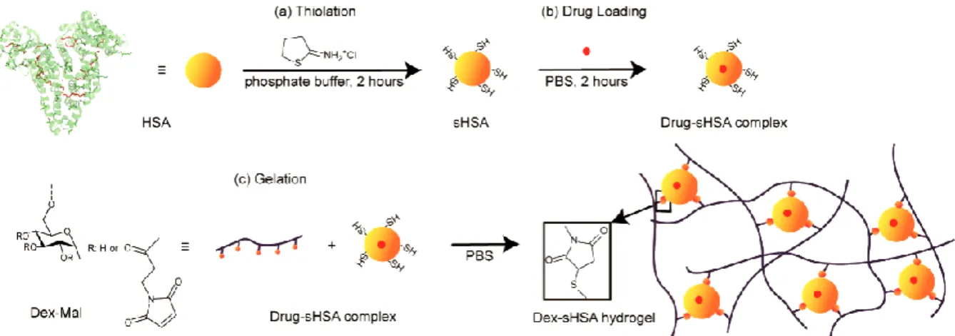
types of (hydrophobic) low molecular weight molecules. Inspired by this natural carrier, a covalently crosslinked dextran-albumin hydrogel system was developed in Chapter 2 to explore the potential of HSA as a hydrophobic-drug cavity and crosslinker for affinity-based drug delivery. Native HSA was modified with 2-iminothiolane (2-IT) to introduce thiol groups at the lysine residues. Circular dichroism spectroscopy revealed that the conformation of thiolated HSA (sHSA) is mostly maintained showing that sHSA was capable of simultaneously being a drug carrier and the crosslinker in the hydrogel system. The hydroxyl groups of dextran were modified with maleimide groups through the formation of ester linkages resulting in functionalized dextran (i.e. Dex-Mal). Next, sHSA was added to Dex-Mal resulting in gelation of the mixture under physiological conditions in a relatively fast manner. The resulting dextran-albumin ( Dex-Mal-sHSA) hydrogel was characterized by oscillatory rheology experiments. In vitro release profiles of three hydrophobic drugs (i.e., ibuprofen, paclitaxel and dexamethasone) from the dextran-albumin hydrogel were recorded, showing high drug loading efficiency and demonstrating sustained release of the corresponding guest molecule.
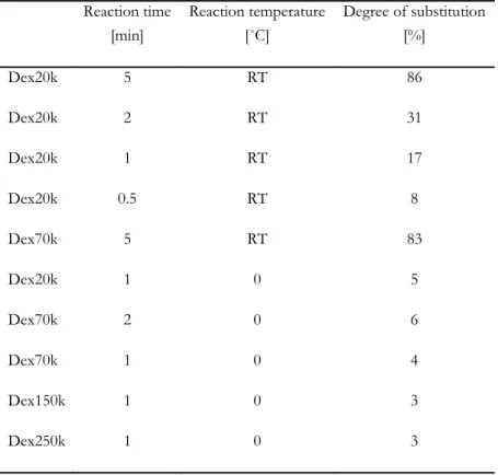
To improve the stability and to gain control over the properties of the abovementioned dextran-albumin hydrogel system (Dex-Mal-sHSA), two new affinity-based drug delivery hydrogel systems were developed in Chapter 3. Firstly, a fraction of the dextran hydroxyl groups were modified with vinyl sulfone. The effect of reaction temperature, reaction time and dextran molecular weight was studied to control degree of vinyl sulfone modification of dextran (i.e. Dex-VS). In comparison to the Dex-Mal polymer in Dex-Mal-sHSA hydrogels used in Chapter 2, the obtained vinyl sulfone functionalized dextran polymers (Dex-VS) showed to be more stable against hydrolysis. Next, a new dextran-albumin hydrogel system (Dex(VS)-sHSA) with improved stability was developed by reacting Dex-VS with sHSA. However, the vinyl sulfone groups show relatively slower reaction kinetics towards thiol groups than the maleimide groups. To accelerate the gelation process while maintaining the concentration of albumin constant, a third macromolecular precursor, poly(ethylene glycol) dithiol (PEG-DT), was introduced into the dextran-albumin hydrogel. By balancing the number of reactive vinyl sulfone groups from Dex-VS and thiol groups from both sHSA and PEG-DT for the thiol-Michael addition reaction, the covalently dual-crosslinked
Chapter 1
12
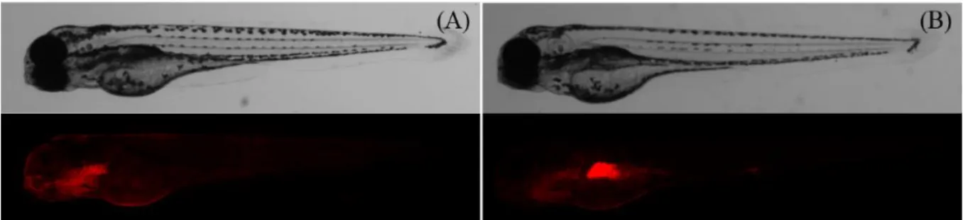
Cyclodextrins are able to form a reversible host-guest complex with suitable hydrophobic molecules. In Chapter 4 the potential of cyclodextrin-modified dextran based drug carriers in a zebrafish embryo toxicity assay was explored. Zebrafish embryos have become a promising model organism for developmental and reproductive toxicity screening studies.17, 19, 74 However, ineffective uptake of certain compounds by zebrafish embryos was reported raising concerns of the predictive capacity of zebrafish embryo based toxicity assays.75 It was reported that zebrafish embryos show a weaker than expected sensitivity for neurotoxic compounds with respect to acute toxicity.76 Here, a previously reported -cyclodextrin decorated dextran-poly(ethylene glycol) Dex-CD/PEG drug carrier system45 was used to enhance the uptake of the model drug valproate in the zebrafish embryo toxicity assay. The polymeric drug carrier was synthesized by crosslinking maleimide modified dextran with poly(ethylene glycol) dithiol, wherein cyclodextrins were conjugated to the dextran backbone to ensure binding of valproate through the formation of a non-covalent host-guest complex. The uptake of fluorescent labelled Dex-CD/PEG drug carriers into the gastrointestinal tract of zebrafish embryos was observed by fluorescent microscopy. Comparison of zebrafish embryos exposed to valproate or valproate loaded Dex-CD/PEG carrier showed that the Dex-CD/PEG carrier improves the valproate uptake via the gastrointestinal tract. Correlations between the composition of Dex-CD/PEG carriers and the viability of 4-dpf zebrafish embryos after 48-hour exposure to valproate loaded Dex-CD/PEG carrier were also studied to investigate the role of Dex-CD/PEG carriers in the toxicity assay.
In Chapter 5, the capacity of hydrogels to guide the formation of giant unilamellar vesicles is explored. Giant unilamellar vesicles (GUVs) are widely used cell membrane model systems for biophysical measurements.77-79 Recently, it was reported that GUVs can be prepared through hydration of a lipid film on a hydrogel consisting of agarose, which was more straightforward and rapid to generate GUVs in solutions of physiologic ionic strength.80 However, using agarose for the hydrogel film results in contamination of the vesicles due to its dissolution during the hydration process. To address this issue, the chemically crosslinked polymer system Dex-Mal-PEG was covalently anchored to a glass slide as a substrate for GUV formation. Using this method, polymer and additive-free GUVs can be prepared rapidly in high yield under physiological ionic strength conditions. Moreover, by varying physiochemical properties of Dex-Mal-PEG hydrogels through controlling the molar ratio of the maleimide and thiol groups, the molecular weight of PEG-DT and the degrees of substitution of Dex-Mal and PEG-DT, the effect of the network on the yield and size distribution of the prepared GUVs was investigated.
In Chapter 6, an attempt was made to direct the assembly of dextran polymers via specific coiled-coil interactions. The coiled-coiled-coiled-coil motif is one of the basic folding motifs in natural proteins, which is a left-handed superhelix formed through the winding of two or more right-handed α-helical peptides around each other.81, 82 Peptide E (amino acid sequence: (EIAALEK)
Chapter 1
13
to dextran-vinyl sulfone polymers to obtain multivalent peptide-dextran conjugates. Two pairs of dextran-peptide E and dextran-peptide K bioconjugates were synthesized, wherein the dextran polymer was attached at either the N- or C-terminus of the peptides. The effect of conjugation on peptide conformation and the interaction between complementary dextran-peptide conjugates was studied by circular dichroism spectroscopy, fluorescence spectroscopy and dynamic light scattering measurements.
Chapter 1
14
R
EFERENCES
1. Malafaya P. B., Silva G. A. and Reis R. L., Natural-origin polymers as carriers and scaffolds for biomolecules and cell delivery in tissue engineering applications, Advanced Drug Delivery Reviews, 2007, 59, 207-233. 2. Yang L. and Zhang L., Chemical structural and chain conformational characterization of some bioactive
polysaccharides isolated from natural sources, Carbohydrate Polymers, 2009, 76, 349-361.
3. Mizrahy S. and Peer D., Polysaccharides as building blocks for nanotherapeutics, Chemical Society Reviews, 2012,
41, 2623-2640.
4. Heinze T., Liebert T., Heublein B., et al., Functional polymers based on dextran, Advances in Polymer Science, 2006, 205, 199-291.
5. Purama R. K., Goswami P., Khan A. T., et al., Structural analysis and properties of dextran produced by leuconostoc mesenteroides nrrl b-640, Carbohydrate Polymers, 2009, 76, 30-35.
6. Ioan C., Aberle T. and Burchard W., Structure properties of dextran. 2. Dilute solution, Macromolecules, 2000,
33, 5730-5739.
7. Ioan C., Aberle T. and Burchard W., Structure properties of dextran. 3. Shrinking factors of individual clusters,
Macromolecules, 2001, 34, 3765-3771.
8. Antoniou E. and Tsianou M., Solution properties of dextran in water and in formamide, Journal of Applied Polymer Science, 2012, 125, 1681-1692.
9. Franssen O., Vos O. P. and Hennink W. E., Delayed release of a model protein from enzymatically-degrading dextran hydrogels, Journal of Controlled Release, 1997, 44, 237-245.
10. Larsen C., Dextran prodrugs—structure and stability in relation to therapeutic activity, Advanced Drug Delivery Reviews, 1989, 3, 103-154.
11. Mehvar R., Dextrans for targeted and sustained delivery of therapeutic and imaging agents, Journal of Controlled Release, 2000, 69, 1-25.
12. Heinze T., Liebert T., Heublein B., et al., in Polysaccharides ii, Springer, 2006, pp. 199-291.
13. Khandare J. and Minko T., Polymer–drug conjugates: Progress in polymeric prodrugs, Progress in Polymer Science, 2006, 31, 359-397.
14. Naessens M., Cerdobbel A., Soetaert W., et al., Leuconostoc dextransucrase and dextran: Production, properties and applications, J Chem Technol Biot, 2005, 80, 845-860.
15. Hill A. J., Teraoka H., Heideman W., et al., Zebrafish as a model vertebrate for investigating chemical toxicity,
Toxicol Sci, 2005, 86, 6-19.
16. Peterson R. E., Theobald H. M. and Kimmel G. L., Developmental and reproductive toxicity of dioxins and related compounds: Cross-species comparisons, Critical Reviews in Toxicology, 1993, 23, 283-335.
17. Strahle U., Scholz S., Geisler R., et al., Zebrafish embryos as an alternative to animal experiments. A commentary on the definition of the onset of protected life stages in animal welfare regulations, Reproductive Toxicology, 2012, 33, 128-132.
18. Mccollum C. W., Ducharme N. A., Bondesson M., et al., Developmental toxicity screening in zebrafish, Birth Defects Research Part C-embryo Today-reviews, 2011, 93, 67-114.
19. He J., Gao J., Huang C., et al., Zebrafish models for assessing developmental and reproductive toxicity,
Neurotoxicology and Teratology, 2014, 42, 35-42.
20. Basu S. and Sachidanandan C., Zebrafish: A multifaceted tool for chemical biologists, Chemical reviews, 2013,
113, 7952-7980.
Chapter 1
15
22. Wasiak I., Kulikowska A., Janczewska M., et al., Dextran nanoparticle synthesis and properties, PLOS ONE, 2016, 11.
23. Varshosaz J., Dextran conjugates in drug delivery, Expert Opinion on Drug Delivery, 2012, 9, 509-523.
24. Nakamura J., Nakajima N., Matsumura K., et al., Water-soluble taxol conjugates with dextran and targets tumor cells by folic acid immobilization, Anticancer Research, 2010, 30, 903-909.
25. Zhao Q., Gottschalk I., Carlsson J., et al., Preparation and purification of an end to end coupled megf-dextran conjugate, Bioconjugate Chemistry, 1997, 8, 927-934.
26. Zhao Q., Tolmachev V., Carlsson J., et al., Effects of dextranation on the pharmacokinetics of short peptides. A pet study on megf, Bioconjugate Chemistry, 1999, 10, 938-946.
27. Hoffman A. S., Hydrogels for biomedical applications, Annals of the New York Academy of Sciences, 2001, 944, 62-73.
28. Cabral J. D. and Moratti S. C., Hydrogels for biomedical applications, Future Medicinal Chemistry, 2011, 3, 1877-1888.
29. Van Tomme S. R., Storm G. and Hennink W. E., In situ gelling hydrogels for pharmaceutical and biomedical applications, International Journal of Pharmaceutics, 2008, 355, 1-18.
30. Ko D. Y., Shinde U. P., Yeon B., et al., Recent progress of in situ formed gels for biomedical applications,
Progress in Polymer Science, 2013, 38, 672-701.
31. Nguyen Q. V., Huynh D. P., Park J. H., et al., Injectable polymeric hydrogels for the delivery of therapeutic agents: A review, European Polymer Journal, 2015, 72, 602-619.
32. Zhou Y., Nie W., Zhao J., et al., Rapidly in situ forming adhesive hydrogel based on a peg-maleimide modified polypeptide through michael addition, Journal of Materials Science: Materials in Medicine, 2013, 24, 2277-2286. 33. Jiang Y., Chen J., Deng C., et al., Click hydrogels, microgels and nanogels: Emerging platforms for drug
delivery and tissue engineering, Biomaterials, 2014, 35, 4969-4985.
34. Nimmo C. M. and Shoichet M. S., Regenerative biomaterials that "click": Simple, aqueous-based protocols for hydrogel synthesis, surface immobilization, and 3d patterning, Bioconjugate Chemistry, 2011, 22, 2199-2209. 35. Wu X., He C., Wu Y., et al., Synergistic therapeutic effects of schiff's base cross-linked injectable hydrogels for local co-delivery of metformin and 5-fluorouracil in a mouse colon carcinoma model, Biomaterials, 2016,
75, 148-162.
36. Canadell J., Goossens H. and Klumperman B., Self-healing materials based on disulfide links, Macromolecules, 2011, 44, 2536-2541.
37. Du X., Zhou J., Shi J., et al., Supramolecular hydrogelators and hydrogels: From soft matter to molecular biomaterials, Chemical Reviews, 2015, 115, 13165-13307.
38. Kim D. Y., Kwon D. Y., Kwon J. S., et al., Stimuli-responsive injectable in situ-forming hydrogels for regenerative medicines, Polymer Reviews, 2015.
39. Buwalda S. J., Vermonden T. and Hennink W. E., Hydrogels for therapeutic delivery: Current developments and future directions, Biomacromolecules, 2017, 18, 316-330.
40. Siepmann J. and Siepmann F., Modeling of diffusion controlled drug delivery, Journal of Controlled Release, 2012,
161, 351-362.
41. Dubose J. W., Cutshall C. and Metters A. T., Controlled release of tethered molecules via engineered hydrogel degradation: Model development and validation, Journal of Biomedical Materials Research Part A, 2005, 74, 104-116.
42. Roy D., Cambre J. N. and Sumerlin B. S., Future perspectives and recent advances in stimuli-responsive materials, Progress in Polymer Science, 2010, 35, 278-301.
Chapter 1
16
44. Zhou J. and Ritter H., Cyclodextrin functionalized polymers as drug delivery systems, Polymer Chemistry, 2010,
1, 1552-1559.
45. Peng K., Cui C., Tomatsu I., et al., Cyclodextrin/dextran based drug carriers for a controlled release of hydrophobic drugs in zebrafish embryos, Soft Matter, 2010, 6, 3778-3783.
46. Peng K., Tomatsu I., Korobko A. V., et al., Cyclodextrin-dextran based in situ hydrogel formation: A carrier for hydrophobic drugs, Soft Matter, 2010, 6, 85-87.
47. Liu C., Zhang Z., Liu X., et al., Gelatin-based hydrogels with [small beta]-cyclodextrin as a dual functional component for enhanced drug loading and controlled release, RSC Advances, 3, 25041-25049.
48. Cai T., Yang W. J., Zhang Z., et al., Preparation of stimuli-responsive hydrogel networks with threaded [small beta]-cyclodextrin end-capped chains via combination of controlled radical polymerization and click chemistry, Soft Matter, 8, 5612-5620.
49. Kratz F., Albumin as a drug carrier: Design of prodrugs, drug conjugates and nanoparticles, Journal of Controlled Release, 2008, 132, 171-183.
50. Cui M., Naczynski D. J., Zevon M., et al., Multifunctional albumin nanoparticles as combination drug carriers for intra-tumoral chemotherapy, Advanced Healthcare Materials, 2013, n/a-n/a.
51. Wu Y., Ihme S., Feuring-Buske M., et al., A core-shell albumin copolymer nanotransporter for high capacity loading and two-step release of doxorubicin with enhanced anti-leukemia activity, Advanced Healthcare Materials, 2012, n/a-n/a.
52. Tada D., Tanabe T., Tachibana A., et al., Drug release from hydrogel containing albumin as crosslinker, Journal of Bioscience and Bioengineering, 2005, 100, 551-555.
53. Nie T., Akins R. E., Jr. and Kiick K. L., Production of heparin-containing hydrogels for modulating cell responses, Acta Biomaterialia, 2009, 5, 865-875.
54. Baldwin A. D., Robinson K. G., Militar J. L., et al., In situ crosslinkable heparin-containing poly(ethylene glycol) hydrogels for sustained anticoagulant release, Journal of Biomedical Materials Research Part A, 100A, 2106-2118. 55. Kiick K. L., Peptide- and protein-mediated assembly of heparinized hydrogels, Soft Matter, 2008, 4, 29-37. 56. Byrne M. E., Park K. and Peppas N. A., Molecular imprinting within hydrogels, Advanced Drug Delivery Reviews,
2002, 54, 149-161.
57. Chen L., Xu S. and Li J., Recent advances in molecular imprinting technology: Current status, challenges and highlighted applications, Chemical Society Reviews, 2011, 40, 2922-2942.
58. Baldwin A. D. and Kiick K. L., Polysaccharide-modified synthetic polymeric biomaterials, Biopolymers, 2010,
94, 128-140.
59. Zelzer M., Todd S. J., Hirst A. R., et al., Enzyme responsive materials: Design strategies and future developments, Biomaterials Science, 2013, 1, 11-39.
60. Hennink W. E., Talsma H., Borchert J. C. H., et al., Controlled release of proteins from dextran hydrogels,
Journal of Controlled Release, 1996, 39, 47-55.
61. Hennink W. E., Franssen O., Van Dijkwolthuis W. N. E., et al., Dextran hydrogels for the controlled release of proteins, Journal of Controlled Release, 1997, 48, 107-114.
62. Van Dijkwolthuis W. N. E., Hoogeboom J. A. M., Van Steenbergen M. J., et al., Degradation and release behavior of dextran-based hydrogels, Macromolecules, 1997, 30, 4639-4645.
63. Liu Y. and Chanpark M. B., A biomimetic hydrogel based on methacrylated dextran-graft-lysine and gelatin for 3d smooth muscle cell culture, Biomaterials, 2010, 31, 1158-1170.
64. Hiemstra C., Der Aa L. J. V., Zhong Z., et al., Rapidly in situ-forming degradable hydrogels from dextran thiols through michael addition, Biomacromolecules, 2007, 8, 1548-1556.
Chapter 1
17
reaction under physiological conditions, Macromolecular Rapid Communications, 2013, 34, 1464-1470.
66. Huh K. M., Ooya T., Lee W. K., et al., Supramolecular-structured hydrogels showing a reversible phase transition by inclusion complexation between poly(ethylene glycol) grafted dextran and α-cyclodextrin,
Macromolecules, 2001, 34, 8657-8662.
67. De Jong S. J., Van Eerdenbrugh B., Van Nostrum C. F., et al., Physically crosslinked dextran hydrogels by stereocomplex formation of lactic acid oligomers: Degradation and protein release behavior, Journal of Controlled Release, 2001, 71, 261-275.
68. Heller D. A., Levi Y., Pelet J. M., et al., Modular ‘click-in-emulsion’ bone-targeted nanogels, Advanced Materials, 2013, 25, 1449-1454.
69. De Geest B. G., Van Camp W., Du Prez F. E., et al., Biodegradable microcapsules designed via 'click' chemistry, Chemical Communications, 2008, 190-192.
70. Ghugare S. V., Chiessi E., Cerroni B., et al., Biodegradable dextran based microgels: A study on network associated water diffusion and enzymatic degradation, Soft Matter, 2012, 8, 2494-2502.
71. Drogoz A., David L., Rochas C., et al., Polyelectrolyte complexes from polysaccharides : Formation and stoichiometry monitoring, Langmuir, 2007, 23, 10950-10958.
72. Balabushevich N. G., Sukhorukov G. B. and Larionova N. I., Polyelectrolyte multilayer microspheres as carriers for bienzyme system: Preparation and characterization, Macromolecular Rapid Communications, 2005, 26, 1168-1172.
73. Jo J., Nagane K., Yamamoto M., et al., Effect of amine type on the expression of plasmid DNA by cationized dextran, Journal of Biomaterials Science-polymer Edition, 2010, 21, 225-236.
74. Parliament E., Directive 2010/63/eu of the european parliament and of the council of 22 september 2010 on the protection of animals used for scientific purposes, 2010, 34-35.
75. Berghmans S., Butler P., Goldsmith P., et al., Zebrafish based assays for the assessment of cardiac, visual and gut function — potential safety screens for early drug discovery, Journal of Pharmacological and Toxicological Methods, 2008, 58, 59-68.
76. Selderslaghs I. W. T., Hooyberghs J., Blust R., et al., Assessment of the developmental neurotoxicity of compounds by measuring locomotor activity in zebrafish embryos and larvae, Neurotoxicology and Teratology, 2013, 37, 44-56.
77. Menger F. M. and Angelova M. I., Giant vesicles: Imitating the cytological processes of cell membranes,
Accounts of chemical research, 1998, 31, 789-797.
78. Walde P., Cosentino K., Engel H., et al., Giant vesicles: Preparations and applications, ChemBioChem, 2010, 11, 848-865.
79. Fenz S. F. and Sengupta K., Giant vesicles as cell models, Integrative Biology, 2012, 4, 982-995.
80. Horger K. S., Estes D. J., Capone R., et al., Films of agarose enable rapid formation of giant liposomes in solutions of physiologic ionic strength, Journal of the American Chemical Society, 2009, 131, 1810-1819.
81. Lupas A., Van Dyke M. and Stock J., Predicting coiled coils from protein sequences, Science, 1991, 252, 1162-1164.
C
HAPTER
2
T
HIOLATED
H
UMAN
S
ERUM
A
LBUMIN
C
ROSSLINKED
D
EXTRAN
H
YDROGEL AS
A
M
ACROSCALE
D
ELIVERY
S
YSTEM
Hydrogels play an important role in macroscale delivery systems by enabling the transport of cells and molecules. Here we present a facile and benign method to prepare a dextran-based hydrogel (Dex-sHSA) using human serum albumin (HSA) as a simultaneous drug carrier and covalent crosslinker. Drug binding affinity of the albumin protein was conserved in the thiolation step using 2-iminothiolane and subsequently, in the in situ gelation step. Oscillation rheometry studies confirmed the formation of a three-dimensional viscoelastic network upon reaction of dextran and the HSA protein. The mechanical properties of Dex-sHSA hydrogel can be tuned by the protein concentration, and the degree of thiolation of sHSA. Sustained release of hydrophobic drugs, such as ibuprofen, paclitaxel and dexamethasone, from the Dex-sHSA network was shown over one week. Hence, this albumin-based dextran hydrogel system demonstrates its potential as a macroscale delivery system of hydrophobic therapeutics for a wide range of biomedical applications.
Chapter 2
21
I
NTRODUCTION
Hydrogels are composed of crosslinked hydrophilic polymer networks, resulting in porous, macroscopic materials of high water content. Their unique physicochemical properties enable them to perform as macroscale delivery systems of bioactive agents ranging from cells to molecules.1, 2 Within such systems, methods to gain spatiotemporal control over release of therapeutic payloads have involved diffusion,3-7 swelling,8-10 degradation,11-13 light14-19 or drug-carrier affinity interactions.20 More specifically, the latter mechanism facilitates controlled release through the strong and reversible association of the drug molecules using non-covalent interactions such as hydrogen bonding, ionic, van der Waals, and hydrophobic interactions within the macroscale material scaffold. In the context of lipophilic drug delivery, the problem of encapsulating a lipophilic drug within a water-rich environment is resolved through the inclusion of hydrophobic side chains,21, 22 hydrophobic drug carriers,23 such as cyclodextrin,24-29 within the polymer or using a molecular imprinting approach30, 31 to prepare the polymeric scaffold. Since it is estimated that 40% of new active chemical entities discovered by pharmaceutical companies are poorly water soluble (aqueous solubility less than 10 µM),32 macroscale drug delivery systems capable of transporting hydrophobic therapeutics are highly relevant.
Inspired by nature, human serum albumin (HSA) is emerging as an attractive drug carrier in biomedical materials.33 HSA is the most abundant plasma protein in human (35-50 g L-1) with a molecular weight of 66.5 kDa and has been shown to be biocompatible, biodegradable, non-immunogenic and non-toxic through its in vivo metabolism by proteases into innocuous degradation products. Naturally, HSA acts as a transport vehicle in the blood for a variety of molecules, ranging from metal ions, fatty acids, amino acids to numerous drug compounds. Furthermore, HSA has 6 potential binding sites including two major hydrophobic cavities34 resulting in numerous possibilities for specific non-covalent interactions with drug compounds. Thus far, various approaches have been explored to develop albumin-based hydrogels for drug delivery, involving albumin crosslinking by coupling agents,35-37 free radical polymerization,38 UV39, 40 or γ-ray irradiation.41 Despite successful implementation of albumin in these drug delivery systems, the biocompatibility of the reported gelation procedures and their effect on the albumin protein remains unclear.
Chapter 2
22
and thiolated albumin (sHSA) in aqueous solution. The conformation of albumin was observed to be retained in circular dichroism (CD) experiments after thiolation and conjugation steps. Oscillatory rheometry confirmed the formation of Dex-sHSA viscoelastic network and revealed that the mechanical properties can be tuned by varying the degrees of thiolation of sHSA and its concentration relative to Dex-Mal. In vitro drug release profiles were evaluated with ibuprofen, paclitaxel and dexamethasone demonstrating sustained release over one week.
Figure 1. Preparation of the Dex-sHSA macroscale delivery system. (a) HSA thiolation. (b) Drug loading. (c) Gelation via maleimide-thiol Michael addition.
R
ESULTS AND DISCUSSION
SYNTHESIS OF SHSA
Chapter 2
23
amount of oxidized Cys-34 increased with the storage time even being stored at -80˚C.44 This value increased to 3.16 free thiol groups per protein when thiolated with 20-fold of 2-IT. As shown in Table 1, the degree of thiolation (DT) can be easily varied by the molar ratio between HSA and 2-IT, thus providing a handle to control the mechanical properties of the final Dex-sHSA hydrogel (vide infra).
Table 1. Controlling the degree of thiolation of sHSA by tuning the molar ratio between HSA and 2-IT.
2-IT/HSA DT
0 0.24
5 0.86
10 1.70
15 2.54
20 3.16
C
ONFORMATION OFHSA,
SHSA,
ANDD
EX-
SHSA
CD spectroscopy is a convenient technique to study the structure of proteins in aqueous solution.45 More specifically, absorption signatures in the far-UV region (below 240 nm) provide insight into protein’s secondary structure while absorption in the near-UV region (260-320 nm) is often correlated with protein’s tertiary structure. Hence, to investigate the effects of the thiolation by 2-IT and the conjugation of the dextran polymer on the conformation of HSA, CD spectra of a series of samples containing native HSA were collected.
Chapter 2
24
More specifically, in the near-UV region, the intensity of [θ] increased slightly after conjugation, on par with the native protein. A similar result was previously reported by Antonov,48 whom suggested that hydrogen bond formation between the hydroxyl groups of dextran with the tryptophan residues locatedon bovine serum albumin leads to the observed increase in CD signal. Gratifyingly, a negligible effect on HSA conformation is observed upon reaction with 2-IT and subsequently, with the dextran polymer by CD spectroscopy.
Figure 2. Far-UV CD (a) and near-UV CD (b) spectra of HSA, sHSA and Dex-sHSA (1:1 maleimide:thiol) in sodium phosphate buffer (50 mM, pH 7).
FORMATION OF DEX-SHSA HYDROGELS
Dex-sHSA hydrogels were formed via maleimide-thiol Michael addition, a rapid, widely used click reaction in biological applications. Since this reaction does not require any initiator, catalyst or crosslinking agent, cell viability can be retained after in situ hydrogel formation.2, 49
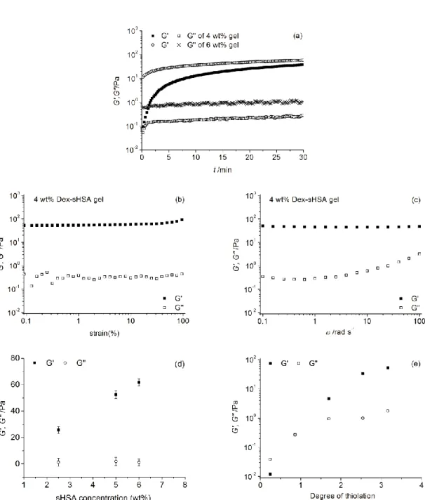
The gelation process of Dex-sHSA hydrogels was monitored by an oscillatory time sweep. After loading and mixing of the Dex-Mal and sHSA solutions on the rheometer plate, the hydrogel was formed on the order of seconds (Figure 3a). Values of storage modulus (G’) were observed above the loss modulus (G”) from the start of the experiment due to the rapid gelation of the sample. Hence, the cross-over point of G’ and G” (generally considered as the gel point) could not be observed. In less than half an hour, G’ reached a plateau indicating that the bulk of the gelation process occurred. Moreover, the gelation time was found to be dependent on the initial concentrations of Dex-Mal and sHSA. In comparison with the 4 wt% Dex-sHSA hydrogel, 6 wt% samples were found undergo a more rapid gelation process, resulting in a greater storage modulus on the initial reading.
Chapter 2
25
Figure 3. Oscillatory rheometry of Dex-sHSA hydrogels at 37 °C. (a) Time sweep of 4 wt% and 6 wt% hydrogels at 1 rad s-1 and 5%
strain. (b) Amplitude sweep from 0.5% to 100% at 1 rad s-1. (c) Angular frequency sweep from 100 rad s-1 to 0.1 rad s-1 and 5% strain.
Effect of (d) the relative concentration of sHSA (DT=3.16) to Dex-Mal, and (e) the degree of thiolation of sHSA on the storage and loss
modulus (G’ and G”) of 5 wt% Dex-sHSA hydrogels.
linear viscoelastic region of the Dex-sHSA hydrogel, which was found to be in the range of 0.1% to 100%. Angular frequency sweep measurements (Figure 3c) showed that G’ was at least an order of magnitude greater than G” over the range of 0.1-100 rad s-1; in agreement with the formation of a three-dimensional crosslinked network.
Chapter 2
26
and the degrees of thiolation (DT) of sHSA can influence the mechanical properties of Dex-sHSA hydrogels. For the former method, the concentration of Dex-Mal was held constant while the concentration of sHSA was varied corresponding to the molar ratio of between thiols and maleimide. As shown in Figure 3d, the storage modulus (G’) of Dex-sHSA gels increased from 25 Pa to 52 Pa when sHSA concentration doubled from 2.5 wt% to 5 wt%. By further increasing the concentration to 6 wt%, G’ of the gels went up to 62 Pa. In the latter method, increasing degrees of thiolation of the sHSA protein also yielded a concomitant increase in the mechanical properties of the resulting Dex-sHSA conjugates.
In Figure 3e, at low DT, sHSA can be covalently ligated to Dex-Mal, but a limited number of thiol groups on the albumin surface prevents the formation of hydrogels. For these samples, G” was greater than G’ in all oscillation measurements exhibiting predominant viscous-behavior. When the DT increased to 1.7 and over, G’ was found to be greater than G” in angular frequency sweep measurements and a viscoelastic material was formed. Hence, both methods, either increasing protein concentration or DT of the protein, provide a facile handle to tune mechanical properties of the macroscale delivery scaffold.
I
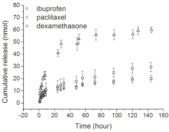
N VITRO DRUG RELEASEDrug release kinetics from HSA binding pockets within the Dex-sHSA hydrogels were evaluated using ibuprofen (IBU), paclitaxel (PTX) and dexamethasone (DXM). IBU is a typical non-steroidal anti-inflammatory drug. PTX is an antimicrotubule chemotherapeutic agent currently used in the treatment of solid tumour malignancies. Both IBU and PTX have been shown to be highly bound to plasma proteins (above 95%).50, 51 DXM is a synthetic corticosteroid widely used as anti-inflammatory and immunosuppressant in clinical treatments, which is moderately bound to plasma proteins (70-80% depending on the test procedure).52 Despite the lipophilic and poor water-soluble character of the three drugs tested (See supporting information Table S1), the drugs were co-dissolved with the HSA drug carrier to form water-soluble drug-HSA complexes and reacted with the hydrogel material. High drug loading efficiencies of IBU, PTX and DXM within the Dex-sHSA material were obtained (91.3%, 87.7% and 92.1%, respectively). By this method, organic solvents such as DMSO become unnecessary for the dispersion of various therapeutics, and further ligation to the hydrogel scaffold enables their prolonged delivery.
Chapter 2
27
curve of DXM exhibited a faster release rate than IBU and PTX, which may arise from differences in hydrophobic character and binding interactions with HSA. However, it is anticipated that these release profiles will change when these networks are presented with proteases due to the enzymatically sensitive HSA crosslinks. Overall, this assay shows that Dex-sHSA hydrogels can be easily loaded with hydrophobic therapeutics and show sustained release.
Figure 4. In vitro drug release profiles of IBU, PTX and DXM from Dex-sHSA hydrogels (4.5 wt%, DT=3.16) in PBS (150 mM, pH 7.4) at 37 °C.
C
ONCLUSION
Chapter 2
28
E
XPERIMENTAL
MATERIALS
Dextran (70 kDa), human serum albumin (HSA), N,N'-diisopropylcarbodiimide (DIC), 2-iminothiolane (2-IT), 5,5’-dithiobis(2-nitrobenzoic acid) (DTNB), dimethyl sulfoxide (DMSO), ethylenediaminetetraacetic acid (EDTA), ibuprofen (IBU), paclitaxel (PTX) and dexamethasone (DXM) were purchased from Sigma-Aldrich. Dextran was dried in a vacuum oven (30 °C) and DMSO was dried over 4Å molecular sieves before use. 3-maleimidopropionic acid and 4-(dimethylamino)pyridinium 4-toluenesulfonate (DPTS) were synthesized as previously reported.53,54 Dialysis membranes (MWCO 3,500-5,000 Da) were obtained from Spectrum Laboratories, Inc.
S
YNTHESIS OFD
EX-M
ALDextran (1 g, 6.17 mmol), 3-maleimidopropionic acid (417 mg, 2.47 mmol), and DPTS (115.7 mg, 0.247 mmol) were dissolved in DMSO (33 mL) followed by the addition of DIC (580 μL, 3.7 mmol). After stirring overnight at room temperature, N, N’-dialkylurea was removed by filtration and the crude product was obtained by precipitation in cold isopropanol. The precipitate was dialyzed against Milli-Q water using a 3500-5000 MWCO cut-off membrane and subsequently lyophilized. 1H NMR (400 MHz, D
2O): δ 3.3-4.0 (m, dextran glucopyranosyl ring protons), 4.9 (s, dextran anomeric proton), 6.9 (s, maleimide).
The degree of substitution of the dextran polymer (DS[Mal]) is defined as the number of maleimide groups per 100 glucopyranose residues. The DS[Mal] is calculated using the ratio: (100x)/(2y), in which x is the integral of the maleimide protons (δ 6.9) and y is the integral of the anomeric proton of dextran (δ 4.9) from the 1H NMR spectra measured by a Bruker AV-400 spectrometer.55 The DS[Mal] of the dextran polymer (Dex-Mal) synthesized in this work was 4.6.
SYNTHESIS OF THE THIOLATED HUMAN SERUM ALBUMIN (SHSA)
HSA was dissolved in 50 mM sodium phosphate buffer (pH 8, including 5 mM EDTA) and stirred gently at 0 °C on ice. Subsequently, an aqueous solution of 2-IT (20 mg mL-1) was added drop-wise to the HSA solution. After 2 hours, the mixture was brought back to room temperature. The product was first dialyzed against 10 mM HEPES buffer (pH 7) three times over 24 hours and then, against Milli-Q water three times over 2 days at 4 °C. Finally, sHSA was lyophilized and stored at -20 °C.
Chapter 2
29
mixed and incubated for one hour at room temperature. During this reaction, the formation of the 2-nitro-5-thiobenzoate anion (TNB2-) resulted in an intense yellow colour with an absorbance at 412 nm by UV-vis spectroscopy (Agilent,Cary-300). The molar extinction coefficient of TNB2- at 412 nm, εTNB2- = 14150 M-1 cm-1, was used to calculated the concentration of free thiol groups in the tested sHSA solution. The concentration of albumin in a control group (200 μL 10 mg mL-1 sHSA and 800 μL 0.1 M sodium phosphate buffer) was determined with the absorbance at 279 nm by UV-vis spectroscopy (εalbumin = 0.531 M−1 cm−1). The DT of sHSA was then calculated by dividing the concentration of free thiol groups by the concentration of albumin.
CIRCULAR DICHROISM (CD)
Circular dichroism spectra were obtained on a Jasco J-815 spectrometer. All spectra were collected with a scan speed of 50 nm min-1 and a response time of 1 second at 20 °C. Each spectrum was averaged over 5 scans. Samples of native HSA, sHSA (DT=3.2), Dex-sHSA conjugate (molar ratio of thiol to maleimide 1:1) were prepared in sodium phosphate buffer (50 mM, pH 7). Native HSA and sHSA solutions were filtered through 0.2 μm syringe filters before CD measurements. For near-UV measurements (250-330 nm), a 1 cm path length quartz cuvette was used for 0.5 mg mL -1 protein samples. For far-UV measurements (200-250 nm), a 0.1 cm path length quartz cuvette was applied for 0.1 mg mL-1 protein samples.
CD data were presented in terms of ellipticity [θ] (degrees) and converted to the mean residue molar ellipticity [θ] in deg cm2 dmol-1 by equation 1, where n is the number of amino acid residues (585), l is the path length of the cuvette, and Cp is the mole fraction.
[𝜃] = 𝜃(𝑚𝑑𝑒𝑔)
𝐶𝑝 ×𝑛×𝑙×10 (1)
α-helix content was calculated from the value of [θ] at 222 nm58 by equation 2.
𝛼 − ℎ𝑒𝑙𝑖𝑥 % = ([𝜃] −2430
30300 ) × 100 (2)
DEX-SHSA HYDROGELS
Dex-Mal and sHSA were dissolved in phosphate buffered saline (PBS, 150 mM, pH 7.4) individually and then mixed together using a pipette to prepare the hydrogel samples (1:1 molar ratio of maleimide to thiol).
Chapter 2
30
were performed to follow gel curing at 1 rad s-1 with 5% strain. The linear viscoelastic regime was determined using an amplitude sweep measurement at 1 rad s-1 from 0.1% to 100% strain. Frequency sweep measurements were performed from 100 to 0.1 rad s-1 with 5% strain in the linear viscoelastic regime.
I
N VITRO DRUG RELEASELipophilic drugs (IBU, PTX or DXM) and sHSA were co-dissolved in PBS (150 mM, pH 7.4, 0.02% NaN3) and incubated for 2 hours at 37 °C prior to gelation. The molar ratio of sHSA and drug was kept at 1:1 in all samples. The drug-sHSA solution was subsequently crosslinked with the Dex-Mal polymer solution to form the drug-loaded hydrogels for in vitro drug release.
To examine the drug loading efficiency, 1 mL 0.25% trypsin solution (25 mM HEPES buffer, pH 7) was added on top of 200 µL drug loaded Dex-sHSA gel (4.5 wt%) and incubated at 37 °C. 2 mL acetonitrile was added to the mixture and the precipitates were removed by ultracentrifuge at 13 000 rpm for 10 minutes. The amount of drug was subsequently quantified by reversed-phase high performance liquid chromatography (RP-HPLC) analysis using two LC-8A pumps, SPD-10AVP UV-VIS and ELSD-LTII detectors from Shimadzu. The separation was performed on a Gemini C18 column at a flow rate of 1 mL min-1 with a linear gradient from 90% B to 10% B, where A was acetonitrile with 0.1 vol% TFA and B was H2O with 0.1 vol% TFA. The eluate was monitored at both 220 and 254 nm. The drug loading efficiency was calculated from the amount of drug recovered from the gel divided by the amount of drug loaded expressed in percent.
Chapter 2
31
R
EFERENCES
1. Kearney C. J. and Mooney D. J., Macroscale delivery systems for molecular and cellular payloads, Nature Materials, 2013, 12, 1004-1017.
2. Kharkar P. M., Kiick K. L. and Kloxin A. M., Designing degradable hydrogels for orthogonal control of cell microenvironments, Chem Soc Rev, 2013, 42, 7335-7372.
3. Uhrich K. E., Cannizzaro S. M., Langer R. S., et al., Polymeric systems for controlled drug release, Chem Rev, 1999, 99, 3181-3198.
4. Censi R., Vermonden T., Deschout H., et al., Photopolymerized thermosensitive poly(hpmalactate)-peg-based hydrogels: Effect of network design on mechanical properties, degradation, and release behavior,
Biomacromolecules, 2010, 11, 2143-2151.
5. Pescosolido L., Feruglio L., Farra R., et al., Mesh size distribution determination of interpenetrating polymer network hydrogels, Soft Matter, 2012, 8, 7708-7715.
6. Dhanasingh A. and Groll J., Polysaccharide based covalently linked multi-membrane hydrogels, Soft Matter, 2012, 8, 1643-1647.
7. Zustiak S. P., Boukari H. and Leach J. B., Solute diffusion and interactions in cross-linked poly(ethylene glycol) hydrogels studied by fluorescence correlation spectroscopy, Soft Matter, 2010, 6, 3609-3618.
8. Lin C. C. and Metters A. T., Hydrogels in controlled release formulations: Network design and mathematical modeling, Adv Drug Deliver Rev, 2006, 58, 1379-1408.
9. Moghadam M. N., Kolesov V., Vogel A., et al., Controlled release from a mechanically-stimulated thermosensitive self-heating composite hydrogel, Biomaterials, 2014, 35, 450-455.
10. Schillemans J. P., Hennink W. E. and van Nostrum C. F., The effect of network charge on the immobilization and release of proteins from chemically crosslinked dextran hydrogels, Eur. J. Pharm. Biopharm., 2010, 76, 329-335.
11. Kiick K. L., Peptide- and protein-mediated assembly of heparinized hydrogels, Soft Matter, 2008, 4, 29-37. 12. Binauld S. and Stenzel M. H., Acid-degradable polymers for drug delivery: A decade of innovation, Chem
Commun, 2013, 49, 2082-2102.
13. Yamaguchi N. and Kiick K. L., Polysaccharide-poly(ethylene glycol) star copolymer as a scaffold for the production of bioactive hydrogels, Biomacromolecules, 2005, 6, 1921-1930.
14. Fairbanks B. D., Singh S. P., Bowman C. N., et al., Photodegradable, photoadaptable hydrogels via radical-mediated disulfide fragmentation reaction, Macromolecules, 2011, 44, 2444-2450.
15. Kloxin A. M., Tibbitt M. W. and Anseth K. S., Synthesis of photodegradable hydrogels as dynamically tunable cell culture platforms, Nature Protocols, 2010, 5, 1867-1887.
16. Tibbitt M. W., Kloxin A. M., Sawicki L. A., et al., Mechanical properties and degradation of chain and step-polymerized photodegradable hydrogels, Macromolecules, 2013, 46, 2785-2792.
17. Peng K., Tomatsu I. and Kros A., Light controlled protein release from a supramolecular hydrogel, Chemical Communications, 2010, 46, 4094-4096.
18. Peng K., Tomatsu I., van den Broek B., et al., Dextran based photodegradable hydrogels formed via a michael addition, Soft Matter, 2011, 7, 4881-4887.
19. Tomatsu I., Peng K. and Kros A., Photoresponsive hydrogels for biomedical applications, Advanced Drug Delivery Reviews, 2011, 63, 1257-1266.
20. Wang N. X. and von Recum H. A., Affinity-based drug delivery, Macromolecular Bioscience, 2011, 11, 321-332. 21. Mullarney M. P., Seery T. A. P. and Weiss R. A., Drug diffusion in hydrophobically modified
Chapter 2
32
22. Liu Y. Y., Shao Y. H. and Lu J., Preparation, properties and controlled release behaviors of ph-induced thermosensitive amphiphilic gels, Biomaterials, 2006, 27, 4016-4024.
23. Hoare T. R. and Kohane D. S., Hydrogels in drug delivery: Progress and challenges, Polymer, 2008, 49, 1993-2007.
24. Thatiparti T. R., Shoffstall A. J. and von Recum H. A., Cyclodextrin-based device coatings for affinity-based release of antibiotics, Biomaterials, 2010, 31, 2335-2347.
25. Zhou J. and Ritter H., Cyclodextrin functionalized polymers as drug delivery systems, Polymer Chemistry, 2010,
1, 1552-1559.
26. Peng K., Cui C., Tomatsu I., et al., Cyclodextrin/dextran based drug carriers for a controlled release of hydrophobic drugs in zebrafish embryos, Soft Matter, 2010, 6, 3778-3783.
27. Peng K., Tomatsu I., Korobko A. V., et al., Cyclodextrin-dextran based in situ hydrogel formation: A carrier for hydrophobic drugs, Soft Matter, 2010, 6, 85-87.
28. Liu C., Zhang Z., Liu X., et al., Gelatin-based hydrogels with [small beta]-cyclodextrin as a dual functional component for enhanced drug loading and controlled release, RSC Advances, 3, 25041-25049.
29. Cai T., Yang W. J., Zhang Z., et al., Preparation of stimuli-responsive hydrogel networks with threaded [small beta]-cyclodextrin end-capped chains via combination of controlled radical polymerization and click chemistry, Soft Matter, 8, 5612-5620.
30. Singh B., Chauhan N. and Sharma V., Design of molecular imprinted hydrogels for controlled release of cisplatin: Evaluation of network density of hydrogels, Industrial & Engineering Chemistry Research, 2011, 50, 13742-13751.
31. Salian V. D. and Byrne M. E., Controlled drug release from weakly crosslinked molecularly imprinted networks: The benefit of living radical polymerization, Macromolecular Chemistry and Physics, 2013, 214, 2355-2366.
32. Heimbach T., Fleisher D. and Kaddoumi A., in Prodrugs, eds. V. Stella, R. Borchardt, M. Hageman, R. Oliyai, H. Maag and J. Tilley, Springer New York, 2007, vol. V, pp. 157-215.
33. Kratz F., Albumin as a drug carrier: Design of prodrugs, drug conjugates and nanoparticles, Journal of Controlled Release, 2008, 132, 171-183.
34. He X. M. and Carter D. C., Atomic-structure and chemistry of human serum-albumin, Nature, 1992, 358, 209-215.
35. Hirose M., Tachibana A. and Tanabe T., Recombinant human serum albumin hydrogel as a novel drug delivery vehicle, Materials Science & Engineering C-Materials for Biological Applications, 2010, 30, 664-669. 36. Kakinoki S., Taguchi T., Saito H., et al., Injectable in situ forming drug delivery system for cancer
chemotherapy using a novel tissue adhesive: Characterization and in vitro evaluation, European Journal of Pharmaceutics and Biopharmaceutics, 2007, 66, 383-390.
37. Tada D., Tanabe T., Tachibana A., et al., Albumin-crosslinked alginate hydrogels as sustained drug release carrier, Materials Science & Engineering C-Biomimetic and Supramolecular Systems, 2007, 27, 870-874.
38. Tada D., Tanabe T., Tachibana A., et al., Drug release from hydrogel containing albumin as crosslinker, Journal of Bioscience and Bioengineering, 2005, 100, 551-555.
39. Oss-Ronen L. and Seliktar D., Photopolymerizable hydrogels made from polymer-conjugated albumin for affinity-based drug delivery, Advanced Engineering Materials, 2010, 12, B45-B52.
40. Oss-Ronen L. and Seliktar D., Polymer-conjugated albumin and fibrinogen composite hydrogels as cell scaffolds designed for affinity-based drug delivery, Acta Biomaterialia, 2011, 7, 163-170.
Chapter 2
33
42. Manjappa A. S., Chaudhari K. R., Venkataraju M. P., et al., Antibody derivatization and conjugation strategies: Application in preparation of stealth immunoliposome to target chemotherapeutics to tumor, Journal of Controlled Release, 2011, 150, 2-22.
43. Weber C., Reiss S. and Langer K., Preparation of surface modified protein nanoparticles by introduction of sulfhydryl groups, Int J Pharm, 2000, 211, 67-78.
44. Funk W. E., Li H., Iavarone A. T., et al., Enrichment of cysteinyl adducts of human serum albumin, Anal Biochem, 2010, 400, 61-68.
45. Kelly S. M., Jess T. J. and Price N. C., How to study proteins by circular dichroism, Bba-Proteins Proteom, 2005,
1751, 119-139.
46. Dockal M., Carter D. C. and Ruker F., The three recombinant domains of human serum albumin - structural characterization and ligand binding properties, Journal of Biological Chemistry, 1999, 274, 29303-29310. 47. Dockal M., Carter D. C. and Ruker F., Conformational transitions of the three recombinant domains of
human serum albumin depending on ph, Journal of Biological Chemistry, 2000, 275, 3042-3050.
48. Antonov Y. A. and Wolf B. A., Calorimetric and structural investigation of the interaction between bovine serum albumin and high molecular weight dextran in water, Biomacromolecules, 2005, 6, 2980-2989.
49. Nair D. P., Podgórski M., Chatani S., et al., The thiol-michael addition click reaction: A powerful and widely used tool in materials chemistry, Chemistry of Materials, 2013.
50. Valko K., Nunhuck S., Bevan C., et al., Fast gradient hplc method to determine compounds binding to human serum albumin. Relationships with octanol/water and immobilized artificial membrane lipophilicity, J Pharm Sci-Us, 2003, 92, 2236-2248.
51. Singla A. K., Garg A. and Aggarwal D., Paclitaxel and its formulations, Int J Pharm, 2002, 235, 179-192. 52. Waters N. J., Jones R., Williams G., et al., Validation of a rapid equilibrium dialysis approach for the
measurement of plasma protein binding, J Pharm Sci-Us, 2008, 97, 4586-4595.
53. de Figueiredo R. M., Oczipka P., Frohlich R., et al., Synthesis of 4-maleimidobutyric acid and related maleimides, Synthesis-Stuttgart, 2008, 1316-1318.
54. Moore J. S. and Stupp S. I., Room-temperature polyesterification, Macromolecules, 1990, 23, 65-70.
55. Vandijkwolthuis W. N. E., Franssen O., Talsma H., et al., Synthesis, characterization, and polymerization of glycidyl methacrylate derivatized dextran, Macromolecules, 1995, 28, 6317-6322.
56. Habeeb A. F. S. A., [37] reaction of protein sulfhydryl groups with ellman's reagent, Methods in Enzymology, 1972, 25, 457-464.
57. Ellman G. L., Tissue sulfhydryl groups, Archives of Biochemistry and Biophysics, 1959, 82, 70-77.
Chapter 2
34
S
UPPORTING INFORMATION
1
H NMR SPECTRA
Figure S1. 1H NMR (D
Chapter 2
35
SWELLING PROPERTIES OF DEX-SHSA HYDROGEL
A Dex-sHSA hydrogel sample (4.5 wt%) was immersed in 150 mM phosphate buffered saline (PBS, pH 7.4, 0.02% sodium azide) at 37 °C. At predetermined time intervals, the entire buffer weight was weighed and fresh buffer was refilled afterwards. The normalized hydrogel weight (NWh) was calculated by the following equation:
𝑁𝑊ℎ =
𝑊𝑡 𝑊0
Where Wt is the hydrated mass at time t and Wo the initial mass of the hydrogel after gelation.
Figure S2. Swelling properties of Dex-sHSA hydrogels.
I
N VITRO DRUG RELEASETable S1. Physicochemical properties of applied drugs.
Drugs Mw Water solubility (25 °C)* logP*
ibuprofen 206.28 21 mg/L 3.97
paclitaxel 853.91 insoluble 3
dexamethasone 392.46 89 mg/L 1.83
Chapter 2
36
In order to quantify the amount of drugs released from the Dex-sHSA gels, calibration curves between drug’s concentration and its peak intensity from RP-HPLC analysis were determined prior to the in vitro drug release study.
C
HAPTER
3
D
UAL
-C
ROSSLINKED
H
UMAN
S
ERUM
A
LBUMIN
-P
OLYMER
H
YDROGELS FOR
A
FFINITY
-B
ASED
D
RUG
D
ELIVERY
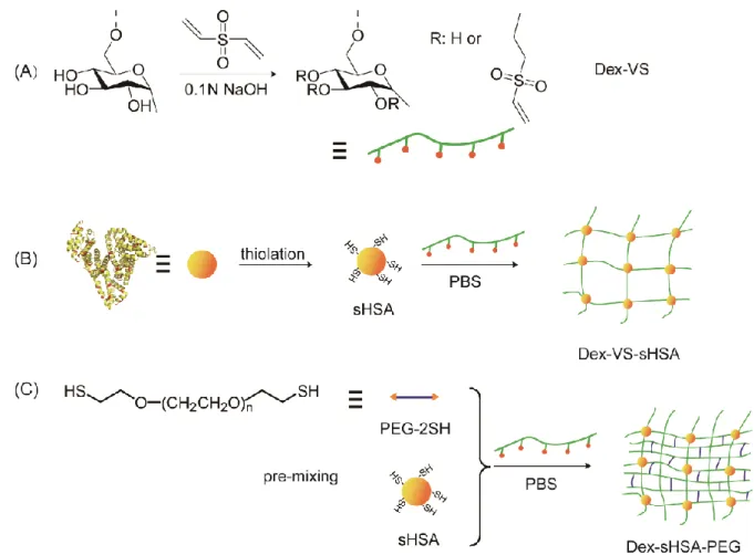
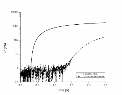
A dual-crosslinked in situ gelling drug delivery scaffold based on dextran, thiolated serum albumin and poly(ethylene glycol) is presented. Dextran-vinyl sulfone conjugates with varied molecular weight and degrees of substitution were synthesized by controlling the reaction time and temperature with divinyl sulfone. Dextran-human serum albumin hydrogels were prepared using a thiol-vinyl sulfone Michael addition reaction with thiolated albumin as the crosslinker. Poly(ethylene glycol) dithiol was added as a third component to the crosslinked dextran-human serum albumin hydrogel to facilitate additional crosslinking; simultaneously reducing the gelation time and increasing the tunability of the physicochemical properties of the Dex-sHSA-PEG network. The onset of gelation of the modular three-component dual-crosslinked hydrogel network ranged from 45 minutes to 1.5 hours depending on both gel constituent concentrations and the gelation temperature (25 °C or 37 °C). All gels remained stable for over a 25-day period under physiological conditions. In vitro drug release assays showed that dual-crosslinked Dex-sHSA-PEG hydrogels can deliver Doxorubicin in a sustained manner over 7 days. Finally, an MTT assay showed the biocompatible nature of the Dex-sHSA-PEG hydrogels and capacity to deliver Doxorubicin successfully to MCF-7 breast cancer cells.
Chapter 3
39
I
NTRODUCTION
Macroscale drug delivery systems have garnered much attention in pharmaceutical research because of their potential to exert control over drug release spatiotemporally.1, 2 Rather than conventional oral or intravenous drug administrations, novel macroscale drug delivery systems are usually implanted at a site of need for use in long-term or drug delivery. By releasing therapeutics locally at a desired site, side effects and toxicity can be diminished due to lower dosing quantities. Typical drug release mechanisms applied in drug delivery systems include diffusion-controlled drug release,3 degradation-controlled drug release4 and stimuli-triggered drug release,5 each with its own inherent advantages and disadvantages in a therapeutic setting.
Affinity-based drug release systems have emerged to be an attractive platform for their delivery due to the capacity to exploit interactions between the delivery system and the therapeutic drug using a combination of non-covalent interactions to control drug release such as hydrophobicity, hydrogen bonding, electrostatics or van der Waals forces.6 Advantages of affinity-based drug delivery systems include improved drug loading, prolonged drug stability and sustained release. Drug-binding hosts can be introduced into the polymeric scaffolds using (macro)molecules such as cyclodextrin,7-11 serum albumin12-15 and heparin,16-18 or by generation of cavities within the bulk polymer material by molecular imprinting.19, 20 A particularly attractive carrier is the most abundant plasma protein (35-50 g L-1) human serum albumin (HSA). HSA has been shown to be biocompatible, biodegradable, non-immunogenic and non-toxic, and is known to bind and transport numerous molecules in the blood circulation, such as fatty acids, bilirubin, hormones, and metal ions.13, 21 Several therapeutic drugs have also been shown to bind various locations of the albumin protein by several non-covalent interactions with high affinity. Consequently, HSA has been exploited for use as a drug depot in a broad range of particulate carriers,22-24 but fewer examples of its use in macroscale hydrogel systems have been reported.12, 25-30 Until now, the synthesis of human serum albumin-based hydrogel materials involves the use of various coupling agents,25-27 free radical polymerization,12 UV29, 30 and γ-ray irradiation28 in order to facilitate gelation of HSA with varying degrees of biocompatibility. However, in order to apply these HSA-based materials in the biomedical domain, benign gelation strategies that enable control over the resultant hydrogel physicochemical properties are still very much needed.