Nontuberculous Mycobacteria
II: Nested-Cohort Study of Impact on Cystic Fibrosis Lung Disease
Kenneth N. Olivier, David J. Weber, Ji-Hyun Lee, Allison Handler, Gail Tudor, Paul L. Molina, Joseph Tomashefski, and Michael R. Knowles, for the Nontuberculous Mycobacteria in Cystic Fibrosis Study Group*
The Cystic Fibrosis/Pulmonary Research and Treatment Center, Departments of Epidemiology, Biostatistics, and Radiology, and the Verne S. Caviness General Clinical Research Center, University of North Carolina at Chapel Hill, Chapel Hill, North Carolina; Pulmonary/Critical Care Medicine Flight, Wilford Hall United States Air Force Medical Center, Lackland Air Force Base, Texas; Department of Pathology, Case Western Reserve University, Cleveland, Ohio
The prevalence of nontuberculous mycobacteria (NTM) is high (ap- function (1). However, the impact of these organisms on the
proximately 13%) in sputum of patients with cystic fibrosis (CF), clinical course of CF lung disease has not been prospectively
but the impact on lung disease is unknown. We followed 60 incident evaluated.
NTM-positive and 99 culture-negative patients with CF for 15 Criteria for the diagnosis of disease caused by NTM in
months and assessed clinical impact of NTM by FEV1 and high- persons without CF have been published (2). Diagnosis
re-resolution computed tomography (HRCT) of the chest.Mycobacte- quires the presence of (1) clinical signs and symptoms; (2) rium aviumcomplex was seen in 75% of NTM-positive subjects. The multiple positive sputum cultures, recovery of NTM in large annual rate of decline in FEV1 was not different among control amounts on microbiologic smears and cultures of
broncho-versus NTM-positive subjects who did not, or did, meet American scopic samples, and/or compatible histopathology with a posi-Thoracic Society microbiologic criteria for NTM disease (3⫾1, 3⫾ tive NTM culture; and (3) compatible radiographic findings. 2, and 5⫾2%, respectively). More subjects with three or more
Defining NTM disease in patients with CF is particularly
chal-positive cultures for NTM had two or more characteristic findings
lenging, given the considerable overlap of the clinical and
ra-on entry HRCT (60%, 9/15) as compared with subjects with two
diographic criteria between disease caused by NTM and CF.
positive cultures or less (32%) or negative cultures (19%; p⬍0.02).
Although case reports of an apparent association between
All subjects with three or more positive cultures and exit HRCTs
NTM and a decline in clinical and radiographic features have
(n⫽6) showed progression of HRCT findings, whereas only 17%
been published, other reports of this relationship suggest
of subjects with two positive cultures or less had progression (p⫽
these organisms may coexist in the lower airways of some
0.0006). In summary, no significant short-term effect on FEV1was
subjects with CF without significant adverse effect (3–13).
detected in patients with multiple positive NTM cultures, but an
abnormal HRCT was predictive of progression. Patients with CF There has been no prospective, rigorous evaluation of the
and multiple positive NTM cultures, characteristic HRCT findings, and impact of NTM in the lower airways on lung function, and
progression of HRCT changes should be monitored closely and radiographic findings of the presence of NTM in CF have
considered for antimycobacterial therapy. not been published. This study was designed to look
systemat-ically at the (short-term) longitudinal effects of NTM on the
Keywords:cystic fibrosis; nontuberculous mycobacteria; Mycobacte- clinical course of CF lung disease, as assessed by changes in rium avium-intracellulare;Mycobacterium abscessus; computed tomogra- spirometry and chest computed tomography (CT), to develop phy of chest
criteria that may indicate a need for further diagnostic evalua-tion and/or initiaevalua-tion of specific antimycobacterial therapy. The prevalence of nontuberculous mycobacteria (NTM) in
the lower respiratory tract of older subjects with cystic fibrosis
METHODS
(CF) is high (approximately 13%) relative to the background population and to other disease groups. We reported, in a
Study Design cross-sectional study, an increase in the prevalence of NTM
A previous multicenter cross-sectional study assessed prevalence of with age and an association with relatively preserved lung
NTM (1) in three sputum samples over a year. The current nested-cohort design identified incident NTM-positive subjects (at least one prior negative culture) from the prevalence study (Figure 1), matched by age, sex, and FEV1to two culture-negative “controls” in that study (Figure 1). Subjects were followed for 15 months, and sputum was
(Received in original form July 9, 2002; accepted in final form November 6, 2002)
tested on each visit. Chest radiographs were performed at entry, 6, and
*The members of the Nontuberculous Mycobacteria in Cystic Fibrosis Study Group
15 months; chest HRCT was performed at entry and 15 months on
are listed in the Appendix.
NTM-positive subjects and 20% of the control subjects.
The views expressed in this article are those of the authors and do not reflect the
official policy of the Department of Defense or other Departments of the U.S. Subjects Government.
We enrolled from 17 U.S. centers, excluding subjects withBurkholderia
Supported by a grant from the Cystic Fibrosis Foundation, CFF A914 and collabora- cepacia. Eleven NTM-negative “control subjects” turned culture-posi-tive grants from participating General Clinical Research Centers, Center for
Re-tive, and re-enrolled as NTM-positive subjects; data from these control
search Resources, National Institutes of Health (individual grant numbers cited in
subjects were excluded. Only one control was available for 10
culture-Appendix).
positive subjects; four controls were transplanted, five died, and two
Correspondence and requests for reprints should be addressed to Lt. Col. Kenneth withdrew. The protocol was approved by Institutional Review Boards N. Olivier, M.D., M.P.H., U.S.A.F., M.C., Wilford Hall, United States Air Force at participating sites.
Medical Center, MSGS/MCCP (Pulmonary/Critical Care), 2200 Bergquist Drive,
Ste 1, Lackland AFB, TX 78236-5300. E-mail: [email protected] Clinical and Laboratory Evaluation
Am J Respir Crit Care Med Vol 167. pp 835–840, 2003
Clinical data were recorded at each visit (14–16). Sputum was processed
Originally Published in Press as DOI: 10.1164/rccm.200207-679OC on November 14, 2002
We used mixed linear regression models to test whether FEV1 de-cline was different between NTM-positive subjects and control subjects and among NTM-positive subjects who did, or did not, meet microbio-logic criteria (23). The first model used data collected at the beginning of the previous prevalence study and during this current study, including covariates (age, sex, and body mass index). The second model covered Figure 1. Timeline depicting the relationship between the previous only data from entry into this study (Figure 1), including additional cross-sectional, prevalence study and this current nested-cohort study, covariates (FEV
1, corticosteroids, and parenteral antibiotics). The effect which was performed in subjects identified by the prevalence study. of NTM status on FEV
1was tested by comparing two models using Subjects with least one negative acid fast bacillus (AFB) culture in the log-likelihood statistics. Pairwise differences were assessed between the prevalence study were entered into the current (nested-cohort) study two categories of NTM-positive subjects and control subjects. at the time of their first positive AFB culture and were matched to two
subjects who were NTM-culture–negative. Both NTM-positive subjects RESULTS and their matched, culture-negative control subjects were assessed
clini-cally every 3 months for 15 months with specimens for mycobacterial Baseline Characteristics
culture obtained at each visit. Sixty NTM-positive and 99 control subjects were enrolled from
17 U.S. CF centers (Table 1). Although NTM-positive subjects tended (p ⫽0.16) to be slightly older (average, 2 years) than control subjects at entry into the cohort study, they also tended reference laboratory (University of Texas Health Center, Tyler, TX)
to have higher FEV1values (approximately 5%, p⫽0.19) and for final identification.
slightly better Birmingham Composite Radiograph Scores
(ap-Radiographic Evaluation proximately 1.0; p ⫽ 0.08). The chest CT findings tended to
parallel the pattern seen with FEV1 values and Birmingham Original radiographs (posteroanterior and lateral chest) were scored
Radiograph Scores. Specifically, more NTM-positive subjects at Chapel Hill (Birmingham Roentgenogram Score) (19). HRCT scans
were performed at respective sites and evaluated in Chapel Hill tended to have at least two chest CT findings that are characteris-(P.L.M.) without knowledge of culture status. HRCTs were assessed tic of NTM disease in subjects without CF as compared with for four characteristic findings associated with NTM in subjects without control subjects (p⫽0.11) (seefurther comments below). There CF: (1) cystic and/or cavitary parenchymal lung disease, (2) subsegmen- were no significant differences in sex, body mass index, or the tal (or larger) parenchymal consolidation, (3) single or multiple pulmo- frequency of pancreatic enzyme use.
nary nodules, and (4) tree-in-bud opacities (20–22).
Mycobacterial Cultures Statistical Analysis
Control subjects had 7.8⫾0.1 (range 4–11) cultures, and Descriptive statistics were used to identify differences between
NTM-positive subjects had 8.9 ⫾ 0.2 (range 5–13) cultures over 16 positive and control (NTM-negative) subjects. The2test was used to
months. Before the first positive culture, 23 (38%) of NTM-compare categoric characteristics at baseline between NTM-positive
positive subjects had one negative culture, 19 (32%) had two and control subjects, and two-tailed Student’s t tests (independent)
were used for continuous variables. negative, and 18 (30%) had three or more negative cultures.
TABLE 1. COMPARISON OF NONTUBERCULOUS MYCOBACTERIA–POSITIVE SUBJECTS AND NONTUBERCULOUS MYCOBACTERIA–NEGATIVE CONTROL SUBJECTS AT ENTRY INTO THE NESTED-COHORT STUDY
NTM-Positive Subjects Control Subjects
Characteristic (n⫽60) (n⫽99) p Value*
Sex, % male 48 45 0.72
Age at enrollment, yr, mean 28.5⫾1.4 26.2⫾0.9 0.16 FEV1, % predicted, mean 57.6⫾2.9 52.8⫾2.2 0.19
Body mass index, kg/m2, mean 20.7⫾0.4 20.0⫾0.3 0.18
Use of pancreatic enzymes, % 95 95 0.99 Birmingham Radiograph Score, mean† 17.6⫾0.5 16.6⫾0.3‡ 0.08
Characteristic CT findings, %§ 40 19 0.11
Definition of abbreviations: CT⫽computed tomography; NTM⫽nontuberculous mycobacteria. *2for dichotomous variables, andttest for continuous variables.
†Composite score. ‡n⫽96.
Figure 2. The number of subjects having positive mycobacterial cul-tures, grouped by mycobacterial species. Thebarsrepresent the number of subjects who did not meet the ATS microbiologic criteria for NTM disease (hatched bars,lower stacks) and those subjects who did meet ATS criteria (solid bars,upper stacks).
The majority of NTM-positive subjects had only a single positive culture (n⫽34, 57%) with only two of these having a positive smear; eight (13%) had two positive cultures, four of whom had positive smears; eight (13%) had three positives with half having positive smears; and 10 (17%) had more than three positive
Figure 3. Potential clinical variables that may confound the relationship cultures with half having at least one positive smear. Of the 60
between NTM and decline in %FEV1(% predicted), plotted by groups
NTM-positive subjects, 22 (37%) met the ATS microbiologic
of subjects (open bars, control subjects;hatched bars, NTM-positive criteria for NTM disease.
subjects who did not meet ATS criteria;solid bars, NTM-positive subjects Mycobacterium aviumcomplex (MAC) was recovered from
who met ATS microbiologic criteria for disease).(A) Presence of hemop-the majority of hemop-the NTM-positive subjects (n⫽45, 75%) with
tysis and use of systemic steroids and parenteral antibiotics (*p⬍0.05, a third (14) of these meeting ATS criteria (Figure 2).
Mycobacte-less than culture-negative control subjects. ‡p ⬍0.05, greater than
rium abscessuswas recovered from seven subjects (12%), and
NTM-positive subjects).(B) Sputum microbiology (*p⬍0.02, greater four of those met ATS microbiologic criteria. Subjects withM. than culture-negative control subjects and NTM-positive subjects who abscessustended to meet ATS NTM criteria more frequently did not meet ATS criteria).
than subjects with MAC or other NTM organisms (odds ratio⫽ 3.2, 95% confidence interval 0.8, 12.9; p⫽ 0.1). One subject had three different organisms—M. abscessus, Mycobacterium
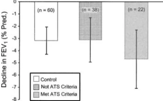
kansasii, andMycobacterium malmoense; three subjects had both not control for differences in baseline FEV1. The decline in FEV1 MAC andM. abscessus; one subject had both MAC andMyco- was modest but significantly different from zero for all three bacterium gordonae(multiple isolates); and one hadMycobacte- groups. However, there was no significant difference in the mean rium pergrinum, one hadMycobacterium lentiflavum, and one annual decline in FEV1(% predicted) among the three groups hadM. kansasii(Figure 2). (1.9⫾ 0.4%, 1.2⫾ 0.6%, and 2.4⫾0.7%, respectively). The second analysis (Model 2) controlled for differences in baseline
Clinical Variables FEV1and the use of parenteral antibiotics and systemic
cortico-There was no significant difference in the number of days with steroids, in addition to the variables controlled for in the initial hemoptysis among the control subjects and NTM-positive sub- analysis. Again, there was no significant difference among the jects that either did not, or did, meet ATS microbiologic criteria groups (3.2⫾1.1%, 3.1⫾1.8%, and 4.7⫾2.4%, respectively) (Figure 3A). Steroids were used less frequently in the NTM- (Figure 4).
positive subjects who met ATS criteria as compared with the
Radiologic Assessment NTM-negative control group (p⬍0.05). Intravenous antibiotics
were used more frequently in the control group compared with Baseline and follow-up chest radiographs were available for the NTM-positive subjects (p⬍0.05, Figure 3A). There were analysis on 53 (54%) of the control subjects, 26 (68%) of the no significant differences among the three groups in the recovery non-ATS criteria NTM-positive subjects, and 10 (45%) of the ofPseudomonas aeruginosa or Aspergillus species during the ATS criteria NTM-positive subjects. There was no significant course of follow-up (Figure 3B).Staphylococcus aureuswas re- change over time in any of the chest radiograph scores (individ-covered from sputum more frequently in NTM-positive subjects ual category and composite) within either control subjects or who met ATS microbiologic criteria than in NTM-positive sub- the two groups of NTM-positive subjects or between the two jects who did not meet ATS criteria and in control subjects (p⬍ culture-positive groups.
0.01) (Figure 3B). Entry HRCTs were available for analysis on 53 (88%) of the NTM-positive subjects and 21 (21%) of the control
(NTM-Longitudinal Decline in Lung Function negative) subjects; exit CTs were available on 24 and 8 of these
study these subjects remained well matched, except for a slightly lower %FEV1in the control subjects (Table 1).
Figure 4. Annual decline in FEV1(% predicted) based on mixed model
analyses, which shows control subjects (open bars), NTM-positive sub- To better assess the effect of NTM on the clinical course of jects who did not meet ATS microbiologic criteria (hatched bars), and CF, we grouped the NTM-positive subjects according to the ATS NTM-positive subjects who met ATS criteria (solid bars). This analysis microbiologic criteria for disease. As reported in the previous (Model 2) incorporated only FEV1data collected after the entry visit prevalence study (1), the majority of the NTM-positive subjects
into this current study, controlled for differences in baseline FEV1, and the had only one positive culture or two positive cultures without
use of systemic corticosteroids and intravenous antibiotics, in addition to smear positivity, and thus did not meet the ATS microbiologic variables in Model 1 (seeMETHODS). criteria for disease. Subjects from whomM. abscessuswas
cul-tured tended to meet microbiologic criteria more frequently than did those with only MAC or other NTM (Figure 2). Subjects who met the microbiologic criteria for NTM were not sicker present at baseline (entry) in a greater fraction (60%, n⫽9)
than either those subjects not meeting criteria or those who of the 15 subjects with three or more positive cultures as
com-remained culture-negative (controls), as assessed by frequency pared with 32% of those subjects with only one or two positive
of hemoptysis, corticosteroid use, intravenous antibiotic use, or cultures and 19% of NTM-negative, control subjects (p⬍0.02).
prevalence ofP. aeruginosa(Figure 3A). Although patients with Comparable differences were noted when analyses included only
mycobacteria tended to have a higher reported prevalence of subjects with MAC andM. abscessus. More subjects who met
Aspergillusthan did culture-negative control subjects, there was ATS criteria and had significant baseline CT findings, and who
no difference inAspergillusprevalence based on ATS microbio-also had exit CTs, had progression of CT findings, as compared
logic criteria. As reported in the previous prevalence study, a with the other subjects with entry and exit CTs. Progression of
strikingly higher prevalence ofS. aureuswas seen in those NTM-CT findings was noted in only 3 of 8 (38%) control subjects,
positive patients who met ATS criteria as compared with NTM-and 4 of 18 (22%) non-ATS criteria NTM-positive subjects,
positive subjects who did not meet ATS criteria and control whereas all 6 (100%) of the subjects with three or more positive
subjects (p⫽0.02) (Figure 3B). cultures had progression (p⬍0.0006).
We tested for change (decline) in FEV1over time to assess the potential effect of NTM on the course of CF lung disease.
DISCUSSION Different models were constructed to control for both key
clini-cal variables and potential confounders. Although NTM-positive We have previously reported a high prevalence of NTM
(approx-subjects who met ATS criteria tended to decline faster than imately 13%) in sputum of persons with CF lung disease. This
both the non–ATS-positive and control subjects, the amount of prevalence is most striking in older subjects with relatively
pre-variability in the slope estimates was large, and no statistically significant differences in the slopes was detected. The rate of decline in FEV1in the CF control subjects was similar to what has been noted in other studies of similar age patients with CF (1–3% per year) (24). There are examples in the literature suggesting a probable adverse effect of NTM on lung function. Some of those indicate a precipitous decline temporally related to recovery of NTM (4, 6, 7), whereas others note a prolonged time span between initial recovery and subsequent adverse ef-fects (9, 12). Perhaps more significant differences in FEV1would be seen with a longer duration of follow-up. For example, we estimated that significant differences would be seen between NTM-positive subjects who met ATS criteria versus control sub-jects if the slopes declined (Figure 4) at similar rates for 6.4 years.
Given the many factors that can affect the change in FEV1 Figure 5. Characteristic HRCT findings in control subjects and
NTM-over relatively short periods of time in patients with CF, airflow positive subjects by culture status at entry (solid bars) into the study.
mechanics may not be the most sensitive or specific indicator of Solid barsrepresent the percent of subjects in each of the three
sub-disease progression related to a specific microorganism, such as groups with at least two of the following characteristics on entry HRCT:
the NTM. Several studies have suggestive characteristic findings (A) areas of cystic and/or cavitary parenchymal lung disease, (B)
subseg-on HRCT scan in patients with NTM and without CF (20–22). mental or larger areas of parenchymal consolidation, (C) single or
multi-More recent studies have also demonstrated progression of these ple pulmonary nodules, and (D) tree-in-bud opacities. More of the NTM
findings over relatively short periods of time; one study noted positive subjects who met ATS Criteria had at least two of these findings
progression at a mean follow-up of 28⫾4 months and another at entry as compared with control subjects and subjects with only one
HRCT findings associated with NTM include cysts or cavities, segmental consolidation, peripheral nodules, and tree-in-bud type infiltrates. Although these radiographic findings may over-lap with those commonly seen in CF, the relative frequency of these findings was higher in patients with CF who met ATS microbiologic criteria for NTM disease, even though their FEV1 values were similar to those of subjects with only one or two positive NTM cultures and culture-negative control subjects. Moreover, all six of the NTM-positive subjects who had three or more positive NTM cultures as well as both entry and exit HRCT scans had progression of at least one of the four HRCT findings. In contrast, only 4 subjects out of 18 with only one or two positive cultures had these HRCT changes. This suggests that HRCT may play a vital role in the evaluation of subjects with CF and with positive NTM sputum cultures and may be an earlier indicator than FEV1of pathogenic infection with NTM; however, additional study will be required for confirmation.
The study was limited by sample size and length of follow-up. Sample size calculations were based on comparison of rate of change in FEV1 for all NTM-positive subjects versus their matched culture-negative control subjects. Given the sampling variability in recovering NTM coupled with the significant
Figure 6. Flow diagram of proposed clinical approach to finding NTM HRCT findings on the entry scan of subjects who eventually
in the lower airway of a patient with CF (seetext for details). Further met ATS microbiologic criteria, it is likely that some subjects
study is needed to validate this approach. entered the study after having had NTM in their airways for a
period of time and thus were prevalent rather than incident positives. A relatively large number of subjects originally
classi-fied as culture-negative control subjects converted to NTM cul- M. abscessus, consideration should be given to beginning specific ture-positive, but it is less likely that subjects were misclassified antimycobacterial treatment, particularly if there is a temporally as control subjects in the final analyses, given the large numbers associated decline in the clinical course; this approach is based of cultures obtained. Finally, both the revised ATS criteria for on prior reports associating this organism with significant disease NTM pulmonary disease and longitudinal studies of the length in CF, (3, 5, 8, 10, 12). If the organism is notM. abscessus, or of time for progression of HRCT findings in patients without if characteristic HRCT findings are not present, and the patient CF and with nodular bronchiectatic disease were published after is clinically stable, a follow-up HRCT should be obtained in 12 the initiation of this investigation (2, 25, 26). It seems unlikely to 15 months, while continuing to collect serial sputum specimens that NTM would have the same impact in subjects with a single for mycobacterial culture. If progression of the characteristic positive culture out of multiple specimens as compared with findings on HRCT is seen and the patient continues to have those subjects who had multiple positive cultures, and this notion positive cultures of the same NTM organism, consideration was borne out in preliminary analyses. Thus, we believed it should be given to specific antimycobacterial treatment. If appropriate to group the NTM-positive subjects, based on the HRCT progression is not noted, continued follow-up with serial ATS microbiologic criteria for disease, to test the clinical impact HRCT scans and cultures should be performed to monitor for of NTM. progression. Given the overlapping coverage of many of the Despite these limitations, this study is compatible with the drugs used to treat NTM with usual CF pathogens such asP. general clinical approach that is evolving to address the presence aeruginosaandS. aureus, airways clearance measures should be of NTM recovered from the sputum of patients with CF intensified, and (other) bacterial lower airway pathogens should (Figure 6). It should be noted that this current approach is based, be treated aggressively to establish a clinical baseline, before in part, on the clinical experience that has been developed in starting specific antimycobacterial treatment, so that the effect the context of NTM in the respiratory tract of individuals with of treatment on mycobacterial disease can be better assessed. non-CF lung disease. Specifically, given the increasing preva- In summary, the presence of NTM in the lower airways is lence of NTM with age (1) and case reports noting the potential common among persons with CF. The majority of persons from for these organisms to cause significant clinical decline, adult whom NTM are recovered will have only a single isolate with patients with CF should have periodic screening cultures. During repeated cultures obtained over one to two years. On the basis periods of clinical decline unresponsive to treatment of conven- of trends in lung function seen in this study, those persons from tional bacterial pathogens, all patients with CF, including chil- whom repeated positive cultures are obtained may be at risk of dren, should be cultured for NTM. If a positive culture for an adverse effect on lung function over a period of months to NTM is obtained, serial specimens should be collected to assess several years. Longer studies of serial lung function would be frequency of recovery of the same species of NTM. If the ATS needed to confirm this. Characteristic HRCT findings of cysts microbiologic criteria for NTM are met, and especially if three or cavities, areas of consolidation, peripheral nodules, or tree-or mtree-ore cultures are positive ftree-or the same species of NTM, a in-bud infiltrates are more prevalent in subjects with CF with baseline HRCT scan should be obtained and assessed for the repeated positive cultures, and these findings may progress over presence of (at least two of the four) characteristic findings of even a relatively short period of 12 to 15 months. Given the NTM, described previously. If the ATS microbiologic criteria large number of confounding factors influencing the course of are not met, periodic follow-up cultures should be obtained to CF lung disease and the relative general indolent nature of these look for increasing prevalence of the organisms. For M. ab- organisms, further validation of the clinical approach described scessus, if baseline HRCT findings are suggestive of mycobacte- previously and assessment of the effectiveness of treating these
puted tomography diagnosis ofMycobacterium aviumcomplex lung
bacteria. Am J Respir Crit Care Med1997;156:S1–S25.
disease: prospective study [abstract].Am J Respir Crit Care Med1995;
3. Boxerbaum B. Isolation of rapidly growing mycobacteria in patients with
151:A476.
cystic fibrosis.J Pediatr1980;96:689–691. 22. Fujita J, Ohtsuki Y, Suemitsu I, Shigeto E, Yamadori I, Obayashi Y,
4. Efthimiou J, Smith MJ, Hodson ME, Batten JC. Fatal pulmonary infec- Miyawaki H, Dobashi N, Matsushima T, Takahara J. Pathological and
tion withMycobacterium fortuitumin cystic fibrosis.Br J Dis Chest radiological changes in resected lung specimens inMycobacterium
1984;78:299–304. avium intracellularecomplex disease.Eur Respir J1999;13:535–540.
5. Smith MJ, Efthimiou J, Hodson ME, Batten JC. Mycobacterial isolations 23. Littell RC, Milliken GA, Stroup WW, Wolfinger RD. SAS system for
in young adults with cystic fibrosis.Thorax1984;39:369–375. mixed models. Cary, NC: SAS Institute, Inc.; 1996.
6. Kinney JS, Little BJ, Yolken RH, Rosenstein BJ.Mycobacterium avium 24. Davis PB. Clinical pathophysiology and manifestations of lung disease.
complex in a patient with cystic fibrosis: disease vs. colonization.Pedi- In: Yankaskas JR, Knowles MR, editors. Cystic fibrosis in adults.
atr Infect Dis J1989;8:393–396. Philadelphia, PA: Lippincott-Raven Publishers; 1999. p. 45–67.
7. Hjelte L, Petrini B, Kaellenius G, Strandvik B. Prospective study of 25. Obayashi Y, Fujita J, Suemitsu I, Kamei T, Nii M, Takahara J. Successive
mycobacterial infections in patients with cystic fibrosis.Thorax1990; follow-up of chest computed tomography in patients with
Mycobacte-45:397–400. rium avium-intracellularecomplex.Respir Med1999;93:11–15.
8. Bange FC, Brown BA, Smaczny C, Wallace RJ Jr, Bottger EC. Lack of 26. Yamazaki Y, Kubo K, Takamizawa A, Yamamoto H, Honda T, Sone S.
transmission ofMycobacterium abscessusamong patients with cystic Markers indicating deterioration of pulmonaryMycobacterium
avium-fibrosis attending a single clinic.Clin Infect Dis2001;32:1648–1650. intracellulareinfection.Am J Respir Crit Care Med1999;160:1851–
9. Kilby JM, Gilligan PH, Yankaskas JR, Highsmith WE Jr, Edwards LJ, 1855.
Knowles MR. Nontuberculous mycobacteria in adult patients with
cystic fibrosis.Chest1992;102:70–75.
APPENDIX
10. Tomashefski JF Jr, Stern RC, Demko CA, Doershuk CF. Nontuberculous
mycobacteria in cystic fibrosis: an autopsy study.Am J Respir Crit Nontuberculous Mycobacteria in Cystic Fibrosis Study Group
Care Med1996;154:523–528. investigators, institutions and NCRR Grant Numbers: Moira
11. Fauroux B, Delaisi B, Clement A, Saizou C, Moissenet D, Truffot-Pernot
Aitken, University of Washington, Seattle (RR0037); G. F. Shay, C, Tournier G, Vu TH. Mycobacterial lung disease in cystic fibrosis:
Kaiser Permanente, Oakland; M. J. Light, University of
Califor-a prospective study.Pediatr Infect Dis J1997;16:354–358.
nia San Diego (RR00827); M. S. Stulbarg, University of
Califor-12. Cullen AR, Cannon CL, Mark EJ, Colin AA.Mycobacterium abscessus
infection in cystic fibrosis: colonization or infection?Am J Respir Crit nia San Francisco (RR00079); J. Wagner, Children’s Hospital,
Care Med2000;161:641–645. Denver (RR00069); M. H. Wagner, University of Florida,
13. Oliver A, Maiz L, Canton R, Escobar H, Baquero F, Gomez-Mampaso Gainesville (RR00082); S. A. McColley, Northwestern
Univer-E. Nontuberculous mycobacteria in patients with cystic fibrosis.Clin sity, Chicago (RR00048); S. H. Davis, Tulane University, New
Infect Dis2001;32:1298–1303.
Orleans (RR05096); B. Rosenstein, Johns Hopkins University, 14. Standardization of spirometry, 1994 update: American Thoracic Society.
Baltimore (RR00052); M. E. Wohl, Children’s Hospital, Boston
Am J Respir Crit Care Med1995;152:1107–1136.
(RR02172); A. Lapey, Massachusetts General Hospital, Boston 15. Hsu KH, Jenkins DE, Hsi BP, Bourhofer E, Thompson V, Tanakawa
N, Hsieh GS. Ventilatory functions of normal children and young (RR01066); W. J. Warwick, C. E. Milla, University of Minnesota,
adults–Mexican-American, white, and black. I: spirometry.J Pediatr Minneapolis (RR00400); K. N. Olivier, C. Pue, M. K. Knowles,
1979;95:14–23. University of North Carolina at Chapel Hill (RR00046); J.
Eisen-16. Knudson RJ, Slatin RC, Lebowitz MD, Burrows B. The maximal expir- berg, Oregon Health Sciences University, Portland (RR00334);
atory flow-volume curve: normal standards, variability, and effects of
S. Fiel, Allegheny University of the Health Sciences, Philadel-age.Am Rev Respir Dis1976;113:587–600.
phia; D. Orenstein, University of Pittsburgh (RR00084); B. Mar-17. Whittier S, Hopfer RL, Knowles MR, Gilligan PH. Improved recovery
shall, University of Utah, Salt Lake City (RR00064); J. Biller, of mycobacteria from respiratory secretions of patients with cystic