MOLECULAR MECHANISMS OF DIRECT CARDIAC REPROGRAMMING
Haley Ruth Chi
A dissertation submitted to the faculty at the University of North Carolina at Chapel Hill in partial fulfillment of the requirements for the degree of Doctor of Philosophy in Department of Pathology and
Laboratory Medicine in the School of Medicine.
Chapel Hill 2020
Approved by: Li Qian
ABSTRACT
Haley Ruth Chi: Molecular Mechanisms of Direct Cardiac Reprogramming (Under the direction of Li Qian)
Direct cardiac reprogramming is a promising approach to cardiac regeneration. Fibroblasts are converted directly into induced cardiomyocytes (iCMs) in vitro and in vivo through exogenous expression of three cardiac lineage transcription factors – Gata4, Mef2c, and Tbx5. Before direct cardiac
reprogramming is ready to be deployed as a clinical therapy, however, additional research is required to improve the efficiency of the reprogramming process and the purity and maturity of the generated iCMs. Our work provides new insight into epigenetic regulation of reprogramming induction and
molecular mechanisms of cell fate conversion to increase reprogramming efficiency and maturation. We first developed a series of cell lines as tools for ongoing research. We developed an αMHC-GFP cardiac reporter fibroblast cell line for convenient reprogramming and rapid detection of newly derived iCMs. Additionally, we generated a tetracycline inducible polycistronic reprogramming factor construct for the temporal regulation of the reprogramming cocktail. We introduced this construct into our cell line to create an inducibly reprogrammable fibroblast cell line. These cell lines have enabled large-scale screens of potential regulatory factors. We next explored the epigenetic regulation of early-stage
depletion de-represses endogenous Gata4 expression and effectively replaces exogenous Gata4 expression from the reprogramming cocktail. Finally, we describe a novel mitochondrial mechanism regulating iCM reprogramming efficiency and maturation. Inhibiting mitochondrial fission by depleting Fis1 enhances the efficiency of direct cardiac reprogramming and increases mitochondrial quantity and respiratory capacity. Overexpression of PGC1α to drive mitochondrial biogenesis similarly enhances reprogramming, confirming that the effect on reprogramming is due to increased mitochondrial content in reprogramming cells. Additional studies are needed to elucidate the mechanism by which Fis1
ACKNOWLEDGEMENTS
I would like to thank my graduate research mentor, Li Qian, for her unwavering and untiring support for me as a researcher and as a person. Her inquisitive mind, irrepressible optimism, and dedication to improving the world within her sphere of influence have been an inspiration and example for me.
I am grateful to all the researchers in the Qian and Liu labs, past and present, for their assistance and camaraderie at the bench. I am particularly indebted to Yang Zhou and Li Wang who generously dedicated much time to training me when I joined the lab.
I appreciate the generosity of my committee who have sat through many long hours of meetings and offered their advice and guidance.
I am indebted to Hong Ma from Jiandong Liu’s lab, Jing Zhang from Qing Zhang’s lab, and Qiang Zhu from Joan Taylor’s lab for technical assistance with the Seahorse experiments.
I would like to thank Felicia Heyward, Sebastien Coquery, Janet Dow, and Nancy Fisher at the UNC Flow Cytometry Core for their assistance learning and troubleshooting flow cytometry.
I must extend my heartfelt gratitude to Rocky Riviella, Tracy Riley, and Dean Riddick in the McAllister Heart Institute office for helping me apply for and manage an external fellowship. I do not know what I would have done without you all.
I would like to offer my sincere thanks to both the former chair of the Pathology Department, Charles Jennette, and the current chair, Russell Broaddus, for their ongoing support of graduate student research.
Other teachers and mentors in graduate school have buoyed me with inspiration and
encouragement: Michael Goy, Cyrus Vaziri, Susan Hadler, Stephanie Montgomery, Patrick Brandt, Anna Ballew O’Connell, Jeremy Purvis.
I am indebted to my undergraduate professors who believed in me and encouraged me to apply to graduate school: Kathleen Alligood, Ed Otto, LaNitra Berger.
I would like to thank the friends I have made in graduate school for enlivening my life outside of lab. I am deeply grateful to Hank Tarlton and UNC InterVarsity Graduate Christian Fellowship for re-orienting my perspective and priorities in life.
TABLE OF CONTENTS
LIST OF FIGURES ... xii
LIST OF TABLES ... xiv
LIST OF ABBREVIATIONS ... xv
CHAPTER 1: INTRODUCTION ... 1
Introduction ... 1
Reprogramming Factor Cocktails ... 2
Reprogramming Factor Stoichiometry ... 5
Epigenetic Barriers to Direct Cardiac Reprogramming ... 7
Repression of Fibroblast Identity ... 11
Intracellular Signaling Pathways ... 14
Growth Factors ... 15
Environmental Cues ... 17
Future Direction ... 20
Addendum ... 22
Present Work... 25
CHAPTER 2: GENERATION OF AN INDUCIBLE FIBROBLAST CELL LINE FOR STUDYING DIRECT CARDIAC REPROGRAMMING ... 27
Introduction ... 27
Materials and Methods... 27
Results ... 32
Development of the mouse embryonic fibroblast cardiac reporter cell line MEF-T. ... 32
Development of the tetracycline-inducible polycistronic MGT reprogramming
factor construct iMGT. ... 35
Development of the tetracycline-inducible direct cardiac reprogramming cell line icMEF. ... 35
Figures ... 37
Discussion... 40
Acknowledgements ... 41
CHAPTER 3: CHARACTERIZATION OF EPIGENETIC BARRIERS DURING DIRECT CARDIAC REPROGRAMMING ... 42
Introduction ... 42
Epigenetic Changes During Reprogramming... 42
Polycomb Repressive Complex 2 ... 43
Experimental Evidence for the Role of PRC2 in Reprogramming ... 44
Polycomb Repressive Complex 1 ... 44
Bmi1 and Cellular Proliferation ... 45
Materials and Methods... 45
Results ... 46
PRC2 subunit depletion does not enhance reprogramming. ... 46
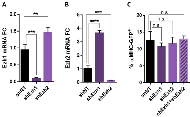
Simultaneous depletion of Ezh1 and Ezh2 does not enhance reprogramming. ... 47
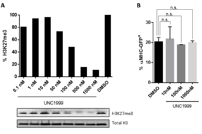
Pharmacological inhibition of Ezh1/2 reduces H3K27me3 but does not enhance reprogramming. ... 47
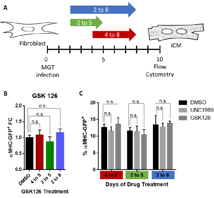
Pharmacological inhibition of Ezh1/2 does not alter early reprogramming events... 48
Pharmacological inhibition of Ezh2 does not increase reprogramming. ... 48
Loss-of-function screen identifies epigenetic regulators likely to function as barriers to reprogramming. ... 49
Bmi1 depletion does not enhance reprogramming through de-repressing cell cycle regulators. ... 49
Bmi1 knockdown primes cells for reprogramming. ... 51
Inhibiting Bmi1 can replace Gata4 in the reprogramming cocktail. ... 51
Figures ... 52
Discussion... 63
CHAPTER 4: STUDYING MITOCHONDRIAL DYNAMICS DURING DIRECT CARDIAC REPROGRAMMING ... 65
Introduction ... 65
Cardiomyocyte Mitochondria ... 65
Mitochondrial Dynamics ... 66
Materials and Methods... 66
Results ... 70
Reprogramming increases mitochondria. ... 70
Reprogramming increases respiratory capacity. ... 71
Inhibiting mitochondrial fission enhances reprogramming. ... 72
Fis1 depletion increases cellular mitochondrial content. ... 72
Reactive Oxygen Species (ROS) mechanism... 72
Inhibiting mitochondrial fission does not increase ROS. ... 73
ROS do not mediate reprogramming... 74
Fis1 depletion increases respiratory capacity. ... 74
Overexpression of PGC1α enhances reprogramming. ... 75
Metaboloepigenetic mechanism. ... 75
Increasing intracellular acetyl-CoA does not enhance reprogramming. ... 76
Proliferation mechanism. ... 76
Timing and duration of Fis1 knockdown. ... 77
Reprogramming factor expression. ... 78
Figures ... 79
Discussion... 95
CHAPTER 5: CONCLUSIONS AND FUTURE DIRECTIONS ... 97
Applications of Our Inducible Fibroblast Cell Lines ... 97
Epigenetic Studies ... 100
Mitochondrial Metaboloepigenetics ... 102
Fis1 Depleted iCM Mitochondrial Morphology ... 102
Fis1 Depleted iCM Maturation ... 103
Cardiac Specificity of Fis1 Depletion ... 103
LIST OF FIGURES
Figure 2.1. Characterization of the MEF-T cell line ... 37
Figure 2.2. Transgene expression in the MEF-T cell line ... 38
Figure 2.3. Inducible reprogramming constructs and cell lines ... 39
Figure 3.1. Direct Cardiac Reprogramming is an Epigenetic Process ... 52
Figure 3.2. Silencing subunits of the PRC2 does not increase reprogramming ... 53
Figure 3.3. Silencing PRC2 catalytic activity does not increase reprogramming ... 54
Figure 3.4. Pharmacological inhibition of PRC2 catalytic activity does not increase reprogramming ... 55
Figure 3.5. Pharmacological inhibition of Ezh1/2 does not accelerate reprogramming ... 56
Figure 3.6. Pharmacological inhibition of Ezh2 does not enhance reprogramming ... 57
Figure 3.7. Loss-of-function screen identifies epigenetic regulators likely to function as barriers to reprogramming ... 58
Figure 3.8. Bmi1 depletion’s enhancement of reprogramming is not mediated through cell cycle regulators ... 59
Figure 3.9. Inhibiting cellular proliferation does not abrogate Bmi1 depletion’s enhancement of reprogramming ... 60
Figure 3.10. Bmi1 depletion primes cells for reprogramming ... 61
Figure 3.11. Bmi1 depletion can substitute for Gata4 factor expression in the reprogramming cocktail... 62
Figure 4.1. Direct cardiac reprogramming increases mitochondria late in reprogramming ... 79
Figure 4.2. Direct cardiac reprogramming increases respiratory capacity ... 80
Figure 4.3. Inhibiting mitochondrial fission enhances reprogramming ... 81
Figure 4.4. Inhibiting mitochondrial fission increases mitochondria ... 82
Figure 4.5. Proposed reactive oxygen species (ROS) signaling pathway ... 83
Figure 4.6. Inhibiting mitochondrial fission does not generate ROS ... 84
Figure 4.7. ROS do not mediate reprogramming ... 85
Figure 4.9. Overexpression of PGC1α enhances reprogramming ... 87
Figure 4.10. Proposed metaboloepigenetic mechanism ... 88
Figure 4.11. Increasing intracellular acetyl-CoA does not enhance reprogramming ... 89
Figure 4.12. Inhibiting mitochondrial fission does not enhance reprogramming through cellular senescence ... 90
Figure 4.13. Timing of mitochondrial fission inhibation ... 91
Figure 4.14. Duration of mitochondrial fission inhibition... 92
Figure 4.15. Reprogramming factor expression ... 93
LIST OF TABLES
LIST OF ABBREVIATIONS
αMHC: alpha myosin heavy chainACL: ATP citrate lyase AKT: protein kinase B
ATP: adenosine triphosphate
bp: base pair
cDNA: complementary DNA
CIC: mitochondrial citrate transporter cTnT: cardiac troponin T
DEG: differentially expressed genes DMNQ: 2,3-dimethoxy-1,4-naphthoquinone DMSO: dimethyl sulfoxide
DZNep: 3-Deazaneplanocin A EdU: 5-Ethynyl-2´-deoxyuridine FAD: flavin adenine dinucleotide fCF: freshly isolated cardiac fibroblast FDR: false discovery rate
FGF: fibroblast growthy factor GFP: green fluorescent protein
GGT: GFP-Gata4-Tbx5 polycistronic construct
GHMT: Gata4, Hand2, Mef2c, and Tbx5 reprogramming cocktail GMT: Gata4, Mef2c, and Tbx5 reprogramming cocktail
H2O2: hydrogen peroxide
H3K27me3: histone 3 lysine 27 trimethylation H3K4me3: histone 3 lysine 4 trimethylation HDAC: histone deacetylase
HNGMT: Hand2, Nkx2.5, Gata4, Mef2c, and Tbx5 reprogramming cocktail HTSF: high throughput sequencing facility
iCM: induced cardiomyocyte
icMEF: iMEF cell line with stable expression of the iMGT construct iMEF: MEF-T cell line with constitutive expression of rtTA
iMGT: inducible polycistronic MGT construct MAPK: mitogen-activated protein kinase MEF: mouse embryonic fibroblast MEF-T: immortalized MEF cell line
MGT: polycistronic Mef2c-Gata4-Tbx5 reprogramming factor construct
miR: microRNA
MMC: mitomycin C
MMP: matrix metalloproteinase NAC: N-acetylcysteine
OAA: oxaloacetate
ROCK: Rho-associated kinase ROS: reactive oxygen species RPK: reads per kilobase
rtTA: engineered reverse tetracycline-controlled transcriptional activator SAM: S-adenosyl methionine
shBmi1: shRNA targeting Bmi1 shCcnd1: shRNA targeting Cyclin D1 shEed: shRNA targeting Eed shEzh1: shRNA targeting Ezh1 shEzh2: shRNA targeting Ezh2 shNT: non-targeting shRNA shRNA: short hairpin RNA shSuz12: shRNA targeting Suz12 siRNA: small interfering RNA TCA: tricarboxylic acid
TGFβ: transforming growth factor beta TPM: transcripts per million
URT: untranslated region
CHAPTER 1: INTRODUCTION
1Introduction
As the leading cause of death in the United States, heart disease accounts for one out of every four mortalities (cdc.gov, 2016a). Contributing to this, every year 735,000 Americans experience a myocardial infarction (cdc.gov, 2016b) which reduces the heart’s pump capacity due to cardiomyocyte death and increases the risk of arrhythmia from the deposition of non-conductive scar tissue. There are currently three primary approaches to cardiac regeneration as a therapy for myocardial infarction: 1) progenitor cell transplantation, 2) induced proliferation of resident cardiomyocytes, and 3) non-myocyte cell fate reprogramming. The first, progenitor cell therapy, is limited by the low viability and integration of transplanted cells. Multiple clinical trials have demonstrated that the engraftment rate of
transplanted cells and the number of cardiomyocytes derived from transplanted progenitors are insufficient to produce a therapeutic effect (Lin and Pu, 2014). However, some clinical benefit is
observed due to paracrine signaling from the transplanted progenitors (Lin and Pu, 2014). Identification of the contributing factors will produce the same effect while bypassing transplantation altogether (Lin and Pu, 2014). The second approach, inducing myocyte cell cycle re-entry, is accompanied by both efficacy and safety concerns. Studies have yet to demonstrate that the proposed methods for stimulating cardiomyocyte proliferation can generate a sufficient quantity of new cardiomyocytes to produce a clinically relevant effect (Lin and Pu, 2014). Furthermore, induced proliferation approaches must demonstrate cardiomyocyte-selective stimulation to preclude oncogenesis (Lin and Pu, 2014). The third approach, cell fate reprogramming of endogenous non-myocytes, is addressed in this review.
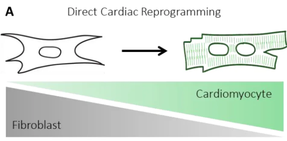
Cell fate reprogramming confers a dual advantage of reducing scar tissue while simultaneously generating new cardiomyocytes. Following coronary artery ligation, direct cardiac reprogramming reduces scar size in murine hearts by over two-fold (Qian et al., 2012; Song et al., 2012). New fibroblast-derived reprogrammed cardiomyocytes comprise 35% of cardiomyocytes in the infarct and border zones (Qian et al., 2012). These newly reprogrammed cardiomyocytes exhibit integration into the working myocardium, with electrical connectivity to endogenous cardiomyocytes and coordinated contraction (Qian et al., 2012; Song et al., 2012). Concomitant with measurably reducing scar size and generating new cardiomyocytes, direct cardiac reprogramming demonstrates substantial therapeutic benefit. Reprogramming therapy improves ejection fraction, stroke volume, and cardiac output in murine hearts following coronary artery ligation (Qian et al., 2012; Song et al., 2012) and sustains improvement up to 12 weeks after myocardial infarction (Song et al., 2012), demonstrating its potential as a therapeutic approach. With the in situ regeneration of mature, functional cardiomyocytes and simultaneous reduction in scar tissue, direct cardiac reprogramming has strong potential as a clinical therapy to restore cardiac function following myocardial infarction. Although significant advances have been made, studies have uncovered numerous molecular barriers to the reprogramming process. This review explores the molecular barriers to cell fate conversion in order to facilitate effective and complete direct cardiac reprogramming.
Reprogramming Factor Cocktails
Observation that only a fraction of the starting cell population fully reprograms into functional cardiomyocytes has prompted numerous initiatives to screen candidate reprogramming factors in order to identify cocktails for optimal reprogramming efficiency. Studies have screened cocktails of
Mohamed et al., 2016), and combinations of the three (Ifkovits et al., 2014; Muraoka et al., 2014; Wang et al., 2014).
Direct cardiac reprogramming was first described using a transcription factor cocktail. Ieda et al. screened a pool of fourteen cardiac lineage transcription factors to define a cocktail of three
reprogramming factors – Gata4, Mef2c, and Tbx5 (GMT) – that are necessary and sufficient to convert fibroblasts to induced cardiomyocyte-like cells (iCMs) both in vitro (Ieda et al., 2010; Wang et al., 2015a) and in vivo (Inagawa et al., 2012; Ma et al., 2015; Qian et al., 2012). Subsequently, Song et al. screened six conserved cardiac lineage transcription factors to identify a cocktail of Gata4, Hand2, Mef2c, and Tbx5 (GHMT) that generates almost five-fold more cTnT/αMHC-GFP double positive iCMs than GMT in vitro (Song et al., 2012). The GHMT cocktail also generates iCMs in murine hearts in vivo, reducing scar
size and improving cardiac function following myocardial infarction (Song et al., 2012). While the above mentioned studies employed sequential subtraction of candidate factors and αMHC-GFP reporter expression to screen cocktails, Protze et al. tested all possible triplet combinations of ten
reprogramming factors and used quantitate PCR for multiple cardiac markers to identify a three factor reprogramming cocktail of Mef2c, Tbx5, and Myocd that induces expression of a broad range of cardiac genes (Protze et al., 2012). Christoforou et al. furthermore demonstrated that a five or a seven factor cocktail adding Myocd, SRF, Mesp1, and SMARCD3 to the three factor GMT cocktail enhances cTnT mRNA expression by almost five-fold (Christoforou et al., 2013). Finally, Addis et al. employed a calcium reporter to functionally screen candidate factors and define a five factor cocktail of Hand2, Nkx2.5, Gata4, Mef2c, and Tbx5 (HNGMT) for maximally efficient reprogramming of functional iCMs (Addis et al., 2013).
permutations of singlet, doublet, and triplet microRNA combinations and screening using quantitative PCR for cardiac markers (Jayawardena et al., 2012). A cocktail of miR-1, miR-133, miR-208, and miR-499 is sufficient to reprogram cardiac fibroblasts both in vitro (Jayawardena et al., 2012) and in vivo
(Jayawardena et al., 2015). The microRNA cocktail generates functional, mature iCMs in vivo and reduces scar size and improves cardiac function in murine hearts following myocardial infarction (Jayawardena et al., 2015). The mechanism of miR-1 and miR-133 enhancement of reprogramming is discussed below in the section “Repression of Fibroblast Identity”.
Chemical cocktails for direct cardiac reprogramming have also been developed, circumventing the genetic manipulation associated risks involved in transcription factor and microRNA cocktails. Fu et al. noted the emergence of spontaneously beating cells during iPSC reprogramming using a cocktail of
small molecule compounds and developed a two-step reprogramming process to induce and stabilize iCM reprogramming (Fu et al., 2015). This two-step chemical cocktail reprogramming generates beating clusters of iCMs that express cardiac markers, assemble contractile sarcomeres, and display
cardiomyocyte-like electrophysical properties without going through a pluripotent stage (Fu et al., 2015).
In addition to pure cocktails of transcription factors, microRNA, or small molecules,
combinations of factors produce a synergistic effect for maximal reprogramming efficiency. Wang et al. directly reprogrammed fibroblasts into iCMs without going through a pluripotent intermediate state using a chemical cocktail plus a single transcription factor Oct4 (Wang et al., 2014). Conversely, Ifkovits et al. used a single small molecule TGFβ inhibitor to enhance reprogramming efficiency of the
that miR-1 or miR-133 together with the transcription factor cocktail GMT produced a six-fold increase over GMT alone (Muraoka et al., 2014). The mechanism of miR-1 and miR-133 enhancement of reprogramming is also discussed below in the section “Repression of Fibroblast Identity”. Jayawardena et al. used a small molecule compound, JAK inhibitor I, in combination with the micoRNA cocktail miR-1,
miR-133, miR-208, and miR-499 to increase reprogramming efficiency by ten-fold (Jayawardena et al., 2012).
Reprogramming Factor Stoichiometry
Decades of research in developmental biology have revealed the fine balance of transcription factor expression that is required to initiate and maintain cardiac lineage commitment. However, in cell fate reprogramming, the forced overexpression of reprogramming factors results in crude, artificial transcription factor dosage. Most studies using the standard Gata4, Mef2c, Tbx5 cocktail utilize
retroviral delivery of the three factors packaged as separate viruses. Starting cells must take up each of the three individual viruses in order to be reprogrammed, leading to low cell fate conversion rates since only a subset of cells receive all three factors. Individual cells also receive different ratios of the three factors. Stochastically, only a small fraction of cells receives the optimal reprogramming factor ratio and dose for cell fate reprogramming.
iCM reporter expression compared to reprogramming with separate viruses (Wang et al., 2015a). The highest reporter expression was achieved using the MGT splice order with concomitantly higher levels of Mef2c and lower levels of Gata4 and Tbx5 (Wang et al., 2015a). The optimal polycistronic MGT vector improved both MHC-GFP reporter expression and cTnT cardiac marker expression compared to separate Gata4, Mef2c, Tbx5 virus reprogramming (Wang et al., 2015a). The polycistronic MGT vector generated reprogrammed cells that formed iCM clusters, assembled cTnT and -Actinin positive sarcomeres, expressed the gap junction protein Connexin 43, exhibited calcium flux, and contracted spontaneously (Wang et al., 2015a). Molecular characterization also shows that the MGT and MTG polycistronic vectors induce higher expression of cardiomyocyte genes and lower expression of cardiac stress genes Nppa and Nppb than reprogramming with separate viruses (Wang et al., 2015a). An optimized protocol for reprogramming using the polycistronic MGT system was described as an additional resource (Wang et al., 2015b).
We also demonstrated that optimal reprogramming factor stoichiometry using the polycistronic MGT vector improves reprogramming efficiency in vivo in a mouse model of myocardial infarction (Ma et al., 2015). Using Periostin-Cre; R26R-lacZ genetic lineage tracing, cells of fibroblast origin were
irreversibly marked with -galactosidase to identify iCMs generated from fibroblasts (Ma et al., 2015). In vivo reprogramming with polycistronic MGT following coronary artery ligation increases the number of
-Actinin/-galactosidase double positive iCMs compared to reprogramming with separate Gata4, Mef2c, Tbx5 viruses (Ma et al., 2015). Additionally, in vivo reprogramming with the polycistronic MGT vector produces greater functional improvement in fractional shortening and ejection fraction and reduces scar size following myocardial infarction than reprogramming with separate viruses (Ma et al., 2015). Optimal stoichiometry of the three transcription factors using a polycistronic vector increases the efficiency of in vivo reprogramming and further improves cardiac function following myocardial
In addition to defining the optimal reprogramming factor stoichiometry, we created an inducible system where polycistronic MGT expression is both temporally and quantitatively regulated through the administration of doxycycline (Vaseghi et al., 2016). Using the TetOn inducible gene expression system, reprogramming factor expression is tightly regulated by the presence of doxycycline in the cell culture media (Vaseghi et al., 2016). Additionally, by titrating the doxycycline concentration, reprogramming factor expression level and dosage can be controlled (Vaseghi et al., 2016).
Two other in vivo studies also created polycistronic reprogramming vectors. Inagawa et al. placed the reprogramming factors in the order Tbx5-Mef2c-Gata4 (Inagawa et al., 2012), while Mathison et al. used the order Gata4-Mef2c-Tbx5 (Mathison et al., 2014). Both studies demonstrate a modest
increase in reprogramming efficiency using their single polycistronic vector compared to three separate viruses, but the improvement is only incremental due to the non-optimal dosage of the reprogramming factors. Inagawa et al. found no difference in the proportion of -Actinin positive reprogrammed cells to infected cells between their polycistronic TMG vector and separate viruses but did see the proportion of -Actinin positive cells that had assembled sarcomeres double from 15% to 30% of infected cells
(Inagawa et al., 2012). Mathison et al. demonstrated increased numbers of reprogrammed cells in vitro and improved cardiac function in vivo using their GMT polycistronic vector (Mathison et al., 2014). The incremental improvement demonstrated by these polycistronic systems is possibly due to the increased delivery of all three reprogramming factors to the starting cell population, although the optimal dosage of the reprogramming factors was not considered.
Epigenetic Barriers to Direct Cardiac Reprogramming
In addition to reprogramming factor dosage and timing, the epigenetic regulation of gene expression programs is critical to cell lineage commitment. Highlighting the role of epigenetic modulation in cell fate determination and conversion, Takeuchi and Bruneau directed ectopic
of the cardiac transcription factor Gata4 and the cardiac specific chromatin remodeling complex subunit, Baf60c (Takeuchi and Bruneau, 2009). Baf60c potentiates the binding of Gata4 to DNA at cardiac loci to turn on ectopic cardiac gene expression (Takeuchi and Bruneau, 2009). Transfection of the transcription factor Tbx5 in addition to Gata4 and Baf60c promotes the complete differentiation of transfected cells into beating cardiomyocytes (Takeuchi and Bruneau, 2009). The transfected cells begin beating even before the endogenous heart field starts to form(Takeuchi and Bruneau, 2009). Baf60c is one of the minimal requirements for this ectopic cardiac differentiation of the embryonic mesoderm,
demonstrating that chromatin remodeling plays a crucial role in transdifferentiation and cardiac fate acquisition. Cellular reprogramming necessarily involves changes to the epigenetic landscape. Epigenetic marks must be erased and re-written to alter chromatin structure and gene expression patterns during reprogramming as the fibroblast signature is repressed and a cardiomyocyte gene expression program is activated. Recent studies have demonstrated that epigenetic manipulation can potentiate the
reprogramming process.
non-specific HDAC inhibitor, enhances direct cardiac reprogramming, increasing the proportion of cTnT or -Actinin positive reprogrammed cells by two-fold (Christoforou et al., 2013). These findings suggest that HDAC inhibition may enhance direct cardiac reprogramming by epigenetically priming cells for cardiac fate acquisition.
Widespread epigenetic repatterning occurs during direct cardiac reprogramming. Zhao et al. performed chromatin immunoprecipitation sequencing of H3K4me2, which marks the promoters and enhancers of transcriptionally active genes, and demonstrated that one week after GHMT cocktail reprogramming 47% of the H3K4me2 peaks had shifted to align with those of primary cardiomyocytes (Zhao et al., 2015). Our lab demonstrated that trimethylation of lysine 27 on histone 3 (H3K27me3), a repressive epigenetic mark, is depleted and trimethylation of lysine 4 on histone 3 (H3K4me3), an activating histone modification, is enriched at cardiac promoters early in GMT cocktail reprogramming and is accompanied by a rapid increase in cardiac gene mRNA expression (Liu et al., 2016b). This early activation of the cardiomyocyte gene expression program is later followed by the increase of H3K27me3 and decrease of H3K4me3 at fibroblast loci and a concomitant decrease in fibroblast gene mRNA
activity reduces H3K27me3 by 30% and increases gene and protein expression of cardiac markers between two- to eight-fold irrespective of microRNA cocktail reprogramming (Dal-Pra et al., 2017). Conversely, knockdown of H3K27 demethylase activity inhibits the induction of cardiac marker
expression during microRNA cocktail reprogramming (Dal-Pra et al., 2017). These findings demonstrate that H3K27 demethylation and de-repression of cardiac loci is essential for direct cardiac
reprogramming.
Given that extensive repatterning of histone modifications occurs during the reprogramming process, modulation of the deposition and removal of histone modifications may promote greater reprogramming efficiency. Hirai et al. used a small molecule inhibitor of Ezh2, the catalytic component of the PRC2 complex catalyzing H3K27me2/3, to demonstrate that Ezh2 inhibition early in
reprogramming increases reprogramming efficiency (Hirai and Kikyo, 2014). The authors also report that late inhibition of the methyltransferase G9a, which catalyzes H3K9me1/2, increases reprogramming efficiency (Hirai and Kikyo, 2014). Conversely, Ifkovits et al. found that pre-treatment of fibroblasts with a G9a histone methyltransferase inhibitor reduced reprogramming efficiency (Ifkovits et al., 2014), demonstrating that the timing of histone methyltransferase inhibition is crucial for its effect on reprogramming. Hirai et al. observed that only specific time windows of drug administration were sufficient to promote an increase in reprogrammed cells and that inhibition at other times resulted in no effect or a decrease in reprogramming efficiency (Hirai and Kikyo, 2014). The two histone
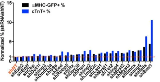
To identify key epigenetic barriers to the reprogramming process, we conducted a
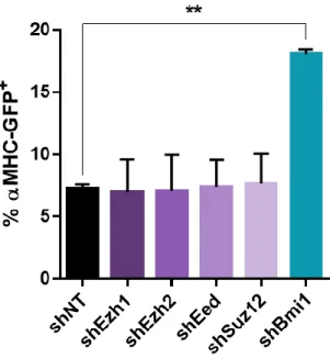
comprehensive loss-of-function screen of epigenetic modifying factors and identified Bmi1 as a critical epigenetic inhibitor of reprogramming (Zhou et al., 2016). Bmi1 is a polycomb group protein that directly binds to cardiac loci and suppresses expression of cardiac genes (Zhou et al., 2016). Depleting Bmi1 increases H3K4me3 and reduces H2AK119ub at cardiac loci and subsequently de-represses cardiac gene expression, priming fibroblasts for reprogramming (Zhou et al., 2016). Bmi1 knockdown plays a role to enhance cardiac fate acquisition early in the reprogramming process, as late Bmi1 depletion does not affect reprogramming (Zhou et al., 2016). Bmi1 also directly binds to the regulatory region and modulates the expression of Gata4 (Zhou et al., 2016). The inhibition of Bmi1 de-represses endogenous Gata4 expression and can therefore replace exogenous Gata4 in the Gata4, Mef2c, Tbx5 reprogramming cocktail (Zhou et al., 2016), permitting two factor-mediated iCM reprogramming to be possible.
In a similar approach, Liu et al. conducted a gain-of-function screen of cardiac development epigenetic modifiers and transcription factors and identified the H3K4 methyltransferase Mll1 as a barrier to direct cardiac reprogramming (Liu et al., 2016a). Pharmacologic inhibition of Mll1 improves both iCM generation with a 1.5-fold increase in MHC-GFP reporter expression and iCM maturation with increased sarcomere assembly and spontaneous beating (Liu et al., 2016a). Inhibition of Mll1 directs cardiomyocyte cell fate specification by suppressing adipocyte lineage transdifferentiation (Liu et al., 2016a). Mll1 inhibition prevents MGT-mediated upregulation of adipocyte genes and adipocyte formation as indicated by Oil Red O staining (Liu et al., 2016a). The adipocyte associated gene Ebf1 is a key target of Mll1, and the reduction in Ebf1 expression caused by Mll1 inhibition mediates the observed increase in reprogramming efficiency (Liu et al., 2016a).
Repression of Fibroblast Identity
Characterization of epigenetic repatterning during reprogramming by Liu et al. demonstrates a gradual repression of fibroblast loci (Liu et al., 2016b). Chromatin immunoprecipitation followed by real time PCR reveals late deposition of the repressive H3K27me3 histone modification at fibroblast marker gene promoters and fibroblast-enriched transcription factor promoters with a concomitant decrease in mRNA expression (Liu et al., 2016b).
Maintenance of residual fibroblast gene expression presents a roadblock to the successful reprogramming of functionally mature cardiomyocytes. MicroRNAs miR-1 and miR-133 enhance direct cardiac reprogramming by suppressing fibrotic gene expression (Muraoka et al., 2014; Zhao et al., 2015). Overexpression of miR-1 and miR-133 together with reprogramming cocktails generates more
spontaneously beating iCMs faster than reprogramming cocktails alone (Muraoka et al., 2014; Zhao et al., 2015). Reprogramming with miR-133 also generates more iCMs exhibiting spontaneous calcium oscillations than transcription factor cocktails alone (Muraoka et al., 2014). A combination of both miR-1 and miR-133 with transcription factor reprogramming cocktails was the most efficient treatment for generating spontaneously beating iCMs (Zhao et al., 2015). MiR-133 overexpression represses fibroblast gene expression through the suppression of Snai1 (Muraoka et al., 2014). Snai1 knockdown suppresses fibroblast gene expression and promotes cardiac gene expression in reprogramming, while Snai1 overexpression maintains fibroblast gene expression and inhibits the development of spontaneous beating in iCMs (Muraoka et al., 2014). These studies indicate that microRNA repression of fibroblast gene expression improves reprogramming speed and iCM functional maturity.
Zhao et al., 2015). Maintenance of pro-fibrotic signaling leads to incompletely converted cells; however, reprogramming factor expression is sufficient to activate pro-fibrotic signaling, which must be
subsequently suppressed for successful conversion (Zhao et al., 2015). Early administration or pre-treatment with a TGF inhibitor silences pro-fibrotic signaling to enhance reprogramming efficiency (Ifkovits et al., 2014; Mohamed et al., 2016). In conjunction with miR-1 and miR-133 overexpression, ROCK or TGF inhibition further represses fibroblast gene expression and increases reprogramming efficiency and speed, suggesting a synergistic barrier to reprogramming between pro-fibrotic signaling and microRNA fibroblast gene regulation (Zhao et al., 2015). Similarly, dual inhibition of TGF and Wnt signaling improves the quantity, maturation, and speed of reprogramming (Mohamed et al., 2016). Early TGF inhibition followed shortly by Wnt inhibition generates an eight-fold increase in MHC-GFP reporter positive iCMs (Mohamed et al., 2016). These iCMs exhibit accelerated reprogramming with beating cells observed as early as one week after reprogramming (Mohamed et al., 2016). TGF/Wnt inhibition during reprogramming produces iCMs that are transcriptionally more similar to adult
cardiomyocytes than iCMs generated in the absence of inhibitors (Mohamed et al., 2016). Inhibition of TGF signaling reduces transcription of fibroblast and extracellular matrix associated genes, while inhibition of Wnt signaling affects chromatin accessibility (Mohamed et al., 2016). Together, the dual inhibition of TGF and Wnt signaling increases expression of mature cardiomyocyte markers including ion channels, calcium handling genes, and components of fatty acid metabolism (Mohamed et al., 2016). These studies demonstrate that residual fibroblast signature is a barrier to complete cell fate conversion and that repressing fibroblast gene expression enhances reprogramming in vitro.
(Mohamed et al., 2016). Histologic sections reveal that TGF/Wnt inhibition reduces scar size and produces thicker bands of fibroblast-derived reprogrammed iCMs re-muscularizing the infarct region (Mohamed et al., 2016). Lineage tracing indicates that TGF/Wnt inhibition during reprogramming produces a five-fold increase in iCM generation compared to reprogramming without inhibitors (Mohamed et al., 2016). TGF/Wnt inhibition also generates iCMs that are functionally more mature, with calcium and contraction kinetics more similar to adult cardiomyocytes (Mohamed et al., 2016).
Intracellular Signaling Pathways
Recent studies have examined the effect of intracellular signaling pathways on direct cardiac reprogramming. Zhou et al. modulated intracellular signaling pathways by screening a library of 192 protein kinases to assess the effect on GHMT transcription factor reprogramming (Zhou et al., 2015). Akt1 activation increases reprogramming efficiency and produces iCMs with a more mature
cardiomyocyte phenotype, exhibiting an increase in calcium flux, spontaneous beating, polynucleation, cellular hypertrophy, mitochondrial function, cardiac marker expression, and sarcomere assembly (Zhou et al., 2015). Akt1 does not enhance the expression of the GHMT reprogramming factors (Zhou et al., 2015). Rather, Akt1 functions through its downstream targets, activating mTOR and inhibiting Foxo3a which have roles in the regulation of mitochondrial metabolism, myocyte development, and gene expression (Zhou et al., 2015).
Abad et al. screened seven small molecule compounds with a demonstrated role in iPSC reprogramming and found that the Notch inhibitor DAPT enhances GHMT transcription factor cocktail reprogramming (Abad et al., 2017). Notch pathway signaling plays an important role in cardiac
sarcomere assembly, and spontaneous beating (Abad et al., 2017). GHMT reprogramming with Akt1 activation and Notch inhibition generated up to 70% conversion efficiency with 45% of the
reprogrammed iCMs exhibiting spontaneously beating (Abad et al., 2017). Notch inhibition modulates transcriptional programs involved in cardiomyocyte differentiation and development by increasing the binding of transcription factor Mef2c to cardiac loci promoters (Abad et al., 2017).
Mohamed et al. screened a library of 5,500 small molecule compounds in an unbiased, high throughput approach to determine cell signaling pathways that modulate reprogramming and identified the WNT and TGF signaling pathways as barriers to reprogramming (Mohamed et al., 2016). Inhibiting both pathways improves GMT transcription factor cocktail reprogramming in vitro and in vivo
(Mohamed et al., 2016). TGF/Wnt inhibition enhances reprogramming efficiency and speed in vitro (Mohamed et al., 2016). GMT reprogramming with both inhibitors produces 30% MHC-GFP reporter expression, while only one inhibitor produces 15% and no inhibitors produces only 4% (Mohamed et al., 2016). GMT reprogramming with both inhibitors generates beating iCMs within one week, while only one inhibitor requires three weeks and no inhibitors requires six to eight weeks (Mohamed et al., 2016). TGF/Wnt inhibition also enhances reprogramming and cardiac function in vivo (Mohamed et al., 2016) (See “Repression of Fibroblast Identity”). RNA sequencing reveals that iCMs reprogrammed in the presence of the TGF inhibitor downregulate fibrotic and extracellular matrix associated genes, while iCMs reprogrammed in the presence of the WNT inhibitor downregulate genes affecting chromatin modulation, nucleosome organization, and DNA packaging.
Growth Factors
Multiple studies have noted greater conversion efficiency or more complete functional
microenvironment such as topographic cues, mechanical forces, growth factors, cytokines, or paracrine signaling play an important role in promoting iCM maturation.
Lack of requisite growth factors constitutes a barrier to reprogramming. Yamakawa et al. noted that under serum-based culture conditions, in vitro reprogramming generated incompletely converted, immature iCMs (Yamakawa et al., 2015). Although many reporter positive cells were observed at early time points, few remained marker positive after four weeks of culture (Yamakawa et al., 2015). The authors screened eight cardiogenic compounds to create a serum-free cell culture media containing fibroblast growth factor (FGF) 2, FGF 10, and vascular endothelial growth factor (VEGF) that greatly enhances the in vitro generation of functionally mature iCMs that contract spontaneously and exhibit calcium oscillations (Yamakawa et al., 2015). The optimized media (FFV) increases the maturity of reprogrammed cells by activating cardiac transcriptional regulators, the p38 MAPK pathway, and the PI3K/AKT pathway (Yamakawa et al., 2015). Additionally, reprogramming with Mef2c and Tbx5 in FFV media upregulates endogenous Gata4 expression and removes the requirement for exogenous Gata4 expression as a reprogramming factor (Yamakawa et al., 2015), similarly to our findings (Zhou et al., 2016). The growth factors in FFV media are critical for late stage maturation but do not affect early reprogramming events (Yamakawa et al., 2015). Christoforou et al. also observed that iCMs cultured in high serum media fail to assemble -Actinin or cTnT positive sarcomeres (Christoforou et al., 2013). Use of low serum growth media increases the assembly of -Actinin/cTnT double positive sarcomeres (Christoforou et al., 2013). However, reprogrammed cells lose striated sarcomere staining over time. By day 30 most double positive cells do not exhibit organized sarcomeres (Christoforou et al., 2013). These studies indicate that the growth factors in serum-free media help promote iCM maturation and
Environmental Cues
In addition to growth factors, environmental cues are important in developing the functional maturity of iCMs. Cultured cardiomyocytes respond differently to the stiffness of the in vitro substrate (Chopra et al., 2012). Polyacrylamide gels with a stiffness between 10-30 kPa favor cardiomyocytes with a spread and elongated morphology that form well organized, polarized sarcomeres. However, stiff substrates produce cells with F-actin stress fibers that lack organized sarcomeres, while soft substrates produce cells with rounded morphology and disorganized sarcomeres. These findings suggest that the cardiomyocyte cytoskeleton remodels based on substrate stiffness (Chopra et al., 2012). However, GMT transcription factor cocktail reprogramming of adult tail tip fibroblasts on substrates of 1, 21, and 62 kPa does not have an effect on reprogramming efficiency even though variation in substrate stiffness
successfully induces a range of morphologies (Sia et al., 2016). Culturing reprogramming cells under conditions of periodic uniaxial stretch also fails to increase reprogramming efficiency, although cells orient in response (Sia et al., 2016).
While direct cardiac reprogramming is unaffected by in vitro substrate stiffness or mechanical stretch, reprogramming does respond to other topographical cues. Morez et al. demonstrated that the forward programming of cardiac progenitor cells using the cardiac lineage transcription factor cocktail Myocardin, Tbx5, and Mef2c is enhanced by topographical cues which modulate histone acetylation (Morez et al., 2015). Sca1+ adult progenitor cells were reprogrammed on flat or microgrooved collagen I coated polydimethylsiloxane substrates. Reprogramming efficiency and sarcomere assembly are
cultured on microgrooved substrates, indicating that culture on microgrooved substrates increases histone acetylation (Morez et al., 2015). Culturing on microgrooved substrates also significantly enhances iCM sarcomere assembly compared to flat substrates (Morez et al., 2015). Unlike
reprogramming efficiency, sarcomere organization is independent of histone 3 acetylation (Morez et al., 2015). These results indicate that topographical cues improve cardiomyocyte reprogramming efficiency and maturation (Morez et al., 2015). Furthermore, Sia et al. demonstrated that GMT reprogrammed adult tail tip fibroblasts cultured on a microgrooved substrate show increased reprogramming and beating through a histone acetylation and transcriptional activation mechanism (Sia et al., 2016). Cells cultured in microgrooves align along the grooves and exhibit an elongated morphology (Sia et al., 2016). Reprogramming in microgrooves generates two-fold more cTnT positive, sarcomere positive, and beating iCMs than reprogramming on flat surfaces (Sia et al., 2016). Microgroove cultured iCMs have 1.5-fold higher nuclear localization of the mechanosensitive transcription factor Mkl1 than iCMs
cultured on flat surfaces (Sia et al., 2016). Blebbistatin treatment prevents Mkl1 nuclear localization and reduces the reprogramming yield of iCMs on microgrooved surfaces to that of flat surfaces (Sia et al., 2016). Jasplakinolide and Cytochalasin D promote Mkl1 nuclear localization and increase the yield of iCMs cultured on flat surfaces to that of grooved surfaces (Sia et al., 2016). However, overexpressing Mkl1 during reprogramming on flat surfaces only partially accounts for the increase in reprogramming seen on grooved surfaces (Sia et al., 2016). Consistent with the findings of Morez et al., Sia et al. show that culturing on microgrooves increases histone 3 acetylation (Sia et al., 2016). Simultaneous HDAC inhibition using VPA and Mkl1 overexpression completely account for the increase in reprogramming on grooved surfaces (Sia et al., 2016).
cardiomyocytes are cultured on fibronectin coated polyacrylamide substrates but not on collagen I coated polyacrylamide (Chopra et al., 2012). Culturing cardiomyocytes on hyaluronic acid instead of polyacrylamide gels partially removes cardiomyocyte dependence on substrate stiffness and allows cells to organize mature sarcomeres on softer substrates (Chopra et al., 2012). These findings suggest that the cardiomyocyte cytoskeleton remodels based on adhesive ligand signaling (Chopra et al., 2012). Consequently, extracellular matrix composition also influences cardiac reprogramming (Kong et al., 2013). Using an indirect reprogramming method that employs de-differentiation followed by directed cardiac differentiation, Kong et al. compared reprograming efficiency on hydrogels incorporating Matrigel, collagen I, or fibrin extracellular matrix proteins (Kong et al., 2013). Reprogramming on fibrin gels yields the greatest number of contractile cardiomyocyte colonies, and supplementation with ascorbic acid, which promotes cellular collagen synthesis, increases contractile colony size (Kong et al., 2013). Contractile colonies stain positive for collagen while non-contractile colonies are negative (Kong et al., 2013). Furthermore, the addition of collagen I to fibrin hydrogels promotes cardiac differentiation and increases the generation of contractile colonies (Kong et al., 2013). These findings demonstrate that the composition of extracellular matrix proteins for in vitro cell culture substrates directly alters
reprogramming efficiency and maturity.
pharmacological inhibition of MMP activity abolishes the increase in reprogramming from fibrin hydrogel 3D culture, indicating that 3D culture enhances reprogramming through a MMP-mediated mechanism (Li et al., 2016). The role of MMPs in enhancing direct cardiac reprogramming in 3D culture suggests that the upregulation of MMPs in infarcted hearts could be a contributing factor to the greater reprogramming efficiency in vivo compared to in vitro (Li et al., 2016).
Manipulation of in vivo conditions also has the potential to improve reprogramming. Promoting angiogenesis through preconditioning with VEGF increases in vivo reprogramming efficiency and improves therapeutic restoration of cardiac function (Mathison et al., 2012). Pro-angiogenic VEGF treatment increases the vascularization of the infarct zone in rat hearts following myocardial infarction (Mathison et al., 2012). VEGF preconditioning also increases the number of Myh7 positive
cardiomyocytes in the infarct zone of GMT treated hearts and increases ejection fraction by four-fold (Mathison et al., 2012). Promoting fibroblast activation and migration through thymosin b4 treatment also enhances in vivo reprogramming efficiency (Qian et al., 2012). Thymosin b4 injection increases fibroblast proliferation in mouse hearts following myocardial infarction (Qian et al., 2012). Thymosin b4 treatment in conjunction with GMT reprogramming increases the generation of iCMs, improves cardiac function, and reduces scar size (Qian et al., 2012).
Future Direction
Additional research is required to translate direct cardiac reprogramming into a clinical therapy. Necessary steps include continued basic research, research in large animal models, improvement in human reprogramming, and bioengineering of delivery mechanisms.
stage studies are aided by the comparative synchrony of cells early in the reprogramming process that provides a large sample population.
Research in large animal models is also required to move direct cardiac reprogramming toward clinical application. The efficiency of housing, breeding, and handling rodents has made them the most widely used animal models in biomedical research. Additionally, a wealth of tools has been developed specifically for murine research, including imaging techniques, in vivo monitoring systems, and genetic manipulation, making the mouse a particularly productive model. However, the mouse exhibits significant cardiovascular differences compared to humans. In addition to obvious differences such as small size and short lifespan, mice differ from humans in a range of anatomical, physiological, energetic, electrophysical, and mechanical properties that include heart rate, coronary artery structure, and contraction/relaxation kinetics. Large animal models such as the dog, sheep, or pig have greater physiologic resemblance to humans with similar body size, heart size, and heart rate. In fact, physiological similarities between pigs and humans are close enough to make the pig an ideal
xenotransplant donor. Recent progress in genetic manipulation of pigs will contribute to the use of the pig as a cardiovascular disease model.
Although significant progress has been achieved in uncovering the molecular barriers to direct reprogramming in mice, research in reprogramming human cells lags far behind. Reprogramming human fibroblasts requires the addition of extra factors but yields far lower conversion efficiency.
Spontaneously beating cells are rare, indicating that more work is required to translate findings from the mouse to human and uncover undiscovered molecular barriers in human reprogramming.
retroviral vectors integrate into the host cell genome, incurring the risk of gene disruption and cellular transformation. The ideal delivery vector would be non-integrating with a high transfection efficiency, specificity for the target cell type, and adequate capacity to accommodate multiple reprogramming factors. Adenoviruses are promising viral vectors for the delivery of reprogramming factors. The most commonly employed vector in clinical trials, adenoviruses are non-integrating, have large capacity and a high transduction efficiency. Additionally, recent research using small molecules to achieve
reprogramming and developments in nanoparticle delivery systems offer potential alternatives to viral vector reprogramming factor delivery.
Addendum
Since publication of this review paper in 2017, significant discoveries have advanced our
understanding the molecular mechanisms and regulation of direct cardiac reprogramming. Using broad, unbiased approaches such as transcriptomics, proteomics, and epigenomics, several key studies have delineated the sequence of molecular events that occur during direct cardiac reprogramming, reconstructing the trajectory and decision points of cell fate conversion. Findings from these studies have uncovered pathways and networks involved in the induction and regulation of direct cardiac reprogramming.
First, Liu et al. used single cell transcriptomics to reconstruct the cell fate trajectory of
2016) by also including epigenetic factors involved in chromatin remodeling and identified several additional epigenetic and splicing factors that are either inhibitory to or necessary for reprogramming.
Using proteomics, Sauls et al. demonstrated that the initiating events of direct cardiac programming follow a series of ordered, temporal steps (Sauls et al., 2018). Sauls et al. performed quantitative mass spectroscopy and identified temporal changes in protein abundance during early stage reprogramming (Sauls et al., 2018). These changes involve specific functional classes of proteins including extracellular matrix proteins, translation factors, and chromatin binding proteins. The study constructed protein relational networks that reveal pathways involved in the induction of iCM reprogramming.
Two studies performed large screens to identify factors that will enhance the reprogramming of refractory adult fibroblasts. Zhou et al. performed a screen of transcription factors and cytokines to identify candidates that would enhance reprogramming of adult mouse fibroblasts (Zhou et al., 2017a). Zhou and colleagues found that zinc finger transcription factor 281 (ZNF281) enhances adult fibroblast reprogramming through association with Gata4 at cardiac enhancers (Zhou et al., 2017a). Similarly, Muraoka et al. screened chemical compounds and found that the non-steroidal anti-inflammatory drug, diclofenac sodium (diclofenac), enhanced reprogramming of adult murine fibroblasts by inhibiting cyclooxygenase-2-mediated prostaglandin E2-prostaglandin E receptor 4 signaling, thereby silencing fibroblast and inflammatory programs (Muraoka et al., 2019).
In addition to these studies employing unbiased experimental approaches, other studies have interrogated specific pathways using hypothesis-driven approaches. While Liu et al. described an inverse relationship between successful fibroblast reprogramming and cellular proliferation rates in their
transcriptomics data (Liu et al., 2017a), Bektik et al. used a hypothesis-driven approach to explore the role of the cell cycle in reprogramming. Bektik et al. used time-lapse imaging to show that
reprogramming is primarily initiated at the late G1 or S phase of the cell cycle and that many
reprogramming cells divide shortly after reprogramming induction (Bektik et al., 2018). However, Bektik et al. show that successfully reprogrammed cells exit the cell cycle during the reprogramming process
and that enhancing cell cycle exit improves reprogramming rates (Bektik et al., 2018). In another
hypothesis-driven approach, Mathison et al. were intrigued by observations that in vivo reprogramming reduces ventricular fibrosis in an effect apparently disproportionate to the number of new
cardiomyocytes generated (Mathison et al., 2017). Mathison and colleagues demonstrated that
collagen1a1, and fibronectin to reduce fibrosis in hearts following direct cardiac reprogramming (Mathison et al., 2017).
In addition to these studies in mouse, significant advances have been made in reprogramming human fibroblasts. Bektik et al. screened additional factors to improve the reprogramming of human fibroblasts and found that the addition of Hand2 and microRNA miR-1 to their 7 factor reprogramming cocktail enhanced reprogramming (Bektik et al., 2017). Zhou et al. performed single cell transcriptomics to analyze the reprogramming trajectory of human fibroblasts reprogrammed with a 4 factor
reprogramming cocktail consisting of polycistronic human Mef2c-Gata4-Tbx5 and microRNA miR-133 (Zhou et al., 2019). Zhou and colleagues found that reprogramming fibroblasts reached a decision point where their trajectory bifurcated, with some regressing back to a fibroblast fate and others continuing on to acquire a cardiomyocyte fate (Zhou et al., 2019). The authors also developed a cell fate index to qualitatively assess the degree of reprogramming progress that can be used in other cell fate transitions.
Finally, advances have been made in improving delivery mechanisms using nanoparticles (Chang et al., 2019) and Sendai virus vectors (Miyamoto et al., 2018).
Present Work
Before direct cardiac reprogramming is ready to be used as a clinical therapy, additional
research is required to improve the efficiency of the reprogramming process and the purity and maturity of the generated iCMs. Naturally, to improve the process, we must first have a detailed understanding of how it works. This was the motivation for my dissertation research. My studies seek to shed new light on epigenetic regulation of reprogramming induction and molecular mechanisms of iCM maturation. I first developed a series of cell lines as tools for ongoing research (Chapter 2). These cell lines have enabled studies performing large-scale screens of potential regulatory factors, including the loss-of-function screen of RNA splicing factors referenced above (Liu et al., 2017a). I next explored the epigenetic
CHAPTER 2: GENERATION OF AN INDUCIBLE FIBROBLAST CELL LINE FOR STUDYING DIRECT
CARDIAC REPROGRAMMING
2Introduction
Direct cardiac reprogramming of fibroblasts into cardiomyocyte-like cells offers additional strategies for cardiac regeneration and disease modeling. The expression of three cardiac-lineage transcription factors – Mef2c, Gata4, and Tbx5 (MGT) – is sufficient to convert fibroblasts directly into induced cardiomyocytes (iCMs) in vitro (Ieda et al., 2010) and in vivo (Qian et al., 2012). A polycistronic construct with the three transcription factors separated by peptide cleavage sites yields
stoichiometrically optimal ratios of the three reprogramming factors (Wang et al., 2015a). This
polycistronic MGT construct produces improved reprogramming efficiency in vitro (Wang et al., 2015a) and improved reprogramming efficiency and cardiac function in vivo (Ma et al., 2015). To facilitate studies in direct cardiac reprogramming, we developed a suite of tools for iCM research including a transformed fibroblast cell line with a cardiac reporter, an inducible polycistronic reprogramming construct, and an inducible reprogrammable fibroblast cell line.
Materials and Methods
All cell lines and constructs described here will be made available to the research community.
Primary Cell Culture and Immortalization.
Mouse embryonic fibroblasts (MEF) were isolated as previously described (Jozefczuk et al., 2012) from αMHC-GFP reporter mice (Ieda et al., 2010; Qian et al., 2012). Animal care was performed in
accordance with the guidelines established by University of North Carolina, Chapel Hill. MEF were seeded at a density of 5x10⁴ cells per well in a 6 well plate coated with 0.01% gelatin. The following day,
cells were lentivirally infected with large T-antigen with Zeocin resistance (AddGene #1779). Two days later, cells were re-plated in media with 300 g/mL Zeocin. Antibiotic selection was maintained until all uninfected cells in control wells had died.
Propidium Iodide Cell Cycle Analysis.
MEF and MEF-T were seeded at a density of 4x10⁴ cells per well in 24 well plates coated with
0.01% gelatin. After 48 hours of culture, cells were dissociated with 0.025% Trypsin-EDTA, washed once with 2% FBS in PBS and once with PBS, and fixed in 70% ethanol overnight at -20°C. Fixed cells were pelleted, washed twice in 1% BSA in PBS, resuspended in PBS with 10 g/mL RNaseA and 50 g/mL PI, and analyzed immediately by flow cytometry. Data was collected on an Accuri C6 cytometer
(BetaDickson) and analyzed using FlowJo software (Tree Star).
Ki67 Nuclear Antibody Staining.
MEF and MEF-T were seeded at a density of 4x10⁴ cells per well in 24 well plates coated with
EdU Incorporation and Staining.
MEF and MEF-T were seeded at a density of 4x10⁴ cells per well in 24 well plates coated with
0.01% gelatin. After 48 hours of culture, EdU was added to the cell culture media to a final
concentration of 10 M and incubated for 2.5 hours. For flow cytometry analysis, cells were harvested and EdU incorporation was visualized using a Click-iT Plus EdU Flow Cytometry Assay Kit (Life
Technologies, C10632) according to the manufacturer’s instructions. Cells were counted on an Accuri C6 cytometer (BetaDickson) and analyzed using FlowJo software (Tree Star). For immunocytochemistry, cultures were washed with 3% BSA in PBS three times and fixed with 4% paraformaldehyde (EMS) at room temperature (RT) for 15 min. After permeabilization with 0.2% Triton in PBS for 20 min at RT, EdU incorporation was visualized with 500 L of Click-iT Plus reaction cocktail (Life Technologies, C10632) according to the manufacturer’s protocol incubated for 30 minutes. After washing with PBS three times, nuclei were stained with Hoechst 33342 (Life Technologies). Images were acquired using EVOS® FL Auto Cell Imaging System (Life Technologies).
Viral Transduction and Efficiency Determination.
MEF and MEF-T were seeded at a density of 2x10⁴ cells per well in 24 well plates coated with
0.01% gelatin and infected with virus the following day in iCM media (4:1 DMEM:M199 with 10% FBS) with 4 g/mL polybrene. Three days post infection, cells were dissociated with 0.025% Trypsin-EDTA, washed and resuspended in PBS. Transduction efficiency was analyzed immediately by flow cytometry. Data was collected on an Accuri C6 cytometer (BetaDickson) and analyzed using FlowJo software (Tree Star).
Characterization of iMEF by Flow Cytometry.
iMEF were seeded at a density of 2x10⁴ cells per well in 24 well plates coated with 0.01% gelatin
Trypsin-EDTA, washed and resuspended in PBS. Transduction efficiency was analyzed immediately by flow cytometry. Data was collected on an Accuri C6 cytometer (BetaDickson) and analyzed using FlowJo software (Tree Star).
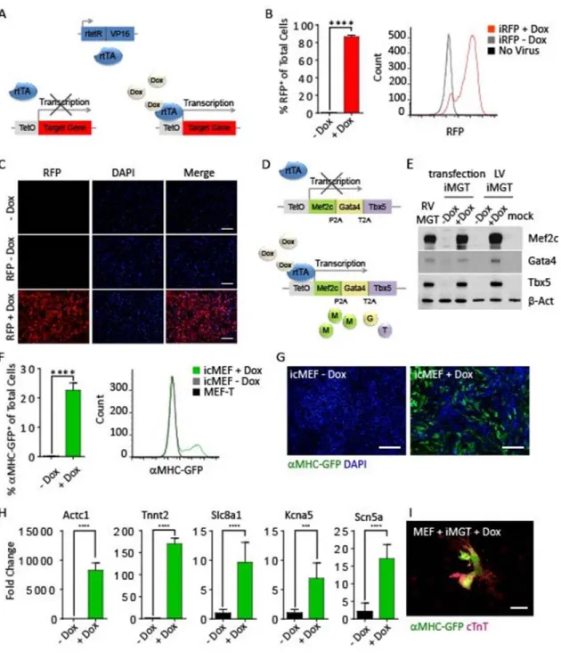
Inducible Polycistronic Reprogramming Construct (iMGT) Cloning
A modified pTRIPZ (Thermo Scientific, now Dharmacon) vector described previously (Zheng et al., 2014) was a kind gift from Qing Zhang. Our polycistronic MGT construct (Wang et al., 2015b) was cloned into the vector behind the tet operator sites and minimal CMV promoter using the AgeI and ClaI restriction enzyme sites. WPRE was amplified from the vector with primers to include flanking KpnI and MluI cleavage sites. Digestion of the vector with the enzymes MluI and KpnI removed a segment from base pairs 4064 to 8019 that included WPRE, rtTA3, PuroR, and the shRNAmir insertion site. This removal of the second KpnI site at bp 6543 leaves a single KpnI site at bp 8019. The WPRE PCR amplicon was digested with KpnI and MluI and ligated into the vector. The resulting construct contains
polycistronic MGT in frame behind the tet operator sites and minimal CMV promoter and lacks rtTA3 and PuroR. For the co-expression of rtTA with the iMGT construct, a pTRIPZ vector was digested with the enzymes XbaI and MluI and re-ligated to remove the tet operator sequences and minimal CMV
promoter. In this construct, rtTA3 is constitutively expressed under the human ubiquitin C promoter. The transcriptional activator rtTA3 is sensitive to doxycycline dosage and can be titrated to regulate transcriptional activity (Das et al., 2004).
Cardiac Reprogramming Using the icMEF Cell Line.
icMEF were seeded at a density of 2x10⁴ cells per well in 24 well plates coated with 0.01%
Growth Curve.
MEF-T were seeded at a density of 1x10⁵ cells per well in 6 well plates coated with 0.01%
gelatin. Wells were harvested in triplicate and cells counted by hemocytometer at 24-hour intervals for eight days.
Immunocytochemistry.
Cells were washed with PBS three times and fixed with 4% paraformaldehyde (EMS) at room temperature (RT) for 15 min. After permeabilization with 0.2% Triton/PBS for 15 min and blocking in 5% BSA for 1 hour, cells were treated with primary antibody at 4°C overnight, secondary antibody for 1 hour at RT, and nuclei staining with Hoechst 33342 (Life Technologies). The following antibodies were used: Rabbit anti-Ki67 (1:500, Abcam), rabbit anti-GFP (1:500, Life Technologies), Alexa Fluor 488–conjugated donkey anti-rabbit IgG (1:500, Jackson ImmunoResearch, Inc.). RFP reporter fluorescence was imaged without antibody staining. Images were acquired using EVOS® FL Auto Cell Imaging System (Life Technologies).
Western Blotting.
Cells were lysed in 2x SDS loading buffer (Bio-Rad). Proteins in cell lysate was separated by SDS-PAGE, transferred to nitrocellulose membranes, and probed with the following antibodies: Mef2c (1:1000, Abcam), Gata4 (1:200, Santa Cruz Biotechnology), Tbx5 (1:200, Santa Cruz Biotechnology), or β-Actin (1:1000, Santa Cruz Biotechnology). The target proteins were detected by chemiluminescence (ECL, Thermo Fisher Scientific). The membranes were stripped with stripping buffer (Sigma) and re-probed with antibody against a second protein or β-Actin as a loading control.
Real Time Quantitative Polymerase Chain Reaction.
protocol. RT-qPCR was performed on a ViiA 7 Real-Time PCR System (Applied Biosystems) with SYBR Green (Thermo Fisher Scientific) or TaqMan (Thermo Fisher Scientific) chemistry.
Statistical Analyses.
The statistical significance of differences between groups was analyzed using a two-way unpaired student’s t-test. A p-value < 0.05 was regarded as significant. Error bars indicate standard deviation.
Results
Development of the mouse embryonic fibroblast cardiac reporter cell line MEF-T.
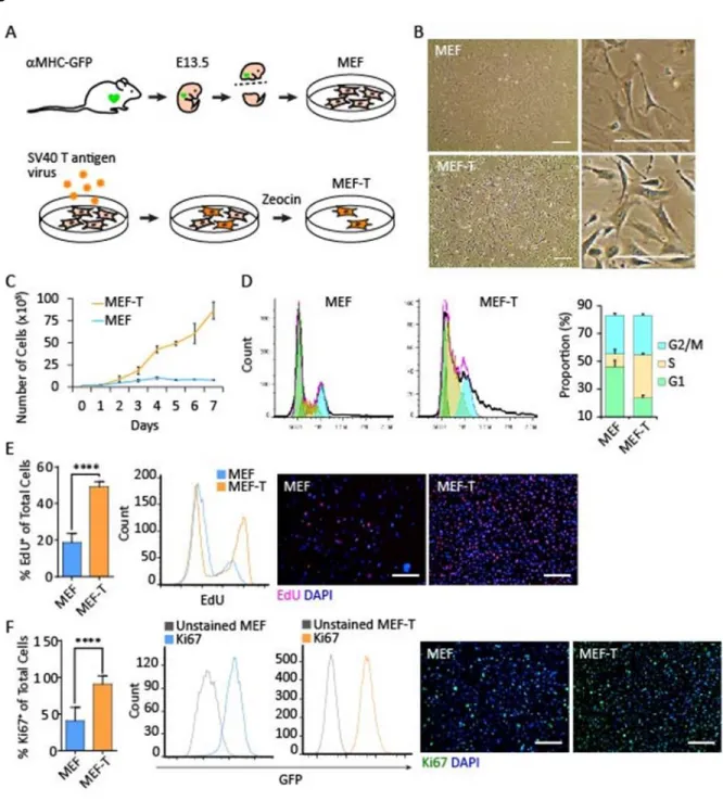
First, we developed a cardiac reporter fibroblast cell line. We isolated mouse embryonic fibroblasts (MEF) at embryonic day 13.5 from myosin heavy chain-green fluorescent protein ( MHC-GFP) cardiac reporter strain mice (Ieda et al., 2010; Qian et al., 2012) (Figure 2.1A). This transgenic strain drives GFP reporter expression with the cardiac MHC promoter. Cardiomyocytes from MHC-GFP mice are GFP positive, while fibroblasts are GFP negative. Consequently, primary MEFs isolated from MHC-GFP embryos are MHC-GFP negative; however, MEFs that have been reprogrammed into iCMs through the forced expression of cardiac lineage specific transcription factor cocktails are GFP positive. We
proliferation marker Ki67 is also over 2-fold higher than primary MEFs (Figure 2.1F). These data demonstrate that MEF-T is a transformed, highly proliferative fibroblast cell line.
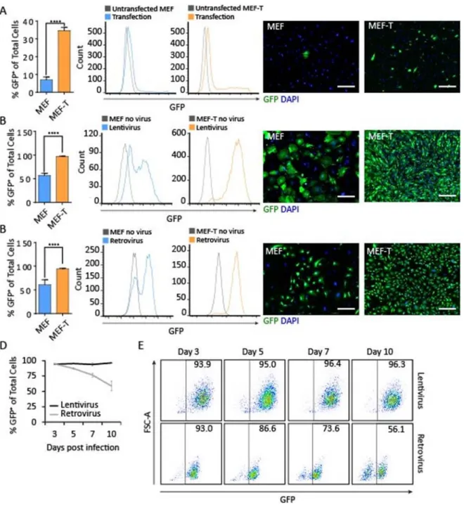
We next determined the potential of the MEF-T cell line for transgene expression using three common laboratory techniques: a lipid-based transfection reagent (Lipofectamine3000), lentiviral transduction, and retroviral transduction. First, we used Lipofectamine3000 to transfect primary MEF and MEF-T. Following transfection with a GFP-expressing plasmid, 35% of MEF-T expressed the GFP transgene compared to 7% of primary MEF (Figure 2.2A), indicating that MEF-T is significantly more susceptible to transfection using lipid-based transfection reagents than primary MEF. Second, we used a lentiviral vector to transduce primary MEF and MEF-T. A GFP-expressing lentivirus transduced 97% of MEF-T but only 57% of primary MEF (Figure 2.2B), indicating that MEF-T is also significantly more susceptible to transduction by lentiviral vectors. Third, to test a large construct encoded in a retroviral construct, we took advantage of our polycistronic system. We designed a retroviral construct to serve as a control for our polycistronic reprogramming construct Mef2c-Gata4-Tbx5 (MGT) (Wang et al., 2015a) by replacing the transcription factor Mef2c with the fluorescent reporter GFP to create the polycistronic construct GFP-Gata4-Tbx5 (GGT). GGT transduced 94% of MEF-T but only 61% of primary MEF (Figure 2.2C). MEF-T is significantly more susceptible to retroviral transduction, lentiviral transduction, and Lipofectamine transfection. Finally, we assessed the duration of transgene expression of constructs of different sizes in the MEF-T cell line. The small GFP lentivirus produced sustained transgene expression, while the large GGT retrovirus produced a transient transgene expression (Figure 2.2D and 2.2E). The cardiac reporter fibroblast cell line MEF-T is the first reported of its kind and will be a valuable tool for in vitro studies involving activation of cardiac markers such as MHC.
Development of the tetracycline-inducible gene expression MEF-T cell line iMEF.
MEF-T cell line to permit the temporal regulation of factor expression during the reprogramming process. With constitutive expression of the engineered reverse tetracycline-controlled transcriptional activator (rtTA), transcription of genes under the control of the tetracycline responsive element tetO in the promoter can be regulated by the addition or removal of the tetracycline derivative doxycycline. In the absence of doxycycline, rtTA does not bind to the tetracycline responsive element in the promoter and fails to initiate transcription of the target genes. However, in the presence of doxycycline, rtTA binds to the promoter and initiates transcription of the target genes. We generated a MEF-T cell line that constitutively expresses rtTA for the temporal regulation of transgene expression, called inducible MEF (iMEF). We tested the TetOn system in iMEF using lentiviral infection with a tetracycline responsive RFP reporter construct (Figure 2.3A). iMEFs transduced with RFP do not express RFP in the absence of doxycycline; however, with the addition of doxycycline to the culture media, 87% of iMEFs robustly express the RFP reporter (Figure 2.3B). The spectral peak of uninfected iMEF overlaps completely with RFP infected iMEFs in the absence of doxycycline, indicating that the TetOn system is not leaky (Figure 2.3B and 2.3C). The iMEF cell line is a valuable tool for temporal regulation of gene expression during reprogramming. One application of the iMEF cell line is to control RNA interference during