Original Research Article
The study of pulmonary function tests in patients with hypothyroidism
Sivaranjani H., Chaitra K. R.*
INTRODUCTION
Hypothyroidism is a very common disease worldwide. It is like a silent epidemic with a prevalence rate of approximately 11% in India.1 It is defined as a clinical
state resulting from insufficient secretion of thyroid hormone from thyroid gland due to structural and /or functional impairment of thyroid hormone production.2,3
It is well known that hypothyroidism affects all organ systems in this body, including the respiratory system.4 It
is seen that the respiratory disease in hypothyroidism ranges from mild dyspnea to a more severe respiratory failure.5,6 This has been attributed to reduction in
expiratory and inspiratory muscle strength, alveolar hypoventilation secondary to depression of hypoxic and hypercapnic ventilatory drives and reduced maximal breathing capacity and diffusion capacity in hypothyroid patients.7-9
Most of the hypothyroid patients complain of that author encounter in this practice complain of easy fatiguability
ABSTRACT
Background: Hypothyroidism is a common disease with a prevalence rate of 11% in India. It affects all organ systems in the body. Patients with hypothyroidism frequently have symptoms of fatigue and exercise intolerance. These symptoms could arise from a reduced pulmonary reserve, cardiac reserve or decreased muscle strength or increased muscle fatigue. This study aims to study the pulmonary function test in patients with hypothyroidism.
Methods: This is a cross sectional study conducted on 100 patients divided into 2 groups (a) newly detected hypothyroids (b) normal control group. Cases were matched with controls in having similar environment exposure and age group. All patients had routine symptom and clinical assessment. Laboratory investigations such as complete blood picture, pulmonary function test, chest x ray and thyroid function test were done. Data was entered and analysed.
Results: In this study conducted on 100 patients, case group had symptoms of easy fatiguability (36%), breathlessness (20%), menstrual abnormality (20%), weight gain (7%) and generalised body aches (5%). Mean FEV1 levels between cases and controls were 1.34 and 1.72 (p value 0.00), mean FVC were 1.88 and 2.09 (p value 0.114), FEV1/FVC ratio of 70.56, 81.98 respectively (p value 0.00). The distribution of PFT pattern was 32% obstructive, 28% mixed pattern and 22% restrictive pattern.
Conclusions: This study shows that hypothyroidism causes significant decrease in FEV1 and FEV1/FVC ratio, thereby suggesting obstructive patterns of lung involvement .Therefore PFT can be used routinely as a screening test for all hypothyroid patients to detect early respiratory dysfunction and thereby optimise treatment especially in obese patients and patients with pre-existing lung disease as hypothyroidism adds to their respiratory dysfunction.
Keywords: Hypothyroid, Obstructive pattern, Pulmonary function test, Restrictive pattern
Department of General Medicine, Bangalore Medical College and Research Institute, Bangalore, Karnataka, India
Received: 24 July 2019
Revised: 02 October 2019
Accepted: 09 October 2019
*Correspondence:
Dr. Chaitra K. R.,
E-mail: [email protected]
Copyright: © the author(s), publisher and licensee Medip Academy. This is an open-access article distributed under the terms of the Creative Commons Attribution Non-Commercial License, which permits unrestricted non-commercial use, distribution, and reproduction in any medium, provided the original work is properly cited.
and intolerance to exercise, these manifestations could be due to various reasons like a limited respiratory reserve, limited cardiac reserve or increased muscle fatigue.10
Another associated complication is difficulties faced during weaning from assisted ventilation in these hypothyroid patients.11 Therefore assessment of
Pulmonary function with spirometry in hypothyroid patients is particularly important.
The effect of hypothyroidism on respiratory system is debatable with studies suggesting both obstructive and restrictive patterns, therefore further studies are warranted to understand the involvement of respiratory system in hypothyroid patients. This study is done to see if there is any impairment in lung function parameters in hypothyroid patients and if present, to see the type of respiratory disease pattern and by doing so we can emphasis the importance of detecting and optimizing hypothyroidism treatment in patients having dyspnea and also in patients facing weaning difficulties during assisted ventilation. The aim and objective of the study were to study pulmonary function tests in patients with hypothyroidism.
METHODS
This study was a case control study conducted in the Department of General medicine, Bangalore medical college from June 2018 to June 2019. The study was approved by institutional ethical committee and written informed consent was taken from all patients who were included in the study. A total of 100 patients with age between 18 to 60 years were included in the study. The study included 2 group, group 1 consisted of 50 newly detected hypothyroids and group 2 with 50 controls who were age, sex matched and from similar environment as that of cases. Group 1 hypothyroid patients include both clinical (TSH >5 milliunits/L with clinical features of hypothyroidism or low fT4) and subclinical hypothyroidism (TSH>5 with no clinical features of hypothyroidism or normal fT4).
Patients already on thyroxine therapy, BMI>23 kg/m2,
history of smoking, anemia, respiratory, cardiac patients and pregnant women were excluded from the study. All patients underwent a detailed clinical examination and routine investigations such as thyroid function test (TSH, fT3 fT4), chest x ray and spirometry were also done. Spirometry was done by vitalograph, a software that is installed in a computer in the department of General medicine in Bangalore medical college, Karnataka.
All patients were asked to rest for 10 to 15 mins in a private quiet room, and they were briefed about the technique. PFT is carried out in a quiet room in siting position with a nose clip. An average of 3 readings were taken. Spirometric parameters recorded for analysis are: Forced vital capacity (FVC), Forced expiratory volume in 1st second (FEV1), FEV1/FVC, Peak expiratory flow
rate (PEFR), Forced expiratory flow 25%-75% (FEF25%-75%).
Inclusion criteria
• Age 18-60 years
• Patients giving informed consent
• Newly detected hypothyroidism both clinical (TSH >5milliunits/L with clinical features of hypothyroidism or low fT4) and Subclinical hypothyroidism (TSH>5 with no clinical features of hypothyroidism or normal fT4)
Exclusion criteria
• Patients already on thyroxine treatment
• BMI >23kg/m2
• H/o smoking, respiratory illness
• Pregnancy
• Anemia
• Cardiac illness
Statistical methods
Data was tabulated in Microsoft Office Excel and statistical analysis was done by using SPSS for windows (version 20.0). The thyroid function test parameters were analysed by paired t test and Pearson’s correlation analysis was used to analyse the relationships between TSH, fT4 with respiratory parameters, P value less than 0.05 was considered statistically significant. r value ranges from -1 to +1 using linear correlation analysis.
RESULTS
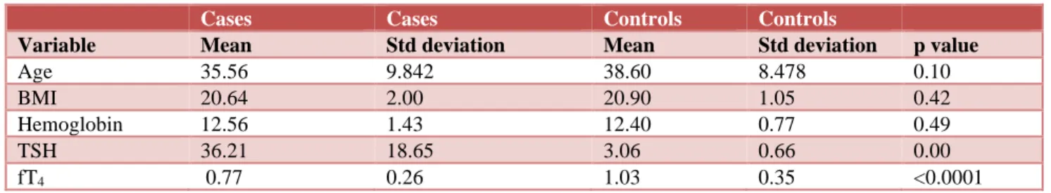
Demonstrates no significant difference in age, BMI in cases and controls. TSH was significantly higher while fT4 was significantly lower in cases compared to controls (Table 1).
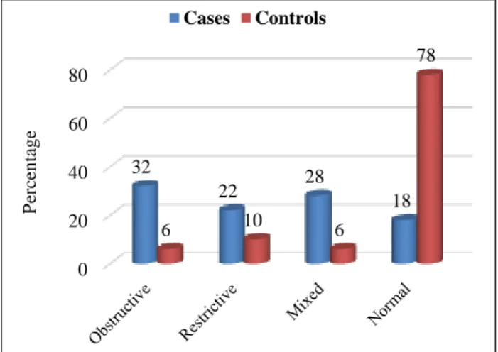
Figure 1: Distribution of cases and controls based on pattern of PFT.
0 20 40 60 80
32
22 28
18
6 10 6
78
P
erc
en
tag
e
In this study author observed a significant decrease in FEV1 and FEV1/FVC ratio in hypothyroids as shown in (Table 2). FVC between cases and controls did not show statistical significance, although the mean FVC was found to be lower in cases (1.88) as compared to controls (2.09).
The various respiratory patterns seen in cases were as follows: obstructive pattern (32%), followed by mixed pattern and restrictive pattern (28%, 22% respectively) (Figure 1).
Furthermore, we observed that there was no significant correlation between TSH or fT4 with FVC, FEV1, FEV1/FVC as seen in (Table 3 and 4).
Table 1: Baseline characteristics of cases and controls.
Cases Cases Controls Controls
Variable Mean Std deviation Mean Std deviation p value
Age 35.56 9.842 38.60 8.478 0.10
BMI 20.64 2.00 20.90 1.05 0.42
Hemoglobin 12.56 1.43 12.40 0.77 0.49
TSH 36.21 18.65 3.06 0.66 0.00
fT4 0.77 0.26 1.03 0.35 <0.0001
Table 2: Pulmonary function tests parameters.
Case Case Control Control
Variable Mean Std deviation Mean Std deviation p value
FVC(L) 1.88 0.717 2.09 0.600 0.144
FEV1 1.34 0.49 1.72 0.57 0.00
EFV1/FVC 70.56 16.93 81.98 11.49 0.00
Table 3: Correlation of FT4 with lung function parameters.
FEV1 FVC FEV1/FVC
FT4 r -0.18 -0.26 -0.01 p 0.20 0.07 0.89
r = Correlation coefficient, p<0.05 considered statistically significant
Table 4: Correlation of TSH with lung function parameters.
FEV1 FVC FEV1/FVC
TSH
Cases r 0.10 0.20 -0.13 p 0.45 0.16 0.36
Controls r 0.09 0.10 -0.06 p 0.49 0.45 0.66
r = Correlation coefficient, p<0.05 considered statistically significant
DISCUSSION
Thyroid hormone acts on various organ systems in this body therefore hypothyroidism can cause varied defects in organ functioning including respiratory system. Thyroid disorders mainly affect females because oestrogen has direct effect on thyroid follicular cells and
increase thyroid binding globulin levels.12 Another most
probable cause explained was the presence of skewed X chromosome inactivation. In this study all 100 patients were females.
Hypothyroidism causes disorders of respiratory function and disturbances of ventilation. Respiratory functioning is affected in the form of hypoventilation and reduction in ventilatory response to hypoxia and hypercapnia.7,13
Hypothyroidism may lead to myopathy, studies have particularly concentrated on inspiratory and expiratory muscles. Supplementation with thyroxine has shown to restore the respiratory muscle function.8
In this study, author observed that there was a significant decrease in FEV1 and FEV1/FVC ratio in hypothyroid patients as shown in table 2, FVC on the other hand was lower in cases (1.88) than controls (2.09) but was not statistically significant (0.114). FEV1 is the volume of air exhaled in 1st second and is the most reproducible and the most important parameter of lung function. The most common respiratory pattern observed was obstructive patterns followed by mixed pattern and restrictive pattern.
This finding is in accordance with the study by Bhuvaneswari T et al, and Cakmak1 who found significant reduction is FEV1/FVC ratio.14,15 But there are
many studies having conflicting reports. The study by Roel S et al, and Sharifi F et al, showed restrictive pattern while study done by Bassi R et al, and Siafakas NM et al, points to a mixed pattern of respiratory disorder in hypothyroidism. These studies also concluded that with thyroxine replacement there was significant improvement in the lung function.8,16,17,19 The pathophysiology that
leads to decline in pulmonary functions is attributed to varied causes, the most important being defective ventilatory drive. Hypoxic drive is affected earlier than hypercapnic drive.
Another explanation is decreased respiratory muscle strength in which the proportion of Type-1 fibers in the diaphragm and intercostal muscles is changed. There can also be the deposition of glycosaminoglycans in pulmonary interstitial tissue causing osmotic oedema and fluid retention.18 Martinez et al, confirmed that
hypothyroid patients develop diaphragmatic dysfunction which can be mild to very severe forms, also low thyroid hormone levels decrease lung elastic tissue and increase work of breathing.19
Considering correlation studies author have observed non significant negative correlation between TSH and FEV1/FVC, fT4 showed nonsignificant positive correlation between FVC, FEV1/FVC and FEV. Valjevac et al, also observed a significant negative correlation between TSH and FVC%.20 Bassi et al, in his study on
newly diagnosed hypothyroids found a highly significant negative correlation of TSH with FEV1 and FEV1/FVC%.18
Sharon et al, also found a statistically non-significant negative correlation of TSH with FVC, FEV1, FEV1/FVC.16 They have also reported a positive
correlation between fT4 with all the lung parameters, but none were statistically significant.
CONCLUSION
In conclusion it is noted that in hypothyroidism there is significant reduction in the dynamic lung functions as compared with controls. Therefore, respiratory system can be affected in hypothyroidism and a simple spirometry can be considered for the evaluation of pulmonary function in these patients.
Limitation of the study included, males were not included in the study due to inadequate numbers and also PFT after levothyroxine therapy was not done to see if the above changes reverse or not.
ACKNOWLEDGEMENTS
Authors are grateful to all the patients who participated in this study.
Funding: No funding sources Conflict of interest: None declared
Ethical approval: The study was approved by the Institutional Ethics Committee
REFERENCES
1. Bagcchi S. Hypothyroidism in India: more to be done. The lancet diabetes and endocrinol. 2014;2(10):778.
2. Dashe JS, Mazziotti A, Press M, Cooper DS. Subclinical hypothyroidism (multiple letters). New Eng J Med. 2001 Dec 20;345(25):1855-6.
3. Kek PC, Ho SC, Khoo DH. Subclinical thyroid disease. Singap Med J. 2003;44(11):595-600. 4. Werner SC, Ingbar SH, Braverman LE, Utiger RD,
editors. Werner and Ingbar's the thyroid: a fundamental and clinical text. Lippincott Williams and Wilkins; 2005.
5. McQuade C, Skugor M, Brennan DM, Hoar B, Stevenson C, Hoogwerf BJ. Hypothyroidism and moderate subclinical hypothyroidism are associated with increased all-cause mortality independent of coronary heart disease risk factors: A PreCIS database study. Thyroid. 2011;21(8):837-43. 6. Resta O, Pannacciulli N, Di Gioia G, Stefano A,
Barbaro MF, De Pergola G. High prevalence of previously unknown subclinical hypothyroidism in obese patients referred to a sleep clinic for sleep disordered breathing. Nutrit, Metabol Cardiovas Dis. 2004;14(5):248-53.
7. Zwillich CW, Pierson DJ, Hofeldt FD, Lufkin EG, Weil JV. Ventilatory control in myxedema and hypothyroidism. New Engl J Med. 1975;292(13):662-5.
8. Siafakas NM, Salesiotou V, Filaditaki V, Tzanakis N, Thalassinos N, Bouros D. Respiratory muscle strength in hypothyroidism. Chest. 1992;102(1):189-94.
9. Brent GA, Davies T. Hypothyroidism and Thyroiditis. In: Melmed S, Polonsky KS, Larsen PR, Kronenberg HM, Editors. Williams Text book of Endocrinology. 12th ed. Philadelphia WB.
Saunders Co; 2011:409.
10. Warren M. Gold M. Pulmonary Function Tests. In: Murray JF, Nadel JA, Editors. Textbook of Respiratory Medicine. 3rd ed. Philadelphia: WB.
Saunders Co.; 2000:781-785.
with severe hypothyroidism. The Am J med. 1993;95(1):29-37.
12. Larsen PR, Kronenberg HM, Melmed, Polonsky KS. William’s Textbook of Endocrinology. 10th ed. Philadelphia: Elsevier Saunders; 2003:424-440. 13. Datta D, Scalise P. Hypothyroidism and failure to
wean in patients receiving prolonged mechanical ventilation at a regional weaning center. Chest. 2004;126(4):1307-12.
14. Bhuvaneswari T, Banu KK. Evaluation of pulmonary functions in patients with hypothyroidism who are on conservative management. Sch J App Med Sci. 2014;2(2A):495-7.
15. Çakmak G, Saler T, Sağlam ZA, Yenigün M, Demir T. Spirometry in patients with clinical and subclinical hypothyroidism. Hypothyroidism: Influences and Treatments. 2007;55(3):266-70. 16. Roel S, Punyabati O, Prasad L, Salam R, Ningshen
K, Shimray AJ, et al. Assessment of functional lung impairment in hypothyroidism. IOSR J Dent Med Sci. 2014;13:4-7.
17. Sharifi F, Amari A. The effect of levothyroxin on pulmonary function tests of hypothyroid patients. Int J Endocrinol Metab. 2005;1:48-51.
18. Bassi R, v Dhillon S, Sharma S, Sharma A, Tapdiya M. Effect of thyroid hormone replacement on respiratory function tests in hypothyroid women. Pak J Physiol. 2012;8(2):20-3.
19. Martinez FJ, Bermudez-Gomez M, Celli BR. Hypothyroidism: a reversible cause of diaphragmatic dysfunction. Chest.1989;96(5):1059-63.
20. Valjevac S, Hadzovic-Dzuvo A, Valjevac A, Kucukalic-Selimovic E, Lepara O. Assessment of lung dysfunction with spirometry in patients with thyroid disorders. Acta Informatica Medica. 2011;19(1):16.