Original Research Article
Differences in survival by race/ethnicity among cutaneous melanoma
patients in the United States over a period from 1982 to 2011
Abdulrahman M. Nasiri
1*, Elharith S. Al-Akeel
1, Nora H. Rayes
2INTRODUCTION
Melanoma is an aggressive skin cancer, which affects pigment-producing cells called melanocytes.1 Although it
accounts for only about 10% of skin cancer cases,
melanoma has a high mortality rate. Furthermore, melanoma has different types, which affect different races and different sites.2 Unlike other types of cancers,
the incidence of melanoma has increased in recent years.3
According to Sandru et al, the likelihood of getting
ABSTRACT
Background: Melanoma is an aggressive skin cancer with a high mortality rate. The incidence of melanoma has increased in recent years from 1:1500 in 1935 to 1:50 in 2011. The aim of this study is to investigate survival by race/ethnicity, taking site into account, among melanoma patients in the United States.
Methods: This study is a secondary analysis of the Surveillance, Epidemiology, and End Results (SEER) Program. SEER collects data through a non-concurrent cohort study design. The sample size was 185219 participants. The chi-square test was used to examine the association between categorical variables. Kaplan-Meier survival analysis was used to estimate the overall survival curve and to estimate the survival curve per race/ethnicity. Collinearity was assessed using Pearson correlation. Cox proportional hazards regression was used to calculate the unadjusted and adjusted hazard ratios (HR).
Results: Non-Hispanic White (NHW) and Other patients were older in age (70 years or older), while non-Hispanic Black (NHB) and Hispanic patients were younger (30-39 years). Melanoma in NHW patients was mostly located in trunk whereas melanoma for NHB, Hispanic and Other patients was mostly located in the lower limbs. For all races/ethnicities except for NHB, more individuals were diagnosed between 2002 and 2011. Patients with melanoma in upper limbs lived more frequently. Fewer women died (6.8%) compared to men (17.1%). Patients who were diagnosed between the ages of 30-39 were more likely to die. NHB had an adjusted HR of 3 (95% CI 2.7, 3.3) compared to NHW. The adjusted HR of lower limb was 1.6 (95% CI 1.5, 1,6) compared to the reference group (Head and Neck). The hazard for trunk and lower limb were about the same as the reference. Those who were 70 years or older had an adjusted HR of 2.2 (95% CI 2.0, 2.4). Women had an adjusted HR of 0.4 (95% CI 0.4, 0.5), and diagnosis during the decade 1982-1991 had an adjusted HR of 2.6 (95% CI 2.4, 2.7).
Conclusions: NHB patients and patients of ages 30-39 years were more likely to die. The poorest survival was for diagnosis between 1982 and 1991. However, more individuals were diagnosed between 2002 and 2011. The lower limb had a worse prognosis with adjusted HR of 1.6 (95% CI 1.5, 1,6), and more men were diagnosed than women.
Keywords: Cancer, Epidemiology and end results (SEER) program, Incidence, Melanoma, Mortality, Races/ethnicities
1College of Medicine, Al Imam Mohammad Ibn Saud Islamic University (IMSIU), Riyadh, Saudi Arabia 2College of Medicine, Princess Nora Bint Abdul Rahman University (PNU), Riyadh, Saudi Arabia
Received: 03 November 2017
Accepted: 29 November 2017
*Correspondence:
Dr. Abdulrahman M. Nasiri, E-mail: dramnasiri@gmail.com
Copyright: © the author(s), publisher and licensee Medip Academy. This is an open-access article distributed under the terms of the Creative Commons Attribution Non-Commercial License, which permits unrestricted non-commercial use, distribution, and reproduction in any medium, provided the original work is properly cited.
melanoma increased from 1:1500 in 1935 to 1:50 in 2011. Lifetime risk of developing melanoma is related to internal and external factors. Internal factors that increase risk include genetics and age, while external factors include fair skin, red hair, light eyes, abundance of freckles, atypical moles or a large number of moles.2 Fare
skin, skin lesions, nevi and advanced age increase risk of developing melanoma.
The Surveillance, Epidemiology, and End Results (SEER) database, a National Cancer Institute program that collects cancer and survival data of U.S. cancer patients, has also shown that the increase in incidence of melanoma varies among different areas and races/ethnicities. The incidence rate as found in SEER shows the highest incidence in Caucasians (19.1 females and 29.7 males per 100,000), followed by Hispanics (4.7 females and 4.4 males per 100,000), followed by Asians and Blacks (1.0 females and 1.1 males per 100,000).4
These numbers are supported by another smaller study.5
Different sites of melanoma have different impact and prognosis. For example, in men, melanoma located on the scalp/neck has a worse prognosis and survival rate compared to other sites.6 In men the most frequent site of
melanoma was the trunk, followed by the lower and upper extremities, while in women, the lower extremities were the predominant site. Common melanoma sites vary by race. Non-Hispanic Whites develop the lesion more frequently in the trunk and upper extremities, while African-Americans, Hispanics, and Asians develop the lesion more frequently in lower extremities.7
Many papers have studied the overall survival for melanoma patients of different races/ethnicities, but with different scopes. Some have studied a specific population while others focused on a specific state. Those who studied the overall survival of melanoma patients by race/ethnicity used a short period of follow-up time or did not analyze recent data.
The aim of this study is to investigate survival by race/ethnicity, taking site into account, among melanoma patients in the United States over a period of 30years (1982-2011). This study will be important in determining whether there have been differences in the survival by race/ethnicity, which could influence the ways interventions for melanoma are targeted.
METHODS
This study is a secondary analysis of data from the SEER Program, which is the largest registry in the world to provide cancer statistics.5,7 SEER collects data through a
non-concurrent cohort study design. The inclusion criteria for this study were adults 18years or older with primary cutaneous melanoma located on head and neck, trunk, upper extremities or lower extremities who were diagnosed in the period of 1982 to 2011.The exclusion criteria were other cancers or sites, secondary lesions,
multiple or unspecified melanoma sites, and patient less than 18years. After excluding duplicate cases (n=20366), those who were less than 18years (n=1300), those who received their diagnosis prior to 1982 (n=16426), and cases that were missing information related to survival (n=25864; 12.3%), the final sample size was 185219 participants.
The variables included in the study were race, age at diagnosis, site, gender and year of diagnosis. Race/ethnicity was divided into four groups: non-Hispanic White (NHW), non-non-Hispanic black (NHB), Hispanic, and Other, which included Asian/Pacific Islander, American Indian/Alaska Native and other/unspecified. Age at diagnosis was grouped into six categories: under 30years, 30 to 39, 40 to 49, 50 to 59, 60 to 69 and 70 and above.8 Site was classified into head and
neck (H/N), trunk, upper limb (UL) and lower limb (LL). Eyelid and ears were considered part of the head and neck category. Both men and women were studied, and year of diagnosis was grouped into three decades: 1982-1991, 1992-2001 and 2002-2011.
Cross-tables were used to compare the different variables. The chi-square test was used to examine the association between categorical variables. A p-value of 0.05 was used as criteria to select confounders. Kaplan-Meier survival analysis was used to estimate the overall survival curve and to estimate the survival curves per race. Collinearity was assessed using Pearson correlation. Cox proportional hazards regression was used to calculate the unadjusted and adjusted hazard ratios (HR) and confidence interval (CI) of 95% was used to indicate the significance of the results. SPSS statistical package for Windows version 22 (IBM) was used to analyze the data.
RESULTS
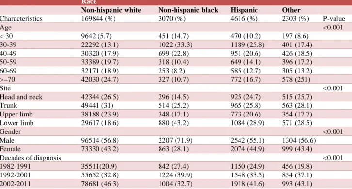
Table 1 describes the demographic characteristics of U.S. adult patients with primary cutaneous melanoma in the period of 1982-2011. NHW as well as Other patients with cutaneous melanoma tended to be older (70 years or older). NHB and Hispanic patients with cutaneous melanoma tended to be younger (30-39years). Melanoma was commonly located in the lower limbs for NHB, Hispanic and Other patients (43.2%, 28.9% and 28.5 respectively), while melanoma in NHW was in the trunk (31%). Unlike other races/ethnicities, NHB men had a greater proportion of cutaneous melanoma than NHB women. More individuals were diagnosed in the last decade (2002-2011) in all races except for NHB.
Table 1: Characteristics of U.S. adult patients with primary cutaneous melanoma, 1982 to 2011 (n=185219).
Race
Non-hispanic white Non-hispanic black Hispanic Other
Characteristics 169844 (%) 3070 (%) 4616 (%) 2303 (%) P-value
Age <0.001
< 30 9642 (5.7) 451 (14.7) 470 (10.2) 197 (8.6) 30-39 22292 (13.1) 1022 (33.3) 1189 (25.8) 401 (17.4) 40-49 30320 (17.9) 699 (22.8) 951 (20.6) 426 (18.5) 50-59 33389 (19.7) 318 (10.4) 649 (14.1) 396 (17.2) 60-69 32171 (18.9) 253 (8.2) 585 (12.7) 305 (13.2) >=70 42030 (24.7) 327 (10.7) 772 (16.7) 578 (251)
Site <0.001
Head and neck 42344 (26.5) 296 (14.5) 925 (24.7) 515 (25.7) Trunk 49441 (31) 514 (25.2) 965 (25.8) 563 (28.1) Upper limb 38188 (23.9) 348 (17.1) 773 (20.6) 354 (17.7) Lower limb 29617 (18.6) 880 (43.2) 1084 (28.9) 571 (28.5)
Gender <0.001
Male 96514 (56.8) 2207 (71.9) 2542 (55.1) 1304 (56.6) Female 73330 (43.2) 863 (28.1) 2074 (44.9) 999 (43.4)
Decades of diagnosis <0.001
1982-1991 35511(20.9) 842 (27.4) 1150 (24.9) 456 (19.8) 1992-2001 55652 (32.8) 1224 (39.9) 1548 (33.5) 854 (37.1) 2002-2011 78681 (46.3) 1004 (32.7) 1918 (41.6) 993 (43.1)
Table 2: Unadjusted association between vital status and risk factors in U.S. adult patients with primary cutaneous melanoma, 1982-2011 (N=185219).
Vital status
Alive Dead
Characteristics 161785 (%) 23434 (%) P-value
Race <0.001
Non-hispanic white 149246 (87.9) 20598 (12.1) Non-hispanic black 1840 (59.9) 1230 (40.1)
Hispanic 3531 (76.5) 1085 (23.5)
Other 1840 (79.9) 463 (20.1)
Age <0.001
< 30 9690 (86.3) 1533 (13.7)
30-39 20376 (79.3) 5328 (20.7)
40-49 29027 (86.7) 4468 (13.3)
50-59 32579 (90.7) 3322 (9.3)
60-69 30897 (90.1) 3380 (9.9)
>=70 39216 (87.9) 5403 (12.1)
Site <0.001
Head and neck 41264 (91.1) 4010 (8.9)
Trunk 48177 (90.6) 4991 (9.4)
Upper limb 38052 (92.8) 2971 (7.2) Lower limb 29600 (89.4) 3515 (10.6) Gender
Male 87375 (82.9) 17970 (17.1)
Female 74410 (93.2) 5464 (6.8)
Decades of diagnosis <0.001
No collinearity was observed in the adjusted model. The median follows up time was 81 months. Survival for each race was calculated using Kaplan-Meier analysis. Table 3 describes unadjusted and adjusted hazard ratios. NHB patients had an adjusted HR of 3 (95% CI 2.7, 3.3) compared to NHW patients; this HR decreased after adjustment. In the unadjusted model, individuals diagnosed between ages 30 to 39years had an HR of 1.6 (95% CI 1.5, 1.7), while those 70years or older had an
HR of 1.2 (95% CI 1.1, 1.2). In the adjusted model, those 70 or older had a HR of 2.2 (95% CI 2.0, 2.4). In the unadjusted model, the HR for individuals diagnosed between ages 40 to 49 was not statistically significant; the estimate became significant in the adjusted model (HR=1.2; 95% CI 1.1, 1.3). The adjusted HR of lower limb was two times higher than that of other sites (HR=1.6; 95% CI 1.5, 1,6). Women had an adjusted HR of 0.4 (95% CI 0.4, 0.5), and diagnosis during the decade 1982-1991 had an adjusted HR of 2.6 (95% CI 2.4, 2.7).
Table 3: Unadjusted and adjusted hazard ratios for primary cutaneous melanoma among adult patients in U.S. in the period of 1982-2011 (n=185219).
Unadjusted Adjusted
Characteristics HR (95% CI) P-value HR (95% CI) P-value
Race
Non-hispanic white REF REF
Non-hispanic black 4.3(4.0, 4.5) <0.001 3.0(2.7, 3.3) <0.001 Hispanic 2.2(2.0, 2.3) <0.001 1.8(1.6, 2.0) <0.001 Other 1.8(1.6, 1.9) <0.001 1.6 (1.4, 1.8) <0.001
Age
< 30 REF REF
30-39 1.6(1.5, 1.7) <0.001 1.4(1.3, 1.5) <0.001 40-49 1.0(1.0, 1.1) 0.475 1.2(1.1, 1.3) <0.001 50-59 0.7(0.7, 0.8) <0.001 1.1(1.0, 1.2) 0.006 60-69 0.8(0.8, 0.9) <0.001 1.3(1.2, 1.4) <0.001 >=70 1.2(1.1, 1.2) <0.001 2.2(2.0, 2.4) <0.001
Site
Head and neck REF REF
Trunk 1.0(0.9, 1.0) 0.02 1.1(1.0, 1.1) <0.001 Upper limb 0.8(0.7, 0.8) <0.001 1.0(0.9, 1.0) 0.041 Lower limb 1.1(1.0, 1.1) 0.001 1.6(1.5, 1,6) <0.001
Gender
Male REF REF
Female 0.4(0.3, 0.4) <0.001 0.4(0.4, 0.5) <0.001
Decades of diagnosis
1982-1991 3.5(3.3, 3.6) <0.001 2.6(2.4, 2.7) <0.001 1992-2001 2.0(1.9, 2.1) <0.001 1.6(1.5, 1.7) <0.001
2002-2011 REF REF
DISCUSSION
This study set out to examine differences in survival by race/ethnicity. NHB patients had 3 times the hazard of dying compared to NHW (HR=3; 95% CI 2.7, 3.3); this hazard was high even before adjusting for other variables (HR=4.3; 95% CI 4.0, 4.5). This finding is similar to that of other studies.6,9,10
In the unadjusted model, patients diagnosed between ages 30 to 39years had an HR of 1.6 (95% CI 1.5, 1.7) in comparison to the reference group, but after adjusting this HR was closer to 1 (HR=1.4; 95% CI 1.3, 1.5) and increased for individuals diagnosed at 70 years or older (HR=2.2; 95% CI 2.0, 2.4). In the unadjusted model, the
hazard ratios by site were similar to the reference group, but in the adjusted model the HR for lower limb increased to 1.6 (95% CI 1.5, 1,6).
The sample for this study included 91.7% NHW, which was in contrast with most of the literature.11 There was an
increase in the number of cases of cutaneous melanoma between 2002-2011.12 The highest number of cutaneous
melanoma cases was among NHW patients 70years or older, which is similar to trends in a previous study conducted in 2012.3,13
lower limb melanoma was more common (43.2% and 28.9%, respectively). This is similar to what Cormier et al found. However, the sample size in this study is larger and the observation period longer than what Cormier et al reported.14,15 More men had melanoma compared to
women, and this was observed for men of all races (56.8% NHW, 71.9% NHB, 55.1% Hispanic and 56.5% Other), which is similar to findings in another study.16
Regarding decade of diagnosis, diagnosis from 1982 to 1991 had an adjusted HR of 2.6 (95% CI 2.4, 2.7).17
One of the strengths of this study is the large population included, which enhances the external validity and applicability to the U.S. population. The secondary nature of this analysis may be considered a limitation. However, the data used were from SEER, which is a high-quality database that follows patients for a long period of time. One limitation was the loss of approximately 12% of cases due to missing information related to survival. However, this didn’t affect the overall result because of the large sample size. Additionally, this study did not take variables such as frequency of outdoor activities, socioeconomic status, ulceration, tumor thickness and tumor width into account. Furthermore, this study did not differentiate sites in the head and neck. One article studied the head and neck in more details by splitting them into additional sites, which showed differences in prognosis.18,19 However, in other studies the head and
neck were studied as one site.20 Finally, the follow up
period was not equal among the three decades of diagnosis. For example, individuals diagnosed in 1982 had the longest possible follow up time, while individuals diagnosed in 2011 had the shortest length of follow up.
CONCLUSION
In conclusion, this study showed that NHB patients had the poorest survival compared to other races. LL, which had a higher HR compared to other sites, was the most common melanoma site among NHB and Hispanic patients, while the trunk was the most common site for NHW patients. These findings add something valuable in the field of melanoma.
Funding: No funding sources Conflict of interest: None declared
Ethical approval: The study was approved by the institutional ethics committee
REFERENCES
1. Higgins HW, Lee KC, Galan A, Leffell DJ. Melanoma in situ: part I. Epidemiology, screening, and clinical features. J Ame Academy Dermatol. 2015;73(2):181-90.
2. Ferri F. Ferri's Clinical Advisor: Expert Consult; 1st Edition. Elsevier. 2015:777-779.
3. Simard EP, Ward EM, Siegel R, Jemal A. Cancers with increasing incidence trends in the United
States: 1999 through 2008. CA: Cancer J Clinicians. 2012;62(2):118-28.
4. Hawryluk EB, Fisher DE. Melanoma Epidemiology, Risk Factors, and Clinical Phenotypes, Advances in Malignant Melanoma - Clinical and Research Perspectives, Dr. April Armstrong (Ed.), In Tech; 2011. Available from: https://www.intechopen.com/books/advances-in- malignant-melanoma-clinical-and-research- perspectives/melanoma-epidemiology-risk-factors-and-clinical-phenotypes.
5. Lachiewicz AM, Berwick M, Wiggins CL, Thomas NE. Survival differences between patients with scalp or neck melanoma and those with melanoma of other sites in the Surveillance, Epidemiology, and End Results (SEER) program. Archives Dermatol. 2008;144(4):515-21.
6. Zell JA, Cinar P, Mobasher M, Ziogas A, Meyskens Jr FL, Anton-Culver H. Survival for patients with invasive cutaneous melanoma among ethnic groups: the effects of socioeconomic status and treatment. J Clinical Oncol. 2008;26(1):66-75.
7. National Cancer Institute. The Surveillance, Epidemiology, and End Results (SEER) Program. Available at http://seer.cancer.gov/. Accessed July 28, 2015.
8. Thörn M, Adami HO, Ringborg U, Bergström R, Krusemo U. The association between anatomic site and survival in malignant melanoma. An analysis of 12,353 cases from the Swedish Cancer Registry. Euro J Cancer Clinic Oncol. 1989;25(3):483-91. 9. Reintgen DS, McCarty KM, Cox E, Seigler HF.
Malignant melanoma in black American and white American populations: a comparative review. Jama. 1982;248(15):1856-9.
10. Hu S, Soza-Vento RM, Parker DF, Kirsner RS. Comparison of stage at diagnosis of melanoma among Hispanic, black, and white patients in Miami-Dade County, Florida. Archives Dermatol. 2006;142(6):704-8.
11. Crombie IK. Racial differences in melanoma incidence. Brit J Cancer. 1979;40(2):185-93. 12. Linos E, Swetter SM, Cockburn MG, Colditz GA,
Clarke CA. Increasing burden of melanoma in the United States. J Investigative Dermatol. 2009;129(7):1666-74.
13. Jemal A, Devesa SS, Hartge P, Tucker MA. Recent trends in cutaneous melanoma incidence among whites in the United States. J National Cancer Inst. 2001;93(9):678-83.
14. Cormier JN, Xing Y, Ding M, Lee JE, Mansfield PF, Gershenwald JE, et al. Ethnic differences among patients with cutaneous melanoma. Archives Internal Medic. 2006;166(17):1907-14.
16. Garbe C, Leiter U. Melanoma epidemiology and trends. Clinics in dermatology. 2009;27(1):3-9. 17. Hall HI, Miller DR, Rogers JD, Bewerse B. Update
on the incidence and mortality from melanoma in the United States. J Ame Academy Dermatol. 1999;40(1):35-42.
18. Gillgren P, Månsson‐Brahme E, Frisell J, Johansson H, Larsson O, Ringborg UA. Prospective Population‐Based Study of Cutaneous Malignant Melanoma of the Head and Neck. Laryngoscope. 2000;110(9):1498-504.
19. Garbe C, Büttner P, Bertz J, Burg G, D'Hoedt B, Drepper H, et al. Primary cutaneous melanoma. Prognostic classification of anatomic location. Cancer. 1995;75(10):2492-8.
20. Wu XC, Eide MJ, King J, Saraiya M, Huang Y, Wiggins C, et al. Racial and ethnic variations in incidence and survival of cutaneous melanoma in the United States, 1999-2006. J Ame Academy Dermatol. 2011;65(5):S26-e1.