IDENTIFYING MECHANISMS REGULATING WNT SIGNALING DURING POSTEMBRYONIC DEVELOPMENT
Kelly Marie Alexandre
A thesis submitted to the faculty of the University of North Carolina at Chapel Hill in partial fulfillment of the requirements for the degree of Master of Science in the
Department of Biology
Chapel Hill 2009
ABSTRACT
Kelly Alexandre: Identifying Mechanisms Regulating Wnt Signaling During Postembryonic Development
(Under the direction of Mark Peifer)
Proper regulation of the Wingless (Wg)/Wnt pathway is essential for development; inappropriate activation of the pathway occurs in many human cancers. Wg signaling stabilizes the effector, Armadillo (Arm) and in its absence Arm is phosphorylated, ubiquitinated, and then degraded. In this work, we take a closer look at Arm regulation. First we investigated the difference in Arm
accumulation in APC, a member of the destruction complex, mutants. We find that while protein levels of Arm are the same, mRNA levels differ between embryos and larvae. Second, published data suggests that the SCF complex is responsible for Arm ubiquitination. The canonical SCF complex is known to contain five parts, including a RING domain protein of the Roc1/Rbx1 family. However, some data has called into question the exactly which Roc protein is part of the complex. Our data suggests that neither Roc1b nor Roc2 are solely responsible for Arm degradation.
iv
ACKNOWLEDGEMENTS
I would like to begin by thanking my advisor, Mark Peifer. His guidance and support have helped me grow as a scientist and a person. I would also like to thank the Peifer lab—Dave, Nasser, Ed, Jess, Nathan, Steph, and Doug. We have rejoiced over scientific accomplishments and commiserated over the failed experiments. Jess and Steph in particular have become a large part of my life in and outside of lab. My committee has been indispensable in my progress as a scientist. Of the people in my life there are a few who I could not be without, whose love and support have shaped who I am today. My parents have played integral parts in shaping my life and helping me along in my journey. I am lucky enough to have four loving, supportive, and amazing parents—Wayne, Debbie, Tom and Maria. They have had four different lives and their diversity has created a wealth of knowledge that I continually tap into. While I was growing up their support and understanding was there for me through some very hard times. Now, as an “adult” I no longer need that constant guidance but I will always appreciate their love. Next, I want to acknowledge Matt. He has been my personal cheerleader for the past three years. Each time I thought things would never get better he was there to show me the light. His love helps me to
understand I am truly blessed. And of course, I have to thank my brother, Chris. The two of us make up a twenty-eight year, two person mutual admiration
vi
TABLE OF CONTENTS
LIST OF FIGURES ... vii
LIST OF ABBREVIATIONS ...viii
Chapter I. INTRODUCTION ...1
II. RESULTS ...10
Armadillo regulation through development ...10
Assessing the roles of different Roc proteins In Arm regulation ...13
III. DISCUSSION...18
MATERIALS AND METHODS ...22
LIST OF FIGURES
Figure
1. APC comparison...3
2. The Wnt pathway...4
3. The canonical SCF complex ...7
4. Armadillo accumulation in APC2g10APC1Q8 mutants during fly development...11
5. Wnt pathway component clones ...12
6. Plane of focus ...12
7. Armadillo levels at different stages of development...13
8. Verification of Roc2 and Roc1b mutants...14
9. Armadillo levels in Roc2 mutants...15
10. Immunoblot comparison of Armadillo levels in Roc mutants...16
LIST OF ABBREVIATIONS
aa amino acid
APC Adenomatous Polyposis Coli
Arm Armadillo
ATP Adenosine triphosphate β-cat β-catenin
bp base pairs
Ci Cubitus interruptous CK1 casein kinase 1
Cul1 Cullin1
DNA deoxyribonucleic acid
Dvl disheveled
FAP familial adenomatous polyposis FLP Flippase recombination enzyme FRT Flippase Recognition Target GFP green fluorescent protein kb kilobase
kd kilodalton
MCR mutation cluster region mRNA messenger ribonucleic acid mt microtubule
PBS Phosphate buffered saline PCR polymerase chain reaction SCF Skip Cullin F-box
RT-PCR reverse transcriptase polymerase chain reaction
Skp Skip
Slmb Slimb
TBST Tris-Buffered Saline Tween-20 TCF T-cell factor
Wg Wingless
Wnt wingless/INT
wt wild type
Introduction
The process of normal development is one of the biggest and most
exciting questions in science. Understanding how a single cell can give rise to a functional, sexually mature adult has long been an important task of scientists. As more has been learned about development, several unifying commonalities have been discovered. One of these is that developmental progression depends on a cell’s ability to produce and respond to various signals. Certain critical signaling pathways are evolutionarily conserved across multiple species. Studying and understanding how these pathways function in different animals gives us a broader insight into development as a whole.
In particular, the Wnt (wingless/INT) pathway is highly conserved and is vital for proper development. Wnt ligands were originally discovered in mice as oncogenes, and simultaneously their roles in development were elucidated in Drosophila melanogaster embryos and adults (Nusse and Varmus, 1982;
Nusslein-Volhard and Wieschaus, 1980; Sharma and Chopra, 1976). The ventral epidermis of the developing fruit fly embryo was a particularly good model
up a gradient (van de Wetering, 1997). The cells that receive Wg produce naked cuticle. Cells that don’t receive Wg produce denticles. When the gradient is missing, is not set up properly, or the receiving cells are unable to respond to the signal, all cells produce denticles (Perrimon and Mahowald 1987). In cases where Wg or its signal transduction pathway are inappropriately activated, all cells secrete naked cuticle. Both circumstances result in embryonic lethality. Wnt signaling has been shown to be important in other animals, as well. For example, Xenopus embryos develop a second body axis when early embryos are injected with active Wnts (McMahon and Moon, 1989).
Subsequently the Wnt pathway has been explored in humans.
Inappropriate activation of key developmental signaling pathways, such as the Wnt, Hedgehog or receptor tyrosine kinase pathways, drive most cancers. Activation of the Wnt pathway is present in many cancers (Polakis 2007). For example, more than ninety percent of colon cancers have a mutation in the Wnt pathway. Typically, the cancerous cells have lost regulation of Wnt signaling so that it is constitutively turned on.
According to the American Cancer Society, colorectal cancers are the third most common type of cancer in the United States (2008). There are two types of colorectal cancer, inherited and sporadic. The inherited type is
for the majority of sporadic colorectal cancers, which comprise the remaining 85% (Polakis, 2007).
s originally characterized in 1991. By analyzing DNA from 40 atients that had FAP, scientists were able to map the APC gene to chromosome q 21-22; from there they narrowed the region down to what is now known as the PC gene (Groden et al., 1991). APC was later shown to be a key component of
the de ly
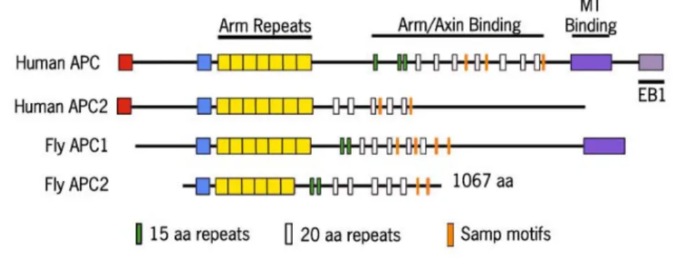
rm/ Figure 1. APC comparison
There are two APCs in humans and in flies. APCs from both species contain Armadillo repeats for protein protein interactions, 15 and 20 amino acid repeats that can bind Arm/β-cat, and SAMP motifs for binding Axin. Human APC and fly APC1 also contain a microtubule binding domain. Human APCs have an oligomerization domain that allows them to bind one another; however, the fly APCs have not been shown to possess such a domain.
APC wa p
5 A
struction complex that forms to target β-catenin (β-cat), the ortholog of f Armadillo (Arm), for destruction (Rubinfeld et al., 1995). Inactivation of APC stabilizes β-catenin and activates Wnt signaling.In many animals, there are two APC family members: APC1 and APC2 (Fig 1). APC proteins are a mosaic of protein binding sites: Armadillo repeats for protein-protein interactions with diverse partners, 15 and 20 amino acid repeats that have the ability to bind A
β-cat, and SAMP motifs that bind Axin, another component of the destruction
complex. Mammalian APC also contains an oligomerization domain and a microtubule binding region. In colon tumors, one allele of APC is usually
inactivated while the other produces a truncated protein (Polakis, 2007). Most of
the truncations found to be critical in cancer occur in a portion of the gene that as been dubbed the Mutational Cluster Region (MCR). This region
ncompasses two of the twenty amino acid repeats, and conserved region B, nd ends just before the SAMP motifs. Other roles for APC beyond canonical
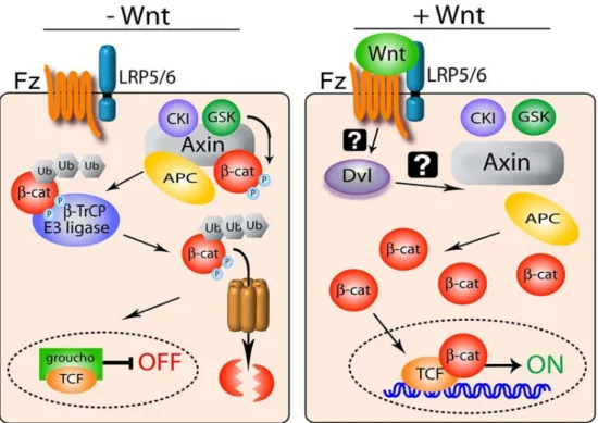
). Figure 2 The Wnt Pathway
The first diagram depicts a cell not receiving Wnt signal. Arm is phosphorylated, ubiquitinated, then degraded. The second half is a cell that has received Wnt. D inactivated the destruction complex, Arm accumulates, moves into the nucleus, a turns on Wnt responsive genes. (adapted from Roberts et al 2007)
vl nd
h e a
The precise details of how the Wnt pathway functions are still being examined. However, the basic steps are known (Cadigan, 2008). The key regulated effector is Arm. It is typically found both in the cytoplasm and at cell-cell jun
t, the r allo
binds Axin and APC, and CK1 and GSK-3
phosph ite
ttled to an
s
ss nt(Cadigan, 2008). In parallel, Disheveled (Dsh) is activated, can interact with Frizzled, and disables
ctions, where it serves a different role in cell adhesion. When a cell receives Wnt signal, Arm is able to relocate into the nucleus. Until this poin HMGbox DNA binding proteins of the T-cell factor (TCF) family and co-represso Groucho are actively repressing transcription of Wnt responsive genes (Cav et al., 1998) . Once inside the nucleus, Arm can displace Groucho, bind with TCF and recruit transcriptional co- activators (Brunner et al., 1997; van de Wetering et al., 1997).
In a cell that is not receiving Wnt, the destruction complex is formed (Cadigan, 2008). This complex is comprised of APC, Axin, Casein kinase 1 (CK1), and GSK-3. Arm
orylate Arm in a series of phosphorylation events, each priming the s for the subsequent phosphorylation. During this period, APC is also
phosphorylated. Once both proteins are phosphorylated, Arm is shu
E3 ubiquitin ligase and ubiquitinated (Aberle et al., 1997). This ubiquitination allows the 26S proteosome to recognize Arm and then degrade it, thu
maintaining repression of Wnt responsive genes.
In a cell that receives Wnt signal, the cell surface receptor LRP5/6 (Arrow), a single-pass transmembrane protein, and Frizzled, a seven–pa transmembrane protein, form a complex and bind W
the de ly
x.
xia , cell cycle transitions,
ns
of
us (for a review see Ho, 2006). Roc1 is a RING domain protein that provides specificity for the E2 protein. Skp binds a member of the large
F-struction complex; the mechanism by which this occurs is not yet ful understood. CK1 also phosphorylates Arrow, which then recruits Axin to the receptor complex. This recruitment helps to disable the destruction comple With no destruction complex to begin the degradation of Arm, Arm accumulates in the cytosol. From there it is able to move into the nucleus, remove the
repressor Groucho, and turn on Wnt responsive genes.
The Wnt pathway is one of many pathways regulated by
ubiquitin-mediated proteolysis. Ubiquitin ligases have many functions in cells(for a review see Petroski, 2008). For example, they have been shown to regulate hypo
responsive transcription factors, mitosis, the cytoskeleton
and protein degradation. The general method of protein degradation begi
when an E1 protein uses ATP to activate a ubiquitin moiety. The ubiquitin is then transferred from the E1 to an E2 protein. Finally, the E3 facilitates the transfer the ubiquitin from the E2 to the substrate. This last step can be accomplished in different ways. In HECT domain-containing E3’s, the ubiquitin is transferred directly to the E3 and then to the substrate. In RING domain-containing E3’s, the transfer is only stimulated by the E3 and the ubiquitin is passed from the E2 to the substrate.
box pr
imaginal discs (Jiang and Struhl 1998; Ou, 2002), Arm accumulates in the
complex is responsible for targeted degradation of Arm. However, work from the Duronio lab showed that when similar loss of function mutant clones are made
part of the canonical SCF complex, and other components in the complex have otein family; in the regulation of β-cat, that F-box protein is Slimb (Slmb). Slmb binds Skp on its amino-terminus and a substrate such as Arm on the carboxyl-terminus. The phosphorylation of Arm by the destruction complex is essential for Arm to be recognized by the SCF complex. β-TrCP, the human ortholog of Slmb, can pull down β-cat and APC in a co-immunoprecipitation assay (Hart et al., 1999).
Consistent with this model, when Slmb or Cul1 mutant clones are made in fly
cytosol, suggesting that it is not being degraded. This implies that the SCF Figure 3 The SCF complex
with Roc1a, Arm does not accumulate (Noureddine et al., 2002). As Roc1a is
already been shown to play a role in the degradation of Arm, these data raise
questions about the mechanism of Arm degradation and the role the canonical SCF complex plays in it.
There are two classes of Roc proteins: Roc1 and Roc2, with most metazoans having one of each (for a review see Yi Sun, 2001). D. melanogas has three Roc proteins: Roc1a, Roc1b and Roc2 (Donaldson et al., 2004, Noureddine, 2002). Roc1
ter
a and hRoc1 have 100% identity in the RING domain. These
rent
n of Arm in
, r destruction and accumulates at very high levels as
compa nt
m, this
orted data have led us to consider two hypotheses to explain the
aforementioned discrepancy. First, it is feasible that the Roc proteins have redundant functions in D. melanogaster. Alternatively, it may be that a diffe Roc protein than Roc1a functions in the fly SCF complex to degrade Arm. We investigated both of these possibilities by analyzing the accumulatio
single Roc mutants.
Our lab has long been interested in the Wnt pathway. While analyzing Drosophila APC mutants, we perceived interesting differences in Arm
accumulation in different tissues. In fly embryos that lack both APC1 and APC2 Arm is not targeted fo
red to wild type (Akong et al., 2002). In contrast, while Apc1 Apc2 muta cells in larval wing discs or brains have an elevated accumulation of Ar
accumulation is much less dramatic than what was seen in the embryos (unpublished data and Hayden, 2007). This was puzzling as it has been rep
difference between the embryo and wing discs phenotypes, testing the possibility that APC might not be fully required for destruction complex function in all
tissues.
Results
Armadillo regulation through development
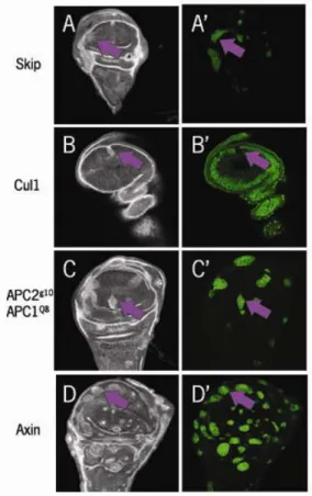
The Wnt signaling pathway has been studied extensively. This wealth of knowledge has allowed us to compare not only data generated in our lab but also other published results. Our lab noticed that APC2G10APC1Q8 mutant clones, APC2 and APC1 null alleles, generated in imaginal discs (Fig 4C) or in the optic lobes of the brain (Fig 4B, Hayden et al, 2007) did not appear to accumulate Arm at levels as high as those reported by other labs who had examined clones mutant for Axin or Slmb (Hamada et al, 1999; Hart et al, 1999; Jiang and Struhl, 1998). In addition, when we compared the levels of Arm accumulation between APC2G10APC1Q8 embryos and APC2G10APC1Q8 mutant wing disc or brain clones
the levels of Arm accumulation in the wing disc clones (figure 4B, C) were much lower than those of the embryos (Fig 4A, McCartney et al 2006, Akong et al 2002). These data suggested the possibility that there could be another protein acting in the same capacity as APC, helping to regulate Arm levels.
localized to the cellular junctions, we now suspect the apparent high level of accumulation others previously reported was due to the plane of focus. Loss of the destruction complex alters the morphology of wing disc cells, and an image taken at the apical portion of the cell will show a higher level of Arm than a more
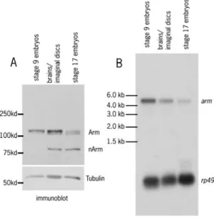
basal section (figure 6); the same is seen in wildtype cells. This still leaves the question about why we observed such a striking difference in Arm accumulation between embryos and larval stages. I addressed this issue. Before I could conduct further analysis of the mutant phenotypes, it was necessary for me to characterize Arm levels through development. I began by comparing Arm protein levels of wild type stage 9 embryos, stage 17 embryos, and wing discs and
brains of third instar larvae (Fig 7A). Arm is known to be upregulated in stage 9 embryos as segment identities are defined. By stage 17, Arm is downregulated in the epidermis as the embryo prepares to hatch (Peifer, 1993; Riggleman B et al., 1989). As anticipated, the levels of Arm protein in the stage 17 embryo were Figure 4 Armadillo accumulation in APC2g10APC1Q8 mutants during fly development
A. (McCartney et al., 2006) Paternally rescued and wild type embryos (stage 9) show similar levels of Arm accumulation; whereas the double mutant embryo has a much higher than normal accumulation. B. mutant clones in the 3rd instar larval brain &C. mutant clones in the 3rd instar larval wing disc; these both display a modest accumulation of arm above normal levels. B. & C. taken by Kuo-Chen Jung
lower than in stage 9. When the stage 9 and the larval tissues were compared however, the amount of Arm was found to be roughly similar (Fig 7A). This result surprised us, because the levels of Arm accumulation in APC2G10APC1Q8
mutants in the two stages are so different.
In order to gain a deeper insight into this process, I next looked at mRNA levels of arm. I made mRNA preparations from wildtype animals from the three stages. I prepared a radioactive probe to arm and probed a Northern blot of all three stages to visualize the levels of mRNA, using the ribosomal protein gene
Figure 6 Plane of focus
Arrows mark the location of a clone taken at the apical part of the cell, note that the levels of all three channels are elevated. Arm is known to be enriched at apical junctions. Arrowheads mark a clone at a more basal section and levels of arm are elevated but at a much lower amount. Taken by Kuo-Chen Jung
rp49 as a loading control (Fig 7B). A STORM machine was used to quantify the amount of mRNA in the three stages by measuring the amount of fluorescence. Levels of arm were normalized to the level of rp49. As expected, the stage 17 mRNA levels of arm were low.
However, when stage 9 and the larval tissues were compared, we found that arm mRNA levels in the embryo were about two times those in the brain and imaginal discs. The difference in mRNA levels suggests that there may be a different requirement for Arm for the embryos than in the larval tissues. It is possible that the rapidity of development in these two different stages changes the amount of arm mRNA that is needed to assure against potential problems.
Figure 7 Armadillo levels at different stages of development A. Immunoblot comparing the amount of Armadillo protein levels. Stage 9 embryos and larval tissues have similar levels of protein.
B. Northern blot comparing the
amount of Armadillo transcript levels. Stage 9 embryos have almost twice as much arm mRNA as the larval tissues.
Assessing the roles of different Roc proteins in Arm regulation
Degradation through ubiquitination is a common method of protein regulation in cells (for a review see Petroski, 2008). E3 ubiquitin ligases form
Figure 8
A. RT-PCR of Roc2KG stocks. B. PCR showing lack of Roc1b DNA in the Roc1bdc3 mutant. C. & D. PCRs showing the lack of Roc2 DNA and presence of P-element DNA Roc2KG. E. location of primers used in B., C., and D.
discrete complexes to target specific proteins. The composition of these
complexes gives them their specificity. The canonical SCF complex consists of a Cul1 scaffold, Skp1 as an accessory protein to bind the F-box protein, an F-box protein that provides specificity for the substrate (in this case Arm), and Roc1a to provide specificity for the E2 protein (for a review see Ho et al., 2006). This SCF complex, with Slmb as the F-box protein, has been implicated in the targeted
Arm degradation. This possibility came into question, however, when it was shown that Roc1a mutant clones in larval wing discs do not show an
accumulation of Arm above wild type levels, but do show accumulation of a different SCF target, the Hedgehog effector Cubitus Interruptus (Noureddine et al., 2002). Given these data, we wanted to find out if there was a different Roc protein in D. melanogaster that acts in the SCF complex, or if the three Rocs were functioning redundantly in this process.
To ascertain the mechanism of Arm destruction, we planned to test these hypotheses by analyzing Arm accumulation in mutants in each of these Roc proteins. The first mutant we looked at was Roc2KG, a null allele of Roc2, which
was generated by a P-element insertion (Reynolds et al., 2008). The mutant is
Figure 9 Armadillo levels in Roc2 mutants
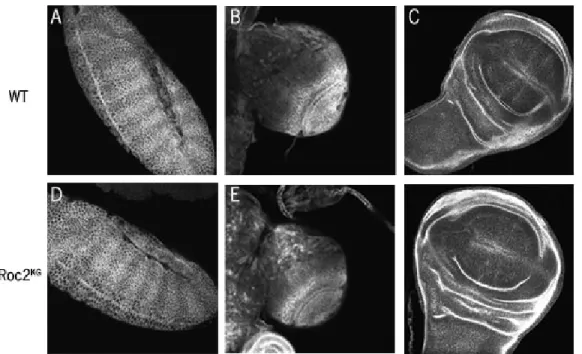
A.-C. are wildtype embryo and larval tissues D.-F. are homozygous mutant
Roc2KG embryos and larval tissues. A. & D. are stage 9 embryos. B. & D., C. & F. are 3rd instar larval brains and wing discs, respectively
homozygous viable and fertile, suggesting that it is not an essential gene. Given the essential role of Wnt signaling and its regulation, this by itself would support the idea that the Roc proteins are acting redundantly, or that Roc2 is not part of the relevant SCF complex. This mutant does not produce a transcript of the gene (Reynolds et al., 2008), and we verified this by PCR and RT-PCR (Fig 8A, C, D). There had been some question about the phenotype of the stock we had in lab, because some of the phenotypes had changed so I tested several stocks to assess which had the correct mutation. The stock from the Bloomington Stock Center proved to be correct. We next immunostained three tissues from Roc2KG
mutants to look at Arm accumulation. When compared to wild type, the mutant embryos, imaginal discs, and larval brains did not show any Arm accumulation beyond normal levels (Fig 9). A Western blot also indicated these animals also had the same amount of Arm protein as wildtype (Fig 10). Together, these data suggest that the SCF complex does not degrade Arm solely through the action of Roc2.
Figure 10
Next, we analyzed Roc1b. The mutant, Roc1bdc3, is a homologous recombination deletion that is homozygous viable but male sterile (Donaldson et al 2004). As with the Roc2 mutant, we verified the mutation by PCR (Fig 8B). It
did not show evidence of Roc1b, supporting the fact that this indeed a Roc1b mutant. Once verified, we immunostained embryos, wing discs, and larval brains to look for Arm accumulation. When compared to the wild type, Roc1bdc3 mutants did not show accumulation above normal (Fig 11). A Western blot confirmed these results (Fig 10). Combined, these data point to Roc1b not being necessary for Arm degradation by itself.
Figure 11 Armadillo levels in Roc1b mutants
A.-C. are wildtype embryo and larval tissues D.-F. are homozygous mutant
Roc1bdc3 embryos and larval tissues. A. & D. are stage 9 embryos. B. & D., C. & F. are 3rd instar larval brains and wing discs, respectively
Discussion
Our lab is fascinated by the process of development and the Wnt signaling
pathway. We were intrigued when we compared published data and our own;
and thought about the questions it brought up. Two of these questions are the
ones I strove to answer with my experiments. The first was why was there a
difference in the levels of Arm in the wing discs and brains of 3rd instar larvae,
versus in embryos, when APC1 and APC2 are inactivated? The second question
was raised by data generated by the Duronio lab; is the canonical SCF complex
the primary mechanism for destroying Arm in Drosophila? These questions are
both complex and we cannot hope to fully answer them in one paper. However,
the experiments I have completed give us insights and ideas for future work that
will one day explain part of the mystery of the Wnt pathway.
As mentioned before, we found a discrepancy between the levels of Arm
accumulation during larval stages, when comparing our own data on APC double
mutants and published data on mutants of other parts of the destruction complex
and the Wnt pathway. We began to address this by recapitulating these clones
in our lab. This revealed that all of these mutants all lead to an elevation of
Armadillo accumulation. In contrast to the previously published data however,
our clones display a much lower accumulation of Armadillo. Our data suggests
the apical portion of the cell. An image taken at this point appears “artificially”
brighter than a more basal section of a neighboring cell. We believe changes in
imaginal disc morphology caused by loss of destruction complex proteins can
lead to this artifact.
It was unexpected that the stage 9 embryos and wing discs/brains would
have the same levels of Arm protein but different levels of arm RNA. During
embryogenesis, there are many vital processes and changes being set up in the
fly. It could be that the embryo keeps extra transcripts waiting to accommodate
the need for very rapid changes in signaling and to accommodate responses in
case something inside the cell goes wrong. The embryos could then quickly
make more Arm protein, without having to wait for additional transcription. This
additional supply might not be necessary in the wing disc because the changes
aren’t occurring as rapidly. Our data suggests it is also a possibility that Arm is
being degraded at a higher rate in the embryo than it is in the larva. There are
points in development that require a large amount of Armadillo. At these points,
the cessation of degradation would allow Armadillo to accumulate at a much
higher rate than if the levels of transcript were lower.
Before undertaking these experiments we postulated that there may be
another protein helping to regulate Arm in the wing discs. None of our data
preclude that from still being a possibility. For example, in embryos there could
be a protein that aids the destruction complex work faster or more efficiently.
Further experiments will have to be done to look into this further
Based on our data thus far, it seems that neither Roc1b nor Roc2 are
solely responsible for Arm degradation in flies. Since Arm degradation is an
essential process during development and both Roc1b and Roc2 are viable,
these data make sense. This does not rule out the possibility that the Roc
proteins are acting redundantly to degrade Arm. To address this possibility, we
are currently creating a fly stock that is mutant for both Roc1b and Roc2.
Preliminary data suggests that these flies are also viable. We are also planning
on creating Roc1a mutant clones in the Roc1b and the Roc2 background, as well
as the double mutant background.
Previous data by other labs have not shown that the Roc proteins can
substitute for one another. Indeed, they have shown that the Rocs generally
have preferred Cullin binding partners and don’t stray from those (Reynolds et
al., 2008). This made it very surprising that none of the three protein deficiencies
by themselves could produce an accumulation of Arm. There remains the
possibility that the original work done on Roc1a in flies might not have shown the
whole picture. In Noureddine 2002, they showed that Roc1a is an essential
gene, so mutant clones must be made in the fly in order to evaluate its affects.
Further, clones lacking Roc1a have growth and proliferation defects. In order to
have clones that were large enough to be able to evaluate the accumulation of
Arm, the Duronio lab was forced to give the cells a very small amount of Roc1a
to get them to live so that they could assess the phenotype. This method worked
when they were testing a protein from the Hedgehog pathway, Cubitus
protein still showed an accumulation of Ci. There is the possibility that the
requirement of Roc1a for Arm degradation is much smaller. It could be that a
very tiny amount is still enough to keep Arm levels low in these clones. David
Roberts has generated data that suggests that Roc1a is responsible for
degrading Arm in Drosophila S2 cultured cells (unpublished). This data has led
us to attempt to recapitulate the Roc1a clones without the addition of Roc1a
protein, to see if this is an artifact of tissue culture or a real possibility.
Materials and Methods
Fly Stocks
All experiments were done at 25°C. Mutations and Balancer chromosomes are described at FlyBase (flybase.bio.indiana.edu).
Generating Mutant Clones
Clones were generated by FLP/FRT mediated mitotic recombination. Larvae were put at 37°C for 3 hours, 3 and 4 days after egg laying. After the heat shock, larvae were returned to 25°C for two days and then dissected.
Immunofluorescence
We used a monoclonal mouse Anti-armadillo 7A1 raised against amino acids 67- 123 (Developmental Studies Hybridoma Bank). Embryos were collected for two hours at 25°C, and then let age 5 hours (to stage 9). For larval collections 3rd instar larvae were dissected and the brains and wing discs were loosened from the cuticle to allow the antibody easier access. Embryos were fixed for 20
minutes in 10% formaldehyde in phosphate buffered saline (PBS). Larval tissues were fixed for 20 minutes in 4% formaldehyde in PBS. All collections were
(PBT). Antibodies were diluted in PBT as follows: α-arm 1:50, for larval brains and wing discs, 1:100 for embryos, α-mouse 1:250(Alexa by Molecular Probes). Primary antibodies were incubated at 4° overnight, secondary antibodies were incubated for three hours at 25°. Prior to mounting brains and wing discs were dissected completely from the cuticle. All samples were mounted in Aqua Poly/Mount (Polysciences). Fixed samples were imaged with a Pascal confocal microscope, using a Zeiss 40X NA 1.3 Plan-Neofluar oil immersion objective, and LSM software at 25°C. Adobe Photoshop CS2 was used to adjust input levels so the main range of signals spanned the entire output grayscale and to adjust brightness and contrast.
Western Blotting
Embryos were collected for two hours and then aged 5 hours (stage 9, for both the Roc mutant western and the developmental western), or 21 hours (stage 17, developmental western). Brains and wing discs were dissected out from 3rd instar larvae. All samples were boiled for 5 minutes in 2x Laemmli buffer, run on an 8% acrylamide gel and transferred to a nitrocellulose membrane. The blot was incubated with α-arm (1:75) and α-tubulin (DM1A, 1:7500, Sigma) for one hour. Washes were done in Tris-Buffered Saline Tween-20 (TBST) at 4 x 15. For detection, the blot was incubated for one hour with horseradish peroxidase conjugated rabbit α-mouse IgG secondary antibody (1:20000, Zymed), and then the ECL-Plus kit (GE Healthcare Amersham) was used.
Northern Blotting
Works Cited
Aberle, H., Bauer, A., Stappert, J., Kispert, A. and Kemler, R. (1997). B-catenin is a target for the ubiquitin-proteasome pathway. The EMBO Journal 16, 3797-3804.
Akong, K., Grevengoed, E. E., Price, M. H., McCartney, B. M., Hayden, M. A., DeNofrio, J. C. and Peifer, M. (2002). Drosophila APC2 and APC1 Play
Overlapping Roles in Wingless Signaling in the Embryo and Imaginal Discs. Developmental Biology 250, 91-100.
American Cancer Society "What Are the Key Statistics for Colorectal Cancer?" 2008. American Cancer Society. June 6,2009 http://www.cancer.org/docroot/cri/ content/cri_2_4_1x_what_are_the_key_statistics_for_colon_and_rectum_cancer. asp
Brunner, E., Peter, O., Schweizer, L. and Basler, K. (1997). pangolin encodes a Lef-1 homolog that acts downstream of Armadillo to transduce the Wingless signal. Nature 385, 829-833.
Cadigan, K. M. (2008). Wnt-[beta]-catenin signaling. Current Biology 18, R943-R947.
Cavallo, R. A., Cox, R. T., Moline, M. M., Roose, J., Polevoy, G. A., Clevers, H., Peifer, M. and Bejsovec, A. (1998). Drosophila Tcf and Groucho interact to repress Wingless signalling activity. 395, 604-608.
Clevers, H. (2006). Wnt/[beta]-Catenin Signaling in Development and Disease. Cell 127, 469-480.
Donaldson, T. D., Noureddine, M. A., Reynolds, P. J., Bradford, W. and Duronio, R. J. (2004). Targeted Disruption of Drosophila Roc1b Reveals Functional
Differences in the Roc Subunit of Cullin-dependent E3 Ubiquitin Ligases. Mol. Biol. Cell 15, 4892-4903.
Groden, J., Thliveris, A., Samowitz, W., Carlson, M., Gelbert, L., Albertsen, H., Joslyn, G., Stevens, J., Spirio, L., Robertson, M. et al. (1991). Identification and characterization of the familial adenomatous polyposis coli gene. Cell 66, 589-600.
Hamada, F., Tomoyasu, Y., Takatsu, Y., Nakamura, M., Nagai, S.-i., Suzuki, A., Fujita, F., Shibuya, H., Toyoshima, K., Ueno, N. et al. (1999). Negative
Regulation of Wingless Signaling by D-Axin, a Drosophila Homolog of Axin 10.1126/science.283.5408.1739. Science 283, 1739-1742.
Hart, M., Concordet, J. P., Lassot, I., Albert, I., del los Santos, R., Durand, H., Perret, C., Rubinfeld, B., Margottin, F., Benarous, R. et al. (1999). The F-box protein [beta]-TrCP associates with phosphorylated [beta]-catenin and regulates its activity in the cell. Current Biology 9, 207-211.
Hayden, M. A., Akong, K. and Peifer, M. (2007). Novel roles for APC family members and Wingless/Wnt signaling during Drosophila brain development. Developmental Biology 305, 358-376.
Ho, M. S., Tsai, P.-I. and Chiang, C.-T. (2006). F-box proteins: the key to protein degradation. Journal of Biomedical Science Volume 13, 181-191.
Jiang, J. and Struhl, G. (1998). Regulation of the Hedgehog and Wingless signalling pathways by the F-box/WD40-repeat protein Slimb. 391, 493-496.
McCartney, B. M., Price, M. H., Webb, R. L., Hayden, M. A., Holot, L. M., Zhou, M., Bejsovec, A. and Peifer, M. (2006). Testing hypotheses for the functions of APC family proteins using null and truncation alleles in Drosophila
10.1242/dev.02398. Development 133, 2407-2418.
McMahon, A. P. and Moon, R. T. (1989). Ectopic expression of the
proto-oncogene int-1 in Xenopus embryos leads to duplication of the embryonic axis. Cell 58, 1075-1084.
Munemitsu, S., Souza, B., Muller, O., Albert, I., Rubinfeld, B. and Polakis, P. (1994). The APC Gene Product Associates with Microtubules in Vivo and Promotes Their Assembly in Vitro. Cancer Res 54, 3676-3681.
Nathke, I. (2006). Cytoskeleton out of the cupboard: colon cancer and cytoskeletal changes induced by loss of APC. Nat Rev Cancer 6, 967-974.
Noureddine, M. A., Donaldson, T. D., Thacker, S. A. and Duronio, R. J. (2002). Drosophila Roc1a Encodes a RING-H2 Protein with a Unique Function in Processing the Hh Signal Transducer Ci by the SCF E3 Ubiquitin Ligase. Developmental Cell 2, 757-770.
Nusse, R. and Varmus, H. E. (1982). Many tumors induced by the mouse mammary tumor virus contain a provirus integrated in the same region of the host genome. Cell 31, 99-109.
Nusslein-Volhard, C. and Wieschaus, E. (1980). Mutations affecting segment number and polarity in Drosophila. 287, 795-801.
Peifer, M. (1993). The product of the Drosophila segment polarity gene armadillo is part of a multi-protein complex resembling the vertebrate adherens junction. J Cell Sci 105, 993-1000.
Petroski, M. (2008). The ubiquitin system, disease, and drug discovery. BMC Biochemistry 9, S7.
Polakis, P. (2007). The many ways of Wnt in cancer. Current Opinion in Genetics & Development Genetic and cellular mechanisms of oncogenesis 17, 45-51.
Reynolds, P. J., Simms, J. R. and Duronio, R. J. (2008). Identifying Determinants of Cullin Binding Specificity Among the Three Functionally Different
<italic>Drosophila melanogaster</italic> Roc Proteins via Domain Swapping. PLoS ONE 3, e2918.
Roberts, D. M., Slep, K. C. and Peifer, M. (2007). It takes more than two to tango: Dishevelled polymerization and Wnt signaling. Nat Struct Mol Biol 14, 463-465.
Riggleman B, Wieschaus E and P., S. (1989). Molecular analysis of the armadillo locus: uniformly distributed transcripts and a protein with novel internal repeats are associated with a Drosophila segment polarity gene. Genes Dev. 3, 96-113.
Rijsewijk, F., Schuermann, M., Wagenaar, E., Parren, P., Weigel, D. and Nusse, R. (1987). The Drosophila homology of the mouse mammary oncogene int-1 is identical to the segment polarity gene wingless. Cell 50, 649-657.
Rubinfeld, B., Souza, B., Albert, I., Munemitsu, S. and Polakis, P. (1995). The APC Protein and E-cadherin Form Similar but Independent Complexes with alpha-Catenin, beta-Catenin, and Plakoglobin. J. Biol. Chem. 270, 5549-5555.
Sharma, R. P. and Chopra, V. L. (1976). Effect of the wingless (wg1) mutation on wing and haltere development in Drosophila melanogaster. Developmental
Biology 48, 461-465.
van de Wetering, M., Cavallo, R., Dooijes, D., van Beest, M., van Es, J., Loureiro, J., Ypma, A., Hursh, D., Jones, T., Bejsovec, A. et al. (1997). Armadillo
Coactivates Transcription Driven by the Product of the Drosophila Segment Polarity Gene dTCF. Cell 88, 789-799.
van den Heuvel, M., Nusse, R., Johnston, P. and Lawrence, P. A. (1989). Distribution of the wingless gene product in drosophila embryos: A protein involved in cell-cell communication. Cell 59, 739-749.
Willems, A. R., Schwab, M. and Tyers, M. (2004). A hitchhiker's guide to the cullin ubiquitin ligases: SCF and its kin. Biochimica et Biophysica Acta (BBA) - Molecular Cell Research 1695, 133-170.
Yi Sun, M. T., Hangjun Duan, Manju Swaroop. (2001). SAG/ROC/Rbx/Hrt, a Zinc RING Finger Gene Family: Molecular Cloning, Biochemical Properties, and Biological Functions. Antioxidants & Redox Signaling 3, 635-650.