THE SELECTION OF HIGHLY STABLE APTAMERS FROM A 2’-FULLY MODIFIED fGmH RNA LIBRARY
Adam David Friedman
A dissertation submitted to the faculty of the University of North Carolina at Chapel Hill in partial fulfillment of the requirements for the degree of Doctor of Philosophy in the
Eshelman School of Pharmacy.
Chapel Hill 2015
Approved by: Andrew Lee April Salama Michael B. Jarstfer Rihe Liu
Zefeng Wang
© 2015
ABSTRACT
ADAM DAVID FRIEDMAN: The Selection of Highly Stable Aptamers Selected from a 2’-fully modified fGmH RNA Library
(Under the direction of Rihe Liu)
When developed as tools for applications within biological systems, RNA aptamers immediately face numerous obstacles, in particular nuclease degradation and post-selection 2’ modification. This study aimed to develop a novel class of highly stable, 2’-fully modified RNA aptamers directly selectable from a fGmH RNA library with improved nuclease
stability. The facile transcription of a fGmH (2’-F-dG, 2’-OMe-dA/dC/dU) RNA library was performed, from which aptamers were directly selected that bind Staphylococcus aureus
Protein A (SpA). The superior nuclease and serum stability of these fGmH aptamers was demonstrated in comparison to 2’-partially modified RNA variants. Characterizations of fGmH RNA aptamers binding to purified SpA and to endogenous SpA present on the surface
of S. aureus cells demonstrated fGmH RNA aptamer selectivity and stability. Significantly,
To my wife, always my better half and eternal love. To my daughter, my new source of youth, joy, and hope. To my mother, the lover of knowledge and nurturer of your little boy.
ACKNOWLEDGEMENTS
My wife, Michelle McCoy Friedman, deserves the most credit in part because of the multifaceted support she has provided me in the attainment of my ambitions, which are partially encapsulated by this dissertation. She trusted me that my decision in 2008 to return to biomedical science was correct, and she served as a constant source of emotional and financial strength as my salary decreased steadily upon transitioning from archaeology to an ORISE ORAU Post-baccalaureate Fellowship at the U.S. Food & Drug Administration, and on into graduate school in the UNC-Chapel Hill Biological and Biomedical Sciences
Program (BBSP). She gave me faith in myself when my confidence flagged. She advised when strategies failed. She soothed when gargantuan stressors opacified my vision of my career. She gave me an incredibly intelligent and beautiful daughter, Jacqueline Anne, and made me realize, quite abruptly and with emotional pangs so intense as to be almost painful, how incomplete my life had been before and how much promise the future held for all of us. She is the love of my life, and this doctorate is for us.
Dr. Rihe Liu, my research advisor, made this student into a scientist. Rihe demands excellence of his students and never lowers the bar – we must rise to his challenge. It is only because of his belief in, and care for, his students that he demands so much. He is a
committed teacher and he invests great energy in teaching us the “methodology of the lab” – hard work, critical thinking, self-reliance, open-mindedness - so that we may teach ourselves. In so doing, he skims away the slag, which can be a painful process for all, but leaves behind a better scientist and a better person.
My doctoral committee chair, Dr. Michael Jarstfer, who was also my BBSP First Year Group mentor before I officially joined the Chemical Biology and Medicinal Chemistry Division, played a huge part in my progression as a student. He could always be counted on as a sounding board for scientific or political matters, or for conversations ranging from “your project”, oxytocin and prosocial behavior in mice, rock climbing, beer styles and their attributes, the upcoming Phish concert, optimistic nihilism (or, as I called it, “Jarstferian Nihilism”), and other subjects too numerous to list. He is a consummate advisor, teacher, and scientist, and my training would not have been complete without his continuous guidance and friendship.
My colleagues in the laboratory did much to help me through the last 5 years. Sarah Claypool, who joined the lab at the same time as I, most of all shared in my anxieties, failures, and triumphs. She always had a knack for instilling needed perspective when
reliable food and Starbucks buddy. Our walks to and from the Starbucks located on Franklin Street were always the panacea that was needed. Dr. Dongwook Kim, the senior postdoc in the Liu Lab during my time here, always served as a source of insight, advice, friendship, and encouragement. I will miss our “super secret projects”, or SSPs, and I hope that I will cross paths with him again following his certain success at Purdue University. Thanks also go to Dr. Jianwei Zou and Ke (Tracey) Mu for their assistance in multiple scientific matters and many a pleasant conversation.
TABLE OF CONTENTS
LIST OF TABLES ... xiii
LIST OF FIGURES ... xiv
LIST OF ABBREVIATIONS ... xvi
CHAPTER 1 ...1
APTAMERS: INNOVATIONS IN NUCLEASE STABILITY, MACROMOLECULAR TARGETING LIGANDS, AND AN
EXPLANATION OF THE UNMET NEEDS TARGETED BY THIS
STUDY ... 1
1.1 Introduction: Aim of the Dissertation ... 1
1.2 Aptamers and Innovations in Nuclease Stability ... 1 1.2.1 RNA Modification ... 3
1.2.2 Incorporating Nuclease Stability into SELEX ... 4
1.2.3 Unmet Needs ... 5
1.3 Macromolecular Targeting Ligands ... 6
1.3.1 Aptamers ... 7
1.3.2 Antibodies ... 13
1.3.3 Protein Domains ... 17
1.3.4 Peptides ... 19
1.4 The Research Approach Pursued in this Dissertation Directly Selected Highly Stable fGmH RNA Aptamers from
fGmH Libraries for Use as Targeting Ligands ... 22
SELECTING fGmH RNA APTAMERS FROM A 2’-‐FULLY
MODIFIED fGmH RNA LIBRARY THAT BIND Staphylococcus
aureus PROTEIN A (SpA) ... 25
2.1 Introduction ... 25
2.2 Results ... 26
2.2.1 The Expression and Purification of LAR T7 RNA
Polymerase ... 26
2.2.2 fGmH RNA Aptamers were Unintentionally Selected against SpA and were Characterized for SpA
Binding ... 28
2.2.3 Selected fGmH RNA Aptamers are Specific for SpA,
and the fGmH RNA Composition is Critical for SpA Affinity ... 31
2.2.4 fmA12 was Structurally Truncated while Preserving SpA Affinity for Potential Future Chemical
Synthesis ... 33
2.2.5 fGmH RNA has High Stability against Serum, Alkaline Hydrolysis, and a Panel of Individual Nucleases
compared to Nucleic Acids of Lower 2’ Modification States ... 35
2.2.6 fmA12 Binds to S. aureus Cells with High SpA
Expression ... 38
2.2.7 Functionalization of AgNPs in Preparation for S.
aureus Antimicrobial Assays ... 39
2.2.8 fmA12 can Deliver AgNPs to S. aureus in a SpA-‐
dependent Manner ... 43
2.3 Discussion ... 45
2.4 Materials and Methods ... 53
2.4.1 His-‐LAR T7 RNA Polymerase Expression,
Purification, and Characterization ... 53
2.4.2 In vitro Selection ... 55
2.4.3 Binding Analysis Using Surface Plasmon
2.4.4 Aptamer Stability Assays ... 59
2.4.5 Confocal Microscopy ... 60
2.4.6 Silver Nanoparticle Stabilization and
Functionalization ... 61
2.4.7 Targeted Killing of S. aureus Using SpA-‐targeted
Silver Nanoparticles ... 62
CHAPTER 3 ... 64
ATTEMPTS TO SELECT fGmH RNA APTAMERS THAT BIND
OTHER TARGETS_ ... 64
3.1 Introduction ... 64
3.1.1 Strategy 1: fGmH RNA Active Site – SELEX
targeting EGFR ... 64
3.1.2 Strategy 2: fGmH RNA SELEX targeting PD-‐1, PD-‐
L1, and PD-‐L2 ... 65
3.2 Results for Strategy 2 ... 67
3.2.1 Selected Aptamer Pools Became Dominated by poly A and poly T Sequences (poly A/T Effect), which is
resolved by exchanging the Elution Buffer for Water ... 67
3.2.2 Selective Pressure Successfully Drives the
Formation of Sequence Motif Groups in an Aptamer Pool Selected against PD-‐L1; a Single, Non-‐binding Sequence
Dominates the PD-‐1 Selected Pool ... 72
3.3 Discussion and Future Directions ... 75
3.4 Long-term Future Studies ... 81
3.4 Materials and Methods ... 83
3.4.1 In vitro Selection ... 83
3.4.2 Binding Analysis Using Biolayer Interferometry
LIST OF TABLES
Table 1.1. Recent examples of targeted nanoparticles using aptamers. ... 8 Table 1.2. Recent examples of targeted nanoparticles using
antibodies. ... 15 Table 1.3. Recent examples of targeted nanoparticles using protein
domains. ... 19 Table 1.4. Recent examples of targeted nanoparticles using short
homing peptides. ... 20 Table 2.1. Dissociation constants of fGmH RNA aptamers binding S.
aureus SpA. ... 31 Table 2.2. Percent survival of fGmH Lib40 RNA against individual
LIST OF FIGURES
Figure 1.1. Scheme for aptamer selection via SELEX. ... 2
Figure 1.2. Diagram of the fGmH RNA described in this work. ... 24
Figure 2.1. Electrophoresis (8%) PAGE gel of the expression and
purification of His-LAR T7 RNA polymerase.. ... 27 Figure 2.2. Transcription efficiency of purified His-LAR T7 RNA
polymerase using different 2’-X-dNTPs ... 28 Figure 2.3. Real-time monitoring of the enrichment of fGmH RNA
aptamers during the selection using Octet BLI. ... 30 Figure 2.4. Percentages of sequences classified as one of the five
sequence motif groups. ... 30 Figure 2.5. The putative secondary structures and random region
sequences of aptamers fmA12, fmF07, fmE09, and fmG12,
representing Groups 1, 2, 3, and 5, respectively. ... 31 Figure 2.6. BLI kinetic characterization of fmA12 using biosensors
immobilized with SpA. ... 31 Figure 2.7. Binding specificity of fmA12 to Protein A, Protein G, and
Protein L. ... 32 Figure 2.8. Protein A (SpA) binding dependence of sequence A12 on
the RNA 2’ modification state (WT A12 (wild-type; rN), bmA12
(fYrR), tmA12 (rGmH), fmA12 (fGmH)). ... 33 Figure 2.9. The predicted secondary structures of fmA12Δ3,
fmA12Δ6, and fmA12Δ9 compared to that of full-length fmA12. ... 34 Figure 2.10. Percent survival of A12 RNAs of varying 2’
modification state in 10% serum (rN (WT A12), fYrR (bmA12),
rGmH (tmA12), fGmH (fmA12)). ... 36 Figure 2.11. Percent survival of A12 RNAs of varying 20
modification state in 50% serum after 2 h and 5 h incubation at
Figure 2.12. Degradation of G12 RNAs of varying 2’ modification
state in alkaline hydrolysis conditions. ... 37 Figure 2.13. Confocal analysis of the binding of functional fmA12
and non-functional tmA12 to S. aureus cells with high (Strain
12598) and low (Strain 10832) SpA expression. ... 40 Figure 2.14. Diagram of the binding of fmA12-functionalized AgNPs
to SpA on the surface of BLI biosensors. ... 42 Figure 2.15. BLI kinetic characterization of fmA12-functionalized
AgNP SpA binding. ... 43 Figure 2.16. S. aureus cell killing effect of fmA12-directed AgNPs
based on CFU analysis. ... 45 Figure 3.1. Sequencing data for PD-1 Round 9 pool demonstrates
poly A/T character (N = 35). ... 68 Figure 3.2. Natural rATP/rUTP contamination check in 2’-O
Me-dATP/dUTP stocks using wild-type T7 RNA polymerase. ... 70 Figure 3.4. Sequencing data for reveals that PD-1 Round 5 pool is
still highly diverse (N = 31). ... 72 Figure 3.5. Sequencing data for reveals that PD-1 Round 6 pool is
still highly diverse (N = 31). ... 73 Figure 3.6. Sequencing data reveals that a single sequence dominates
the PD-1 Round 7 pool (N = 31). ... 73 Figure 3.7. Sequencing data for reveals that a single sequence
dominates the PD-1 Round 7 pool (N = 31). ... 74 Figure 3.8. Sequencing data for reveals that multiple sequence motif
groups are present in PD-L1 Round 8 pool (N = 64). ... 75 Figure 3.9. Crystal structures of the PD-1 immune checkpoint
LIST OF ABBREVIATIONS
2’-OMe 2’-O-methyl
2’-F 2’-fluoro
2’-OH 2’-hydroxyl
Ab antibody
AgNP silver nanoparticle ASO antisense oligonucleotide AuNP gold nanoparticle
BLI biolayer interferometry
BPH-1 human prostate benign hyperplastic epithelial cell line
cDNA complementary DNA
CFU colony forming unit
CTC circulating tumor cell DEPC diethyl pyrocarbonate ddH2O double-distilled water DNA deoxyribonucleic acid
DNase deoxyribonuclease
DNase I DNase RQ1
dNTP deoxynucleotide
DSPE 1,2-Distearoyl-sn-glycero-3-phosphoethanolamine
E. coli Escherichia coli
EDC 1-Ethyl-3-(3-dimethylaminopropyl)carbodiimide EGFR epidermal growth factor receptor
EPR enhanced permeability and retention Exo I Exodeoxyribonuclease I
Exo III Exodeoxyribonuclease III
fGmH 2’-F-dG, 2’-OMe-dA/dC/dU RNA FMN flavin mononucleotide
fYrR 2’-F-pyrimdine, 2’-OH-purine RNA
GMP guanosine monophosphate
HDAC histone deacetylase
Hep G2 human hepatocellular carcinoma
HER3 human epidermal growth factor receptor 3
LB Luria-Bertani medium
LNCaP androgen-dependent human prostate adenocarcinoma
mAb monoclonal antibody
MBCSP magnetic-based core-shell particles
MCF-7 human mammary gland, breast; derived from metastatic site M-MLV Moloney Murine Leukemia Virus
MNA 2’-OMe-dN RNA
MRSA Methicillin/Multiple-Resistant S. aureus
MWCO molecular weight cutoff NHS N-hydroxysulfosuccinimide
NP nanoparticle
NTA nitriloacetic acid
PAGE polyacrylamide gel electrophoresis
PC3 androgen-independent human prostate adenocarcinoma cells PCR polymerase chain reaction
PD-1 programmed death 1 receptor PDAC pancreatic ductal adenocarcinoma
PD-L1 PD-1 ligand 1
PD-L2 PD-1 ligand 2
PDX patient-derived xenograft
PEG polyethylene glycol
PLGA poly(lactic-co-glycolic acid)
PpL Protein L
PrEC human prostate normal epithelial cell line PSMA prostate-specific membrane antigen
rGmH 2’-OH-dG, 2’-OMe-dA/dC/dU RNA
rN 2’-OH-rN RNA
RNA ribonucleic acid
RNase ribonuclease
rNTP ribonucleotide
ROS reactive oxygen species
RWPE-1 human prostate normal epithelial cell line
SasG Protein G
S. aureus Staphylococcus aureus
SCBC single-cell barcode chip
SCCHN squamous cell carcinoma of head and neck scFv single chain variable fragment
SELEX Systematic Evolution of Ligands by EXponential enrichment
SpA Protein A
SPION superparamagnetic iron oxide nanoparticle SPR surface plasmon resonance
ssDNA single-stranded DNA T7 Exo T7 Exodeoxyribonuclease
TBE Tris/Borate/EDTA
99mTc metastable nuclear isomer of Technetium-99 TKI tyrosine kinase inhibitor
CHAPTER 1
APTAMERS: INNOVATIONS IN NUCLEASE STABILITY, MACROMOLECULAR TARGETING LIGANDS, AND AN EXPLANATION OF THE UNMET NEEDS
TARGETED BY THIS STUDY 1.1 Introduction: Aim of the Dissertation
The aim of this dissertation is to investigate a novel aptamer class with considerable potential to improve the in vivo applicability of nucleic acid-based affinity molecules. In the process of performing the first proof-of-concept fGmH (RNA composed of F-dG and
2’-O-methyl-dC/dA/dU) RNA aptamer selection, fGmH RNA stability against serum and a panel of individual nucleases was assessed, and a selected fGmH RNA aptamer, fmA12, was used as a targeting ligand for the specific delivery of antimicrobial silver nanoparticles (AgNPs) to Staphylococcus aureus. This chapter will provide an overview of nuclease stability issues of various nucleic acids, the use of aptamers as targeting ligands as compared to other macromolecules, and a description of the research approach pursued during the course of this dissertation.
1.2 Aptamers and Innovations in Nuclease Stability
Developed in the 1990s by the Szostak, Gold, and Joyce groups through an in vitro
acid library, where every sequence within the library contains 20 to 50 random residues, which determines the diversity of the potential aptamer pool. Although the theoretical
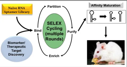
diversity is an astronomical figure, the realistic diversity that can be achieved experimentally is typically in the range of 1 × 10¹³ to 1 × 10¹⁵ unique sequences. The high sequence and conformational diversity of the initial nucleic acid pools ensures a high probability of discovering aptamers that bind to numerous targets of interest (5, 6). Aptamer selection, oftentimes called the Systematic Evolution of Ligands by EXponential enrichment (SELEX), involves iterative rounds of binding to a target of interest, partitioning between binding versus non-binding sequences, and amplification of the enriched target binding aptamers for the next round of selection (Fig. 1.1).
Figure 1.1. Scheme for aptamer selection via SELEX.
and many nonspecific nucleases, yielding half-lives in the range of 30 to 60 minutes (5). This section will discuss relevant advances in nucleic acid stabilization, use of nuclease-stabilized naïve aptamer libraries in SELEX, and unmet needs.
1.2.1 RNA Modification
RNA with 2’ modifications exhibits increased resistance to many nucleases by reducing hydrolysis of the phosphodiester backbone (7).In vitro, natural RNA exhibits half-lives of seconds to a few minutes in various biological fluids including human serum,
whereas RNAs partially modified at the 2’ position have extended half-lives of 5 to 15 hours (5, 7, 8). Before the advent of aptamers, the nuclease stabilizing effect of 2’-fluoro (2’-F) and 2’-amino modifications on RNA was well-known, and this knowledge was applied to
hammerhead ribozymes (i.e. RNA enzymes) after their discovery by Cech and coworkers (9-11). Without disrupting catalytic activity, Eckstein and coworkers replaced a subset of the hammerhead ribozyme’s nucleotides with 2’-F or 2’-amino equivalents, thus increasing ribozyme nuclease stability without disrupting catalytic activity (12). Yang and coworkers continued this work using 2’-O-methyl (2’-OMe) modifications on a subset of nucleotides, and were able to both maintain catalytic activity and increase nuclease resistance 1000-fold (13). Similarly, Paolella and coworkers achieved similar success using 2’-O-allyl
In addition to conferring nuclease resistance, 2’-OMe and 2’-F modifications stabilize base pairing. Residues bearing a 2’-OMe prefer a C3’-endo pucker due to steric repulsion with the 2-carbonyl, the 2’-OMe, and 3’-phosphate (16, 17). Due to the high
electronegativity of fluorine, nucleosides bearing a 2’-F moiety also adopt an RNA-type C3’-endo sugar conformation, but this is partially due to the 2’-F exerting a gauche effect on the sugar (18). RNA residues bearing a 2’-F moiety potently increase the hybridization stability between oligonucleotides and RNA at approximately 1.8 °C per residue (a possible result of enthalphy effects and enhanced stacking interactions resulting from the high electronegativity of fluorine) compared to about 1.3 °C and 1.0 °C per residue when 2’-OMe and 2’-OH residues are used, respectively (19, 20).
1.2.2 Incorporating Nuclease Stability into SELEX
2’-OMe and 2’-F modifications on pyrimidines and some purines have been
commonly used for stabilizing aptamers (5, 7, 8, 21, 22). When aptamers intended for an in vivo application are first selected from natural or 2’-partially modified RNA libraries, further stabilization of these aptamers by installation of additional 2’-modified residues is desirable yet challenging. 2’-hydroxyls can participate in aptamer:target interactions or in the structural folding of functional aptamers via hydrogen bonding. Therefore, post-selection replacement of residues bearing 2’-hydroxyls with 2’-modified variants carries a high risk of disrupting the structure and target affinity of the aptamer.
replaced with a 2’-OMe purine analog one at a time. Each NX1838 variant bearing a new single 2’-OMe purine analog was chemically synthesized and tested for target binding. This systematic testing found that all but two purines bearing 2’-hydroxyls could be replaced with 2’-OMe purines and maintain target-binding affinity and inhibitory activity against
VEGF165-mediated signaling (23, 24). Though ultimately successful, the chemical synthesis and affinity testing performed via this strategy was exceptionally tedious, time-consuming, and still limited in addressing the problem.
1.2.3 Unmet Needs
post-selection functional testing using chemically synthesized 2’-OMe modified aptamer variants should be performed. In addition, it is unknown whether this selection would have been successful if the chemically-synthesized, all 2’-OMe-modified library, with a quality typically much lower than that of enzymatic synthesis, had not been included in the overall first round.
1.3 Macromolecular Targeting Ligands
The use of aptamers as targeting ligands both in vitro and in vivo has been pursued in the literature with vigor since shortly after their development in 1990 by Ellington and Szostak. As macromolecular targeting ligands, aptamers and polypeptide-based targeting ligands each have unique features that can make the former advantageous over the latter, and
vice versa. The attractiveness of aptamers as biopolymers for “smart” targeting, particularly
for in vivo applications, derives from multiple properties that allow aptamers to outperform
Importantly, it is possible to synthesize aptamers with a specific functional moiety, such as a carboxylate, amino, sulfhydryl, or aldehyde, at only one end of the nucleic acid aptamer. This ensures, and greatly facilitates, site-specific conjugation with a wide variety of biomaterials and prevents the formation of heterogeneous mixtures.
As a prelude to Chapter 2, wherein the deployment of S. aureus Protein A-binding aptamer fmA12 as a silver nanoparticle targeting ligand will be described, this section will discuss and compare the uses of aptamers and polypeptides as targeting ligands.
1.3.1 Aptamers
Aptamers are well-suited to serving as targeting ligands. It is possible to synthesize aptamers with a specific functional moiety, such as a carboxylate, amino, sulfhydryl or aldehyde, at only one end of the nucleic acid aptamer. This ensures and greatly facilitates site-specific conjugation and prevents the formation of heterogeneous mixtures. Aptamers are typically non-immunogenic (34). Many other attributes make aptamers attractive for nanoparticle targeting, such as being non-toxic (34-36) and modifiable for stability in circulation (37). They can be selected in vitro and in vivo, and be repeatedly and reversibly denatured. Moreover, as aptamers are not dependent on animals or their immune responses, aptamers can be selected against weakly immunogenic targets and toxins. The ability to chemically synthesize aptamers infers little batch variation (29). They are much smaller than antibodies and can form compact structures, allowing them to bind clefts, binding sites, and enzymatic active sites, which is difficult, if not impossible, for antibodies to achieve (38).
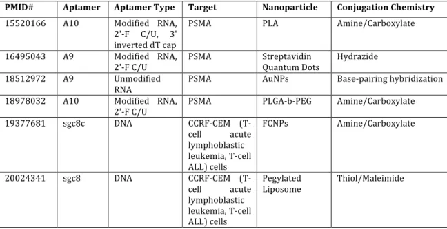
nanoparticles to tumor tissues. Of the reviewed studies (Table 1.1) that used aptamers as nanoparticle targeting ligands for cell surface biomarkers, the aptamers were DNA (62%), unmodified RNA (17%), or modified RNA (21%). Compared to numerous RNA nucleases, there are relatively few DNA nucleases in vivo. DNA aptamers do, however, suffer from characteristics that can complicate their in vitro selection via SELEX, such as the formation of hard to manage G-tetrads.
Of those studies using unmodified RNA aptamers, two chose nanoparticles that confer nuclease resistance to the aptamers. Li and coworkers employed gold nanoparticles (AuNPs), which maintain a halocline immediately surrounding the AuNP and provide a blanketing solution layer of high ionic character that discourages nuclease activity (39, 40). In another instance, Lee and coworkers found that PSMA-specific RNA aptamer A9, when conjugated to dendrimers, was nuclease-resistant 24 hours post-exposure (41). Exploiting A9 nuclease sensitivity, Yu and coworkers intentionally used nucleases as a means of
releasing intercalated doxorubicin from A9 aptamers targeting nanoparticles to PSMA (42). Table 1.1. Recent examples of targeted nanoparticles using aptamers.
PMID# Aptamer Aptamer Type Target Nanoparticle Conjugation Chemistry 15520166 A10 Modified RNA,
2'-‐F C/U, 3' inverted dT cap
PSMA PLA Amine/Carboxylate
16495043 A9 Modified RNA,
2'-‐F C/U PSMA Streptavidin Quantum Dots Hydrazide 18512972 A9 Unmodified
RNA PSMA AuNPs Base-‐pairing hybridization 18978032 A10 Modified RNA,
2'-‐F C/U PSMA PLGA-‐b-‐PEG Amine/Carboxylate 19377681 sgc8c DNA CCRF-‐CEM (T-‐
cell acute lymphoblastic leukemia, T-‐cell ALL) cells
FCNPs Amine/Carboxylate
20024341 sgc8 DNA CCRF-‐CEM (T-‐ cell acute lymphoblastic leukemia, T-‐cell ALL) cells
Pegylated
PMID# Aptamer Aptamer Type Target Nanoparticle Conjugation Chemistry 20066302 J18 Unmodified
RNA EGFR AuNPs Base-‐pairing hybridization 20080797 TDO5 DNA immunoglobin
heavy mu chain receptor
Aptamer-‐PEG-‐
Lipid NPs Non-‐covalent interaction
20947949 GB-‐10 DNA tenascin-‐c Dextran
Magnetic NPs Amine/Carboxylate 21233423 A10 Modified RNA,
2'-‐F C/U, 3' inverted dT cap
PSMA PLGA-‐b-‐PEG Amine/Carboxylate
21281497 apt1 Unmodified RNA, 2’-‐OMe termini
CD30 PEI-‐citrate Non-‐covalent interaction
21342659 MUC1 DNA MUC1 Quantum Dot Amine/Carboxylate 21530479 Ky2 DNA Kanamycin,
kanamycin B, tobramycin
AuNPs Non-‐covalent interaction
21641946 A9 Unmodified
RNA PSMA PAMAM dendrimer Base-‐pairing hybridization 21648076 A9 Unmodified
RNA PSMA TCL-‐SPION Amine/Carboxylate 21732610 MUC1 DNA MUC1 Three-‐
dimensional (3D) DNA polyhedra
Self-‐assembly
21788069 AS1411 DNA nucleolin PEG-‐PLGA Amine/Carboxylate 21888350 sgc8 DNA CCRF-‐CEM cell
line PHMNP Amine/Carboxylate 21912664 MUC1 DNA MUC1 PLGA Amine/Carboxylate 21936502 A10 Unmodified
RNA PSMA QD–PMAT–PEI Amine/Carboxylate, Thiol/Maleimide 21942498 sgc8c DNA CCRF-‐CEM cell
line AuNPs Gold/Thiol 21944470 AS1411 DNA nucleolin Magnetic
Fluorescence NP (MF)
Amine/Carboxylate
22214176 XEO2
mini Modified RNA, 2’-‐OMe C/A/U PC3, LNCaP DSPE-‐PLGA Thiol/Maleimide 22424140 sgc8c DNA CCRF-‐CEM cell
line Streptavidin-‐coated MNPs Biotin/Streptavidin 22424140 TDO5 DNA Ramos
leukemia cell line
Streptavidin-‐
coated MNPs Biotin/Streptavidin
22424140 T2-‐
KK1B10 DNA K562 leukemia cell line Streptavidin-‐coated MNPs Biotin/Streptavidin 22424140 KDED2a-‐
3 DNA DLD1 colon cell line Streptavidin-‐coated MNPs Biotin/Streptavidin 22424140 KCHA10 DNA HCT116 colon
cell line Streptavidin-‐coated MNPs Biotin/Streptavidin 22424140 TLS11a DNA LH86 liver cell
line Streptavidin-‐coated MNPs Biotin/Streptavidin
nanoparticles, Levy and coworkers described the conjugation of a thrombin aptamer to CdTe nanocrystal Quantum Dots (QDs) for use as a thrombin sensor via FRET (43). The design involved the synthesis of a secondary sequence that would hybridize with the QD-conjugated thrombin aptamer and disrupt conformation. The secondary sequence featured a quencher that obfuscated QD fluorescence. Binding of thrombin deoppilates the secondary sequence quencher, stabilizes proper thrombin aptamer structure, and permits QD fluorescence and thrombin binding detection.
The possibility of aptamer-QD conjugation and targeting was extended by Chu and coworkers upon the use of two PSMA aptamers, A9 and A10, previously discovered by Lupold and coworkers (44, 45). These aptamers, which were partially-modified 2’-F-dCTP/dUTP, were subjected to a procedure developed by Qin and Pyle (1999) involving 3’ oxidation with sodium periodate followed by reaction with biotin hydrazide to complete aptamer 3’ biotin labeling (46). The biotin labels permitted aptamer loading onto
streptavidin-conjugated QD525 nanocrystals. In another iteration, the nanocrystals were PEGylated with HS-PEG 2000 and HS-PEG-biotin 3400. Biotin-labeled aptamers were loaded onto the nanocrystals following avidin coating. Aptamer-QD nanoparticles proved able to label androgen-dependent human prostate adenocarcinoma cells (LNCaP), which overexpress PSMA. The nanocrystals were also tested against three-dimensional
organotypic (RAFT) LNCaP and PC3 (androgen-independent human prostate
nanocrystals were able to penetrate the LNCaP RAFT culture to a depth of 200 µm, which suggested their suitability in labeling biomarkers deep within tissues.
AuNPs were found miscible with aptamer targeting approaches by Javier and
coworkers (47). The A9 PSMA aptamer was used with a 24 nucleotide extension that could hybridize with a thiol-functionalized capture oligo, featuring a C6 S-S spacer and
hexaethyleneglycol 18-atom spacer on the 5’ end, conjugated to the AuNPs via Au-thiol chemistry. This strategy of loading the A9 aptamer maintained the folded conformation of A9 and permitted the specific labeling of LNCaP cells to demonstrate the use of these targeted nanoparticles as clinical imaging contrast agents. In addition, AuNP internalization was achieved by Li and coworkers via conjugation of the nanoparticles with EGFR aptamers (48). This strategy took advantage of specific EGFR receptor-mediated endocytosis,
overcoming the non-specific internalization effect observed with DNA-conjugated AuNPs (49).
More recently, attempts have been made to improve the delivery of chemotoxin-loaded nanoparticles to solid tumors relying on the FDA-approved strategy of enhanced permeability and retention (EPR). Mann and coworkers designed an amino PEGylated liposome and a thiophosphate-backbone modified aptamer (ESTA) against E-selectin, which is upregulated in inflamed vasculature, such as that of angiogenic solid tumors (50). The strategy aided EPR nanoparticle tumor accumulation via E-selectin aptamer targeting. Conjugation occurred via EDC:NHS amine reactive chemistry with approximately 50% of available amines occupied post-reaction. Pharmacokinetics revealed no significant
difference in initial volume distribution between naked PEG-liposomes and
T½ increased from 23 ± 4 hrs for PEG-liposome to 32 ± 7 hrs. Without any significant
reduction in bioavailability, Mann and coworkers assert this approach may allow the
reduction in dosing and drug toxicity. Similarly, Yu and coworkers used DNA aptamer S2.2 to target poly(D,L-lactide-co-glycolide) PLGA nanoparticles to MUC1, a transmembrane glycoprotein overexpressed in many malignant adenocarcinomas (51). Aptamer S2.2, with a 48 nucleotide spacer terminating in an amino group, was conjugated to paclitaxel-loaded PLGA nanoparticles via EDC:NHS amine reactive chemistry. Cell uptake of MUC1-targeted nanoparticles was 71% higher in MCF-7 cells (MUC1 overexpressing) compared to HepG2 (MUC1 non-overexpressing). Confocal microscopy qualitatively confirmed the presence of nanoparticles in MCF-7 cytoplasm. Aptamer targeting influenced MCF-7 cell viability: about 60% versus near-100% in HepG2 cells. Interestingly, free paclitaxel had almost identical effects on MCF-7 and HepG2, yielding cell viabilities of about 90%.
The direct selection of prostate cancer (PCa) cell internalizing 2’-O
Me-dCTP/dUTP/dATP (rGmH) aptamers was achieved by Xiao and coworkers (52). Counter selection against RWPE-1 (prostate normal epithelial cell line), PrEC (prostate normal epithelial cell line), and BPH-1 (prostate benign hyperplastic epithelial cell line) was performed prior to selection against PC3 and LNCaP. The aptamers that did not internalize into prostate normal cells were collected and, for the first six rounds of selection, PC3 or LNCaP cells were lysed to harvest internalized aptamers. For rounds 7-12, selection
were conjugated to hybrid lipid-polymer nanoparticles composed of a PLGA core (for drug encapsulation), a lipid monolayer, and a PEG shell conjugated to 1,2-distearoyl-sn -glycero-3-phosphoethanolamine (DSPE) for association with the lipid monolayer. Comparison of docetaxel-loaded nanoparticles to aptamer-targeted docetaxel-loaded nanoparticles yielded similar toxicities in non-target cells of about 75.33 ± 2.21% and 71.45 ± 3.60% cell viability, respectively. Aptamer targeting yielded enhanced nanoparticle performance with cell
viabilities of 85.47 ± 3.65% in non-targeted versus 63.10 ± 5.81% in targeted cells. Across all reviewed studies, aptamers were conjugated to a variety of nanoparticles successfully, as has been done with peptides and antibodies. Unlike peptides and antibodies, however, the maintenance of proper aptamer orientation was rarely a problem. Through the use of capture oligonucleotides conjugated first to the nanoparticle, followed by
hybridization of the aptamer via a consensus sequence, or through the synthesis of targeting aptamers with a terminal biotin, thiol, or amine, directional conjugation was easily achieved. 1.3.2 Antibodies
The idea of using nanoparticle technology as a drug delivery platform antedates aptamers, originating from the development and investigation of polyalkylcyanoacrylate nanoparticles in the early 1980s (53, 54). Antibodies were used in this pioneering research because, at this time, the work of Pimm and coworkers in the early 1980s notably also antedated the development of phage display screening of short peptide libraries (55), yet hybridoma technology had existed for almost a decade (56).
Another method, described by Russell and Lonberg, created a transgenic mouse whose murine antibody genes were replaced by human versions (59, 60). Vectibix, a human anti-EGFR that can be used to treat colorectal cancer, was developed using transgenic mice (61). Other approaches focused on the modification of existing murine mAbs into murine/human chimeras for the purpose of improving their pharmacokinetics (62).
Antibodies, which were considered originally as targeting ligands due to their availability for research and their attributes as specific, in vivo targeting ligands without reliance on tumor EPR, gained greater utility as nanoparticle targeting ligands from the aforementioned advances. For in vivo therapeutics, the continued use of antibodies as nanoparticle targeting ligands is due largely to various developments that have overcome the problems of cross-species antibody immunogenicity. Consequently, therapeutic mAb
development for the purpose of translation to the clinic remains an active field and, therefore, mAbs persist as major nanoparticle targeting ligands.
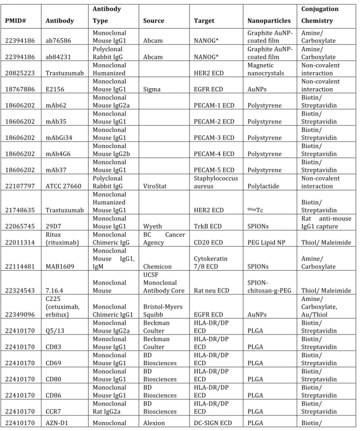
A review of papers describing antibody-guided nanoparticles (Table 1.2) reveals that most targeting antibodies are monoclonal and mostly murine, though some antibodies from other species, and polyclonals from rabbit, have been effective, as well as some chimeric (63-65) and humanized antibodies (66-68). The majority of these antibodies target the
employed lack directionality, presumably due to the presence of multiple reactive functional groups on antibodies, yielding heterogeneous antibody orientations on the nanoparticles. Table 1.2. Recent examples of targeted nanoparticles using antibodies.
PMID# Antibody
Antibody
Type Source Target Nanoparticles
Conjugation Chemistry
22394186 ab76586 Monoclonal Mouse IgG1 Abcam NANOG* Graphite AuNP-‐coated film Amine/ Carboxylate
22394186 ab84231 Polyclonal Rabbit IgG Abcam NANOG* Graphite AuNP-‐coated film Amine/ Carboxylate
20825223 Trastuzumab Monoclonal Humanized HER2 ECD Magnetic nanocrystals Non-‐covalent interaction
18767886 E2156 Monoclonal Mouse IgG1 Sigma EGFR ECD AuNPs Non-‐covalent interaction
18606202 mAb62 Monoclonal Mouse IgG2a PECAM-‐1 ECD Polystyrene Biotin/ Streptavidin
18606202 mAb35 Monoclonal Mouse IgG1 PECAM-‐2 ECD Polystyrene Biotin/ Streptavidin
18606202 mAbGi34 Monoclonal Mouse IgG1 PECAM-‐3 ECD Polystyrene Biotin/ Streptavidin
18606202 mAb4G6 Monoclonal Mouse IgG2b PECAM-‐4 ECD Polystyrene Biotin/ Streptavidin
18606202 mAb37 Monoclonal Mouse IgG1 PECAM-‐5 ECD Polystyrene Biotin/ Streptavidin
22107797 ATCC 27660 Polyclonal Rabbit IgG ViroStat Staphylococcus aureus Polylactide Non-‐covalent interaction
21748635 Trastuzumab
Monoclonal Humanized
Mouse IgG1 HER2 ECD 99mTc
Biotin/ Streptavidin
22065745 29D7 Monoclonal Mouse IgG1 Wyeth TrkB ECD SPIONs Rat anti-‐mouse IgG1 capture
22011314 Ritux (rituximab) Monoclonal Chimeric IgG BC Agency Cancer CD20 ECD PEG Lipid NP Thiol/ Maleimide
22114481 MAB1609
Monoclonal Mouse IgG1,
IgM Chemicon Cytokeratin 7/8 ECD SPIONs Amine/ Carboxylate
22324543 7.16.4 Monoclonal Mouse
UCSF Monoclonal
Antibody Core Rat neu ECD SPION-‐chitosan-‐g-‐PEG Thiol/ Maleimide
22349096
C225 (cetuximab,
erbitux) Monoclonal Chimeric IgG1 Bristol-‐Myers Squibb EGFR ECD AuNPs
Amine/ Carboxylate, Au/Thiol
22410170 Q5/13 Monoclonal Mouse IgG2a Beckman Coulter HLA-‐DR/DP ECD PLGA Biotin/ Streptavidin
22410170 CD83 Monoclonal Mouse IgG1 Beckman Coulter HLA-‐DR/DP ECD PLGA Biotin/ Streptavidin
22410170 CD69 Monoclonal Mouse IgG1 BD Biosciences HLA-‐DR/DP ECD PLGA Biotin/ Streptavidin
22410170 CD80 Monoclonal Mouse IgG1 BD Biosciences HLA-‐DR/DP ECD PLGA Biotin/ Streptavidin
22410170 CD86 Monoclonal Mouse IgG1 BD Biosciences HLA-‐DR/DP ECD PLGA Biotin/ Streptavidin
PMID# Antibody
Antibody
Type Source Target Nanoparticles
Conjugation Chemistry (αDC-‐SIGN1) Mouse hybrid
IgG2/IgG4 Pharmaceuticals Streptavidin
22410170 AZN-‐D2 Monoclonal Mouse IgG2
Alexion Pharmaceutic
als DC-‐SIGN ECD PLGA Biotin/ Streptavidin
22410170 Ab (αDC-‐SIGN2) hD1
Monoclonal Humanized IgG2/4
Alexion Pharmaceutic
als DC-‐SIGN ECD PLGA Biotin/ Streptavidin
22410170 H200 (αDC-‐SIGN3) Polyclonal Rabbit IgG Santa Biotechnology Cruz DC-‐SIGN ECD PLGA Biotin/ Streptavidin 22464249 Colo205 Monoclonal FMMU, China EpCAM ECD Silica NPs NaIO4 oxidation 22464249 sw480 Monoclonal FMMU, China EpCAM ECD Silica NPs NaIO4 oxidation 22464249 NCM460 Monoclonal FMMU, China EpCAM ECD Silica NPs NaIO4 oxidation
22469295 9B9 Monoclonal Rat ShJU, China EGFR ECD PHPA-‐PEI Non-‐covalent interaction
22494888 Anti-‐CD44 Monoclonal BD Biosciences CD44 ECD PEG-‐Liposome Thiol/ Maleimide
22471719
C225 (cetuximab,
erbitux) Monoclonal Chimeric EGFR ECD PLGA-‐ZnS:Mn2+
Amine/ Carboxylate
22515817 Monoclonal BD Biosciences IL-‐6 Fe3O4@SiO2 Glutaraldehyde
22515817 Monoclonal BD Biosciences IFN-‐γ Fe3O4@SiO3 Glutaraldehyde
22515817 Monoclonal BD Biosciences AFP fetoprotein) (alpha-‐ Fe3O4@SiO4 Glutaraldehyde
18239128 FIB504 Monoclonal Rat IgG2a β7 ECD integrin Multilamellar Liposome Amine/Carboxylate
14512622 Polyclonal PSA ECD AuNPs Non-‐covalent interaction
14512622 Monoclonal PSA ECD Iron Oxide NPs Glutaraldehyde/Amine
21838300 anti-‐TM34-‐211 Monoclonal Rat IgG2a
Murine thrombomodul
in ECD PEO-‐filomicelles Biotin/ Streptavidin
21838300 anti-‐TM201-‐411 Monoclonal Rat IgG2a
Murine thrombomodul
in ECD PEO-‐filomicelles Biotin/ Streptavidin
21976974 Trastuzumab
Monoclonal Humanized
Mouse IgG1 Roche HER2 ECD Chitosan NPs Thiol/Maleimide
21976975 anti-‐DR5
Monoclonal Humanized
IgG2b Santa Biotechnology Cruz DR5 ECD PLA Amine/ Carboxylate
22072868 LCCS Abs Polyclonal Mouse
liver cancer cell surface-‐specific
(LCCS) AuNPs Non-‐covalent interaction
22035507 Anti-‐Her2 Ab Monoclonal Mouse IgG1
Bender MedSystems
(eBioscience) HER2 Iron Oxide NPs Amine/ Carboxylate
21980236 Anti-‐EGFR Ab Monoclonal Chimera
Beijing Zhong Shan
Company EGFR Quantum Dots (QD800) Thiol/Maleimide
Antibody targeting of nanoparticles faces several major challenges: antigen binding (the mAb must have high target specificity and affinity and the linker, as well as NPs, must not perturb the desired specificity), conjugation (the Ab-NP linkage must be highly efficient and site-specific), and circulation time (the mAb-NP conjugate linker must be stable during circulation). In addition, immunogenicity and purity are other concerns. The body can perceive antibodies as foreign proteins and clear them, nullifying the action of the targeted NPs. Many conjugation techniques, such as those exploiting lysine side-chain amines and cysteine sulfhydryl groups, incite immunogenic responses by yielding heterogeneous mixtures of targeted NPs, each with differing Ab:NP molar ratios, conjugation sites, pharmacokinetics, and safety profiles.
1.3.3 Protein Domains
Targeting ligands based on full-length antibodies have several intrinsic disadvantages compared to ligands with much smaller sizes. First, the large sizes of the full-length
bacteria with much lower manufacturing costs. In addition, it should possess functional residues that facilitate conjugation with nanoparticles, preferably in a site-specific manner.
The use of antibody fragments represents an interesting compromise in the selection of target-specific affinity molecules. The smallest antibody fragments are those based on a single domain, such as naturally occurring heavy-chain antibodies found in camelids
(nanobody) and humans (VH domains) (70-81). These single-domain antibody fragments are well expressed, quite soluble and stable, and yet still able to maintain the specificity and affinity comparable to scFvs.
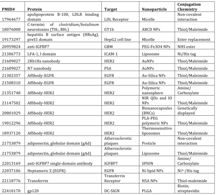
A different approach is to select single domain antibody mimics from partially randomized libraries based on protein scaffolds related or not related to natural antibodies (82). Scaffolds that have been used to construct single domain protein libraries include immunoglobulin-like b-barrel, zinc fingers, a-helical bundles, Src homology domains, PDZ domains, various repeat proteins, protease inhibitors, and disulfide-bond constrained small toxins (82-89). Among them, targeting ligands based on FN3 (the tenth type III domain of human fibronectin), Z domain, and DARPins are most promising (90). Several examples of protein domain-based ligands that are suitable for targeted delivery of nanoparticles include FN3-based ligands (monobody) that recognize VEGF receptor and integrin avb3, Z domain based ligands (affibody) that recognize EGFR and HER2, and DARPin based ligands that recognize HER2.
Compared to the number of nanoparticle applications utilizing antibodies and peptides for targeting, relatively few studies have employed protein domains or
protein domain nanoparticle targeting is highly promising, as compared to antibodies and peptides, for targeted delivery of nanoparticles.
Table 1.3. Recent examples of targeted nanoparticles using protein domains.
PMID# Protein Target Nanoparticle Conjugation Chemistry
17964677 apolipoprotein B-‐100, LDLR binding domain LDL Receptor Micelle Non-‐covalent interaction
18076008 C-‐termini of neurotoxins (THC, BHclostridium/botulinumC) GT1b ABCD NPs Thiol/Maleimide
19173297 hepatitis B surface antigen (HBsAg), preS1 domain HepG2 cell line Micelle Ester replacement 20959824 anti-‐IGFBP7 GBM PEG-‐Fe3O4 NPs NHS ester 21306773 LFA-‐1, I domain ICAM-‐1 Liposome Ni/His tag
21609027 2Rb18a nanobody HER2 AuNPs Thiol/Maleimide
21609027 N7 nanobody PSA AuNPs Thiol/Maleimide
21302357 Affibody-‐EGFR EGFR Au-‐Silica NPs Thiol/Maleimide 21508310 Affibody-‐EGFR EGFR Au-‐Silica NPs Thiol/Maleimide
21351748 Affibody-‐HER2 HER2 Polymeric nanosphere Amine/ Carboxylate
21147502 Affibody-‐HER2 HER2 NIR QDs and IO NPs Thiol/Maleimide
20801029 Affibody-‐HER2 HER2 Bionanocapsules (BNCs) Genetically displayed
19012296 Affibody-‐HER2 HER2 PLA-‐PEG polymeric NPs Thiol/Maleimide
18937120 Affibody-‐HER2 HER2 Thermosensitive liposomes Thiol/Maleimide
21753879 adiponectin, globular domain (gAd) Atherosclerotic plaques Proticle Non-‐covalent interaction
21753879 adiponectin, globular domain (gAd) Atherosclerotic plaques Liposome Thiol/Maleimide
22013169 anti-‐IGFBP7 single-‐domain antibody IGFBP7 SPION Amine/ Carboxylate 22037106 Heptameric Z (EGFR) EGFR Ni-‐lipid NPs Ni2+/His tag
22118776 Transferrin Transferrin Receptor HSA NPs Thiol-‐maleimide
22410170 gp120 DC-‐SIGN PLGA Biotin, streptavidin
1.3.4 Peptides
Much smaller than antibodies but larger than small molecules, short homing peptides offer additional nanoparticle targeting options and certain advantages over the
small molecules and antibodies, short peptides provide smaller size, as well as high specificity and affinity.
Targeting homing peptides are typically discovered via phage display, first developed in 1985. Phage display is a screening tool for peptides, allowing selection of peptide
sequences with increased affinities to a specific target of choice (91, 92). The phage display system is a cyclic selection process where the purified target molecules or specific cell types are incubated with a randomized library of peptide sequences displayed on bacteriophage capsids. Some peptides on the capsids bind to the target protein. Non-binders are washed away while capsids displaying binding peptides are eluted. Eluted bacteriophages are collected, used to infect E. Coli, are amplified, and sent to the next cycle of selection. Selected peptides have been used as molecular probes for imaging and can be applied as therapeutics as well.
There are numerous publications using short homing peptides to target nanoparticles during the past decade. Studies using peptides as nanoparticle targeting ligands (Table 1.4) predominantly utilized ligands discovered via phage display. Some used natural peptides, such as EGF (93, 94), CANF (95), and Angiopep-2 (96). About 30% of reviewed papers used cyclic peptides, though this percentage is influenced by the popularity of the RGD peptide as a targeting ligand to αvβ3 integrin (97-102). All studies targeted cell surface proteins. As with antibodies, the selected peptides were successfully conjugated to a variety of nanoparticles, such as metallic NPs (e.g. gadolinium oxide, SPIONs, AuNPs, MBCSPs), micelles and polymers (e.g. chitosan, PLGA, poly(methyl methacrylate)), and dendrimers. Table 1.4. Recent examples of targeted nanoparticles using short homing peptides.
PMID# Peptide Sequence Cyclic Target Nanoparticle Conjugation Chemistry polysiloxane
shell
21740042 EGF EGFR ECD Vault
nanoparticles
Recombinant fusion
21763734 NGR NGR CD13 ECD ELP Micelle Non-covalent interaction 21781994 S2P CRTLTVRK
C Stabilin-2 ECD Chitosan Amine/Carboxylate, Thiol/Maleimide 21871505 EGF EGFR ECD GemC18 NPs Thiol/Maleimide 21945679 I4R CRKRLDR
NC IL-4R ECD Hydrophobically modified glycol chitosan
Amine/Carboxylate
21987727 AH1 SPSYVYHQ F
MHC Class II ECD G5-PAMAM dendrimer
Thiol/Maleimide
21987727 TRP2180–188 SVYDFFV
WL MHC Class II ECD G5-PAMAM dendrimer Thiol/Maleimide 21987727 PAn DR
epitope (PADRE)
aKXVAAW TLKAAaZC
MHC Class II ECD G5-PAMAM dendrimer
Thiol/Maleimide
21987727 HA110–120 SFERFEIFP
KEC MHC Class II ECD G5-PAMAM dendrimer Thiol/Maleimide 22014944 iRGD CRGDKGP
DC αvβ3, αvβ5 PLGA-PLL-PEG Thiol/Maleimide 22049461 CANF NPR-C ECD poly (methyl
methacrylate)-PEG
Methacrylate/ Acetylene
22087004 KLWVLPK
GGGC-Am Collagen IV PLGA PEG Thiol/Maleimide 22093292 CSK CSKSSDYQ
C Goblet cells trimethyl chitosan chloride (TMC)
Amine/Carboxylate
22118776 TRAIL hTRAIL(114
–281) DR4, DR5 ECD HSA NPs Thiol/Maleimide 22133551 Angiopep-2 TFFYGGSR
GKRNNFK TEEY
LRP Receptor ECD PEG-co-poly(ε -caprolactone)
Thiol/Maleimide
22179825 tLyp-1 CGNKRTR Yes Neuropilin-1/2 ECD Iron oxide nanoworms
(NWs)
Biotin/Neutravidin
22196766 Pep 1 CHVLWST
RC Yes Pancreatic islet capillary endothelial cells PLGA-b-PEG Amine/Carboxylate 22197725 CLL1-L1 CDLRSAA
VC Yes CLL1 nanomicelle PEG telodendrimer
Click Chemistry: Alkyne/Azide
22375916 RGD cRGDfK Yes αvβ3 integrin AuNPs Amine/Carboxylate 22396491 OA02
cdG-HoCit- GPQc-Ebes-K-alkyne
α-3 integrin PEG telodendrimer
Click Chemistry: Alkyne/Azide
22403681 Tet-1 HLNILSTL
WKYR Motor neurons PLGA Amine/Carboxylate 22497548 RGD cyclic RGD Yes αvβ3 integrin PEG-PEI Electrostatic
PMID# Peptide Sequence Cyclic Target Nanoparticle Conjugation Chemistry 22561668 GRGDS GRGDS αvβ3 integrin MBCSP
(PLGA-magnetite)
Amine/Carboxylate
Han and coworkers provide an interesting alternative to the typical nanoparticle formulations and conjugation paradigms (93). They expressed targeting peptides recombinantly fused to the 97 kDa major vault protein (MVP), which self-assembles into Vault Nanoparticles – naturally-occurring nanoparticles present in cell cytoplasm composed of ribonucleoproteins. At times, the orientation of the conjugated peptides in the reviewed studies can be
problematic but this is controlled in some applications at the level of peptide synthesis, typically through the additional of a unique functional group to peptide termini, allowing for site-specific conjugation with nanoparticles.
There have been numerous effective in vitro peptides (e.g. targeting protein kinase CK2, glioma, FGF receptor, and many others). Finding peptides that work in an in vivo
setting, however, appears more challenging as they are prone to proteolysis, glomerular transit, feature varying toxicities and differential effects on cell signaling, can encourage allergic sensitization, and are not amenable to oral bioavailability (103-105). In addition, the costs of peptide synthesis can be prohibitive for some applications (103).
1.4 The Research Approach Pursued in this Dissertation Directly Selected Highly
Stable fGmH RNA Aptamers from fGmH Libraries for Use as Targeting Ligands
that we confirmed had no discernible effect on catalysis (25, 106). Unlike the Sousa Variant, however, this polymerase also bore the Y639L mutation, rather than Y639F. The “LAR” T7 RNA polymerase used in this study was an attempt to address problems of inefficient non-canonical ribonucleotide incorporation during in vitro transcription. LAR does not require doping of the 2’-X-dNTP pool with 2’-OH-rGTP to produce fGmH RNA with up to 30 to 40 copies and high quality homogeneous fGmH RNA libraries that are truly directly selectable.
The selection of fGmH RNA aptamers focused on those that would have the
properties of enhanced hybridization and nuclease stability, as discussed above, inherent in their functionality. The resulting fGmH RNA has some unique features that would greatly enhance their suitability for in vivo applications: enhanced stability against many nucleases and an inability of most endogenous polymerases to utilize the metabolic products of eventual fGmH RNA degradation (Fig. 1.2). In addition, the use of 2’-F residues in RNA does not greatly enhance nuclease resistance, which limited the desired number of 2’-F residues in the RNA composition pursued in this study. However, the superior ability of 2’-F residues to stabilize base pair interactions compared to 2’-OMe prompted us to hypothesize that the fGmH RNA composition of one 2’-F and three 2’-OMe residues would have a higher probability of achieving greater conformational and nuclease stability for translational
applications.
PD-1 Ligand 2. Overall, this work will demonstrate the direct selection of aptamers from fGmH RNA libraries, provide serum stability comparisons between fGmH RNA aptamers and RNAs with fewer 2’ modifications, and employ fGmH RNA aptamers as targeting ligands.
CHAPTER 2
SELECTING fGmH RNA APTAMERS FROM A 2’-FULLY MODIFIED fGmH RNA LIBRARY THAT BIND Staphylococcus aureus PROTEIN A (SpA)
2.1 Introduction
The original plan guiding this selection was to select fGmH RNA aptamers in a proof-of-concept selection against epidermal growth factor receptor (EGFR) via a
selection strategy called Active Site – SELEX. This strategy will be described in Chapter 3, the results of which formed the study described in this chapter. The strategy, however, did not select fGmH aptamers against the EGF ligand binding site of EGFR but, instead, against the Protein A (SpA) coating the magnetic sepharose beads used for Fc-EGFR immobilization. fGmH aptamers that bound SpA were therefore used as proof-of-concept for fGmH RNA aptamer technology, as described in the rest of this chapter.
A fGmH RNA library with a diversity of 7 × 10¹³ was used in this selection. SpA itself is actually an attractive, if not unintentional, selection target because its structure and interaction mode with IgG Fc have been well characterized, and because S. aureus
provides a convenient and relevant biological system for biomaterial development. In addition, SpA has become increasingly recognized over the last ten years as a key determinant of S. aureus and, especially, MRSA pathogenesis. SpA has binding
VH3 class Fabs (107). Most well-known is the protective role that SpA confers to S.
aureus by neutralizing the humoral response via IgG-Fc binding, thereby preventing
opsonization. It is clear that highly stable fGmH RNA aptamer anti-SpA nanoparticle ligands are desirable as potential therapeutics and reagents for the study of various aspects of S. aureus biology.
2.2 Results
2.2.1 The Expression and Purification of LAR T7 RNA Polymerase
As discussed above, it is of great interest to develop a novel class of 2’-fully modified RNA aptamers as enhanced biomaterial targeting ligands consisting of a fGmH RNA composition. To generate a T7 RNA polymerase mutant that can efficiently accept 2’-F-dGTP and 2’-OMe-dATP/dCTP/dUTP as substrates, Chuan (Lawrence) Fu
rNTPs was examined on a 20% denaturing PAGE gel. Incubation of 5 pmol aliquots of natural RNA with and without purified LAR T7 RNA polymerase did not indicate the presence of contaminating nucleases.
As shown in Lanes 1 to 3 in Figure 2.2, when primed with GMP in the presence of PEG-8000, a transcriptional initiator and molecular crowding adjuvant, respectively, His-LAR T7 RNA polymerase variant accepted 2’-F-dGTP and 2’-O
Me-dATP/dCTP/dUTP very well, with a relative transcription efficiency of 96% compared to an LAR in vitro transcription reaction using all natural 2’-OH-rGTP/rATP/rCTP/rUTP.
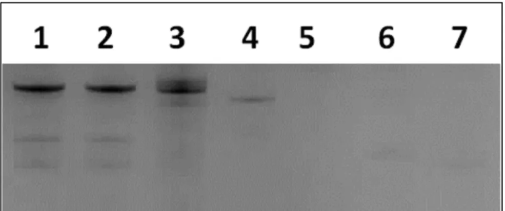
Figure 2.1. Electrophoresis (8%) PAGE gel of the expression and purification of His-LAR T7 RNA polymerase. Lanes 1) Induced insoluble fraction, 2) Induced soluble fraction, 3) Uninduced whole cell lysate, 4) Molecular weight marker, 5) Molecular weight marker, 6) Induced soluble fraction, 7) Flow-through of Talon metal affinity column, 8) Buffer A Wash (No imidazole), 9) Buffer B Wash (20 mM imidazole), 10) Buffer C Elution 1 (200 mM imidazole), 11) Buffer C Elution 2 (200 mM imidazole).
This contrasts with the various R425 T7 RNA polymerase mutants developed by
transcription products were obtained. With the availability of such a T7 RNA polymerase mutant, an fGmH RNA library with a diversity of 7 × 10¹³ unique sequences was
enzymatically synthesized and purified for in vitro selection.
Figure 2.2. Transcription efficiency of purified His-LAR T7 RNA polymerase using different 2’-X-dNTPs. Lane 1) fGmH RNA using 2’-F-dGTP and 2’-OMe-dA/U/CTP, 2) fGmH RNA repeat, 3) natural RNA using 2’-OH-rG/A/U/CTP, 4) 2’-OMe-dA/U/CTP, no 2’-F-dGTP, 5) 2’-OMe-dU/CTP, 2’-F-dGTP, no 2’-OMe-dATP, 6) 2’-OMe-dA/CTP, 2’-F-dGTP, no 2’-OMe-dUTP, 7) 2’-OMe-dA/UTP, 2’-F-dGTP, no 2’-OMe-dCTP.
2.2.2 fGmH RNA Aptamers were Unintentionally Selected against SpA and were
Characterized for SpA Binding
To investigate that 2’-fully modified aptamers can be directly selected from an fGmH RNA library enzymatically synthesized by LAR T7 RNA polymerase, we performed a proof-of-concept selection originally intended against EGFR. However, fGmH RNA aptamers were instead selected against S. aureus SpA. SpA serves as a model target because of its well-characterized structure, its role as a virulence factor for both antibiotic sensitive and resistant S. aureus, and because S. aureus offered a
specifications. For each round of selection, the input fGmH RNA library, at ~0.7 µM, was incubated with 25 µL (1 column volume) of Protein A beads. Binding was performed in 6 column volumes of 1× Aptamer Selection Buffer for 1 h at 4 °C. Beads were then washed twice in 6 column volumes of 1× Aptamer Selection Buffer, each for 5 min at 4 °C. Presumably, the washing (12 column volumes total) removed sequences bound non-specifically, as well as target-binding sequences with rapid dissociation rates. Aptamers were eluted three times from the beads in a total of 18 column volumes of 1× Aptamer Selection Buffer, each for 20 min at 4 °C. This longer and extensive elution was designed to select aptamers with slower dissociation rates.
We used the Octet BLI method to monitor the evolution of desired binding characteristics within the selected pools during the selection. As shown in Figure 2.3, enrichment of the SpA-binding aptamers was not detectable by this binding assay before Round 4. Binding was detected in Round 5, with a limited further increase in Round 6. Such phenomena are typical for SELEX. Cloning and sequencing, as described, were performed on Rounds 5, 6, 7, and 8 selected pools (N = 154) (Fig. 2.4). Despite the small sequencing sample size, the evolution of five distinct primary sequence groups was evident across these pools.
Sequences A12, F07, E09, E03, and G12, representing primary sequence groups 1-5 (Fig. 2.5), respectively, were prepared for kinetic characterization via PCR
not shown). These results were confirmed via Octet BLI, and the binding curves measured for fmA12, the aptamer extensively used in the remainder of this study, are provided in Figure 2.6 and demonstrate saturable and monophasic binding. Due to the requirement of examining many different sequences and their variants, subsequent kinetic characterizations were performed using Octet BLI, which allowed the binding of 8
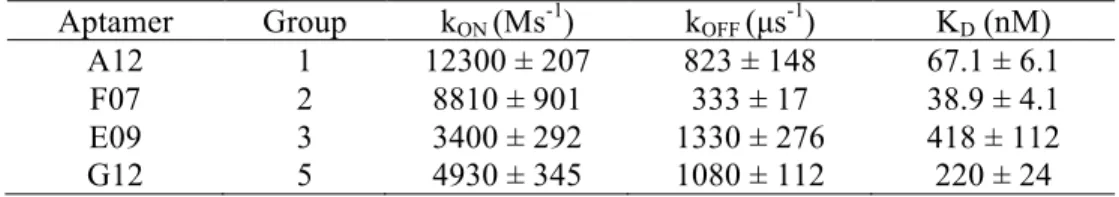
samples to SpA to be measured and analyzed in parallel. As listed in Table 2.1, fmA12 and fmG12 on Octet yielded KDs of 67.1 ± 6.1 nM and 220 ± 24 nM, respectively, which are comparable with those determined by Biacore. BLI analysis for fmF07 and fmE09 resulted in KDs of 38.9 ± 4.1 nM and 418 ± 112 nM, respectively, whereas fmE03 was found to be a non-binding sequence. These fGmH aptamers feature KDs that are reproducible in quadruplicate: a property that likely resulted from fGmH sequences forming stable structures.
Figure 2.3. Real-time monitoring of the enrichment of fGmH RNA aptamers during the selection using Octet BLI.
Figure 2.5. The putative secondary structures and random region sequences of aptamers fmA12, fmF07, fmE09, and fmG12, representing Groups 1, 2, 3, and 5, respectively.
Figure 2.6. BLI kinetic characterization of fmA12 using biosensors immobilized with SpA.
Table 2.1. Dissociation constants of fGmH RNA aptamers binding S. aureus SpA.
2.2.3 Selected fGmH RNA Aptamers are Specific for SpA, and the fGmH RNA
Composition is Critical for SpA Affinity
To test the target-binding specificity of the selected fGmH RNA aptamers, we used Octet BLI binding assays to examine the affinity of these aptamers to S. aureus SpA and Protein G (SasG), and P. magnus Protein L (PpL), three bacterial proteins that have similar IgG binding features. As shown in Figure 2.7, the previously characterized SpA-binding aptamer fmA12 did not exhibit measurable affinity for SasG or PpL. fmE09 and fmG12 yielded similar results (data not shown). fmF07, however, displayed minimal
Aptamer Group kON (Ms-1) kOFF (µs-1) KD (nM) A12 1 12300 ± 207 823 ± 148 67.1 ± 6.1
F07 2 8810 ± 901 333 ± 17 38.9 ± 4.1
E09 3 3400 ± 292 1330 ± 276 418 ± 112
binding to both SasG and PpL, though with much lower affinity than SpA (2.76 ± 2.39 µM for SasG and 452 ± 92 nM for PpL, compared to 38.9 ± 4.1 nM for SpA). It is presumed that fmF07 is capable of binding a structurally conserved region present in all three proteins. However, the results for fmA12, fmE09, and fmG12 clearly indicate that selected fGmH RNA aptamers are capable of interacting specifically with the target used in selection.
Figure 2.7. Binding specificity of fmA12 to Protein A, Protein G, and Protein L.
results indicate that once selected, fGmH RNA aptamers must maintain their 2’
modification state to preserve target binding. As such, fGmH RNA aptamers constitute a distinct aptamer class. The lack of detectable tmA12 functionality also ruled out the concern that fmA12 guanine residues have heterogeneous 2’-hydroxyls, potentially from trace contamination of 2’-OH-rGTP in 2’-F-dGTP or rGMP used for transcription.
Figure 2.8. Protein A (SpA) binding dependence of sequence A12 on the RNA 2’ modification state (WT A12 (wild-type; rN), bmA12 (fYrR), tmA12 (rGmH), fmA12 (fGmH)).
2.2.4 fmA12 was Structurally Truncated while Preserving SpA Affinity for
Potential Future Chemical Synthesis
All identified SpA-binding aptamers have full-length sequences of 85 residues. To map the target-binding regions and facilitate future large-scale chemical synthesis using an oligonucleotide synthesizer, the high affinity SpA-binding aptamer fmA12 was shortened via comprehensive testing of nine truncation and stem stabilization variants (fmA12Δ1 through Δ9). Truncations fmA12Δ1/2/4/5/7/8 resulted in global disruption of the putative fmA12 secondary structure, as predicted by Mfold (data not shown).
last five residues at the 3’ end (red sequence in Figure 2.9), which appeared not to be involved in the stem or loop secondary structures as predicted by Mfold. Indeed, fmA12Δ3 affinity for SpA was preserved compared to full-length fmA12 with a KD of 69.7 ± 3.5 nM. Truncation fmA12Δ6 maintained Stem-loop Domain I (blue sequence in Figure 2.9) but completely removed Stem-loop Domain II (green sequence in Figure 2.9), resulting in more than a 1,000-fold reduction in binding affinity (KD of 63.1 ± 7.3 µM). Truncation fmA12Δ9 was designed with the fmA12Δ3 acaulescent Stem-loop Domain II and the 17-residue single-stranded region at the 5’ end removed (orange sequence in Figure 2.9). The resulting fmA12Δ9, with a length of 61 residues, exhibited preserved SpA binding with a KD of 69.1 ± 16.7 nM - almost identical to that of the full-length fmA12. Sequence truncation analysis indicated, in summary, that Domain I alone is insufficient for binding, and, presumably, that both domains are necessary to achieve high affinity SpA binding. Overall, sequence truncation found a version of full-length fmA12 (fmA12Δ9) that could be chemically synthesized if future need arose. For the purposes of this study, no further truncation was necessary though future applications may require different sequence length optimizations dependent on the features desired from the aptamer.