HIGH-THROUGHPUT EXPERIMENT DRIVEN MODELING OF RNA INTERACTIONS AND STRUCTURES
Greggory Mathew Rice
A dissertation submitted to the University of North Carolina at Chapel Hill in partial fulfillment of the requirements for the degree of Doctor of Philosophy in the Department of Chemistry
Chapel Hill 2015
©2015
ABSTRACT
GREGGORY MATHEW RICE: High-throughput Experiment Driven Modeling of RNA Interactions and Structures
(Under the direction of Kevin M. Weeks)
ACKNOWLEDGEMENTS
First, I would like to thank my advisor Kevin. Thank you for giving me the freedom to try new ideas (that often didn’t work) and letting me learn by doing. I will miss going into your office to bounce a new idea off you. I have become a better writer and scientist because of you.
I would also like to thank the wonderful teachers and mentors that I’ve had at UNC, specifically Barry, Gary, Alain, Nikolay, and many others. You have given me the foundation and provided the support for me to succeed in graduate school and beyond.
Next, I would like to thank the members of the Weeks group past and present. I couldn’t have asked for a better group of scientists (and friends) to work with. Your collective wisdom is a force to be reckoned with.
To team SHAPE-MaP, thank you for reminding of me the value of close collaboration. Together we accomplished something that alone would not have been as great. At times it was challenging, but in the end, we made it.
To the lifelong friends that I have made since coming to UNC, you guys rock. Matt, Fatima, Kathleen, Tim, and Hannah you have become a second family for me. Graduate school has been easier because of you all. Wherever I am in the world, you will always have a place to stay.
TABLE OF CONTENTS
LIST OF TABLES ... xi
LIST OF FIGURES ... xii
LIST OF ABBREVIATIONS AND SYMBOLS ... xiv
CHAPTER 1: THE IMPORTANCE OF RNA STRUCTURE ... 1
Establishment of the structure-function relationship in ribonucleic acid ... 1
High resolution methods for determining RNA structures ... 2
RNA foot-printing methods for determining structure ... 4
Secondary and structure prediction methods ... 6
Rise of deep sequencing and coupling to structure probing methods ... 7
Research overview ... 10
Perspective ... 11
References ... 13
CHAPTER 2: CHEMICAL INTERACTIONS OF SHAPE ADDUCTS WITH RNA IN THREE DIMENSIONAL SPACE ... 17
Introduction ... 17
Results ... 19
Fingerprinting RNA structure using multiple SHAPE reagents ... 19
Stacking interactions between reagent and nucleobase influence SHAPE reactivity ... 22
HMX overview ... 24
Molecular overlap model for HMX intensities. ... 26
HMX experiment and outlook. ... 29
Methods ... 30
1M6 and NMIA free energy calculations ... 30
Modeling of adduct disruption of native RNA tertiary structure (HMX). ... 31
References ... 32
CHAPTER 3: RNA SECONDARY STRUCTURE MODELING AT CONSISTENT HIGH ACCURACY USING DIFFERENTIAL SHAPE ... 34
Introduction ... 34
Results ... 38
Selection of a challenging test set. ... 38
Incorporation of differential SHAPE into secondary structure modeling. ... 40
Impact of ∆GDiff on structure modeling. ... 41
Responsive and non-responsive RNAs. ... 44
Discussion ... 45
Limitations and Perspective ... 47
Methods ... 48
Chemical probing by differential SHAPE. ... 48
Differential SHAPE data analysis. ... 49
Differential SHAPE pseudo-free energy change penalty. ... 50
Exploration of simpler differential SHAPE energy potentials. ... 51
Implementation in RNAstructure Fold and ShapeKnots. ... 53
Plots and figures. ... 54
References ... 55
CHAPTER 4: AUTOMATED MOTIF DISCOVERY IN LARGE RNAS USING SHAPE-MAP AND SUPERFOLD ... 58
Results ... 59
The MaP strategy ... 59
Structure modeling: validation ... 61
A second-generation model for an HIV-1 RNA genome ... 65
Development of SuperFold, a large RNA folding algorithm ... 65
De novo identification of well-determined structures ... 68
Motif discovery and deconvolution of structural polymorphism ... 70
Discussion ... 75
Methods ... 76
SHAPE-MaP experimental overview. ... 76
SHAPE-MaP development and efficiency ... 76
SHAPE-MaP using fragmented samples. ... 79
SHAPE-MaP using targeted gene-specific primers ... 80
Filtering by Z-factor for differential SHAPE data ... 81
Structure modeling ... 81
Error analysis and determining a minimum number of reads required for accurate RNA structure modeling ... 84
Algorithmic discovery of HIV-1 regions with low Shannon entropy and low SHAPE reactivity ... 85
HIV competition assays ... 85
Calculation of differences in SHAPE reactivities in pseudoknot mutants ... 86
References ... 88
CHAPTER 5: RIBOSOME DYNAMICS VISUALIZED BY CORRELATED CHEMICAL PROBING IN LIVING CELLS ... 92
Introduction ... 92
Development of a general analysis framework for randomly primed reads. ... 98
Correlated chemical probing reveals distinct structural networks ... 101
Network analysis reveals distinct communities in the small subunit with structural hubs ... 101
Discussion ... 105
Methods ... 106
DMS modification of extracted ribosomal RNA ... 106
Antibiotic treatment for in cell samples ... 106
Dimethyl sulfate treatment and purification of ribosomal RNAs ... 107
Reverse transcription screening for improved MaP conditions ... 108
Library preparation and sequencing ... 108
Data processing and alignment ... 109
Correlation analysis of randomly primed reads ... 109
Network analysis of correlations in the small ribosomal subunit ... 110
LIST OF TABLES
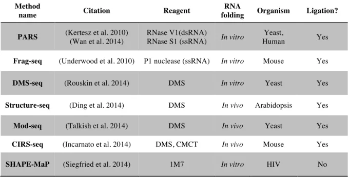
Table 1.1: A selection of deep sequencing structure probing methods. ... 9
Table 3.1: RNA secondary structure modeling accuracies with 1M7 and differential SHAPE
information. ... 39
Table 3.2: RNA secondary structure modeling accuracies comparing three-reagent differential
SHAPE to related recent works. ... 46
Table 3.3: RNA secondary structure modeling accuracies for a two-reagent differential SHAPE
LIST OF FIGURES
Figure 1.1: Explaination of the levels of RNA structure. ... 3
Figure 1.2: Chemical probing methods and detection by capillary electrophoresis. ... 5
Figure 1.3: Explaination of paired end sequencing. ... 8
Figure 2.1: RNA SHAPE chemistry. ... 18
Figure 2.2: SHAPE analysis of the ligand-bound state of the TPP riboswitch. ... 21
Figure 2.3: Conformations and structural contexts for nucleotides exhibiting differential reactivities in the ligand-bound state of the TPP riboswitch. ... 22
Figure 2.4: Effect of varying the substituents on SHAPE reactivity and electronic structure calculations for the 1M6- and NMIA-nucleotide complex stabilities. ... 23
Figure 2.5: Visualization of HMX interference information on accepted three-dimensional structures. ... 25
Figure 2.6: Physical model for 2'-hydroxyl molecular interference. ... 27
Figure 3.1: Accuracy of an RNA structure model and its usefulness for understanding structure-function interrelationships. ... 36
Figure 3.2: Differential SHAPE analysis of the E. coli 5S rRNA. ... 37
Figure 3.3: Statistical determination of the ∆GDiff free energy change penalty. ... 40
Figure 3.4: Representative secondary structure modeling for the 5S rRNA without and with SHAPE data. ... 42
Figure 3.5: Circle plots illustrating SHAPE-directed structure modeling. ... 43
Figure 3.6: Circle plots illustrating SHAPE-directed structure modeling for Tetrahymena group I intron. ... 44
Figure 3.7: Comparison of the statistically determined pseudo-free energy change term with the grid-search optimized ln-form ∆GSHAPE. ... 52
Figure 4.1: Overview of the SHAPE-MaP approach. ... 60
Figure 4.2: Nucleotide-resolution interrogation of RNA structure and ligand-induced conformational changes. ... 61
Figure 4.3: Mutation rate histograms for paired and non-paired nucleotides in the 16S rRNA. ... 62
Figure 4.4: SHAPE-MaP replicates of E. coli 16S rRNA. ... 63
Figure 4.6: SHAPE-MaP analysis of the HIV-1 NL4-3 genome. ... 66
Figure 4.7: Overview of the SuperFold pipeline. ... 67
Figure 4.8: Functional and structural validation of newly discovered HIV-1 RNA motifs. ... 71
Figure 4.9: Pseudoknot SHAPE-MaP profiles for ENVPK and CAPK. ... 74
Figure 4.10: Detection of 2'-O-adducts by mutational profiling. ... 78
Figure 5.1: Overview of DMS modification and RING-MaP experiment. ... 93
Figure 5.2: Structural organization of the bacterial ribosome. ... 95
Figure 5.3: DMS reactivity across the small ribosomal subunit. ... 97
Figure 5.4: Computational approach for analyzing randomly primed read data. ... 99
Figure 5.5: RING-MaP correlations within the small ribosomal subunit. ... 100
Figure 5.6 Correlation network diagrams separated into communities. ... 103
LIST OF ABBREVIATIONS AND SYMBOLS
2'OH ribose 2'-hydroxyl
1M6 1-methyl-6-nitroisatoic anhydride 1M7 1-methyl-7-nitroisatoic anhydride
A adenosine
Asp aspartic acid
C cytidine
CA capsid
cDNA complementary DNA
CE capillary electrophoresis
CMCT N-cyclohexyl-N’-(2-morpholinoethyl)carbodiimide metho-p-toluenesulfonate CO2 carbon dioxide
cryo-EM cyro-electron microscopy
dA deoxyadenosine
DMD discrete molecular dynamics
DMS dimethyl sulfate
DMSO dimethyl sulfoxide DNA deoxyribonucleic acid
EDTA ethylenediaminetetraacetic acid
ENV Envelope
G guanosine
g gram
g gravity
HCV hepatitis C virus
HMX 2'-hydroxyl molecular interference IRES internal ribosome entry site
kB Boltzmann constant
KCl potassium chloride
kcal kilocalorie
ln natural log
LNA locked nucleic acid
M molar
MaP mutational profiling
MDa mega Dalton
MgCl2 magnesium chloride
min minute
mL milliliter
mM millimolar
mol mole
NaCl sodium chloride
ng nanogram
NGS next-generation sequencing
nM nanomolar
NMIA N-methylisatoic anhydride
NMR nuclear magnetic resonance spectroscopy
nt nucleotide
OD600 optical density at 600 nanometers
PK pseudoknot
PPT polypurine tract
ppv positive predictive value
Rif rifampicin
RING-MaP RNA interaction groups identified by mutation profiling RNA ribonucleic acid ribonuclease
rpm revolutions per minute RRE Rev response element
rRNA ribosomal RNA
RT reverse transcriptase
sens sensitivity
SDS sodium dodecyl sulfate
SHAPE selective 2’-hydroxyl acylation analyzed by primer extension
SHAPE-MaP selective 2’-hydroxyl acylation analyzed by primer extension and mutational profiling
SHAPE-seq selective 2’-hydroxyl acylation analyzed by primer extension sequencing
Spc spectinomycin
STMV satellite tobacco mosaic virus TPP thiamine pyrophosphate
T temperature
TAR trans-activation response element
TE 10 mM Tris (pH 8.0), 1 mM EDTA
TPP thiamine pyrophosphate
tRNA transfer RNA
U uridine
UTR untranslated region
°C degree Celsius
ΔG Gibbs free energy change
μg microgram
μL microliter
CHAPTER 1: THE IMPORTANCE OF RNA STRUCTURE Establishment of the structure-function relationship in ribonucleic acid
During the early years of molecular biology in the 1950s, ribonucleic acid (RNA) was considered a passive intermediate carrying information coding for the sequence of proteins stored in deoxyribonucleic acid (DNA) to the ribosome (Crick 1970). In the intervening decades since the postulation of this now-famous central dogma, RNA has been found to be a far more versatile molecule than first thought. RNA is able to affect gene expression through a number of complex systems including alternative exon splicing (Amaral et al. 2008), microRNA (Ha and Kim 2014) and RNA interference (Fire et al. 1998), and non-coding RNAs (Storz et al. 2011; Geisler and Coller 2013).
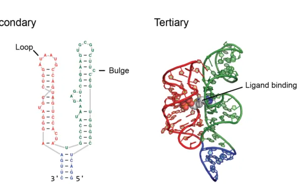
The ability of an RNA to fold back on itself and form higher order structures likely plays an essential role in many of these biological systems (Sharp 2009). RNA often folds three-dimensionally into a single functionally relevant structure. RNA structure can be described on three different hierarchical levels, with each level encoding essential and increasingly complex information. The first, and simplest level is the primary sequence which consists of the linear linkage of the four RNA nucleotides: adenine (A), cytosine (C), guanine (G), and uracil (U) (Leontis et al. 2006). Many biological systems operate solely on the primary sequence of an RNA, such as RNA interference. However, the next level of RNA structure –secondary structure, can modulate the efficiency of these systems.
typically described as loops, bulges, and single stranded regions (Fig. 1.1) (Tinoco and Bustamante 1999; Leontis et al. 2006). Together, the helices of an RNA arrange to form tertiary structure. RNA tertiary structure is stabilized by non-canonical base pairing, base stacking, and hydrogen bonding (Fig. 1.1b). The tertiary structure of an RNA can also play important functional roles in gene regulation, splicing, and even protein synthesis. In prokaryotes (and some eukaryotes), riboswitches are small RNA elements that change their structure based on recognition of small molecule ligands (Serganov et al. 2006; Dethoff et al. 2012). In the case of riboswitches, the proper formation of tertiary structure is essential for ligand binding and gene regulation.
High resolution methods for determining RNA structures
After the discovery of functional RNAs, high-resolution structure determination methods that were first applied to proteins were applied to RNA. One such technique is X-ray crystallography, which, when suitable crystals are obtained, can often determine the atomic-level tertiary structure of a molecule based on the diffraction pattern produced by X-rays traveling through the crystallized molecule of interest. X-ray crystallography was first applied to RNA in order to determine the structure of 76-nucleotide tRNA in 1974 (Kim et al. 1974; Klug et al. 1974). Decades later, crystallography has revealed molecular details of the ~3000-nucleotide large ribosomal subunit (Ban et al. 2000) and the small and large ribosomal subunit complex (Korostelev et al. 2006). Nuclear magnetic resonance spectroscopy (NMR), which works on solutions of molecules, was applied to studying RNA structure later than crystallography. NMR revealed not only structural features, but the dynamic motion of RNA as well (something that crystallography is unable to do) (Cheong et al. 1990; Al-Hashimi and Walter 2008).
different nucleobases, NMR is limited to small RNAs –typically less than 100 nucleotides in length– without heroic efforts of partially enriching segments of RNA molecules with NMR active atomic isotopes (Lu et al. 2011). Because of these drawbacks, both NMR and X-ray crystallography are only useful to a small fraction of all RNAs that are of functional interest.
RNA foot-printing methods for determining structure
Determining the secondary structure of an RNA is often key to understanding its function. However flexibility, structural heterogeneity, and time constraints make high-resolution methods intractable for many RNAs. Experimental evidence for base pairing can be obtained using ribonuclease enzymes (RNases) or chemical agents that are sensitive to base pairing and structural flexibility. Some RNases can cleave RNA in a structure specific manner: RNase V1 cleaves at base paired nucleotides whereas RNase S1 cleaves at single stranded nucleotides (Ehresmann et al. 1987). Despite their structure selectivity, RNase enzymes are large relative to small molecules and often have a sequence bias in the sites they will cleave. One small molecule reagent that reacts with RNA in at unpaired nucleotides is dimethyl sulfate (DMS), which reacts most detectably at the pairing face of A and C nucleobases to form a methyl adduct (Fig. 1.2a). However, determining locations of DMS reactivity is difficult at G nucleotides and DMS does not react broadly at U nucleotides.
and DMS are able to pass through cell membranes and can be used to probe the structure of RNA in living cells (Spitale et al. 2013; Tyrrell et al. 2013; Ding et al. 2014; McGinnis and Weeks 2014). Both nuclease and chemical probing experiments are quantified using reverse transcription primer extension (Low and Weeks 2010; Karabiber et al. 2013). Using this detection method, a fluroescently labeled primer is extended from the 3 prime end of an RNA by a reverse transcriptase enzyme (Fig. 1.2c). When reverse transcriptase encounters an adduct, or a break in the RNA in the case of nucleases, it is unable to proceed and dissociates. The cDNA products are resolved using capillary electrophoresis and quantified based on their intensity. In order to account for inherent reverse transcription pauses, a no reagent control is used to subtract the background signal away from the reagent signal.
Secondary and structure prediction methods
ligand binding are difficult to extract from sequence alone and can significantly decrease the accuracy of RNA structure modeling. In order to compensate for these interactions, some RNA modeling programs have successfully incorporated SHAPE and DMS chemical probing data (Deigan et al. 2009; Hajdin et al. 2013) (Cordero et al. 2012) to significantly increase the accuracy of generated models.
Rise of deep sequencing and coupling to structure probing methods
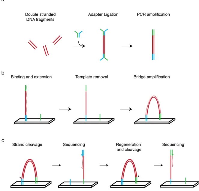
One of the consequences of the human genome project was the rapid development of platforms that enable rapid sequencing of DNA bases (International Human Genome Sequencing Consortium 2004). Rather than sequencing a single stretch of DNA, deep sequencing platforms sequence millions of individual DNA fragments simultaneously, on a massively parallel scale. The current state of the art, and widely used platform is marketed by Illumina. This platform works using a sequencing by synthesis approach with reversible terminator chemistry (Bentley et al. 2008)(Fig. 1.3), and requires that all DNA fragments have the same sequences on the 5' and 3' ends of the molecules to act as molecular “handles” for PCR amplification and sequencing primer binding sites.
Method
name Citation Reagent
RNA
folding Organism Ligation?
PARS (Kertesz et al. 2010)
(Wan et al. 2014)
RNase V1(dsRNA)
RNase S1 (ssRNA) In vitro
Yeast,
Human Yes
Frag-seq (Underwood et al. 2010) P1 nuclease (ssRNA) In vitro Mouse Yes
DMS-seq (Rouskin et al. 2014) DMS In vitro Yeast Yes
Structure-seq (Ding et al. 2014) DMS In vivo Arabidopsis Yes
Mod-seq (Talkish et al. 2014) DMS In vivo Yeast Yes
CIRS-seq (Incarnato et al. 2014) DMS, CMCT In vivo Mouse Yes
SHAPE-MaP (Siegfried et al. 2014) 1M7 In vitro HIV No
Research overview
The overall goals of this project are three-fold. First, to use computational modeling in order to help interpret chemical probing experiments and second, to create new approaches that enable the study of RNA structure at large scales. Finally, I want to use the understanding and new technical achievements gained in the first two goals to find and solve important, biologically relevant problems. In Chapter 2, I highlight my endeavors to give a molecular interpretation of two different chemical probing phenomenon by applying principles of molecular modeling. The first part of the chapter is devoted to explaining the preferential SHAPE reactivity of 1-methyl-6-nitroisatoic (1M6) to positions in folded RNAs that have available base stacking interactions. Using density functional theory (DFT) calculations I showed that 1M6 interacts more favorable at these positions relative to other reagents. In the second part of the chapter I developed a molecular overlap model to predict how disruptive an adduct at the 2’-hydroxyl position of the ribose would be to the folding of the tertiary structure of an RNA. My model gave a correlation relative to a real experiment as high as R=0.7 in several RNAs.
RNA secondary structure prediction is a challenging problem that only increases in difficulty as the length of the RNA increases. In Chapter 3, I define a novel energy potential to constrain RNA modeling predictions using information gained from position specific differences in reactivity by different SHAPE reagents. This energy potential is able to increase the accuracy of already “good” predictions to above 90% (“excellent”) across several RNAs with structures that were previously difficult to predict accurately. This increase in prediction accuracy is important since most RNAs of global interest, such as HIV-1, HCV, and Dengue virus, are thousands of nucleotides in length.
ability to quickly generate large amounts of data. Using these tools we uncover and validate three previously unreported pseudoknots in HIV-1 –a remarkable feat for a virus that has been intensely studied for more than twenty years.
Finally, in Chapter 5, I extend the RING-MaP (Homan et al. 2014) experiment and analysis with random priming in order to uncover structural dynamics in the bacterial ribosome. Previously, the RING-MaP experiment was limited to only small RNAs due to inefficiencies in reverse transcription and challenges in the analysis. In order to address these challenges, I found new conditions to dramatically improve the efficiency of reverse transcription in the presence of adducts and created a new algorithm that enables the rapid analysis of long RNAs (>10,000 nts). This extension will become an excellent tool for validating new structures in long RNAs.
Perspective
In this project I apply principles from biochemistry, physical chemistry, and molecular biology in order to address the challenge of modeling RNA structures (some of which are thousands of nucleotides in length) at large scales. I use chemical and molecular modeling to give support for observed biochemical phenomena and create a new energy potential based on elements of tertiary structure detected from biochemical probing experiments in order to increase the accuracy of RNA secondary structure models. Additionally I validate a new technique (SHAPE-MaP) to couple RNA structure probing with deep sequencing, create a structure modeling package (SuperFold) to de novo discover well structured regions in long RNAs, and extend the RING-MaP approach (both experiment and analysis) to work with random priming and long RNAs in order to uncover structural dynamics within the small subunit of the ribosome.
REFERENCES
Al-Hashimi HM, Walter NG. 2008. RNA dynamics: it is about time. Curr Opin Struct Biol 18: 321– 329.
Amaral PP, Dinger ME, Mercer TR, Mattick JS. 2008. The eukaryotic genome as an RNA machine. Science 319: 1787–1789.
Ban N, Nissen P, Hansen J, Moore PB, Steitz TA. 2000. The complete atomic structure of the large ribosomal subunit at 2.4 A resolution. Science 289: 905–920.
Bentley DR, Balasubramanian S, Swerdlow HP, Smith GP, Milton J, Brown CG, Hall KP, Evers DJ, Barnes CL, Bignell HR, et al. 2008. Accurate whole human genome sequencing using reversible terminator chemistry. Nature 456: 53–59.
Cheong C, Varani G, Tinoco I. 1990. Solution structure of an unusually stable RNA hairpin, 5'GGAC(UUCG)GUCC. Nature 346: 680–682.
Cordero P, Kladwang W, VanLang CC, Das R. 2012. Quantitative dimethyl sulfate mapping for automated RNA secondary structure inference. Biochemistry 51: 7037–7039.
Crick F. 1970. Central dogma of molecular biology. Nature 227: 561–563.
Deigan KE, Li TW, Mathews DH, Weeks KM. 2009. Accurate SHAPE-directed RNA structure determination. Proc Natl Acad Sci 106: 97–102.
Dethoff EA, Chugh J, Mustoe AM, Al-Hashimi HM. 2012. Functional complexity and regulation through RNA dynamics. Nature 482: 322–330.
Ding Y, Tang Y, Kwok CK, Zhang Y, Bevilacqua PC, Assmann SM. 2014. In vivo genome-wide profiling of RNA secondary structure reveals novel regulatory features. Nature 505: 696–700. Ehresmann C, Baudin F, Mougel M, Romby P, Ebel JP, Ehresmann B. 1987. Probing the structure of
RNAs in solution. Nucleic Acids Res 15: 9109–9128.
Fire A, Xu S, Montgomery MK, Kostas SA, Driver SE, Mello CC. 1998. Potent and specific genetic interference by double-stranded RNA in Caenorhabditis elegans. Nature 391: 806–811. Geisler S, Coller J. 2013. RNA in unexpected places: long non-coding RNA functions in diverse
cellular contexts. Nat Rev Mol Cell Biol 14: 699–712.
Gherghe CM, Shajani Z, Wilkinson KA, Varani G, Weeks KM. 2008. Strong correlation between SHAPE chemistry and the generalized NMR order parameter (S2) in RNA. J Am Chem Soc 130: 12244–12245.
Ha M, Kim VN. 2014. Regulation of microRNA biogenesis. Nat Rev Mol Cell Biol 15: 509–524. Hajdin CE, Bellaousov S, Huggins W, Leonard CW, Mathews DH, Weeks KM. 2013. Accurate
Homan PJ, Favorov OV, Lavender CA, Kursun O, Ge X, Busan S, Dokholyan NV, Weeks KM. 2014. Single-molecule correlated chemical probing of RNA. Proc Natl Acad Sci 111: 13858– 13863.
Incarnato D, Neri F, Anselmi F, Oliviero S. 2014. Genome-wide profiling of mouse RNA secondary structures reveals key features of the mammalian transcriptome. Genome Biol 15: 491. International Human Genome Sequencing Consortium. 2004. Finishing the euchromatic sequence of
the human genome. Nature 431: 931–945.
Karabiber F, McGinnis JL, Favorov OV, Weeks KM. 2013. QuShape: rapid, accurate, and best-practices quantification of nucleic acid probing information, resolved by capillary electrophoresis. RNA 19: 63–73.
Kertesz M, Wan Y, Mazor E, Rinn JL, Nutter RC, Chang HY, Segal E. 2010. Genome-wide measurement of RNA secondary structure in yeast. Nature 467: 103–107.
Kim SH, Suddath FL, Quigley GJ, McPherson A, Sussman JL, Wang AH, Seeman NC, Rich A. 1974. Three-dimensional tertiary structure of yeast phenylalanine transfer RNA. Science 185: 435–440.
Klug A, Robertus JD, Ladner JE, Brown RS, Finch JT. 1974. Conversation of the Molecular Structure of Yeast Phenylalanine Transfer RNA in Two Crystal Forms. Proc Natl Acad Sci 71: 3711–3715.
Korostelev A, Trakhanov S, Laurberg M, Noller HF. 2006. Crystal structure of a 70S ribosome-tRNA complex reveals functional interactions and rearrangements. Cell 126: 1065–1077.
Leontis NB, Lescoute A, Westhof E. 2006. The building blocks and motifs of RNA architecture. Curr Opin Struct Biol 16: 279–287.
Low JT, Weeks KM. 2010. SHAPE-directed RNA secondary structure prediction. Methods 52: 150– 158.
Lu K, Heng X, Garyu L, Monti S, Garcia EL, Kharytonchyk S, Dorjsuren B, Kulandaivel G, Jones S, Hiremath A, et al. 2011. NMR detection of structures in the HIV-1 5'-leader RNA that regulate genome packaging. Science 334: 242–245.
Mathews DH, Sabina J, Zuker M, Turner DH. 1999. Expanded sequence dependence of thermodynamic parameters improves prediction of RNA secondary structure. J Mol Biol 288: 911–940.
Mathews DH, Turner DH. 2006. Prediction of RNA secondary structure by free energy minimization. Curr Opin Struct Biol 16: 270–278.
McGinnis JL, Dunkle JA, Cate JHD, Weeks KM. 2012. The mechanisms of RNA SHAPE chemistry. J Am Chem Soc 134: 6617–6624.
Merino EJ, Wilkinson KA, Coughlan JL, Weeks KM. 2005. RNA structure analysis at single nucleotide resolution by selective 2'-hydroxyl acylation and primer extension (SHAPE). J Am Chem Soc 127: 4223–4231.
Nicholson BL, White KA. 2014. Functional long-range RNA-RNA interactions in positive-strand RNA viruses. Nat Rev Micro 12: 493–504.
Reuter JS, Mathews DH. 2010. RNAstructure: software for RNA secondary structure prediction and analysis. BMC Bioinf 11: 129.
Rouskin S, Zubradt M, Washietl S, Kellis M, Weissman JS. 2014. Genome-wide probing of RNA structure reveals active unfolding of mRNA structures in vivo. Nature 505: 701–705.
Serganov A, Huang L, Patel DJ. 2008. Structural insights into amino acid binding and gene control by a lysine riboswitch. Nature 455: 1263–1267.
Serganov A, Polonskaia A, Phan AT, Breaker RR, Patel DJ. 2006. Structural basis for gene regulation by a thiamine pyrophosphate-sensing riboswitch. Nature 441: 1167–1171.
Sharp PA. 2009. The centrality of RNA. Cell 136: 577–580.
Siegfried NA, Busan S, Rice GM, Nelson JAE, Weeks KM. 2014. RNA motif discovery by SHAPE and mutational profiling (SHAPE-MaP). Nat Meth 11: 959–965.
Spitale RC, Crisalli P, Flynn RA, Torre EA, Kool ET, Chang HY. 2013. RNA SHAPE analysis in living cells. Nat Chem Biol 9: 18–20.
Staple DW, Butcher SE. 2005. Pseudoknots: RNA structures with diverse functions. PLoS Biol 3: e213.
Storz G, Vogel J, Wassarman KM. 2011. Regulation by small RNAs in bacteria: expanding frontiers. Mol Cell 43: 880–891.
Talkish J, May G, Lin Y, Woolford JL, McManus CJ. 2014. Mod-seq: high-throughput sequencing for chemical probing of RNA structure. RNA 20: 713–720.
Tinoco I, Bustamante C. 1999. How RNA folds. J Mol Biol 293: 271–281.
Tyrrell J, McGinnis JL, Weeks KM, Pielak GJ. 2013. The cellular environment stabilizes adenine riboswitch RNA structure. Biochemistry 52: 8777–8785.
Underwood JG, Uzilov AV, Katzman S, Onodera CS, Mainzer JE, Mathews DH, Lowe TM, Salama SR, Haussler D. 2010. FragSeq: transcriptome-wide RNA structure probing using high-throughput sequencing. Nat Meth 7: 995–1001.
Wan Y, Qu K, Zhang QC, Flynn RA, Manor O, Ouyang Z, Zhang J, Spitale RC, Snyder MP, Segal E, et al. 2014. Landscape and variation of RNA secondary structure across the human transcriptome. Nature 505: 706–709.
Weeks KM, Mauger DM. 2011. Exploring RNA structural codes with SHAPE chemistry. Acc Chem Res 44: 1280–1291.
CHAPTER 2: CHEMICAL INTERACTIONS OF SHAPE ADDUCTS WITH RNA IN THREE DIMENSIONAL SPACE1
Introduction
RNA is a central information carrier in biology (Sharp 2009). Information directing the function of an RNA is encoded at several levels. The RNA primary sequence composed of the four nucleotide alphabet arranges into base pairs to form helices. These helices further pack and arrange in order to form specific higher-order three-dimensional structure structures (Leontis et al. 2006). Higher-order RNA structures are typically comprised of secondary structure elements held together by a few key tertiary interactions (Weeks 2010; Butcher and Pyle 2011) including long-range stacking, loop-loop and loop-helix contacts, and pseudoknots. Regions of an RNA that contain significant tertiary structures ultimately have numerous important functional roles.
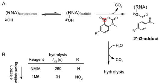
Nucleotides that participate in either base pairing or stable higher-order tertiary structure interactions can be detected by protection from solution-phase chemical probing reagents, whereas single-stranded and relatively unstructured elements are reactive (Wilkinson et al. 2006). Selective 2ʹ-hydroxyl acylation analyzed by primer extension (SHAPE) has emerged as an especially informative approach for probing RNA structure and dynamics (Gherghe et al. 2008; Mortimer and Weeks 2009). SHAPE chemistry exploits the discovery that the reactivity of the ribose 2ʹ-hydroxyl is highly sensitive to local nucleotide flexibility (Fig. 2.1A). Flexible nucleotides sample many conformations, a few of which preferentially react with hydroxyl-selective, electrophilic reagents to form 2ʹ
1 The text and figures from this chapter was adapted from modeling experiments undertaken in collaboration
with other students for two different projects. My critical contribution to the differential SHAPE project was to confirm the hypothesis that 1M6 more favorably interacts at available base stacks for RNA nucleotides compared to NMIA using density functional theory (DFT). In the HMX experiment I worked to establish the expected disruption a SHAPE adduct could cause to a folded (packed) RNA based on principles of molecular overlap. Figures and text from this chapter originally appeared in Steen, K.-A., Rice, G. M., & Weeks, K. M.
(2012). Fingerprinting noncanonical and tertiary RNA structures by differential SHAPE reactivity. Journal of
the American Chemical Society, 134(32), 13160–13163. doi:10.1021/ja304027m and Homan, P. J., Tandon, A.,
adducts (Fig. 2.1A). However, it is not obvious based on the chemical reactivity of a nucleotide whether a given constraining interaction reflects a base pairing or tertiary interaction. Nucleotides involved in tertiary interactions often have unusual backbone or stacking geometries (Holbrook 2008; Butcher and Pyle 2011), adopt the syn conformation (Sokoloski et al. 2011), or undergo conformational changes on slow timescales (Gherghe et al. 2008; Mortimer and Weeks 2009).
Figure 2.1: RNA SHAPE chemistry. (A) Mechanism in the context of the concurrent hydrolysis reaction. The red circle denotes the reactive center of the reagent. (B) SHAPE reagents and hydrolysis half-lives.
Clarke 1999; Ryder and Strobel 1999; Strobel 1999). These approaches generally require multiple distinct experiments to interrogate the tertiary environment of every nucleotide in an RNA.
Here we describe two strategies in which 2'-hydroxyl-selective reagents are used to interrogate higher order structures in RNA. The first strategy, which we term differential SHAPE, compares differences in the position specific SHAPE reactivity in order to determine features of higher-order RNA structure from chemical probing experiments. The second strategy, which we call 2'-hydroxyl molecular interference (HMX), a hydroxyl-selective reagent is used to create a pool of RNAs with evenly distributed 2'-O-ester adducts. Next, a structure-selective pressure, such as RNA folding, is placed on the pool of modified RNAs. A subset of 2'-O-ester groups will interfere with molecular interactions and prevent native structure formation. By partitioning the sample into folded and unfolded states, nucleotides whose modification disrupts tertiary interactions are identified. This information is used to characterize the internal packing interactions that define higher-order RNA structure.
Results
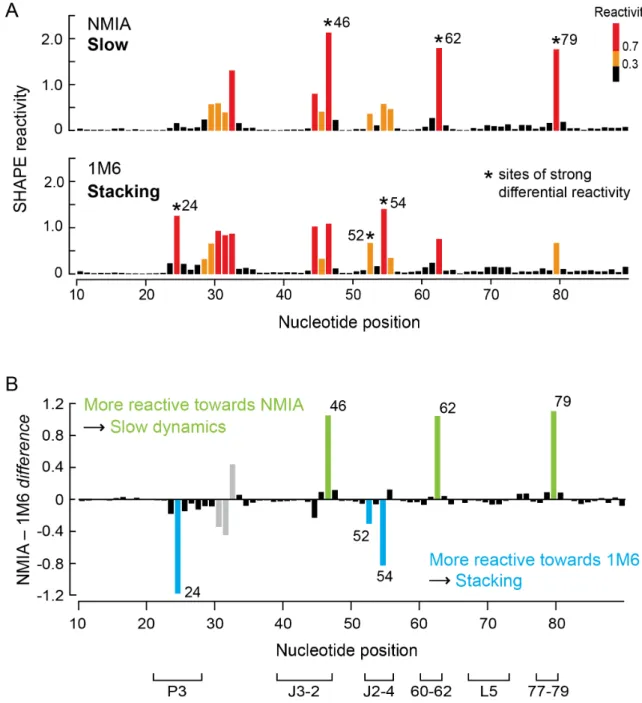
Fingerprinting RNA structure using multiple SHAPE reagents
We initially screened potential SHAPE reagents for the ability to “fingerprint” RNA tertiary structure motifs using the aptamer domain of the TPP riboswitch in the ligand-bound state. The TPP riboswitch has been extensively characterized by crystallography (Serganov et al. 2006; Haller et al. 2013) and SHAPE chemistry (Steen et al. 2011). This RNA contains many tertiary structure features, especially at or near the ligand binding pocket, that are common to highly structured RNAs including base stacking, long-range docking interactions, and tight turns in the RNA backbone.
local conformational changes on slow timescales (Fig. 2.1B) (Gherghe et al. 2008). These nucleotides are usually in the relatively rare C2ʹ-endo conformation and, in some cases, govern the folding of entire RNA domains (Mortimer and Weeks 2009). The second reagent, 1M6, differs from NMIA by a single nitro (–NO2) group on the double ring system (Fig. 2.1B). This modification changes the
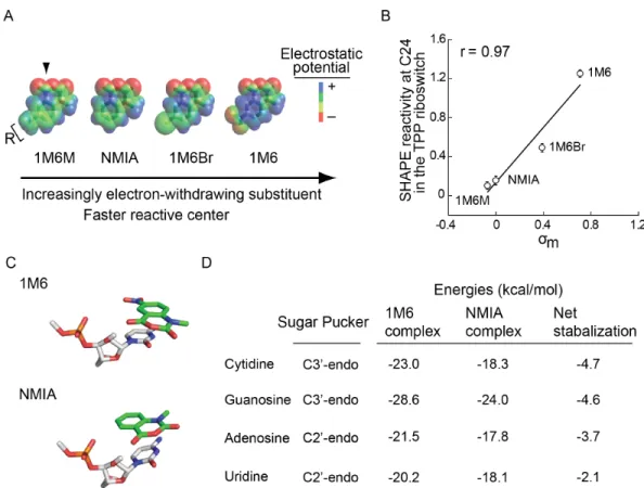
chemical behavior of 1M6 in two ways relative to that of NMIA. Addition of the electron-withdrawing group increases the electrophilicity of the reactive center (Fig. 2.1A, red circle), and consequently 1M6 reacts more rapidly than NMIA. Second, the –NO2 group significantly changes the
electronic distribution of the reagent ring system which, we will show below, allows 1M6 to stack with RNA nucleobases.
Stacking interactions between reagent and nucleobase influence SHAPE reactivity
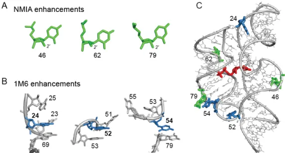
Each of the three nucleotides that reacted preferentially with NMIA (Fig. 2.3A) adopts the relatively rare C2ʹ-endo conformation, consistent with previous studies(Gherghe et al. 2008). However, the mechanism by which nucleotides might react preferentially with 1M6 has not been previously explored. The three nucleotides that reacted preferentially with 1M6 are located in diverse local structural environments but share the characteristic that one face of the nucleobase is available for π-π stacking interactions with a small molecule like 1M6 (Fig. 2.3B). This conformation is unusual because, both in A-form helices and in most highly folded RNAs, base-base stacking is nearly fully saturated (Leontis et al. 2006; Butcher and Pyle 2011). Only a few nucleotides in special structural contexts – especially at bulges, turns and the termini of some helices – form “one-sided” stacking interactions.
Figure 2.4: Effect of varying the substituents on SHAPE reactivity and electronic structure calculations for the 1M6- and NMIA-nucleotide complex stabilities. Increasing the electron-withdrawing ability of the functional group results in a more electrophilic reactive center and changes in the overall electrostatic profile of the reagent. (A) Electrostatic potential maps for each reagent. (B) Correlation between SHAPE reactivity at C24 (which presents an available open stacking face; see Figure 3B) and A45 (which has pre-existing stacking interactions at both nucleobase faces) in the TPP riboswitch and the electron withdrawing potential as measured by the Hammett coefficient (σm)
of the reagent R-group. (C) The most stable stacking conformations for the cytidine-1M6 and -NMIA complexes. (D) Representative open-faced stacking complex energies and net stabilization energy as a function of nucleotide, ribose conformation, and reagent.
We hypothesized that the –NO2 substituent polarizes the two-ring system which stabilizes the
0.97; Fig. 2.4B). In contrast, this trend was not observed for A45, which is also reactive towards SHAPE reagents but forms stacking interactions on both sides of the adenine base. These reactivity patterns are consistent with the formation of increasingly favorable reagent-nucleobase stacking interactions (Mignon et al. 2005), which are possible at C24 but not A45.
Since the effect of electron-withdrawing groups on the stacking interaction and resulting SHAPE reactivity is quantitative, we estimated representative electronic contributions associated with this interaction from first principles for the four RNA nucleotide types (Fig. 2.4C). Complexes formed between 1M6 and the four RNA nucleotides were -2 to -5 kcal/mol more stable than those formed with NMIA (Fig. 2.4D). These values are significant when compared to the approximate net stabilization energy of a two base pair stack of 2-3 kcal/mol (Petersheim and Turner 1983) and likely reflect the upper limit of favorable interaction. Favorable stacking appears to enhance 1M6 reactivity by increasing the effective local concentration of the reagent at nucleotides where one face is available for the one-sided stacking interaction.
HMX overview
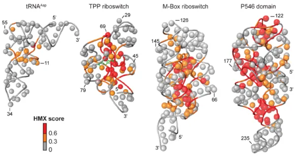
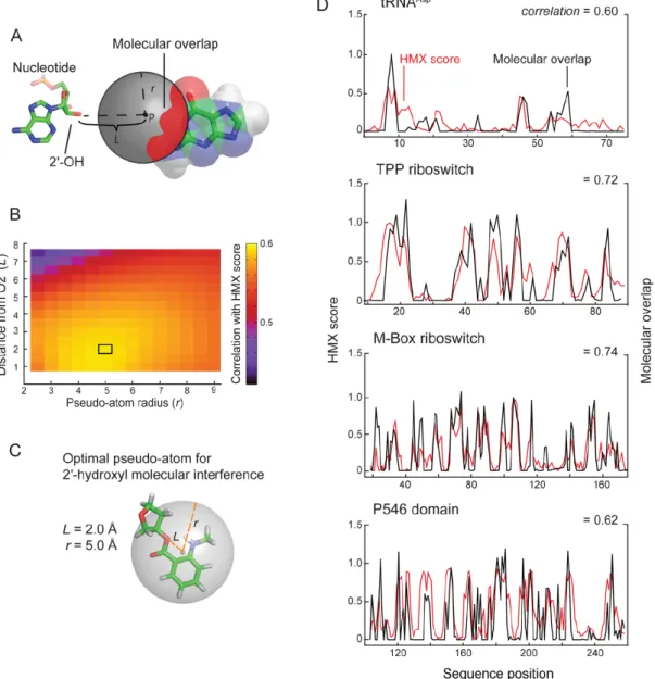
natively folded from the unfolded structures based on mobility in non-denaturing acrylamide gels, although many other selection strategies are compatible with this approach. After partitioning folded and unfolded populations, positions of modified nucleotides were detected as stops using reverse transcription- mediated primer extension. Adducts that disrupted folding were identified by comparing the profiles of the unfolded and folded RNA at each position. HMX scores for each nucleotide were calculated as the difference between the normalized profiles for the folded and unfolded RNA.
After partitioning, sites of modification were identified in the folded and unfolded populations by reverse transcription-mediated primer extension. 2'-O-ester adducts that prevent folding were over represented in the unfolded band and underrepresented in the folded band. The resulting modified RNA data were normalized using a cross-correlation approach to create an HMX score that allowed identification of nucleotides preferentially modified in the unfolded population relative to the folded population. The HMX score takes into account that the separation of unfolded and folded populations using 2'-O-adduct molecular interference is imperfect and that there is some noise in the separated signals. Positions with medium and high interference scores were visualized on the known three- dimensional structures of each RNA (Fig. 2.5). Nucleotides with high HMX scores corresponded to nucleotides directly involved in tertiary interactions and to nucleotides within densely packed regions of the RNA. Because the 2'-O-ribose modification occurs in the RNA backbone and likely does not significantly destabilize helix formation (Lesnik et al. 1993; Lesnik and Freier 1998), interfering positions corresponded almost exclusively to higher-order interactions and not to canonically base-paired nucleotides (Fig. 2.5).
Molecular overlap model for HMX intensities.
A pseudo-atom with these parameters tightly, and fully, encapsulates the NMIA adduct ester at a ribose ring (Fig. 2.6C).
The correlations between the experimental interference scores and the molecular overlap calculations for each RNA are high (Fig. 2.6D), indicating that the 2'-O-ester adduct disrupts RNA structure by sterically blocking RNA interactions in crowded regions of the RNA. For example, interfering nucleotides in the TPP riboswitch interact directly with ligand and other nucleotides form RNA-RNA contacts. The HMX experiment was sensitive to both types of interactions, indicating that HMX will be useful for examining intramolecular and intermolecular RNA contacts and protein and small molecule ligand interactions with RNA. Critically, as judged by visualizing interfering positions in three dimensions (Fig. 2.5) and from molecular overlap analysis (Fig. 2.6D), HMX analysis is exquisitely sensitive to higher-order molecular interactions in RNA, with essentially no detection of false positive interactions.
Discussion
Differential SHAPE outlook
2.3B), the correlation between strength of electron-withdrawing substituent and SHAPE reactivity (Fig. 2.4B), and high-level density functional theory calculations (Figure 2.4D).
HMX experiment and outlook.
HMX measures the effect of introducing a molecular perturbation at the ribose 2'-OH position on RNA folding. Modifications at the 2'-ribose position, which lie on the exterior of an RNA duplex, generally do not substantially destabilize simple RNA secondary structures (Lesnik et al. 1993; Lesnik and Freier 1998). Thus, the 2'-O-ester molecular interference measurement is exquisitely and specifically sensitive to interactions that govern RNA tertiary folding. For the five RNA evaluated in this work – tRNAAsp, the TPP and M-Box riboswitch aptamer domains, and the
P546 domain RNA – the interfering nucleotides identified by HMX correspond closely to the densely packed interior of these structures (Fig. 2.5). This relationship is quantitative. Molecular interference by the 2'-O-ester group was highly correlated with a sphere of defined location relative to the RNA ribose group (Fig. 2.6). We anticipate that 2'-O-ester mediated molecular interference will prove broadly useful in evaluating higher-order RNA packing in the context of large RNAs and RNA-protein complexes.
Methods
1M6 and NMIA free energy calculations
Models of SHAPE reagents and nucleic acids were created separately using the modeling program Avogadro (Hanwell et al. 2012). The simplified nucleic acid model system consisting of a nucleoside with a phosphate at the 3' position and an alcohol at the 5' position of the sugar was created for each of the four RNA bases. The charge of the base was neutralized at the phosphate to simplify the gas phase optimization. Nucleotide and SHAPE molecule complexes were based off of modeling of the 1M6 stacking interaction at C24 in the TPP riboswitch. The orientation of the SHAPE molecule was chosen to maximize the overlap of the ring systems and substituent interactions between the molecules, which has been shown prior to enforce the stacking interactions (Florian et al. 1999).
Structures were optimized using the default method implemented in the Gaussian 09 package (Frisch et al. 2009) in gas phase using M06-2X/6-311G*. The M06-2X functional has been shown to be robust enough to model stacking interactions of aromatic systems (Churchill and Wetmore 2011) and consistently recover the CCSD(T) CBS π-π interaction energy(Rutledge and Wetmore 2010). This 6-311G* basis set was chosen for its computational efficiency for a system of this size for optimizing the geometry of the system. Once the models were optimized, a high level single point energy calculation on the optimized structure using M06-2X/6-311+G(2d,p) was used to obtain more accurate structure energies.
Interaction energies for the different SHAPE complexes were calculated using the SCF energy of the structures to get the energy difference at infinite distance allowing for direct comparisons between different complexes:
Modeling of adduct disruption of native RNA tertiary structure (HMX).
REFERENCES
Butcher SE, Pyle AM. 2011. The molecular interactions that stabilize RNA tertiary structure: RNA motifs, patterns, and networks. Acc Chem Res 44: 1302–1311.
Chen VB, Arendall WB, Headd JJ, Keedy DA, Immormino RM, Kapral GJ, Murray LW, Richardson JS, Richardson DC. 2010. MolProbity: all-atom structure validation for macromolecular crystallography. Acta Crystallogr D Biol Crystallogr 66: 12–21.
Churchill CDM, Wetmore SD. 2011. Developing a computational model that accurately reproduces the structural features of a dinucleoside monophosphate unit within B-DNA. Phys Chem Chem Phys 13: 16373–16383.
Clarke PA. 1999. RNA footprinting and modification interference analysis. Methods Mol Biol 118: 73–91.
Conway L, Wickens M. 1989. Modification interference analysis of reactions using RNA substrates. Meth Enzymol 180: 369–379.
Florian J, Šponer J, Warshel A. 1999. Thermodynamic parameters for stacking and hydrogen bonding of nucleic acid bases in aqueous solution: ab initio/Langevin dipoles study. The Journal of Physical Chemistry B 103: 884–892.
Frisch MJ, Trucks GW, Schlegel HB, Scuseria GE, Robb MA, Cheeseman JR, Scalmani G, Barone V, Mennucci B, Petersson GA, et al. 2009. Gaussian 09.
Gherghe CM, Mortimer SA, Krahn JM, Thompson NL, Weeks KM. 2008. Slow conformational dynamics at C2'-endo nucleotides in RNA. J Am Chem Soc 130: 8884–8885.
Haller A, Altman RB, Soulière MF, Blanchard SC, Micura R. 2013. Folding and ligand recognition of the TPP riboswitch aptamer at single-molecule resolution. Proc Natl Acad Sci 110: 4188– 4193.
Hammett LP. 1937. The effect of structure upon the reactions of organic compounds. Benzene derivatives. J Am Chem Soc 59: 96–103.
Hanwell MD, Curtis DE, Lonie DC, Vandermeersch T, Zurek E, Hutchison GR. 2012. Avogadro: an advanced semantic chemical editor, visualization, and analysis platform. J Cheminform 4: 17. Holbrook SR. 2008. Structural principles from large RNAs. Annu Rev Biophys 37: 445–464.
Leontis NB, Lescoute A, Westhof E. 2006. The building blocks and motifs of RNA architecture. Curr Opin Struct Biol 16: 279–287.
Lesnik EA, Freier SM. 1998. What affects the effect of 2“-alkoxy modifications? 1. Stabilization effect of 2-”methoxy substitutions in uniformly modified DNA oligonucleotides. Biochemistry 37: 6991–6997.
Merino EJ, Wilkinson KA, Coughlan JL, Weeks KM. 2005. RNA structure analysis at single nucleotide resolution by selective 2'-hydroxyl acylation and primer extension (SHAPE). J Am Chem Soc 127: 4223–4231.
Mignon P, Loverix S, Steyaert J, Geerlings P. 2005. Influence of the pi-pi interaction on the hydrogen bonding capacity of stacked DNA/RNA bases. Nucleic Acids Res 33: 1779–1789.
Mortimer SA, Weeks KM. 2009. C2'-endo nucleotides as molecular timers suggested by the folding of an RNA domain. Proc Natl Acad Sci 106: 15622–15627.
Petersheim M, Turner DH. 1983. Base-stacking and base-pairing contributions to helix stability: thermodynamics of double-helix formation with CCGG, CCGGp, CCGGAp, ACCGGp, CCGGUp, and ACCGGUp. Biochemistry 22: 256–263.
Rutledge LRRR, Wetmore SDWD. 2010. The assessment of density functionals for DNA–protein stacked and T-shaped complexes. http://dxdoiorg/101139/V10-046 88: 815–830.
Ryder SP, Strobel SA. 1999. Nucleotide analog interference mapping. Methods 18: 38–50.
Serganov A, Polonskaia A, Phan AT, Breaker RR, Patel DJ. 2006. Structural basis for gene regulation by a thiamine pyrophosphate-sensing riboswitch. Nature 441: 1167–1171.
Sharp PA. 2009. The centrality of RNA. Cell 136: 577–580.
Sokoloski JE, Godfrey SA, Dombrowski SE, Bevilacqua PC. 2011. Prevalence of syn nucleobases in the active sites of functional RNAs. RNA 17: 1775–1787.
Steen K-A, Siegfried NA, Weeks KM. 2011. Selective 2'-hydroxyl acylation analyzed by protection from exoribonuclease (RNase-detected SHAPE) for direct analysis of covalent adducts and of nucleotide flexibility in RNA. Nature Protocols 6: 1683–1694.
Strobel SA. 1999. A chemogenetic approach to RNA function/structure analysis. Curr Opin Struct Biol 9: 346–352.
Weeks KM. 2010. Advances in RNA structure analysis by chemical probing. Curr Opin Struct Biol 20: 295–304.
CHAPTER 3: RNA SECONDARY STRUCTURE MODELING AT CONSISTENT HIGH ACCURACY USING DIFFERENTIAL SHAPE2
Introduction
RNA is a central information carrier in biology (Sharp 2009). Information is encoded in RNA at two distinct levels: in its primary sequence and in its ability to fold into higher order structures (Leontis et al. 2006; Dethoff et al. 2012). The most fundamental level of higher order structure is the pattern of base pairing or secondary structure. Defining the secondary structure of an RNA is also a critical first step in tertiary structure modeling (Hajdin et al. 2010; Weeks 2010; Bailor et al. 2011). The structures of RNA molecules modulate the numerous functions of RNA and the interactions of RNAs with proteins, small molecules, and other RNAs in splicing, translation, and other regulatory machineries (Mauger et al. 2013).
Accurate, de novo modeling of RNA secondary structure is challenging: In the absence of experimental restraints, current algorithms predict base pairing patterns that contain, on average, 50-70% of the canonical (G-C, A-U, and G-U) pairs in secondary structures established through phylogenetic analysis or high-resolution experimental methods (Mathews et al. 2004; Hajdin et al. 2013). The modeling challenge results from the fact that there are only four RNA nucleotides and that these nucleotides have the potential to arrange into many, often energetically similar, RNA secondary structures, even though many RNAs adopt a few or only single structures (Tinoco and Bustamante 1999). Features that are difficult to extract solely from the sequence – such as kinetic pathways, protein facilitators, and ligand binding – also influence RNA folding. Identification of the correct RNA secondary structure also becomes much more difficult as the length of the RNA increases.
Selective 2'-hydroxyl acylation analyzed by primer extension (SHAPE) reagents can be used to interrogate the flexibility of nearly every nucleotide in an RNA (Merino et al. 2005; McGinnis et al. 2012). Reactivity at the 2'-hydroxyl toward the reagent 1-methyl-7-nitroisatoic anhydride (1M7) measures local nucleotide flexibility. Because base-paired nucleotides are also structurally constrained, SHAPE reactivity is roughly inversely proportional to the probability that a nucleotide is paired. Incorporation of SHAPE reactivity information into RNA folding algorithms results in accuracies above 90% for most RNAs including those with pseudoknots (Deigan et al. 2009; Hajdin et al. 2013). SHAPE has been used to create nucleotide-resolution models for the viral genomes of HIV-1 (Watts et al. 2009) and STMV (Archer et al. 2013) and to analyze conformational changes in HIV-1 (Wilkinson et al. 2008) and the Moloney murine leukemia virus (Grohman et al. 2013). Although SHAPE-directed folding yields near-perfect models for many RNAs, there remain a few RNAs whose structures are difficult to recover using a single structure probing experiment (Cordero et al. 2012; Leonard et al. 2013). These “hard” RNAs are modeled with sensitivities in the 75-85% range.
On average, SHAPE-directed modeling currently recovers approximately 93% of accepted base pairs in challenging sets of RNA molecules. This level of sensitivity is sufficient for generation of robust biological hypotheses and for three-dimensional structure modeling. Many of the models generated at this level of accuracy differ from the accepted models by a few base pairs and should be considered nearly perfect (Fig. 3.1, upper structures). Improving accuracies to above the 90% level for all RNAs is the current challenge in experimentally-directed secondary structure modeling. Inclusion of additional comprehensive and information-rich biochemical information could further inform, and potentially solve, the RNA secondary structure modeling problem.
We recently described an approach that we call differential SHAPE that reveals local non-canonical and tertiary structure interactions based on simple biochemical probing experiments (Steen et al. 2012). In this strategy, the position-specific reactivities of two reagents, N-methylisatoic anhydride (NMIA) and 1-methyl-6-nitroisatoic anhydride (1M6), are compared. The first reagent, NMIA, has a relatively long half-life in solution and reacts preferentially with nucleotides that experience slow dynamics. Often these nucleotides are in the rare C2'-endo ribose conformation and have been implicated as molecular timers capable of governing folding in large RNAs (Gherghe et al. 2008; Mortimer and Weeks 2009). For the second reagent, the nitro group of 1M6 makes the two-ring system electron-poor, and this reagent is able to stack with RNA nucleobases that are not protected by interactions with other nucleotides in an RNA structure (Steen et al. 2012). This conformation is unusual since most nucleobases stack with other bases on both faces (Leontis et al. 2006). By taking the difference in reactivity profiles for these two 2'-hydroxyl selective reagents, nucleotides involved in structurally distinctive interactions within an RNA structure can be identified (Fig. 3.2). Because the differential SHAPE analysis is specifically sensitive to non-canonical and tertiary interactions in RNA (Steen et al. 2012), this approach can help to identify nucleotides that are constrained (and thus unreactive to 1M7-SHAPE) but do not participate in canonical base pairing. Here we develop a pseudo-free energy term that includes information from the slow and stacking differential SHAPE reactivities to yield nearly perfect secondary structure models in a concise experiment that scales to RNAs of any size.
Results
Selection of a challenging test set.
riboswitch aptamer domains that require ligand binding to fold into their correct structures (the TPP, adenine, glycine, cyclic-di-GMP, M-Box, and lysine riboswitches); four RNAs longer than 300 nucleotides, including several domains of the E. coli 16S and 23S ribosomal RNAs; four pseudoknot-containing RNAs; and every other RNA of which we are aware that contains up to one pseudoknot for which the single-reagent 1M7 prediction accuracy is less than 90% (Table 3.1) (Cordero et al. 2012; Leonard et al. 2013; Hajdin et al. 2013).
Incorporation of differential SHAPE into secondary structure modeling.
SHAPE experiments were performed with 1M7, NMIA, and 1M6 on RNAs pre-incubated in the presence of cognate ligand if appropriate but without protein. Based on pilot work on three short RNAs, SHAPE reactivity signals from NMIA and 1M6 correlate strongly at most positions (Steen et al. 2012). We therefore used a windowed scaling algorithm to locally normalize NMIA and 1M6 SHAPE profiles to each other (see Methods) and then subtracted the normalized profiles to generate differential SHAPE reactivity traces (Fig. 3.2).
Figure 3.3: Statistical determination of the ∆GDiff free energy change penalty. (A) Differential reactivities were binned as a function of base pairing status in the accepted structure. Paired and non-paired nucleotides were each fit to a gamma distribution. (B) Final ∆Gdiff energy function calculated
∆GDiff = d × (positive amplitude differential signal) (1)
where d is 2.11 kcal/mol. This energy penalty was added to the standard 1M7-based pseudo-free energy as implemented in ShapeKnots (Low and Weeks 2010; Hajdin et al. 2013); inclusion of this penalty improved predictions for many RNAs. For each RNA model, we report the accuracy of a secondary structure prediction in terms of it sensitivity (sens, fraction of base pairs in the accepted structure predicted correctly) and positive predictive value (ppv, the fraction of predicted pairs that occur in the accepted structure).
Impact of
∆
G
Diffon structure modeling.
In the absence of experimental restraints, the mfold algorithm predicts only 10 of the 35 base pairs (29%) in the accepted structure of the E. coli 5S rRNA (Fig. 3.4, left structure). Addition of 1M7-SHAPE constraints yielded a substantial improvement: 86% of the accepted base pairs were present in the SHAPE-directed model. As is common for predictions at this level of accuracy, most of the structure is modeled correctly. The exceptions are base pairs in one element, a helix at a three-way junction (Fig. 3.4, middle structure, positions 102-107). When differential SHAPE data were added as constraints, a substantially improved structural model was obtained (Fig. 3.4, right structure). The errors in the differential SHAPE-based model are minor and involve the addition of a few base pairs in the second helix of the structure near nucleotide 30. These base pairs may in fact form under our probing conditions, given that this RNA was probed in the absence of ribosomal subunits and proteins.
and 112) and eliminated the false positive pseudoknot. In addition, lower magnitude differential reactivities shifted the folding landscape of nucleotides 39-49 to result in agreement of the predicted and accepted structures.
Figure 3.4: Representative secondary structure modeling for the 5S rRNA without and with SHAPE data. Base pair predictions are illustrated with colored lines (green, purple, and red denoting correct, incorrect, and missing base pairs, respectively) on conventional secondary structure representations (top) and circle plots (bottom). Nucleotides are colored according to their SHAPE reactivity on a black, yellow, red scale for low, medium, and strong reactivity. Nucleotides showing strong preferential reactivity with NMIA (>0.3 units) are indicated with a delta symbol.
5' and 3' ends of the RNA (Fig. 3.5, bottom left). Nucleotides in this helix are moderately reactive toward SHAPE reagents, suggesting that the P1 helix is not especially stable under the conditions used for structure probing. In the crystal structure that is the basis for the accepted model, the P1 helix is stabilized by three G-C base pairs (Dann et al. 2007) that were not present in the transcript analyzed by SHAPE. SHAPE data suggest that the native sequence P1 helix is conformationally dynamic. For the sequence of RNA probed in this work, the SHAPE-constrained structure is essentially correct.
Figure 3.6: Circle plots illustrating SHAPE-directed structure modeling for Tetrahymena group I intron. 1M7 SHAPE data (left) and with 1M7 and differential reactivity data (right). Reactive nucleotides in the P7 helix are shown in an expanded view (right); × symbols indicate structurally significant mis-predictions relative to the accepted structure. Scheme for illustrating base-pair accuracy (relative to crystallographic structures) and nucleotide SHAPE reactivities are as outlined in Figure 3.3; positions with positive-amplitude differential reactivities (favoring NMIA) are indicated with a delta symbol.
Responsive and non-responsive RNAs.
restraints, none of the predictions became substantially worse with the exception of the Tetrahymena group I intron (Table 3.1).
The modeled structure for the Tetrahymena group I intron became less like the accepted structure upon inclusion of differential reactivity information: The sensitivity decreased from 93% to 85% (Table 3.1 and Fig. 3.6). The P7 helix comprises a pseudoknot in the accepted RNA structure. One strand of the P7 helix is reactive by SHAPE and is not present in the SHAPE-directed model (Fig. 3.6). Our data suggest that the P7 helix is conformationally dynamic under the solution probing conditions used in this work.
Discussion
that each set of restraints – nearest neighbor parameters, 1M7-SHAPE, and differential SHAPE – provides information that is orthogonal to the others, roughly corresponding to local secondary structure, non-nearest neighbor interactions, and non-canonical and tertiary interactions, respectively.
Table 3.2: RNA secondary structure modeling accuracies comparing three-reagent differential SHAPE to related recent works. Approaches that allow pseudoknots are indicated with an asterisk. Methods that used parameters optimized using small datasets are indicated with a dagger.
2011a). The differential SHAPE data thus have high information content that is obtained in a concise experiment that scales easily to large RNAs.
Using differential SHAPE for RNA secondary structure prediction represents a significant advance in RNA structure modeling. With differential SHAPE information, the structures of some of the RNA molecules that were previously viewed as the most challenging, including the 5S rRNA, the glycine riboswitch, and some ribosomal domains, were modeled in nearly perfect agreement with the accepted structures (Table 3.1). An intriguing trend was that the RNAs that were most responsive to the differential reactivity penalty were those with structures predicted most poorly in the absence of differential SHAPE experimental. We speculate that RNAs in this class have non-canonical interactions that are incompletely described by the nearest neighbor algorithm or single-reagent data. SHAPE-driven predicted structures that disagree with the accepted structures – those of the M-Box and lysine riboswitches and the Tetrahymena group I intron – “errors” appear to reflect differences between in-crystal and in-solution conformations for these RNAs.
Limitations and Perspective
currently restrict base pairing partner to within 600 nucleotides. In general, this is a good assumption and, for example, allows full-length ribosomal RNAs to be modeled at high accuracy (Deigan et al. 2009). However, there are important RNA-RNA interactions that occur over distances of a thousand nucleotides or more (Alvarez et al. 2005; Jin et al. 2011) that will not be detected with the current approach. Finally, SHAPE reactivities always reflect the structural ensemble present in solution at the time of probing. If an RNA is partially misfolded or samples multiple conformations, the resulting SHAPE profile will reflect these contributions.
The highly accurate RNA secondary structure modeling reported here involves straightforward experiments with three reagents 1M7 (Mortimer and Weeks 2007), 1M6, and NMIA (Steen et al. 2012). In this work we examined complex RNA structures, including more than 3800 nucleotides, and specifically focused on those RNAs thought to comprise the most difficult known modeling challenges. The limitations outlined above notwithstanding, we believe that three-reagent SHAPE is approaching the upper limit that solution-phase RNA structure probing can accomplish. Three-reagent SHAPE structure probing is experimentally concise, yields consistently accurate RNA structural models, and can be applied to RNAs of any complexity and size, including complete viral genomes and the constituents of entire transcriptomes.
Methods