Structural and Functional Models for
[NiFe] Hydrogenase
Coverpage illustration:
Crystal structure of [Ni6(cpss)12] (Chapter 6). Ni, green; S, red; Cl, yellow; C, gray.
Printed by:
Structural and Functional Models for
[NiFe] Hydrogenase
PROEFSCHRIFT
ter verkrijging van de graad van Doctor aan de Universiteit Leiden,
op gezag van Rector Magnificus prof. mr. P. F. van der Heijden,
volgens besluit van het College voor Promoties
te verdedigen op woensdag 14 oktober 2009
klokke 16.15 uur
door
Raja Angamuthu
Promotor Prof. Dr. J. Reedijk
Copromotor Dr. E. Bouwman
Overige leden Prof. Dr. M. Schröder (The University of Nottingham, UK)
Prof. Dr. M. Fontecave (Université Joseph Fourier, Grenoble, France)
Dr. M. C. Feiters (Radboud Universiteit Nijmegen)
Prof. Dr. M. T. M. Koper
Prof. Dr. J. Brouwer
Table of Contents
List of Abbreviations i
1
General Introduction 0012
Ligand Design, Synthetic Procedures and Experimental Methods 0333
[Ni(S4)Fe(C5H5)(CO)](PF6) Complexes Containing Tetradentate S2S’2-donor Ligands: Synthesis, Characterization and Electrocatalytic DihydrogenProduction 051
4
Synthesis, Characterization and Electrocatalytic Properties of[Ni(S4)Fe(C5H5)(CO)](PF6) Complexes Containing Bidentate SS’-donor Ligands 071
5
Heterodinuclear [NiRu] Complexes Comprising Ruthenium Bis-Bipyridine: Synthesis, Characterisation and Electrocatalytic Dihydrogen Production 0876
Hexanuclear (Ni6-)Metallacrown as Functional Model of [NiFe] Hydrogenase 1037
A Molecular Cage of Ni(II) and Cu(I) Resembling the Active Site ofNi-Containing Enzymes 121
8
Light-Induced C–S Bond Cleavage in a Nickel Thiolate Complex: Relevance tothe Function of Methyl Coenzyme M Reductase (MCR) 131
9
Summary, Conclusions and Future Perspectives 143 Samenvatting (Summary in Dutch) 151
Curriculum Vitae (English) 156
Curriculum Vitae (Tamil) 157
Publications 158
ACN Acetonitrile
APT Attached Proton Test
b Broad
bpy 2,2’-Bipyridine
CoM Coenzyme M; 2-thioethane sulfonate COSY Correlation Spectroscopy
CV Cyclic voltammetry; cyclic volammogram
Cys Cysteine
d Doublet
dedtc Diethyldithiocarbamate
DMF N,N-Dimethylformamide
dpa Dipicolylamine
dppe 1,2-Bis(diphenylphosphino)ethane Epa Anodic potential; oxidation potential Epc Cathodic potential; reduction potential EPPG Edge plane pyrolytic graphite (electrode)
eq. Equivalent
ESI-MS Electrospray ionization mass spectrometry
Et Ethyl
FTIR Fourier transform infra red
GC Glassy carbon
Glu Glutamic acid
GSH Glutathione
H2ase Hydrogenase
H2bdt Benzene-1,2-dithiol
H2bme*-daco 1,5-bis(mercaptoethyl)-1,5-diazacyclooctane H2pdt Propane-1,3-dithiol
H2tpdt 2-Thiapropane-1,3-dithiol Hacac Acetylacetone, 2,4-pentanedione
HCp 1,3-Cyclopentadiene
HER Dihydrogen evolution reaction HG-GSH Hemithioacetal
His Histidine
J Coupling constant
LMCT Ligand-to-metal charge transfer
m Multiplet in NMR; medium in IR
m/z Ratio of mass upon charge
Md Distal metal
Me Methyl
MeCoM Methyl–coenzyme M; 2-(methylthio)ethanesulfonate MLCT Metal-to-ligand charge transfer
Mp Proximal metal
NMR Nuclear magnetic resonance
NOESY Nuclear Overhauser Effect Spectroscopy OTf– Trifluoridomethanesulfonate
PEM Proton exchange membrane
Ph Phenyl
PMe3 Trimethylphosphine
PPh3 Triphenylphosphine
ppm Parts per million
ROESY Rotating-Frame NOE Spectroscopy s Singlet in NMR; strong in IR SCE Standard calomel electrode SHE Standard hydrogen electrode
t Triplet
tBu tertiary-Butyl
TEA·HCl Triethylamine hydrochloride
THF Tetrahydrofuran
TMS Tetramethylsilane
tmtu 1,1,3,3-tetramethyl-2-thiourea TOCSY Total Correlation Spectroscopy
tpa Tripicolylamine
TsOH⋅H2O para-Toluenesulfonic Acid monohydrate UV-Vis Ultra violet and visible spectrocopy
Val Valine
w Weak
δ Chemical shift
1
1.
General Introduction
Abstract. The main goal of the research presented in this thesis is the synthesis of suitable structural and functional models for the enzyme [NiFe] hydrogenase, which can reduce protons† into dihydrogen. This chapter starts with a brief survey of the roles of all the known nickel-containing enzymes in biological systems with a focus on the [NiFe] hydrogenases. Structure, function, physicochemical and catalytic properties of the [NiFe] hydrogenase itself and of the reported model complexes are presented. This chapter concludes with the goal of the research and modeling strategies, followed by an outline of the thesis.
1.1.
A Prelude to Nickel Biochemistry
Nickel is a relatively abundant element, constituting approximately 8% of the earth’s core and 0.01% of the earth’s crust. Organisms in nature have obtained nickel by leaching the most abundant form of nickel, Ni(II), from the earth’s crust. It is perhaps puzzling then as to why no protein or enzymatic system containing functionally significant nickel was known until 1975, despite the fact that nickel is readily available.1‐4
Currently there are only nine proteins or enzymatic systems known in nature that encompass functionally significant nickel; the environment around nickel within each protein is different. Several aspects in the bioinorganic chemistry of nickel‐containing enzymes are unusual in the context of known coordination chemistry of common nickel salts, as well as the functions in which these enzymes are involved (See Scheme 1.1).5,6
1.2.
Nickel-Containing Enzymes and their Role in Biological Systems
1.2.1.
Introduction
Nickel‐containing enzymes catalyze many critical and distinct biological processes. Most of them are industrially and environmentally significant, such as (1) hydrolysis of urea into ammonia and carbamate,7‐10 (2) reversible interconversion of carbon monoxide
and carbon dioxide, (3) decomposition of the acetyl group into separate one‐carbon units in some cells or catalyzing acetate synthesis using one‐carbon precursors in others,11‐19 (4) detoxification of cytotoxic methylglyoxal (MG) via the isomerization of hemithioacetal,4,20,21 (5) oxidation of 1,2‐dihydroxy‐3‐keto‐5‐methylthiopentane (aci‐
reductone) into methylthio propionic acid, formic acid and carbon monoxide,22‐26 (6)
degradation of methylenediurea (slow release fertilizer),27 (7) methane generation,28 (8)
dismutation of toxic and cell damaging superoxide radical anions into harmless molecular oxygen29,30 and on top of all, (9) reversible interconversion of dihydrogen into protons
and electrons (Scheme 1.1).31‐35 The first eight enzymes are briefly discussed here. The
hydrogenase enzymes are described in more detail in Section 1.3.
1.2.2.
Urease
Urease (urea amidohydrolase) catalyzes the hydrolysis of urea to form ammonia and carbamate at approximately 1014 times the rate of the uncatalyzed reaction.36 The
carbamate formed spontaneously degrades in vivo to form a second molecule of ammonia and hydrogen carbonate.37 This urease‐catalyzed hydrolysis is in contrast with the
uncatalyzed reaction, which affords ammonia and cyanic acid.38 James B. Sumner
successfully crystallized the enzyme urease from Jack bean in 1926 after almost nine years of hard work, as the first enzyme to be isolated in crystalline form.7
The presence of a nickel center in the active site was discovered only fifty years after the isolation of the crystalline urease.8 It took almost seventy years before the first
crystal structure of a urease was reported.6,9,10 The crystal structure of urease shows the
active centre to contain a homodinuclear Ni2 center. Each nickel ion is coordinated to a
water molecule and two histidine nitrogen donors apart from the two bridges between the nickel centers formed by a hydroxido group and a carbamylated lysine (Fig. 1.1). Numerous dinuclear nickel(II) complexes have been reported in recent literature to mimic the structure and function of urease.39‐54
1.2.3.
CO Dehydrogenase/Acetyl-Coenzyme A Synthase (CODH/ACS)
The bifunctional enzyme CODH/ACS has an important role in the global carbon cycle as the C‐cluster, an Ni–Fe–S centre, of CODH reduces carbon dioxide to carbon monoxide and the A‐cluster, another Ni–Fe–S centre, of ACS assembles acetyl‐CoA from a methyl group, coenzyme‐A and the CO generated by the C‐cluster (Scheme 1.1).11,14,17,55‐59
The A‐cluster is a complex metallocofactor, containing an Fe4S4 group connected by
cysteine bridging to Mp of a dinuclear [MpNid] site. The proximal metal Mp is
predominantly Cu in the as‐isolated enzyme from native Moorella thermoacetica, but [NiNi] and [ZnNi] forms are also known to be isolated and well studied (Fig. 1.2).55‐57,59,60
Fig. 1.2. Perspective view of the A-cluster of ACS (left, 1MJG) and the C-cluster of CODH (right, 1JJY).
The distal nickel ion Nid is in a square‐planar (NiN2S2) geometry derived from two
backbone carboxamido nitrogens and two Cys‐S residues. The Nid centre is bridged
through the two Cys‐S donors to the proximal metal Mp that is in a tetrahedral
coordination environment. A fourth nonprotein ligand (CO/acetyl) is bound to Mp to
iron ion, which is extraneous to the cuboidal‐like core, through a bridging sulfide (Fig. 1.2). Numerous model complexes mimicking the structure and functions of CODH/ACS, involving methyl transfer61,62 and CO insertion63‐68 reactions, have been reported in
recent years and have been recently reviewed.69‐73
1.2.4.
Glyoxalase I (GlxI)
Glyoxalase I, a member of the metalloglutathione transferase superfamily, catalyzes the first step in the detoxification of cytotoxic methyglyoxal (MG) via the conversion of nonenzymatically‐produced hemithioacetals (HG‐GSH) into S‐D‐ lactoylglutathione and thereby plays a critical detoxification role in cells (Scheme 1.2). Yet, the mechanism of nickel incorporation into GlxI remains hard to pin down.4,20,21 The
three‐dimensional structure of the enzyme is homodimeric with what appears to be two identical active sites (Fig. 1.3).20 Each active site contains two histidine and two glutamic
acid residues that coordinate to the metal ion along with two water molecules so that the catalytic metal ion has a distorted octahedral geometry.
Scheme 1.2. Formation and isomerization of hemithioacetal.
Fig. 1.3. Active site structure of glyoxalase (1F9Z).20
1.2.5.
Aci-reductone Dioxygenase (ARD)
Aci‐reductone dioxygenase was first discovered in 1993 in a study of the methionine salvage pathway in Klebsiella pneumoniae.74 ARD was found to cleave the key
en‐3‐one) and its analogues.75,76 Investigations using K. pneumoniae unveiled that aci‐
reductone is oxidized to two different sets of products. In the productive case, a dioxygenase activity produces formic acid and the α‐ketoacid precursor of methionine.25,75 In addition, a second, non‐productive dioxygenase activity converts the
aci‐reductone into formate, carbon monoxide, and methylthiopropionic acid. Remarkably, these activities belong to the same protein (ARD), but result from the differences in metal content (Scheme 1.3). The reason for the presence of two isoforms of a protein with different metals is a mystery. Further investigations using recombinant protein confirmed that the productive activity is due to the iron‐containing ARD and the non‐productive activity is from the Ni– or Co–containing ARD.75
Scheme 1.3. Metal-dependent reactions carried out by ARD.
The global structure of ARD was elucidated employing high‐resolution NMR spectroscopy,24,26 while the active site structure was studied with by X‐ray absorption
spectroscopy.23 The active site appears to have an octahedral geometry with three
nitrogen donors provided by His96, His98 and His140 together with three oxygen donors provided by Glu102 and two water molecules. Among these six ligands, His96 and Glu102 are trans located at the paramagnetic nickel(II) ion.23 A limited number of structural77
and functional78 models have been reported recently in an effort to understand the
catalytic mechanism of ARD.
1.2.6.
Methylenediurease (MDUase)
Methylenediurease (MDUase), isolated from Burkholderia, was found to be a nickel‐dependent enzyme, which is able to degrade methylenediurea into urea and formaldehyde with ammonia and carbon dioxide as byproducts (Scheme 1.4).27
Methyleneureas or ureaforms are condensation products of urea and aldehyde [(H2N‐(CO–NH–CH2–NH)n–CO–NH2); n=1 for methylenediurea] which are potentially
applied as slow‐release fertilizers in bioremediation processes (more than 300,000 tons per year).79‐81 Significantly, the methylenediurease activity was resolved by anion
exchange chromatography from urease activity of the same microorganism, and each enzyme was found to be specific toward its own substrate, such as Ralstonia paucula (methyleneureas),80 Burkholderia (methylenediurea and dimethylenetriurea),79
Further studies are necessary to characterize the structure and functional mechanism of this enzyme.
Scheme 1.4. Degradation pathway of methylenediurea by MDUase.
1.2.7.
Methyl-Coenzyme M Reductase (MCR)
Methyl‐coenzyme M reductase (MCR) is the key enzyme in biological methane formation by methanogenic archaea.28,83,84 In the MCR active site, the nickel ion is present
in the tightly, but non‐covalently, bound tetrahydrocorphinoid complex called coenzyme F‐430 (Fig. 1.4). The upper face of the F‐430 cofactor forms the floor of a narrow hydrophobic well leading to the surface of the protein. The nickel ion is coordinated by the four pyrrolic nitrogens in the equatorial plane and by an oxygen of the glutamine side‐chain in the lower axial position. The upper axial position contains either the thiolate or the sulfonate group of CoM, depending on the form of MCR isolated.
Fig. 1.4. Schematic view of coenzyme F-430 of MCR showing the extensively reduced tetrapyrrole ring in which the π chromophore only extends over three of the four nitrogens.
Scheme 1.5. Catalytic cycle involving the coenzyme F-430-assisted methane formation in methanogenic archaea (Adapted from the literature).85
Scheme 1.6. Mechanism of F-430-catalyzed methane formation (Adapted from the literature).85-87
EPR spectrum and UV‐visible absorption maxima at 380 and 750 nm. Conventional purification of MCR leads to an inactive enzyme that contains the metal in the Ni(II) valence state. The first isolation of highly active enzyme preparations from reductively preconditioned cells and the reductive reactivation of the so‐called MCRox1 state to active
enzyme (MCRred1) demonstrated that the enzyme is active only if the metal center of
coenzyme F‐430 is in the Ni(I) form.88
The mechanism shown in Scheme 1.6 postulates the formation of Ni(I) and a thiyl radical. The formed thiyl radical attacks the Me‐CoM to form the sulfuranyl radical. The unpaired electron from the Ni(I) dx2‐y2 orbital transfers to the C‐S σ* orbital and induces
the homolytic cleavage of the C‐S bond to form the Ni(II) methyl‐substituted coenzyme F‐430 and the unsymmetrical disulfide. This methyl‐substituted coenzyme F‐430 is further attacked by HSCoB to release methane.
1.2.8.
Nickel Superoxide Dismutase (Ni-SOD)
Nickel superoxide dismutase (Ni‐SOD) is a recently discovered member of the nickel‐containing metalloenzymes and of the SOD class of enzymes that catalyze the disproportionation of highly toxic superoxide (O2•−) into peroxide (O22−) and molecular
oxygen.29,30,89 Ni‐SOD is the fourth member of this class of enzymes; the other known
SODs containing Fe, Mn and Cu/Zn. Reduced Ni‐SOD contains nickel(II) in a square‐ planar N2S2 coordination environment derived from the backbone terminal amino group
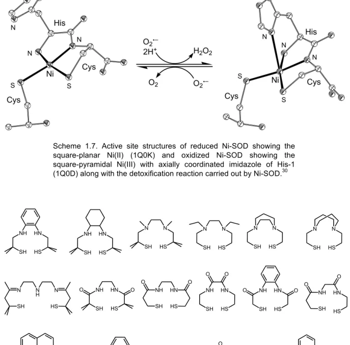
of His1, the amide group, and the thiolate groups of Cys2 and Cys6 (Scheme 1.7).30,90,91
The Nδ and Nε nitrogens of His1 are not involved in coordination; they are involved in hydrogen‐bonding to the main‐chain oxygen atom of Val8 (Nδ) and to Glu17 (Nε) of a neighboring subunit.30 Oxidized Ni‐SOD contains a Ni(III) ion in a distorted
square‐pyramidal N3S2 coordination environment derived from same units as reduced
Ni‐SOD and in addition the Nδ nitrogen of His1 (Scheme 1.7).
The presence of thiolate donors makes the Ni‐SOD different from other SODs and the stabilization of these two thiolate ligands against sulfur‐based oxidation in the presence of the highly active radical substrate remains elusive. The monomeric unit of this enzyme is a 4‐helix bundle accommodating the active site at the N‐terminus and six of these bundles make the whole molecule of a Ni‐SOD as a homohexamer. The proposed mechanism of dismutation shows that the electron transfer from the nickel(II) ion to the axially bound superoxide must be coupled with a proton transfer to generate the dihydrogen peroxide. Site‐specific mutagenesis studies confirm the significance of the histidine ligand, as altering this site tremendously decreases the dismutase activity.
A number of nickel complexes with N2S2 and N3S2 (bis‐amide or bis‐amine)
crystal structure of a nickel‐containing superoxide dismutase (Fig. 1.5).92‐96 NiN2S2
complexes can be reactive toward both H2O2 and O2, often yielding S‐based oxygenation
products.97 Synthetic studies have demonstrated that NiN2S2 complexes in bis‐amine
ligand environments are more stable toward oxygen than the corresponding bis‐amide complexes.70,92
Scheme 1.7. Active site structures of reduced Ni-SOD showing the square-planar Ni(II) (1Q0K) and oxidized Ni-SOD showing the square-pyramidal Ni(III) with axially coordinated imidazole of His-1 (1Q0D) along with the detoxification reaction carried out by Ni-SOD.30
NH SH HN HS O O N HS N H N SH NH SH HN HS O O NH SH HN HS O O NH SH HN HS O O NH SH HN HS O O NH SH HN HS NH SH HN HS N SH N HS N SH N HS N SH N HS N SH N HS HN HS NH SH O O N H NH SH HN HS NH2 O O O O NH O N HN O SH HS HN HS NH SH O O
Recently the first NiN2S2 complex [NiII(beamm)]– [H2beamm = N‐{2‐[benzyl(2‐
mercapto‐2‐methylpropyl)amino]ethyl}‐2‐mercapto‐2‐methylpropionamide] (Fig. 1.6) containing amine/amide coordination has been reported with the studies on the difference between amine/amide and bisamide coordination on the models of Ni‐SOD.98
Bis‐amine‐coordinated NiN2S2 complex [NiII(bmedach)] [H2bmedach = N,N’‐bis(2‐
mercaptoethyl)‐ 1,4‐diazacycloheptane] (Fig. 1.6) possess a NiII/NiIII redox potential far
too positive to reduce superoxide (E1/2 > 1.2 V vs Ag/Ag+), while bis‐amide‐coordinated
NiN2S2 complex [NiII(emi)]2− [H2emi = N,N’‐ethylenebis(2‐mercaptoisobutyramide)] (Fig.
1.6) is incapable of oxidizing superoxide after accessing the NiIII oxidation state.
Fig. 1.6. Comparison of NiN2S2 complexes with different environments.98
A model complex for Ni‐SOD should have the NiII/NiIII redox potential between
0.04 V and 1.09 V vs Ag/Ag+, obviously because the oxidation and reduction potentials of
the superoxide radical anion are respectively 0.04 V and 1.09 V vs Ag/Ag+.99 It has been
postulated that the combination of amine and amide in an NiIIN2S2 coordination
environment ensures a Ni‐centered one‐electron oxidation process, appropriately tunes the NiII/NiIII redox potential for SOD catalysis, and secures the thiolate donors from
oxygenation by O2.98 However, [NiII(beamm)]− is not reactive towards O2•−, even though it
has an amine/amide mixed environment around the nickel ion; this suggests that the fifth axial coordination might be a key component for the SOD activity, as suggested by the site‐specific mutagenesis studies.
1.3.
Hydrogenases (H2ases)
1.3.1.
Introduction
Hydrogenases are a class of enzymes, which catalyze the interconversion of protons and electrons with molecular hydrogen (H2 H+ + H– 2H+).100 The recent
surge towards the development of cheap and clean alternatives for fossil fuels has drawn tremendous attention on the research concerning the active site structure of the hydrogenases and the mechanism behind their catalytic function.101,102 Furthermore, the
presence of biologically unusual ligands in the active sites of hydrogenases has drawn particular attention from the coordination and bioinorganic chemists.103‐110
active site, namely (1) [FeFe] hydrogenases, (2) [NiFe] hydrogenases and (3) [Fe] hydrogenases or iron‐sulfur‐cluster free hydrogenases. Although the main focus of this thesis is on the modeling of Ni‐containing enzymes, all three classes are briefly discussed in the following sections.
1.3.2.
[FeFe] Hydrogenases
[FeFe] hydrogenases and their model complexes are the most studied among the three types of hydrogenases.111 The periplasmic [FeFe] hydrogenase is involved in H2
uptake while the cytoplasmic [FeFe] hydrogenase is involved in dihydrogen production.112
Fig. 1.7. Active site structure of [FeFe] hydrogenase (1FEH).
The H‐cluster of [FeFe] hydrogenase active site is built up from two parts, namely, an [4Fe4S] cubane and a binuclear [2Fe2S] metal center bridged by a dithiolate ligand, linked to each other by a cysteinyl residue.113,114 The metal centers in the binuclear site
are bridged by the biologically unusual carbonyl ligand and a set of carbonyl and cyanide groups coordinate to each iron center. The coordination environment of the active site of
[FeFe] hydrogenase can be simply formulated as
[(H2O)(CN)(CO)Fe(SCH2XCH2S)Fe(CN)(CO)(µ‐SCys)(Fe4S4)] (X = CH2, NH, O). More
detailed information on [FeFe] hydrogenase and its model complexes are available in recent reviews.100,106,112,115‐117
1.3.3.
[Fe] Hydrogenases
[Fe] hydrogenase is the relatively new member in the hydrogenase family, and is present in some methanogenic archaea.118 [Fe] hydrogenase is also called iron‐sulfur
only a mononuclear iron center in the active site.119 The [Fe] hydrogenase has been
abbreviated as “Hmd” (H2‐forming methylenetetrahydromethanopterin), as it catalyzes
the reversible reduction of methenyltetrahydromethanopterin (methenyl‐H4MPT+) with
dihydrogen to methylenetetrahydromethanopterin (methene‐H4MPT), which is an
intermediary step in the biological conversion of CO2 to methane (Scheme 1.8).
The iron ion in the active site of Hmd has a square‐pyramidal geometry comprised of a pyridine‐type nitrogen of the guanylylpyridone derivative coordinated apically to the iron center. The basal‐plane comprises two cis‐carbonyl ligands, a cysteinyl thiolate and an unknown ligand.119 A water molecule is located trans to the pyridone derivative within
a distance of 2.7 Å. The oxidation state of the iron center remains elusive; high‐spin Fe(II) has been excluded, as the as‐isolated form of the enzyme is not EPR active and Mössbauer experiments suggest low‐spin Fe(0) or Fe(II).120 More detailed information on the
enzyme Hmd118,119,121 and of its model complexes122‐125 are available in recent
literature.111
Scheme 1.8. (A) Reaction catalyzed by the [Fe] hydrogenase. (B) Schematic representation of the active site of the [Fe] hydrogenase showing the iron guanylylpridone cofactor (FeGP cofactor) from M. jannaschii (3DAG).119
1.3.4.
[NiFe] Hydrogenases
[NiFe] hydrogenases are interesting among the three types of hydrogenases due the presence of the heterodinuclear active site (Fig. 1.8). They are further divided into four subclasses according to the functions in which they are involved namely, (1) H2‐uptake, (2) H2‐evolution, (3) bidirectional H2‐activation and (4) H2‐sensing.
High‐resolution X‐ray crystal structures are available for the [NiFe] hydrogenases isolated from D. gigas,31,126 D. vulgaris,32,127‐129 D. fructosovorans,33,130,131 D. sulfuricans35
Fig. 1.8. Active site structure of [NiFe] hydrogenase from D. gigas (2FRV).
All the known X‐ray structures have revealed a heterodinuclear active site which can be formulated as [(Cys–S)2Ni(μ‐S–Cys)2Fe(CN)2(CO)]; it contains a NiS4 center with
four S‐donors derived from cysteine residues, two of which bridge the nickel and iron center (Fig. 1.8). Surprisingly, the low‐spin iron center is further coordinated by biologically unusual carbonyl and two cyanide groups. Even though the earlier studies speculated that these carbonyl and cyanide ligands are part of the catalytic center,132 the
X‐ray structural studies suggest that these groups may just maintain the oxidation state of iron at 2+ in order to preserve its low‐spin nature.31
As the “gas channel” from the surface of the enzyme ends at nickel33,133 and the
inhibitors CO and HOO– bind at nickel,127,128,130 the nickel ion is suggested to be the
binding site of dihydrogen; yet DFT studies have shown the possibility of iron being the H2 binding site.117,134 Numerous studies suggest that only the nickel center is responsible
for the redox state changes in the active site. All the observed redox states of the enzyme together present a highly complicated scheme of the catalytic cycle as shown in Scheme 1.9. The enzyme’s different redox states in its active and inactive forms are distinguished by different notations in literature as shown in Scheme 1.9.
Ni-A Ni-B
Ni-SU Ni-SIII Ni-SII
Ni(III) Ni(III)
Ni(II) Ni(II) Ni(II)
Niu* Nir*
Niu−S Nia−S Nir−S
Ni-C
Ni(III)
Nia−C*
Ni-L
Ni(I)
Nia−L*
Ni-CO
Ni(II)
Nia−SCO
Ni-RI
Ni(II)
Nia−SRI Ni-RII
Ni(II)
Nia−SRII
high−spin e−, H+
Activation
O2 O2
e−
H+
e−, H+
hν
30K
CO 200K
e−, H+
H+
Scheme 1.9. Overview of different redox states proposed for [NiFe] hydrogenase showing various redox states of the enzyme (u, unready; r, ready; a, active; S, SI, EPR-silent).117,134,136 EPR-active species are shown in green.111 Diamagnetic species are shown in red. Alternative notations are denoted in blue. X-ray crystallographically characterized species are underlined.34,126-130,137 In some reports Ni-SII and Ni-SIII are denoted as Ni-SI(b) and Ni-SI(a), respectively.138
1.4.
Modeling the Structure of [NiFe] Hydrogenases
1.4.1.
Introduction
The report of the first X‐ray crystal structure of a [NiFe] hydrogenase enzyme watered the surge towards better structural and functional models.31 A large number of
small molecular models comprising heterodinuclear [NiFe] complexes have been reported since the first structure report in 1996.139 The field of heterodinuclear
complexes modeling [NiFe] hydrogenases has been first reviewed in the year 2001,110 a
later review in the year 2003 focuses on both [NiFe] and [Fe‐only] hydrogenases.138 In
enzyme was reported.103 A large number of reports appeared in special issues of
Chemical Reviews,100,102,111,112,116,117,134,135,140 Coordination Chemistry
Reviews103,104,108,109,141‐143 and Chemical Society Reviews,144 that are helpful for the
readers to obtain an overview of the biochemistry and structural properties of the enzyme, the model complexes and the techniques used to assess the functional activity of the model compounds. Some remarkable model systems and the major functional studies of the mimics are discussed in the following sections.
1.4.2.
[NiFe] Complexes
A large number of heterodinuclear [NiFe] complexes have been reported as structural models for [NiFe] hydrogenase, since the first report of the X‐ray crystal structure of the enzyme.103,110,138,145 Darensbourg and coworkers reported the first
reasonably accurate structural model for the [NiFe] hydrogenase, comprising of a Ni(II) ion in an N2S2 environment with one of the two thiolates bridging to an Fe(CO)4 moiety
(Fig. 1.9A); the Ni⋅⋅⋅Fe distance (3.76 Å) is rather large compared to the biological system (2.6‐2.9 Å).139
Fig. 1.9. [NiFe] complexes reported as mimics for [NiFe] hydrogenase by Darensbourg et al. (A),139 Pohl et al. (B)146 and Evans et al. (C).147
Pohl and coworkers reported the first [NiFe] complex in which two thiolates of a NiN2S2 metalloligand are bridging to the iron moiety resulting in a Ni to Fe distance of 2.8
Å (Fig. 1.9B).146 Evans and coworkers reported the first [NiFe] complex containing two
thiolates bridging to the iron center containing carbonyl ligands with a Ni⋅⋅⋅Fe distance of 3.3 Å (Fig. 1.9C).147 This complex introduced the utilization of soft P‐donor ligands
instead of N‐donor ligands to mimic the S‐donor cysteinates of the [NiFe] hydrogenase.
Sellman and coworkers have reported a large series of transition‐metal complexes of S‐donor ligands. The first [NiFe] complex (Ni⋅⋅⋅Fe = 3.3 Å) comprising an NiS4
coordination sphere with two thiolates bridging to the iron moiety with a carbonyl ligand was reported by Sellman and coworkers in 2002 (Fig. 1.10A).148 In the same year,
(1,2‐bis(4‐mercapto‐3,3‐dimethyl‐2‐thiabutyl)benzene) and its mononuclear low‐spin nickel complex149 which was the basis of a number of structural150 and functional145,151,152
models for [NiFe] hydrogenase. The compounds [Ni(xbsms)Fe(NO)2] (Fig. 1.10B),
[Ni(xbsms)Fe(CO)4] (Fig. 1.10C) and [Ni(xbsms)Fe(CO)2I2] (Fig. 1.10D) were derived
from [Ni(xbsms)] by reaction of the nickel complex with [Fe(CO)2(NO)2], [Fe2(CO)9] and
[Fe(CO)4I2], respectively.150
Fig. 1.10. Heterodinuclear [NiFe] complexes reported as mimics for [NiFe] hydrogenase by Sellman et al. (A)148 and Bouwman et al. (B-D).149,150
Fig. 1.11. Heterodinuclear [NiFe] complexes reported by Schröder et al.153
Breakthrough model complexes were reported by Schröder et al.153 and Tatsumi
et al.154 in the year 2005. Where all the known [NiFe] complexes contain square‐planar or
square‐based geometry around the nickel center, the complex [(dppe)Ni(µ‐pdt)Fe(CO)3]
(dppe, 1,2bis(diphenylphosphino)ethane; pdt, propane‐1,3‐dithiolate) (Fig. 1.11A) interestingly was reported to have a distorted tetrahedral NiS2P2 coordination
nickel center in the square‐planar precursor [Ni(pdt)(dppe)] has undergone a complete tetrahedral twist on binding of the Fe(CO)3 moiety. The complex
[(dppe)Ni(µ‐pdt)Fe(CO)3] (Fig. 1.11A) is unstable in solution and affords
[(CO)Ni(µ‐dppe)(µ‐pdt)Fe(CO)2] (Fig. 1.11B) upon rearrangement in benzene; this
compound also contains a diamagnetic nickel center with the same Ni⋅⋅⋅Fe distance (2.47 Å). In addition, several other [NiFe] complexes were reported, obtained from [Fe(Cp)(CO)2I] as a precursor. The [NiFe] complexes [(dppe)Ni(µ‐pdt)Fe(Cp)(CO)]+ (Fig.
1.11C), [Ni(N2S3)Fe(Cp)]+ (Fig. 1.11D) and [Ni(S4)Fe(Cp)(CO)]+ (Fig. 1.11E) were reported
with Ni⋅⋅⋅Fe distances of 2.78, 2.54 and 3.17 Å, respectively.153
Fig. 1.12. Heterodinuclear and oligonuclear [NiFe] complexes reported by Tatsumi et al.154,155
The complex [(dedtc)Ni(µ‐pdt)Fe(CO)2(CN)2]– (Fig. 1.12A) was reported as the
closest yet structural model of that time comprising most of the elements matching the active site of [NiFe] hydrogenase; an S4 coordination geometry around the nickel center,
two thiolates bridging to the iron center (Ni⋅⋅⋅Fe = 3.05 Å) which coordinates to diatomic ligands (CO and CN).154 Recently, Tatsumi et al. reported a number of [NiFe] complexes
(Fig. 1.12B‐E) formed from the reaction between the tetranuclear [Ni2Fe2] cluster
[(CO)3Fe(µ‐StBu)3Ni(µ‐Br)]2 (Fig. 1.12G) and various S‐donor ligands such as SC(NMe2)2,
NaS(CH2)2SMe, NaSC6H4SMe and NaOSC6H4SMe.155 The linear clusters
[(CO)3Fe(µ‐SPh)3Ni(µ‐SPh)3Fe(CO)3] (Fig. 1.12F) and [(CO)3Fe(µ‐StBu)3Ni(µ‐Br)]2 (Fig.
1.12G) were obtained from the reaction between FeBr2(CO)4 and NiBr2(C2H5OH)4 in the
resulting [NiFe] complexes were characterized by X‐ray crystallography, despite the fact that these complexes were synthesized and manipulated at –40 °C.
Fig. 1.13. [NiFe] complexes reported by Sellman et al. (A)156,157 and Schröder et al. (B,C).158-160
The trinuclear [Ni2Fe] complex [(bdt)(NiPMe3)2Fe(CO)(bdt)2]
[bdt = benzene‐1,2‐dithiolate] (Fig. 1.13A) was reported as the first functional model for [NiFe] hydrogenase; upon reaction with HBF4 this compound evolved molecular
hydrogen and formed the stable one electron oxidized paramagnetic complex [(bdt)(NiPMe3)2Fe2(CO)2(bdt)2]+.156 The dinuclear complex [Ni(N2S2)Fe(CO)3] (Fig.
1.13B) was reported by Schröder et al. with a diimine‐dithiolato ligand coordinated to the nickel(II) ion and with the iron center of the Fe(CO)3 moiety coordinated to the C=N π
bond (Ni⋅⋅⋅Fe = 2.89 Å).159 The two trinuclear complexes [(bdt)(NiPMe3)2Fe(CO)(bdt)2]
(Fig. 1.13A) and [Ni(S4)Fe2(CO)6] (Fig. 1.13C)160 are so far the only [NiFe] complexes
which show electrocatalytic activity in the reduction of protons into molecular hydrogen at –0.48 V vs. NHE157 and –1.03 V vs. Fc/Fc+,158 respectively.
More recently, Schröder et al. reported a [NiFe2] cluster (Fig. 1.14A) with
interesting structural features formed from the reaction between the mononuclear nickel complex [Ni(S5)] [H2S5 = bis(2‐((2‐mercaptophenyl)thiol)ethyl)sulfide] and [Fe3(CO)12]
as a result of C–S and S–Ni bond cleavages.161 The origin of and the mechanism by which
the bridging sulfide ion is formed are unclear. The [NiFe2] cluster comprises a NiS3
moiety connected to two Fe(CO)3 moieties by direct Ni‐Fe bonds and a sulfide ion capping
the [NiFe2] equilateral triangle forming a trigonal pyramid (Fig. 1.14A).
Tatsumi et al.162 reported the [NiFe] complex [(dedtc)Ni(µ‐tpdt)Fe(CN)2(CO)]–
(Fig. 1.14B) formed from the reaction between [(CN)2(CO)Fe(SCH2CH2SCH2CH2SK)]– and
dithiolate); the CN/CO bands in the IR spectrum of this complex reproduce those of the Ni–A, Ni–B and Ni–SU states of the [NiFe] hydrogenases.
Fig. 1.14. [NiFe] complexes recently reported by Schröder et al. (A)161 and Tatsumi et al. (B)162.
A number of nickel‐ruthenium complexes145,151,152,163‐167 have also been reported
as models for [NiFe] hydrogenases (Fig. 1.15A‐D), since the first report of [Ni(S2N2)RuCp*]2(OTf)2 (Fig. 1.15A) and [Ni(bme*‐daco)RuCp*(NCMe)]OTf (Fig. 1.15B)
with NiN2S2 coordination geometry as models for ACS and [NiFe] hydrogenase.163 A very
recent review111 from Pickett et al. covers many aspects of [Fe], [FeFe] and [NiFe]
hydrogenase enzymes and of their model complexes. The review contains tables of CO stretching frequencies, metal‐to‐metal distances, redox potentials of selected complexes and other important features that may be useful for the interested readers.111
Fig. 1.15. [NiRu] complexes reported by Rauchfuss et al.163
1.5.
Modeling the Function of Hydrogenases
1.5.1.
Introduction
reactivity was not reported. Various groups have synthesized many complexes solely in the aim of mimicking the functions of hydrogenases. Although model complexes have been reported as catalysts for proton reduction, activation of dihydrogen and H/D exchange reactions, only the complexes that are active catalysts for proton reduction are discussed in detail, in the view of the aim of this thesis.
1.5.2.
Electrocatalysts for Proton Reduction
Until recently, the complexes [(bdt)(NiPMe3)2Fe(CO)(bdt)2] (Fig. 1.13A)156 and
[Ni(S4)Fe2(CO)6] (Fig. 1.13C)160 were the only [NiFe] complexes reported to be active as
electrocatalyst for proton reduction. The recent report from Rauchfuss et al.168 presents a
new type of synthetic approach towards [NiFe] complexes, with a bridging hydride ion as shown in Scheme 1.10. The hydride complex [(CO)(dppe)Fe(pdt)(µ‐H)Ni(dppe)]+
synthesized by this approach catalyses the reduction of protons at –1.37 V vs Fc/Fc+
using trifluoroacetic acid as the proton source. Ni Fe S S P P C C CO O O Ph PhPh Ph Ni Fe S S P P C C C O O O Ph PhPh Ph Ni Fe S S P P C O Ph Ph Ph Ph
HBF4 hν, dppe
H H + + P P Ph Ph Ph Ph
[(CO)(dppe)Fe(pdt)(µ−H)Ni(dppe)]+
Scheme 1.10. [Ni(µ-H)Fe] complexes reported by Rauchfuss et al.168
Due to the pronounced stability of coordination complexes of chelating ligands, there has been considerable interest in stable and efficient electrocatalysts, such as nickel and cobalt complexes of macrocycles and multinuclear metallacrowns, as they can be potentially employed in PEM (Proton Exchange Membrane) water electrolysis cells.169‐172
A handful of transition‐metal complexes, away from the interest of modeling the active site of hydrogenases, have also been reported to reduce protons into dihydrogen effectively with various overpotentials ranging between –1.5 and –0.2 V vs. SCE.151,169,170,173‐181
A series of cobalt difluoroboryl‐diglyoximate complexes have been reported recently to catalyze the electrochemical dihydrogen evolution at overpotentials as low as –0.20 V vs. SCE in acetonitrile.170‐172,182 The dinuclear complex [(CpMo‐μ‐S)2S2CH2] has
source.173 The oxothiomolybdenum wheel Li2[Mo8S8O8(OH)8(oxalate)] has recently been
shown to be a electrocatalyst producing dihydrogen from HClO4, p‐toluenesulfonic acid,
trifluoroacetic acid and acetic acid at –1 V vs. SCE.169
Fig. 1.16. [NiRu] complexes reported by Fontecave et al. as electrocatalysts for H2 production.151,152
[Ni(xbsms)Ru(CO)2Cl2] (Fig. 1.16A) was the first [NiRu] complex reported as a
functional model for [NiFe] hydrogenase showing electrocatalytic properties to produce H2 from a DMF solution of TEA⋅HCl at –1.50 V vs Ag/Ag+.152 (NEt4)2[Ni(emi)Ru(CO)2Cl2]
(Fig. 1.16B), [Ni(xbsms)Ru(p‐cymene)Cl]BF4 (Fig. 1.16C) and (NEt4)[Ni(emi)Ru(p‐
cymene)Cl] (Fig. 1.16D) were further reported with similar comparable H2‐evolution
properties.151 However, these complexes are leaving the researchers with the interesting
question whether similar [NiFe] complexes can be used as electrocatalysts. A recent review from Artero et al.141 provides detailed information about electrocatalysts for the
proton‐reduction reaction along with mechanistic details, while another review from Pickett et al.111 tabulates the working potentials of a large selection electrocatalysts.
1.5.3.
Photocatalysts for Proton Reduction
Due to the fact that dihydrogen production by cheaper and efficient sources is important in the context of the quest for alternative fuels, researchers have successfully made use of light‐sensitive materials assisting redox systems in proton reduction. Recent reports from Artero et al.,183,184 Reek et al.,185 Song et al.186,187 and Sun et al.188‐193 have
demonstrated the utilization of photoactive complexes in photocatalytic dihydrogen production. These photosystems can be classified into three different types according to their constitution: (1) Photosensitizing systems, e.g. Ru(bpy)3 covalently linked to a
redox active center, such as a diiron moiety (Fig. 1.21A);192‐195 (2) Photosensitizing
systems linked to a redox active system through non‐covalent linkage such as metalloporphyrins (Fig. 1.21B);185,186,189 (3) Homogeneous solutions containing
undergo the proton reduction.183‐185,190 Recent reviews provide detailed information on
photocatalytic proton reduction.107,111,188
N
N N
N N N
Ru N
N N
N Zn
Fe Fe
S S
N
OC CO CO OC
OC CO O
HN
O O
Fe Fe
S S
OC
CO CO OC
OC CO
N
O O
A
B
Fig. 1.17. Illustration of photocatalysts in the photoreduction of protons reported by Sun et al.189,194
1.5.4.
Other Functional Models
Recently, the water soluble [NiRu] complex [Ni(N2S2)Ru(H2O)(C6Me6)](NO3)2 (Fig.
1.18A) has been reported to form the hydride‐bridged complex [Ni(N2S2)(H2O)(µ‐H)Ru(C6Me6)](NO3) (Fig. 1.18B) by the reaction with H2 in water. The
latter complex catalyses the H/D exchange in acidic medium (pH 4‐6).166 Furthermore,
[Ni(N2S2)Ru(H2O)(C6Me6)](NO3)2 produces the hydroxido bridged complex
[Ni(N2S2)Ru(OH)(C6Me6)](NO3)2 in basic medium (pH 7‐10), which catalyses the
hydrogenation of carbonyl compounds.167
1.6.
Aim and Outline of the Research
1.6.1.
Aim
The aim of the research described in this thesis is the synthesis of new structural and functional models for the enzyme [NiFe] hydrogenase. By varying the steric and electronic properties of the ligands, attempts will be undertaken to tune the structural and redox properties of the [Ni] and [NiFe] complexes. Owing to the fact that the research towards the models for [NiFe] hydrogenase has led to a handful of unexpected and exciting findings, this thesis also reports structural and/or functional models of other Ni‐containing enzymes such as ACS/CODH and MCR.
1.6.2.
Modeling Strategy
The travel along the literature on the models complexes of hydrogenases that appeared after the report of the crystal structure of D. gigas provides a clear view of the gradual developments and the interesting facts about this particular discipline of chemistry.
The first phase of the modeling was to make stable mononuclear nickel complexes with NiS4 coordination spheres, inspired by the complex [Ni(xbsms)]149 introduced by my
predecessor, which formed the basis for many stable interesting [NiFe]150,196 and
[NiRu]145,151,152 complexes as structural and functional models. A library of tetradentate
chelating S4‐donor ligands containing two thioether and two thiolate donors were
designed/selected (Fig. 1.19) to be used in the synthesis of stable low‐spin nickel(II) complexes. The variation in the bridges (C2, C3 and C4) and the dimethyl substitution were introduced in the view of controlling steric and electronic properties of the complexes.
The second phase was to use new bidentate S2‐donor ligands for the synthesis of
Ni(S2)2 complexes thereby providing flexibility around the Ni center (Fig. 1.20). The R
groups were varied and the dimethyl groups were introduced in the view of fine‐tuning the geometrical and electronic properties of the complexes.
R S SH
Cl H3C
R S SH
R =
Fig. 1.20. Bidentate chelating SS’-donor ligands selected in the present study.
The third phase was using the low‐spin nickel complexes synthesized with the S2S’2‐donor and SS’‐donor ligands in the synthesis of heterodinuclear [NiFe] complexes
by reacting them with Fe moieties such as [Fe(Cp)(CO)]2+ (Cp, cyclopentadienyl).
The final phase was to use Ru‐containing moieties such as [Ru(bpy)2]2+ and
[Ru(tpa)]2+ instead of iron‐containing moieties in order to enhance the stability of the
model systems. Further to study the effect of attaching photosensitive groups directly to the redox active center (Fig. 1.21) in contrast to the conventional methods (Fig. 1.17). S S S S Ni Ru N N N N 2+ R R R R S S S S Ni Ru N N N N 2+ R R R R
Fig. 1.21. Heterodinuclear [NiRu] complexes planned; R = H or Me; Bipyridine or tripicolylamine are used as N-donor ligands.
1.6.3.
Outline of the Thesis
A library of new low‐spin nickel complexes of new tetradentate dithioether‐dithiolate ligands are reported in Chapter 3. These low‐spin nickel complexes were reacted with [Fe(C5H5)(CO)I] to obtain [NiFe] complexes, including one reported
complex; their electrocatalytic properties towards proton reduction are also reported in Chapter 3. Chapter 4 is devoted to analogous [NiFe] complexes based on new [Ni(S2)2]
complexes. The [Ni(S2)2] complexes reported in this chapter were obtained by the
reaction of Ni(acac)2 with bidentate thioether‐thiolate ligands reported in Chapter 2.
The reactivity of four [Ni(S4)] complexes with the [Ru(bpy)2(EtOH)2] moiety in
order to make [NiRu] complexes and of their proton reducing abilities are presented in Chapter 5. A serendipitously obtained hexanuclear Ni6‐thiolate metallacrown, its
reactivity with iodine, protonation studies and the proton reduction abilities are presented in Chapter 6. Reactivity of a new [Ni(S2)2] complex reported in Chapter 4
towards CuI yielded a heterooctanuclear cage possessing interesting structural features including Ni–H anagostic interactions, which is reported in Chapter 7. A light‐induced C–S bond cleavage in a nickel thiolate complex with relevance to the function of methyl‐coenzyme M reductase (MCR) is presented in Chapter 8.
In Chapter 9, a summary of all the results reported in the previous chapters, important general conclusions drawn from the studies and future prospects for further research are provided. Parts of this thesis have been published,197‐199 or have been
submitted for publication;200 some manuscripts are under preparation.201‐207
1.7.
References
1. J. C. Fontecilla‐Camps and S. W. Ragsdale, Adv. Inorg. Chem., 1999, 47, 283‐333.
2. J. J. R. Frausto da Silva and R. J. P. Williams, The Biological Chemistry of the Elements, Clarendon Press, Oxford, 1991.
3. W. Kaim and B. Schwederski, Bioinorganic Chemistry: Inorganic Elements in the Chemistry of Life, John Wiley & Sons Ltd., Chichester, 1994.
4. N. Sukdeo, E. Daub and J. F. Honek, Biochemistry of the Nickel‐Dependent Glyoxalase I Enzymes, in Nickel and Its Surprising Impact in Nature, eds. A. Sigel, H. Sigel and R. K. O. Sigel, John Wiley & Sons, New York, 2007, pp. 445‐471.
5. S. W. Ragsdale, Curr. Opin. Chem. Biol., 1998, 2, 208‐215. 6. R. P. Hausinger, J. Biol. Inorg. Chem., 1997, 2, 279‐286. 7. J. B. Sumner, J. Biol. Chem., 1926, 69, 435‐441.
8. N. E. Dixon, C. Gazzola, R. L. Blakeley and B. Zerner, J. Am. Chem. Soc., 1975, 97, 4131‐4133. 9. P. A. Karplus, M. A. Pearson and R. P. Hausinger, Acc. Chem. Res., 1997, 30, 330‐337.
10. E. Jabrie, M. B. Carr, R. P. Hausinger and P. A. Karplus, Science, 1995, 268, 998‐1004.
11. T. I. Doukov, L. C. Blasiak, J. Seravalli, S. W. Ragsdale and C. L. Drennan, Biochemistry, 2008, 47, 3474‐3483.
12. T. I. Doukov, H. Hemmi, C. L. Drennan and S. W. Ragsdale, J. Biol. Chem., 2007, 282, 6609‐ 6618.
13. C. L. Drennan, T. I. Doukov and S. W. Ragsdale, J. Biol. Inorg. Chem., 2004, 9, 511‐515.
15. C. L. Drennan, J. Y. Heo, M. D. Sintchak, E. Schreiter and P. W. Ludden, Proc. Natl. Acad. Sci. U. S. A., 2001, 98, 11973‐11978.
16. H. Dobbek, V. Svetlitchnyi, L. Gremer, R. Huber and O. Meyer, Science, 2001, 293, 1281‐ 1285.
17. J. Seravalli, W. W. Gu, A. Tam, E. Strauss, T. P. Begley, S. P. Cramer and S. W. Ragsdale, Proc. Natl. Acad. Sci. U. S. A., 2003, 100, 3689‐3694.
18. P. A. Lindahl, Biochemistry, 2002, 41, 2097‐2105.
19. E. L. Maynard and P. A. Lindahl, J. Am. Chem. Soc., 1999, 121, 9221‐9222.
20. M. M. He, S. L. Clugston, J. F. Honek and B. W. Matthews, Biochemistry, 2000, 39, 8719‐8727. 21. Z. D. Su, N. Sukdeo and J. F. Honek, Biochemistry, 2008, 47, 13232‐13241.
22. G. D. Straganz and B. Nidetzky, ChemBioChem, 2006, 7, 1536‐1548.
23. F. Al‐Mjeni, T. Ju, T. C. Pochapsky and M. J. Maroney, Biochemistry, 2002, 41, 6761‐6769. 24. T. C. Pochapsky, S. S. Pochapsky, T. T. Ju, H. P. Mo, F. Al‐Mjeni and M. J. Maroney, Nat. Struct.
Biol., 2002, 9, 966‐972.
25. Y. Dai, P. C. Wensink and R. H. Abeles, J. Biol. Chem., 1999, 274, 1193‐1195.
26. H. P. Mo, Y. Dai, S. S. Pochapsky and T. C. Pochapsky, J. Biomol. NMR, 1999, 14, 287‐288. 27. T. Jahns, R. Schepp, C. Siersdorfer and H. Kaltwasser, Acta Biol. Hung., 1998, 49, 449‐454. 28. U. Ermler, W. Grabarse, S. Shima, M. Goubeaud and R. K. Thauer, Science, 1997, 278, 1457‐
1462.
29. H. D. Youn, E. J. Kim, J. H. Roe, Y. C. Hah and S. O. Kang, Biochem. J., 1996, 318, 889‐896. 30. J. Wuerges, J. W. Lee, Y. I. Yim, H. S. Yim, S. O. Kang and K. D. Carugo, Proc. Natl. Acad. Sci. U.
S. A., 2004, 101, 8569‐8574.
31. A. Volbeda, M. H. Charon, C. Piras, E. C. Hatchikian, M. Frey and J. C. Fontecilla‐Camps, Nature, 1995, 373, 580‐587.
32. Y. Higuchi, T. Yagi and N. Yasuoka, Structure, 1997, 5, 1671‐1680.
33. Y. Montet, P. Amara, A. Volbeda, X. Vernede, E. C. Hatchikian, M. J. Field, M. Frey and J. C. FontecillaCamps, Nat. Struct. Biol., 1997, 4, 523‐526.
34. E. Garcin, X. Vernede, E. C. Hatchikian, A. Volbeda, M. Frey and J. C. Fontecilla‐Camps, Structure, 1999, 7, 557‐566.
35. P. M. Matias, C. M. Soares, L. M. Saraiva, R. Coelho, J. Morais, J. Le Gall and M. A. Carrondo, J. Biol. Inorg. Chem., 2001, 6, 63‐81.
36. L. Pauling, The Nature of the Chemical Bond, Cornel university, Ithaca, New York, 1948. 37. R. L. Blakeley, J. A. Hinds, H. E. Kunze, E. C. Webb and B. Zerner, Biochemistry, 1969, 8,
1991‐2000.
38. R. L. Blakeley, A. Treston, R. K. Andrews and B. Zerner, J. Am. Chem. Soc., 1982, 104, 612‐ 614.
39. G. Estiu and K. M. Merz, J. Am. Chem. Soc., 2004, 126, 11832‐11842.
40. S. Mukherjee, T. Weyhermuller, E. Bothe, K. Wieghardt and P. Chaudhuri, Eur. J. Inorg. Chem., 2003, 863‐875.
41. D. Gaynor, Z. A. Starikova, S. Ostrovsky, W. Haase and K. B. Nolan, Chem. Commun., 2002, 506‐507.
42. A. M. Barrios and S. J. Lippard, Inorg. Chem., 2001, 40, 1250‐1255. 43. A. M. Barrios and S. J. Lippard, J. Am. Chem. Soc., 2000, 122, 9172‐9177.
44. S. Benini, W. R. Rypniewski, K. S. Wilson, S. Miletti, S. Ciurli and S. Mangani, J. Biol. Inorg. Chem., 2000, 5, 110‐118.
45. A. M. Barrios and S. J. Lippard, J. Am. Chem. Soc., 1999, 121, 11751‐11757.
46. F. Meyer, E. Kaifer, P. Kircher, K. Heinze and H. Pritzkow, Chem.Eur. J., 1999, 5, 1617‐1630. 47. D. A. Brown, L. P. Cuffe, O. Deeg, W. Errington, N. J. Fitzpatrick, W. K. Glass, K. Herlihy, T. J.
Kemp and H. Nimir, Chem. Commun., 1998, 2433‐2434.
48. T. Koga, H. Furutachi, T. Nakamura, N. Fukita, M. Ohba, K. Takahashi and H. Okawa, Inorg. Chem., 1998, 37, 989‐996.
![Fig. 1.12. Heterodinuclear and oligonuclear [NiFe] complexes reported by Tatsumi et al](https://thumb-us.123doks.com/thumbv2/123dok_us/8309823.2200726/26.892.93.776.384.728/fig-heterodinuclear-oligonuclear-nife-complexes-reported-tatsumi-et.webp)
![Fig. 1.16. [NiRu] complexes reported by Fontecave et al. as electrocatalysts for H 2 production](https://thumb-us.123doks.com/thumbv2/123dok_us/8309823.2200726/30.892.117.755.222.394/fig-niru-complexes-reported-fontecave-et-electrocatalysts-production.webp)
![Fig. 1.18. Heterodinuclear [NiRu] complexes reported by Ogo et al. 166](https://thumb-us.123doks.com/thumbv2/123dok_us/8309823.2200726/31.892.165.769.867.1047/fig-heterodinuclear-niru-complexes-reported-ogo-et-al.webp)


![Fig. 3.2. Perspective views of the molecular structures of [Ni(ebsms)] 2 (left) and [Ni(pbsms)] (right)](https://thumb-us.123doks.com/thumbv2/123dok_us/8309823.2200726/63.892.106.812.155.740/fig-perspective-views-molecular-structures-ebsms-pbsms-right.webp)
![Fig. 3.3. Molecular structures of [Ni(S 2 S’ 2 )] complexes. The complexes [Ni(ebsms)] 2 , [Ni(pbss)], 37 [Ni(pbsms)] and [Ni(xbsms)] 33 have been characterised by X-ray crystallography.](https://thumb-us.123doks.com/thumbv2/123dok_us/8309823.2200726/64.892.96.809.586.754/molecular-structures-complexes-complexes-pbsms-xbsms-characterised-crystallography.webp)
![Table 3.5. Electronic absorption maxima for the [NiFe] complexes measured using acetonitrile solutions](https://thumb-us.123doks.com/thumbv2/123dok_us/8309823.2200726/69.892.117.816.711.980/table-electronic-absorption-maxima-complexes-measured-acetonitrile-solutions.webp)
