PROTEIN FUNCTION PREDICTION USING FAMILY-SPECIFIC STRUCTURAL MOTIFS
Tanarat Kietsakorn
A thesis submitted to the faculty of the University of North Carolina at Chapel Hill in partial fulfillment of the requirements for the degree of Master of Science in the School of
Pharmacy (Division of Chemical Biology and Medicinal Chemistry).
Chapel Hill 2011
Approved by:
iii ABSTRACT
TANARAT KIETSAKORN: Protein Function Prediction using Family-specific Structural Motifs
(Under the direction of Alexander Tropsha, Ph.D.)
Protein function prediction using structural motifs is expected to be more reliable and informative than using global sequences/structures or sequence motifs.
In the first part of this thesis, we report a novel application of two structural motif-based methods, FFSM and CASIM, for predicting family-specific structural motifs and conserved key residues in Metallo-dependent phosphatase (Metallophos) structures. We also introduced the novel function prediction approach based on 3D-1D Cumulative Support Profiles, which represents degree of conservation of amino acid residues specific to Metallophos family.
In the second part of this thesis, we present novel structural motif-based approaches for function annotation of protein tyrosine kinase (PTK) sequences. This is the first report of non-traditional function inference, from structure to sequence to function.
iv
ACKNOWLEDGEMENTS
First of all I would like to express my appreciation to my thesis advisor, Dr. Alexander Tropsha. My graduate study could not be completed without him. He has introduced me to the field of bioinformatics, my area of interest, and provided me with guidance, encouragement and patience throughout my graduate study.
Special thanks go to Dr. Denis Fourches. I am grateful for his time and effort in assisting this research project. His valuable help involved in nearly all aspects of my work; from the early state of developing my proposal until the final state of proofreading the thesis. In addition, I would like to thank him for allowing me to use his computational tool called ‘CASIM’, which effectively solved many interesting and challenging bioinformatics problems that I encountered during my thesis.
As well, I would like to thank our inter-departmental collaborations; Dr.Wei Wang, Dr. Jun Huan, Dr. Deepak Bandyopadhyay and all members in motifSpace project in UNC Computer Science Department. Their hard work and focus in developing the ‘FFSM’ method, another main bioinformatics tool used in my study, deserved to be noted.
I would like to thank the other two of my committee members; Dr. Michael Jarstfer and Dr. Scott Singleton for their support and valuable scientific advice.
v
vi
TABLE OF CONTENTS
LIST OF TABLES ... ix
LIST OF FIGURES ... x
LIST OF ABBREVIATIONS ... xii
Chapter 1. INTRODUCTION 1
1.1 Introduction to protein function prediction ... 1
1.1.1 Automated Function Prediction requires the ‘gold standard’ of functional label. ... 1
1.1.2 Insufficiency of global sequence or structural similarities for protein function inference. ... 4
1.1.3 The importance of motifs (local similarity) for function inference ... 5
1.2 Overview of Chapter 2 ... 7
1.3 Overview of Chapter 3 ... 8
1.4 Introduction to Delaunay and Almost-Delaunay tessellations ... 9
1.5 Introduction to FFSM ... 11
1.6 Introduction to CASIM ... 12
vii
2.2 Methods... 19
2.2.1 Training set of Metallo-dependent Phosphatases ... 19
2.2.2 Selection of the test set from the external dataset containing Metallophos members ... 21
2.2.3 Background dataset ... 22
2.2.4 Identification of Metallophos-specific structural motifs using FFSM ... 22
2.2.5 Identification of Metallophos-specific structural motifs using CASIM ... 24
2.2.6 Cumulative Support Profiles for protein function inference ... 26
2.3 Results ... 27
2.3.1 Metallophos-specific structural motifs identified by FFSM and CASIM ... 27
2.3.2 Validation on test proteins of known function ... 33
2.3.3 Predicting Metallophos function and conserved key residues in proteins of unconfirmed function ... 36
2.3.4 Cumulative Support Profiles: test case to YfcE ... 40
2.4 Discussion ... 43
2.5 Conclusions ... 53
2.6 Supplementary data ... 54
3. A NOVEL APPROACH FOR PROTEIN FUNCTION PREDICTION AT THE SEQUENCE LEVEL BASED ON FAMILY-SPECIFIC STRUCTURAL MOTIFS 56
3.1 Introduction ... 56
3.2 Methods... 58
3.2.1 Training set of PTK structures ... 58
viii
3.2.3 Identification of structural motifs from
PTK-training set using FFSM ... 61
3.2.4 Transformation of structural motifs into sequence signatures ... 62
3.2.5 Test set of protein sequences ... 63
3.2.6 Determination of specific-pattern conservation using precision and recall ... 63
3.2.7 Using family-specific sequence fingerprints for function prediction of protein sequences ... 64
3.2.8 Benchmark methods... 64
3.2.9 Benchmarking analysis ... 66
3.3 Results ... 67
3.3.1 PTK-specific structural motifs and their related sequence signatures ... 67
3.3.2 Conservation of the sequence signatures in PTKs ... 67
3.3.3 Prediction accuracy of FFSM-based models using PTK-specific sequence fingerprints for function inference of PTK sequences ... 68
3.3.4 Function inference of new PTK entries ... 71
3.3.5 Comparing prediction accuracy of FFSM-based and benchmark methods ... 71
3.4 Discussion ... 75
3.5 Conclusions ... 81
4. SUMMARY AND FUTURE DIRECTIONS 83
4.1 Summary ... 83
4.2 Future directions ... 86
ix
LIST OF TABLES
Table
2.1 Metallophos training-set containing 10 protein chains ... 20
2.2 Number of Metallophos-specific structural motifs ... 28
2.3 Metallophos-specific structural motifs retrieved by CASIM ... 31
2.4 Function prediction on test proteins of known function using AFP methods. ... 34
2.5 Conserved key residues in test proteins of known PTK function ... 36
2.6 Function prediction on test proteins of unconfirmed Metallophos functions using CASIM-FFSM ... 40
3.1 PTK training-set containing 24 proteins ... 60
3.2 Number of PTK-specific structural motifs ... 67
3.3 Penalty score comparison of FFSM-based methods and benchmark methods ... 75
4.1 SCOP classification of phospholipase A2 (sPLA2 and aPLA2) ... 89
4.2 SCOP classification of β-lactamases (class A, B, C and D) ... 90
x
LIST OF FIGURES
Figures
1.1 Voronoi tessellation and Delaunay tessellation (DT) in 2D space ... 10 1.2 An Almost Delaunay (AD) graph of a protein structure ... 12 1.3 CASIM structural packing motifs retrieved from the DT
tessellation of a protein ... 14 1.4 The 2nd order tessellation allows CASIM to retrieve
complex neighborhood relationships for the 1st order
Delaunay tetrahedra ... 15 2.1 Distribution of pair-wise sequence identities in the
Metallophos training-set ... 21 2.2 The overall workflow to identify family-specific structural
motifs using the CASIM approach ... 25 2.3 Distribution of Metallophos-specific structural motifs ... 29 2.4 Metallophos-specific motifs retrieved by CASIM ... 30 2.5 Visualization of the Metallophos-specific motif retrieved
by CASIM for all training set members ... 32 2.6 Visualization of the Metallophos-specific motif retrieved
by CASIM for protein YfcE ... 39 2.7 The CSP profiles of the serine/threonine-protein phosphatase
2B and hypothetical protein YfcE ... 41 2.8 The CSP profile generated from background dataset is not
present in the test protein YfcE ... 42 2.9 Metallophos-specific motifs retrieved by CASIM-FFSM
correspond to structurally conserved protein regions at the metal binding sites ... 46 2.10 Measurement of the distances between two residues (histidine
xi
involved in Metallophos-specific structural motifs retrieved by
CASIM-FFSM. ... 52
3.1 A protein sequence P42679 consists of 3 domains (SH3, SH2 and tyrosine kinase (Tyr pkinase) domains) ... 59
3.2 Distribution of pair-wise sequence identities in the PTK training-set ... 61
3.3 Transformation of a structural motif into corresponding sequence signatures ... 62
3.4 Prediction accuracy of sequence signatures derived from structural motifs at f=1.0 ... 68
3.5 Design of PTK-specific fingerprints and FFSM-based models ... 69
3.6 Model selection using PR curves ... 70
3.7 PR curves of models preserving recall almost 90% and precision more than 90% ... 70
3.8 Precision and recall comparison of FFSM-based methods and motif searches of PROSITE and PRINTS ... 72
3.9 PR curves of FFSM-based models and benchmark methods ... 73
3.10 A structural motif and its sequence signature ... 78
3.11 PTK-specific sequence fingerprints mapped on the structure of PTK-training set ... 79
3.12 PTK-specific patterns on protein LCK ... 81
4.1 Structures of sPLA2 and cPLA2 ... 88
4.2 The structure of the ADH enzyme family ... 91
xii
LIST OF ABBREVIATIONS
1D one-dimensional
3D three-dimensional
AAT Aspartate aminotransferase
AD Almost Delaunay
ADH Alcohol dehydrogenase
AFP Automated Function Prediction
b Maximum background occurrence
BSGC the Berkeley Structural Genomics Center
Cα alpha carbon
CASIM Conserved Adjacent Simplex Miner cPLA2 cytosolic phospholipase A2
CSA Catalytic Site Atlas
CRP Catalytic Residue Prediction CSP Cumulative Support
DMSO Dimethyl sulfoxide DT Delaunay tessellation
EC Enzyme Classification scheme ESA Exposed surface areas
ETA Evolutionary Trace Annotation
f Minimum support
FFSM Fast Frequent Subgraph Mining
xiii
GO Gene Ontology
HMM hidden Markov models
Metallophos Metallo-dependent phosphatases NAD+ Nicotinamide adenine dinucleotide PDB Protein databank
PLA2 Phospholipase A2 PROTMAN PROTein MANager PTK Protein tyrosine kinases RMSD Root-mean-square deviation
SCOP Structural Classification of Proteins
CHAPTER I INTRODUCTION
1.1 Introduction to protein function prediction
The knowledge of protein function is necessary to understand the machinery of life and translate this knowledge into drug discovery. There has been an exponential increase in the number of available protein sequences and structures resulting from genome sequencing and structural genomics projects, respectively; however, the function of many proteins still remain unknown. Consequently, there is a growing challenge of developing computational tools to predict functions of these proteins of unknown functions and focus the costly and time-consuming experimental work towards hypothesis validation rather than random (or serendipitous) exploration.
1.1.1 Automated Function Prediction requires the ‘gold standard’ of functional label.
2
important to provide the machine with a standardized functional term.
Databases of protein function classification have been created using specific terms for different aspects of protein function. The two most widely-used schemes for protein sequences are Gene Ontology (GO)2 and Enzyme Classification scheme (EC)3. GO categorizes protein function by controlled protein annotation vocabularies in terms of molecular function, biological process and cellular component. Molecular function is referred to the task performed by an individual protein whereas biological process composes of a variety of molecular functions, and cellular component indirectly addresses protein function in the context of sub-cellular structures, location and macromolecular complexes. EC scheme is a 4-level hierarchical functional classification for enzymes, based on the type of chemical reactions they catalyze. Each protein is associated with an EC number, which consists of 4 digits, 1 for each level. The first digit represents 6 main chemical reactions that the enzymes catalyze (oxidoreductases, transferases, hydrolases, lyases, isomerases or ligases). The second and third numbers describe the subclass and sub-subclass of the overall reaction, whereas the last number usually reflects the substrate specificity of the reaction. While GO is applicable for a variety of proteins, EC is limited to enzymes only. However, most approaches for enzyme function prediction rely on EC annotation4-8. That is because EC annotation provides higher enzyme-annotation coverage, and has been used as a gold standard in most enzyme databases. Moreover, EC annotation can be related to GO annotation using the web service referred as “ec2go” provided by GO website2.
3
corresponding UniProt sequences. In another effort, Bandyopadhyay10 reported the application of Fast Frequent Subgraph Mining (FFSM) for function inference of protein structures using family and superfamily definitions of Structural Classification of Proteins (SCOP) database11 to define protein function. SCOP classifies proteins based on their three-dimensional structural similarity through the levels of class, fold, superfamily, family, domain and species. Actually, the relationship between SCOP classification and protein function is not obvious. SCOP classification is based on global structural (fold) similarity of a single domain, not functional similarity. A single SCOP family may be related to more than one function. For instance, the SCOP family of AAT-like (AAT: Aspartate aminotransferase) corresponds to two remote functions; lygase and transferase. In some other cases, a given function occurs in different SCOP folds (Beta-lactamase). I believe that proteins can perform multiple tasks and some of them are performed by their substructure (e.g. motifs), which explains why one function can be detected in proteins with different SCOP folds. However, SCOP does not allow this interconnection. Since SCOP is a hierarchical classification, proteins with different fold types will always be classified into different superfamily and family. Therefore, SCOP fold, superfamily and family are not ideal levels for investigating the relationships between structures and functions. Although domains are the basic unit of protein structure, function and evolution, using SCOP-domain level for function annotation is appropriate for single domain proteins only. In this thesis, I report novel approaches for
predicting function of two protein groups; Metallo-dependent phosphatases (Metallophos) in
Chapter 2 and protein tyrosine kinases (PTKs) in Chapter 3. EC annotation has been used in
a case of PTKs. However, SCOP annotation has been applied to describe function of
4
studied, (2) Methallophos family contains only one-domain proteins, and (3) their SCOP
definition is well adopted by most bioinformatic studies.
1.1.2 Insufficiency of global sequence or structural similarities for protein function inference
Most of AFP tools assume that proteins with similar sequences or structures usually share common function. Consequently, the function of a protein of unknown function is typically inferred from its homologous proteins of known function. This classical approach for inferring protein function typically relies on sequence similarity analysis, also known as homology-based annotation transfer12. The most popular methods in this category are sequence similarity search tools such as BLAST13, or profile-based similarity search tools based on profile hidden Markov models (profile HMMs)14. Thornton suggested that function inference by sequence similarity is most reliable when the pair-wise sequence identity is above 40%15-17. Skolnick reported a threshold of 40% and 60% sequence identity as cutoffs for accurate function transfer between proteins that respectively share first three digits and all four digits in EC classification scheme4. Therefore, the major limitation of homology-based annotation transfer appears when the sequence similarity falls below a certain similarity cutoff. However, there are known exceptions to those recommended global similarity rules. For instance, melamine deaminase and atrazine chlorohydrolase share 98% sequence identity, but catalyze different reactions18. The authors suggested that thenine amino acids that differ between those two proteins are indeed responsible for their functional difference.
5
approaches fail or become unreliable. For instance, MJ0882, a hypothetical protein from
Mehanococcus jannaschii has no detectable sequence similarity to any protein sequence in
the Protein Databank (PDB). However, global structure comparison based on fold similarity by DALI20 suggested that the protein was probably a methyltransferase because its crystal structure had a similar fold to many methyltransferases in the PDB, and this activity was subsequently confirmed by biochemical experiments21. However, it should be pointed out that proteins with similar folds may also have different functions15. For instance, proteins with the TIM barrel fold may carry out more than 60 different enzymatic functions. On the other hand, fold similarity does not always imply similar function; for instance, different o-glycosyl glucosidases belong to seven fold types19. Obviously, neither sequence nor global structure similarity is globally applicable for reliable function inference. A probable cause for those exceptions are as the following: proteins with highly similar sequences or structures may not share the same function because of divergent evolution where residues responsible for function have changed while most of their sequences or structures remained unchanged. In contrast, two proteins with low overall sequence or structural similarity may have the same function because their active sites could have remained conserved throughout the evolution unlike their remaining regions. This assumption leads to the emerging concept of
function inference through motifs (local similarity), the main focus of this thesis.
1.1.3 The importance of motifs (local similarity) for function inference
6
proteins with similar function either at the sequence or structure levels. Motifs could be defined as highly conserved sets of residues that form similar patterns and often represent functionally important regions such as active or binding sites, or regions defining the overall protein fold. Therefore, local similarity analysis to identify either sequence or structural motifs could be useful for predicting protein function and/or identifying functionally significant sites.
Typically, sequence motifs are derived from multiple sequence alignments of proteins with similar function. Of the approaches implementing these motifs, PROSITE patterns22 is the most widely used for inferring function. Other methods such as PRINTS23 and Scan2S24 were aimed to improve the predictive performances of PROSITE patterns. PRINTS uses the occurrence of multiple motifs (forming fingerprints) to reach better sensitivity whereas Scan2S includes secondary structure constrains to achieve better precision. However, all of these methods are capable of detecting only sequence-ordered motifs.
7
systematic analysis of their 3D structures. For example, the Genetic Algorithm Search for Patterns in Structures (GASPS)27 deduces motifs from multiple sequence alignments of homologous proteins. These motifs are then converted into 3D patterns by SPASM28, which represents each residue in the motif by two points: the Cα carbon atom and the side-chain geometrical centroid. MSDmotif29 uses enriched motifs integrating 3D structurally conserved patterns and super-secondary structural and sequence motifs; these motifs are classified into 13 types, based on specific patterns of hydrogen bonding, ϕ/ψ and χ angles. Evolutionary Trace Annotation (ETA)30 identifies evolutionary important residues from phylogenetic trees of homologous protein sequences, and then maps those residues onto the structure to generate 3D templates. However, most of the structural motif based approaches described above rely on multiple sequence alignments. Thus, these methods inherit the limitations of sequence-motif based approaches. The sequence-independent AFP methods have been aimed to obtain information missing at the sequence level. Only few methods are in this category: (1) the 3D template searches31, 32 (enzyme active site template, ligand binding site template, DNA-binding site template and reverse template searches), and (2) FFSM 33, 34.
1.2 Overview of Chapter 2
Structural motifs are considered much more conserved and informative than their corresponding sequence motifs. However, only few structural motif-based approaches have been addressed the problem of Automated Function Prediction (AFP) using the information of 3D protein structures alone. The limitation is due to the difficulty of local similarity comparison. In this chapter, we report an application of two sequence-independent methods,
family-8
specific-structural motifs and conserved key residues. These two methods were implemented
based on computational geometry technique known as Delaunay Tessellation (DT)36, 37. FFSM was developed earlier in collaboration with colleagues in the UNC Computer Science Department. Currently, the method was applied for predicting protein family-specific structural motifs only10, 33, 34, 38, 39. CASIM has been developed and implemented in the PROTMAN (PROTein MANager) program in our research group, and its application is first reported herein. We present a successful case study of Metallophos family. We are able to identify the Metallophos family specific residue packing patterns (Metallophos-specific motifs) using FFSM and CASIM. The identified Metallophos-specific motifs were found at the metal-binding active sites in the training-set members and the test proteins of known functions. We discuss the complementarities between the two approaches for the identification of family specific packing motifs and their use for the automated predicting function and conserved key residues (likely functionally important residues) for proteins of unconfirmed functions.
1.3 Overview of Chapter 3
9
from protein structures to predict function and functionally important residues of protein sequences. We applied FFSM to identify structural motifs (frequent subgraphs) conserved in a given protein family. However, structural motifs represent three-dimensional structures; thus they cannot be directly mapped onto the linear string of protein sequences. We converted those identified structural motifs into sequence patterns, which can be easily matched on protein sequences by uncomplicated text mining algorithm. Our approaches were successfully applied for function inference of PTK family.
1.4 Introduction to Delaunay and Almost-Delaunay tessellations
Delaunay tessellation (DT) is a fundamental computational geometry structure related to the Voronoi tessellation. In Voronoi diagram40, the space is partitioned into cells, each of which consists of one node and the points that are nearest to that node than to any other nodes.DT connects nodes in Voronoi diagram. DT and Voronoi diagram in two dimensions are illustrated in Figure 1.1A. In three-dimensional space, DT generates an aggregate of space-filling, non-overlapping irregular tetrahedra or simplices, preserving an empty sphere property. Each Delaunay simplex defines objectively and uniquely four nearest neighbors as vertices of a tetrahedron. Logically, the entire Delaunay structure could be described as a network of contacts between nodes thus forming a connected graph.
structural family can then be described by a family of labeled graphs where each graph represents a protein member of the family
imprecise. The errors may occur due to measurement imprecision or atomic motions. Since DT represents a node as a certain point, it is not robust to perturbation. Small change in point coordinates may change the set of ne
structure analysis, Bandyopadhyay and Snoeyi
Almost Delaunay (AD)41. Instead of presenting each amino acid as a p allows the movement of a point with parameter
property. The protein graphs constructed by AD are termed AD edge graphs ( 1.1C), which contains both DT edges and the new AD edges. It is re
approach helps recover greater number of more specific motifs that DT with a relatively minor loss in computationally efficiency.
Figure 1.1: (A): Voronoi tessellation and Delaunay tessellation
polihedra is shown by thin lines and the corresponding DT is shown by thick lines crambin (PDB ID: 1crn) in 3D space
(A)
10
structural family can then be described by a family of labeled graphs where each graph represents a protein member of the family. However, protein structure coordinates are imprecise. The errors may occur due to measurement imprecision or atomic motions. Since DT represents a node as a certain point, it is not robust to perturbation. Small change in point coordinates may change the set of nearest neighbors. To improve DT algorithm
Bandyopadhyay and Snoeyink introduced a DT-based approach
. Instead of presenting each amino acid as a precise point, AD allows the movement of a point with parameter ε while still preserving the empty sphere property. The protein graphs constructed by AD are termed AD edge graphs (
), which contains both DT edges and the new AD edges. It is reported that the AD approach helps recover greater number of more specific motifs that DT with a relatively minor loss in computationally efficiency.
Voronoi tessellation and Delaunay tessellation (DT) in 2D space polihedra is shown by thin lines and the corresponding DT is shown by thick lines
crambin (PDB ID: 1crn) in 3D space42. The backbone of the protein is shown by thick lines whereas (B)
(C)
structural family can then be described by a family of labeled graphs where each graph . However, protein structure coordinates are imprecise. The errors may occur due to measurement imprecision or atomic motions. Since DT represents a node as a certain point, it is not robust to perturbation. Small change in point DT algorithm for protein based approach called recise point, AD while still preserving the empty sphere property. The protein graphs constructed by AD are termed AD edge graphs (see Figure ported that the AD approach helps recover greater number of more specific motifs that DT with a relatively
11
the DT is shown by thin lines. (C): Illustration of AD edges; vertex can move within bounding sphere with radius ε41.
1.5 Introduction to FFSM
Based on the assumption that amino acid residues responsible for protein function are encoded in family structural motifs, Huan et al at UNC developed the FFSM method focusing on finding structural motifs in protein families33, 34. FFSM identifies recurrent
frequent subgraphs from family members modeled as AD graphs. Accordingly, those
family-specific subgraphs or fingerprints correspond to structural motifs in protein structures. It is shown that this method was capable of capturing local packing motifs characteristic of protein structural and functional families10, 33, 34, 38, 39. The concept of FFSM can be briefly described as follow. FFSM represents each protein structure in the family of interest as an AD graph consisting of nodes and edges. Every node in the graph characterizes distinct amino acid residues in that protein and has the residue type as its label. Edges are distinguished and labeled according to AD algorithm and their lengths.
FFSM restricts the subgraph (the sub-structural pattern of a protein graph; see Figure 1.2) to a fully rigid interconnected subgraph referred as a clique. A clique is a graph where each node has degree n-1 where n is the number of nodes and degree is the number of edges incident with it. According to FFSM implementation, the sub-structural patterns identified by FFSM are not limited to only quadruplets. To eliminate the redundant subgraphs, FFSM selects only the maximal frequent subgraph (a graph that is not part of any larger frequent subgraph).
12
subgraphs or fingerprints if and only if they are rarely found in the other proteins of a diverse ‘background’ database (other proteins outside a target family).
Figure 1.2: An Almost Delaunay (AD) graph of a protein structure; a subgraph DSGP (showed in red) is a sub-structural pattern of that protein graph43.
1.6 Introduction to CASIM
The novel CASIM approach, implemented in the PROTMAN (PROTein MANager) program package by Fourches35, has been developed to improve the performance of DT/AD based approaches for effective identification of family structural motifs.
13
of all these Delaunay quadruplets is determined using a second Delaunay tessellation of the tetrahedron centroid. Each tetrahedron has a unique nomenclature based on the alphabetical order of its residue-vertices. Similarly, motifs involving several tetrahedra possess a unique and single nomenclature based on their composition and the alphabetical order as well. For instance, a motif shown in Figure 1.4 involves four neighboring Delaunay tetrahedra encompassing eight residues: DGGL, GGLL, GHIL and CHIL; thus, its unique name is CHIL-DGGL-GGLL-GHIL. Moreover, the motifs retrieved by CASIM provide additional information. Each CASIM motif is characterized by a series of constitutive and geometrical descriptors to enhance its specificity: the motif’s exposed surface areas (ESA); its overall volume; number of involved residues; contact types between residues (peptide bond or geometrical proximity edge); the chirality of its constitutive tetrahedra; the overall SNAPP score44; chain characteristics (single chain or interfacial motif); presence/absence of organic ligands inside or in the proximity of the motif. In addition, all combinations of sub-motifs [CHIL-DGGL, DGGL-GGLL, CHIL-DGGL-GGLL, etc. for the example shown in Figure 1.3] involving one, two or three tetrahedra are also investigated to define families of motifs.
Figure 1.3: CASIM structural packing
motif CHIL-DGGL-GGLL-GHIL involving four neighboring simplicial
14
CASIM structural packing motifs retrieved from the DT of a protein: example of the GHIL involving four neighboring simplicial tetrahedral.
15
Figure 1.4: The 2nd order tessellation allows CASIM to retrieve complex neighborhood relationships for the 1st order Delaunay tetrahedra. Different types of tetrahedra are retrieved. They can share: (A) a common face; (B) a common edge; (C) a common node); (D) nothing.
W
I
G
L
G
A
T
C
H
C
D
T
C
H
C
A
D
T
C
H
C
A
A
D
C
W
(D)
Distance threshold used for the
tessellation
(C) (A)
(B)
CHAPTER 2
PROTEIN FUNCTION PREDICTION AT THE STRUCTURAL LEVEL BASED ON PROTEIN FAMILY-SPECIFIC STRUCTURAL MOTIFS AND
CONSERVED KEY RESIDUES
2.1 Introduction
17
properties, amino acid composition and atomic density. From our standpoint, it is also challenging to address this problem based on structural data alone. Third, predicting function of proteins of unknown functions especially those with low sequence identity (less than 20%) to proteins of known functions are still the ultimate aim for all AFP researches.
The goal of this study is to investigate those three challenging problems. We focus on a structure-based function inference using both structural motifs and functionally important residues. We also applied this strategy for predicting function of proteins of unknown functions having low sequence identity (less than 20%) to proteins of known functions. We report an application of two sequence-independent structure-based methods, FFSM and a novel CASIM for predicting both family-specific structural motifs and conserved key residues. Currently, function inference by FFSM reported earlier was based on the occurrence of family-specific structural motifs only10, 33, 34, 38, 39. We extended the application of FFSM for predicting conserved key residues as well. In order to improve the efficiency and specificity of DT graph mining approach, we have incorporated FFSM with a novel CASIM approach (report herein for the first time). CASIM defines a novel type of structural packing motifs as an ensemble of neighboring Delaunay tetrahedra (where vertices are side chain centroids of amino acid residues). In addition, CASIM has been implemented to provide more comprehensive information for the identified family motifs.
18
Metallophos family members include both mono- and diphosphoesterases possessing two catalytically essential metal cations (e.g., magnesium, manganese, iron, zinc) in their active sites48. These enzymes play a critical role in a number of cellular processes49-52 especially in the propagation of intracellular signals making them viable drug targets for such diseases as diabetes, cancer, cardiovascular disorders and others as discussed in a recent important review53.
19
having support data from the primary literatures. We compared our predicting performance with several publicly available methods such as a sequence-based search (Pfam)14, 3D template searches31, 32, 54 (i.e. enzyme active site template and reverse template searches) and the Catalytic Residue Prediction (CRP)45. Furthermore, we predicted function and conserved key residues of proteins of unconfirmed Metallophos function having sequence identities less than 20% (midnight zone) compared to the training set. The studies reported herein showed that our predicted results are in agreement with the published results and are comparable to those from the benchmark methods. This observation illustrates the power of our methodologies for addressing the challenging issues of predicting function and key residues of proteins of unconfirmed function based on structure information alone.
2.2 Methods
2.2.1 Training set of Metallo-dependent Phosphatases
20
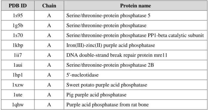
three iterations) or the crystal structure resolution. In this study, we used a 90% sequence identity cutoff, resolution less than 3 Å and R-value better than 0.3. After the curation, the training set consisted of ten PDB chains (see Table 2.1): 1s95A (PDB code: 1s95; chain A), 1g5bA, 1s70A, 1kbpA, 1ii7A, 1auiA, 1xzwA, 1uteA and 1qhwA sharing no more than 85% pair-wise sequence identities (see Figure 2.1).
Table 2.1: Metallophos training-set containing 10 protein chains
PDB ID Chain Protein name
1s95 A Serine/threonine-protein phosphatase 5 1g5b A Serine/threonine-protein phosphatase
1s70 A Serine/threonine-protein phosphatase PP1-beta catalytic subunit 1kbp A Iron(III)-zinc(II) purple acid phosphatase
1ii7 A DNA double-strand break repair protein mre11 1aui A Serine/threonine-protein phosphatase 2B
1hp1 A 5'-nucleotidase
1xzw A Sweet potato purple acid phosphatase 1ute A Pig purple acid phosphatase
21
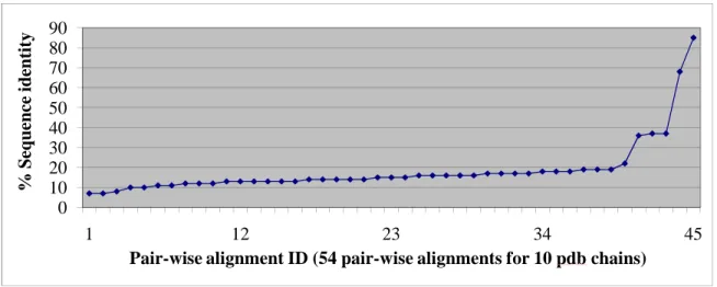
Figure 2.1: Distribution of pair-wise sequence identities in the Metallophos training-set: (1) sequence length: average = b= 311 amino acid residues, minimum = 219 amino acid residues, maximum = 378 amino acid residues; (2) sequence identity: average = 18.6%, minimum = 7%; maximum = 85%, only 5 pair-wise alignments have sequence identities > 30%.
2.2.2 Selection of the test set from the external dataset containing Metallophos members We found that the majority of proteins in each of the other five Metallophos families in SCOP 1.7.1 (families of YfcE-like, TT1561-like, Hypothetical protein aq_1666, DR1281-like and Phosphoesterase-related) were proteins of unconfirmed Metallphos function. We included all members in those five families into the external dataset. We combined this dataset with new 74 Metallophos entries added in the new release of SCOP 1.7.3 and SCOP1.7.5. Then, the representative proteins were retrieved using the same PISCES criteria applied to generate the training set (see Section 2.2.1). In order to illustrate the performance of our approach on remote homology detection, we selected only proteins having sequence identity less than 20% when compared to the training-set members. We retrieved 12 proteins into the test set. Three of them have known Metallophos functions according to the literature information (PDB chains: 3d03A (PDB code: 3d03; chain A)56, 1s3lA57 and 1t70A58). One had a function suggested from structures (2nxfA)59 whereas the rest were proteins of
0 10 20 30 40 50 60 70 80 90
1 12 23 34 45
% S eq u en ce i d en ti ty
22
unconfirmed Metallophos function (3ck2A, 1su1A60, 1xm7A, 1t71A, 2cv9A, 2yvtA, 1nnwA and 1uf3A).
2.2.3 Background dataset
In our subgraph mining-based approaches, frequent subgraphs retrieved from the training set of proteins of interest become common subgraphs if and only if they are rarely found in the proteins of a “background” database. In this study, the same PISCES criteria used to curate the training set (see Section 2.2.1) were applied to the PDB (May 2007 release) to build a background dataset. This dataset included 6,605 non-redundant protein chains excluding the 84 Metallophos proteins in SCOP 1.7.1 (see Section 2.2.1).
2.2.4 Identification of Metallophos-specific structural motifs using FFSM
The FFSM approach33, 34 (see Section 1.3-1.5) was applied to mine Metallophos-specific structural motifs (non-redundant frequent common subgraphs) from a training set of 10 Metallophos proteins. Each protein structure in the training set was modeled as AD (Almost Delaunay) graph consisting of nodes and edges. In this study, motifs were restricted to fully interconnected subgraphs in which all nodes connect to each other. Other parameters were set for mining motifs from the graph representations of protein structures in the training set as follows:
• Nodes represent alpha carbons (Cα) of amino-acid residues. There are 20 possible types of nodes based on the 20 natural types of amino acid residues.
23
edges (edge length for type1 to type5 are 0-4, 4-6, 6-8.5, 8.5-10 and 10.5-12.5 Å, respectively) and 5 types of distance constraints between non-contacting residues (edge length for type6 to type10 are 0-4, 4-6, 6-8.5, 8.5-10 and 10.5-12.5 Å, respectively).
• Minimum size of the motif was set to 4 amino acid residues
• Minimum support (f) of that subgraph is the minimum fraction of family members in the training set that must contain that subgraph
• Maximum background occurrence (b) is the maximum fraction of proteins in the background dataset that contain a subgraph of interest. The value of b was set to 0.1% by default.
A subgraph is considered frequent if its ‘minimum support’ (f) value is higher than a user-defined threshold (e.g., f=0.9; the motif presents in at least 90% of the family members). However, those frequent subgraphs become frequent common subgraphs (motifs) if and only if they are rarely (below certain frequency threshold) found in proteins of a ‘background’ dataset (b= 0.1%: found in no more than seven proteins out of 6,605 non-redundant protein chains in the background dataset).
24
2.2.5 Identification of Metallophos-specific structural motifs using CASIM
CASIM (see paragraph 1.6) was applied to mine Metallophos-specific structural motifs (frequent common Delaunay tetrahedral) from a training set of 10 Metallophos proteins. Each protein structure was modeled as DT (Delaunay tessallation) graph consisting of nodes and edges. Nodes represent side chain centroids of amino acid residues. There are 20 possible types of nodes based on the 20 natural types of amino acid residues. Unlike FFSM that defines a motif as a fully interconnected subgraph, CASIM describes motifs as ensembles of neighboring Delaunay tetrahedral. Thus, we expected to recover motifs missed by FFSM or vice versa.
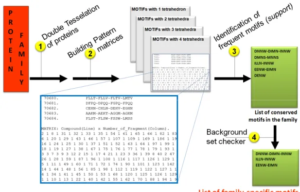
To reach the goal of efficient and fast function annotation, CASIM was applied to identify Metallophos-specific motifs as follows (see Figure 2.2): 1) each family member in the training set is tessellated to obtain a list of its constitutive CASIM structural motifs, 2) The lists of motifs for all family members are processed to build pattern matrices where each row corresponds to a protein, and each column corresponds to the type of the motif. These matrices contain the occurrences of each motif’s type in every protein of the training set, using a specific sparse matrix implementation (only non-zero values are stored for efficiency). All motifs included in a given pattern matrix involve the same number of constituent Delaunay tetrahedra.
imply that they are specific to this family. Each family
with high support (i.e., in significant number of protein members of a family) but low background (i.e., in a very small number of all other proteins). Therefore, the algorithm applies a ‘background’ frequency filter to obtain the list of conserv
are retained if and only if they are rarely found in the ‘background’ dataset background occurrence (b) was
database). The background checker implemented
standard Dual-Core PC) requires less than a second to retrieve all necessary information concerning the motifs in the background set; this high computational efficiency is achieved because all possible motifs present in
stored in a database.
Figure 2.2: The overall workflow to identify family approach.
25
imply that they are specific to this family. Each family-specific motif is required
with high support (i.e., in significant number of protein members of a family) but low background (i.e., in a very small number of all other proteins). Therefore, the algorithm applies a ‘background’ frequency filter to obtain the list of conserved motifs. Frequent motifs are retained if and only if they are rarely found in the ‘background’ dataset
background occurrence (b) was set to 0.1% by default (less than 7 proteins with our current database). The background checker implemented in the CASIM software (running on a Core PC) requires less than a second to retrieve all necessary information concerning the motifs in the background set; this high computational efficiency is achieved because all possible motifs present in the background proteins have been pre
The overall workflow to identify family-specific structural motifs using the CASIM
specific motif is required to occur with high support (i.e., in significant number of protein members of a family) but low background (i.e., in a very small number of all other proteins). Therefore, the algorithm ed motifs. Frequent motifs are retained if and only if they are rarely found in the ‘background’ dataset. The maximum set to 0.1% by default (less than 7 proteins with our current in the CASIM software (running on a Core PC) requires less than a second to retrieve all necessary information concerning the motifs in the background set; this high computational efficiency is achieved the background proteins have been pre-calculated and
26
2.2.6 Cumulative Support Profiles for protein function inference
Since each amino acid residue in a protein is surrounded by other residues, there is an interesting question of characterizing the environment of each residue and investigating the structural similarity between the neighborhoods of each residue (especially the functionally significant ones) for a given target protein vs. a set of proteins (such as a family of proteins with the same function).
One simple yet powerful approach to comparing residue environments between protein structures is the use of so called 3D-1D profiles. Originally proposed by Eisenberg61, this approach translates various parameters of a residue’s environment in 3D to a sequence-specific profile where each residue in the sequence is given some sort of score reflecting its 3D environment. 3D-1D profiles have been used in fold recognition61 or protein model quality assessment62. In our previous studies, we employed similar concept to compare proteins using profiles based on four-body statistical potentials generated with the help of Delaunay tessellation63.
27
frequencies but as numbers of occurrences). For example, if the quadruplet DGGH has a support value equal to 6 (i.e., it occurs in 6 out of 10 proteins in the training set) and the motif DGGH-GGHN has a support of 2, the partial cumulative support of DGGH is 6+2 = 8. This procedure is repeated for all motifs involving the quadruplet DGGH to calculate a total value of the cumulative support for this quadruplet.
This score can be calculated for any Delaunay quadruplet in any protein of the training set. If a quadruplet occurs frequently in a family, and so are its neighbors, its cumulative support is expected to be high. Thus, the cumulative support provides a quantitative assessment of the conservation of each Delaunay quadruplet of residues within a protein family. Similar consideration could then be applied to each amino acid residue to calculate its individual cumulative support: the latter is equal to the sum of the cumulative supports of all quadruplets involving this particular residue. Finally, the cumulative support values for each residue can be plotted against the residue number in the sequence to obtain the protein cumulative support profile (see Figure 2.7) where the peaks correspond to residues with the highest conserved 3D environment in the protein family.
2.3 Results
2.3.1 Metallophos-specific structural motifs identified by FFSM and CASIM
Both FFSM and CASIM approaches were independently utilized for identifying structural motifs conserved in the training set of Metallophos members but found in no more than 0.1% (b=0.1) in the background dataset of 6605 protein chains.
28
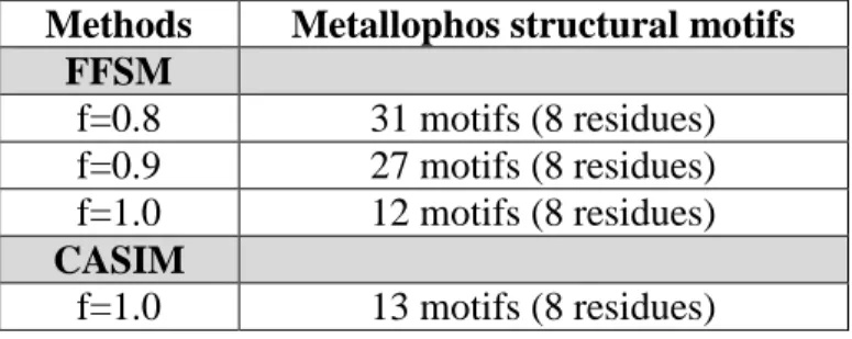
value was increased from 0.8 to 1.0, respectively; (2) CASIM retrieved 13 motifs at f = 1.0. Both FFSM and CASIM detected the same set of eight residues in the training-set members.
Table 2.2: Number of Metallophos-specific structural motifs retrieved from the family training set (column 2) at given support (f) values
Methods Metallophos structural motifs FFSM
f=0.8 31 motifs (8 residues) f=0.9 27 motifs (8 residues) f=1.0 12 motifs (8 residues) CASIM
f=1.0 13 motifs (8 residues)
server via client scripts under
calculations presented in this study were performed on a Dual
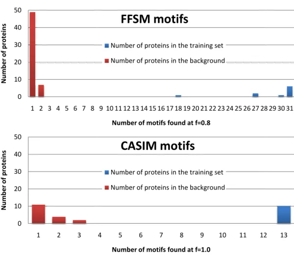
Figure 2.3: Distribution of Metallophos CASIM (bottom) in the training set
The examples of two Metallophos the Table 2.3, specifically their annotations: type 1122 (called motif A) and
(called motif B; cf. Figure 2.4
residues involved in four neighboring Delaunay tetrahedra: for instance, motif A involves the tetrahedra DDHH, DGGH, D
included in these two motifs are shown in 0 10 20 30 40 50
1 2 3 4 5 6 7 8
N u m b e r o f p ro te in s 0 10 20 30 40 50
1 2 3
N u m b e r o f p ro te in s 29
client scripts under Linux. On the contrary, CASIM is executed locally (all calculations presented in this study were performed on a Dual-Core PC).
Distribution of Metallophos-specific structural motifs retrieved by FFSM in the training set (blue) and the background dataset (red).
The examples of two Metallophos-specific motifs detected by CASIM are given in , specifically their annotations: DDHH-DGGH-DGHN-DHHN
) and DDGH-DDHH-DGGH-DGHN connectivity type ; cf. Figure 2.4). Briefly, these two CASIM motifs A and B included seven residues involved in four neighboring Delaunay tetrahedra: for instance, motif A involves the tetrahedra DDHH, DGGH, DGHN and DHHN. Metallophos protein residues which are included in these two motifs are shown in Table 2.3. A rapid analysis suggests that motifs A
9 10 11 12 13 14 15 16 17 18 19 20 21 22 23 24 25 26 27 28
Number of motifs found at f=0.8
FFSM motifs
Number of proteins in the training set
Number of proteins in the background
4 5 6 7 8 9 10 11 12
Number of motifs found at f=1.0
CASIM motifs
Number of proteins in the training set
Number of proteins in the background
is executed locally (all
specific structural motifs retrieved by FFSM (top) and
specific motifs detected by CASIM are given in DHHN of connectivity connectivity type 1222 ). Briefly, these two CASIM motifs A and B included seven residues involved in four neighboring Delaunay tetrahedra: for instance, motif A involves the GHN and DHHN. Metallophos protein residues which are . A rapid analysis suggests that motifs A
28 29 30 31
and B involve exactly the same residues but importantly, their types are different (1122 for motif A, 1222 for motif B): the tetrahedral connectivity between these residues is different because of the types of graph edges (i.e., an edge represents either a peptidic bond or geometrical proximity in 3D space) between the vertex
2.4). The nomenclature 1122 reflects that two Delaunay tetrahedra out of four included in motif A are of type 1 and the other two are of type 2. Here the tetrahedron DDHH type 1 (type 1 means that all four vertex
motif A not present in the motif B, whereas the tetrahedron DDHH type 2 (type 2 means that two out of four vertex-residues are consecutive in the protein sequence) is present in motif B.
Figure 2.4: Metallophos-specific motifs (DDHH DGHN) retrieved by CASIM plotted on
a training-set member): both motifs involve the same residues but different constitutive Delaunay neighbor tetrahedra, improving their specificity to recognize Metallophos activity
30
and B involve exactly the same residues but importantly, their types are different (1122 for motif B): the tetrahedral connectivity between these residues is different because of the types of graph edges (i.e., an edge represents either a peptidic bond or geometrical proximity in 3D space) between the vertex-residues of the tetrahedra (see
). The nomenclature 1122 reflects that two Delaunay tetrahedra out of four included in motif A are of type 1 and the other two are of type 2. Here the tetrahedron DDHH type 1 (type 1 means that all four vertex-residues are not consecutive in the protein sequence) in the motif A not present in the motif B, whereas the tetrahedron DDHH type 2 (type 2 means that residues are consecutive in the protein sequence) is present in motif B.
specific motifs (DDHH-DGGH-DGHN-DHHN and DDGH
DGHN) retrieved by CASIM plotted on serine/threonine-protein phosphatase 2B (PDB chain: 1au1A, : both motifs involve the same residues but different constitutive Delaunay
ng their specificity to recognize Metallophos activity.
and B involve exactly the same residues but importantly, their types are different (1122 for motif B): the tetrahedral connectivity between these residues is different because of the types of graph edges (i.e., an edge represents either a peptidic bond or residues of the tetrahedra (see Figure ). The nomenclature 1122 reflects that two Delaunay tetrahedra out of four included in motif A are of type 1 and the other two are of type 2. Here the tetrahedron DDHH type 1 in sequence) in the motif A not present in the motif B, whereas the tetrahedron DDHH type 2 (type 2 means that residues are consecutive in the protein sequence) is present in motif B.
31
Table 2.3: Metallophos-specific structural motifs retrieved by CASIM in the ten training-set members and also in hypothetical protein YfcE (PDB chain: 1su1A). ESA = Exposed Surface Area (Å2), ESA1; Volume in Å3.
MOTIF DDHH-DGGH-DGHN-DHHN
Protein Volume ESA TYPE 1122
1auiA 37.3 131.4 ASP77 GLY104 ASP105 GLY136 ASN137 HIS186 HIS268
1g5bA 41.4 139.4 ASP20 GLY48 ASP49 GLY74 ASN75 HIS139 HIS186 1hp1A 45.0 148.5 ASP16 GLY58 ASP59 GLY90 ASN91 HIS192 HIS227
1ii7A 45.1 149.8 ASP8 GLY48 ASP49 GLY83 ASN84 HIS173 HIS206
1kbpA 60.5 192.2 ASP15 GLY43 ASP44 GLY80 ASN81 HIS166 HIS203
1qhwA 38.0 131.0 ASP10 GLY47 ASP48 GLY86 ASN87 HIS182 HIS217
1s70A 54.3 177.3 ASP64 GLY91 ASP92 GLY123 ASN124 HIS173 HIS248 1s95A 37.4 129.8 ASP67 GLY95 ASP96 GLY127 ASN128 HIS177 HIS252 1uteA 65.7 201.5 ASP12 GLY49 ASP50 GLY88 ASN89 HIS184 HIS219
1xzwA 58.4 188.1 ASP16 GLY44 ASP45 GLY81 ASN82 HIS167 HIS204
1su1A 33.8 122.0 ASP9 GLY36 ASP37 GLY72 ASN73 HIS105 HIS127
MOTIF DDGH-DDHH-DGGH-DGHN
Protein Volume ESA TYPE 1222
1auiA 37.3 131.4 ASP77 GLY104 ASP105 GLY136 ASN137 HIS186 HIS268
1g5bA 41.4 139.4 ASP20 GLY48 ASP49 GLY74 ASN75 HIS139 HIS186
1hp1A 60.9 181.0 ASP16 GLY58 ASP59 GLY90 ASN91 HIS192 HIS227
1ii7A 45.1 149.8 ASP8 GLY48 ASP49 GLY83 ASN84 HIS173 HIS206
1kbpA 38.2 131.4 ASP15 GLY43 ASP44 GLY80 ASN81 HIS166 HIS203
1qhwA 55.1 170.5 ASP10 GLY47 ASP48 GLY86 ASN87 HIS182 HIS217
1s70A 56.4 184.3 ASP64 GLY91 ASP92 GLY123 ASN124 HIS173 HIS248 1s95A 52.8 167.3 ASP67 GLY95 ASP96 GLY127 ASN128 HIS177 HIS252
1uteA 65.7 201.5 ASP12 GLY49 ASP50 GLY88 ASN89 HIS184 HIS219
1xzwA 50.9 161.9 ASP16 GLY44 ASP45 GLY81 ASN82 HIS167 HIS204
1su1A 58.2 176.9 ASP9 GLY36 ASP37 GLY72 ASN73 HIS105 HIS127
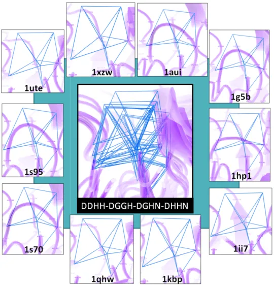
motifs (performed by the TMAlign program
training-set members revealed a very good local alignment of the seven residues involved in the motifs. In Figure 2.5, the motif DDHH
PyMol program under the control of PROTMAN via python scripts. For each residue within the motif, its representative vertices (corresponding to side chain centroïds) were fairly well superimposed. The RMSD values for each residue were in the range 0.41
overall RMSD value was equal to 0.59
Figure 2.5: Visualization of the Metallophos retrieved by CASIM for all training set members.
32
s (performed by the TMAlign program64 executed via PROTMAN interface) for the set members revealed a very good local alignment of the seven residues involved in
, the motif DDHH-DGGH-DGHN-DHHN is visualized
PyMol program under the control of PROTMAN via python scripts. For each residue within the motif, its representative vertices (corresponding to side chain centroïds) were fairly well
ed. The RMSD values for each residue were in the range 0.41-0.73 overall RMSD value was equal to 0.59Å for the whole DDHH-DGGH-DGHN
Visualization of the Metallophos-specific motif (DDHH-DGGH-DGHN by CASIM for all training set members.
executed via PROTMAN interface) for the set members revealed a very good local alignment of the seven residues involved in is visualized in the PyMol program under the control of PROTMAN via python scripts. For each residue within the motif, its representative vertices (corresponding to side chain centroïds) were fairly well 0.73Å whereas the DGHN-DHHN motif.
33 2.3.2 Validation on test proteins of known function
The three test protein structures (PDB chains: 3d03A, 1z2wA and 1t70A) had known Metallophos functions according to the published results from experimental analyses (see Supplementary data). In addition, the catalytic site residues were also suggested for the test proteins by the authors, mostly from structure analysis and few from mutation analysis. These test proteins have only 9-19% sequence identity to the training set.
In this study, function inferences by CASIM and FFSM were relied on the appearance of Metallophos-specific motifs and the conserved key residues involved with those motifs (see Table 2.4 and Table 2.5). As reported in Section 3.1, the family motifs and conserved key residues detected by CASIM (f=1.0) and FFSM (f=0.8) were 13 motifs with 8 residues and 31 motifs with 8 residues, respectively. CASIM was capable of detecting all family motifs in 3d03A whereas FFSM detected the majority of the motifs (25 from 31 motifs and 7 from 8 residues) in this protein. Compared to FFSM, CASIM captured the larger fraction of family motifs in 1z2wA. However, CASIM could not identify any family motifs in 1t70A, which was in turn identified by FFSM. Thus, by combining CASIM and FFSM (see Table 2.4, column 6, CASIM-FFSM), we could retrieve the family motifs in all test proteins. This observation suggested the benefit of combining two methods to recover more motifs from graph space.
34
We evaluated the prediction performance of CASIM-FFSM with the published results and those from publicly available methods (see Table 2.4 and Table 2.5); Pfam, reverse template and enzyme active site template searches, and Catalytic Residue Prediction (CRP). The common highlights between reverse template search and CASIM-FFSM are that they aim to predict protein function based on structure data alone and do not require any prior knowledge of functionally important residues. The reverse template was generated by breaking the query protein itself into many three-residue templates of neighboring residues. Then, these small templates were scanned against a representative set of structures in PDB. The enzyme active site template search and CRP assigned active site residues based on the data obtained from the Catalytic Site Atlas (CSA). Enzyme active site templates were manually derived templates of three to six residues. Each template consisted of one, two or three residues known to be catalytic, and one or more additional conserved residues relative to the catalytic residues. CRP was mainly developed for predicting catalytic residues instead of predicting protein function. The method predicted key residues from sequence and structure feathers using SVM.
Table 2.4: Function prediction on test proteins of known function using AFP methods. For reverse
template and enzyme active site template searches, we reported only the first hit of known function having the highest scoring template and are not found in our external set; resi = number of amino acid residues.
PDB chain
Protein name % Seq iden to the training set CASIM (f=1) FFSM (f=0.8) CASIM-FFSM
Pfam Reverse
template Enzyme active site template 3d03A Glycerophos phodiesterase
10-16 13 motifs (8 resi) 25 motifs (7 resi) 38 motifs (8 resi) Metallophos (2.3e-13) 1qhw (Metallophos) 4kbp (Metallophos)
1z2wA Vsp29 11-19 5 motifs (8 resi) 1 motif (4 resi) 6 motifs (8 resi) Metallophos (0.00047) 3dsd (Metallophos) 4kbp (Metallophos)
1t70A DR1281 9-17 0 1 motif
35
We found that CASIM-FFSM was able to detect the majority of published key residues in 3d03A and 1z2wA, and two published key residues in 1t70A. It is important to underline that other residues detected by us that were not found in the literatures were all neighbors of the published key residues, and were found at the metal-binding sites.
Pfam can also infer Metallophos function to all test proteins with high confidence (E-value less than 0.001). This function inference by Pfam was based on the presence of the HMM profile of Metallophos family (PF00149) obtained from the publicly available Pfam database.
The reverse template and enzyme active site template searches were applied for both function inference and catalytic residue prediction. In our study, we reported only a hit (matched structure) having the highest score template and were not members of our external dataset. We found that reverse template search and enzyme active site template search were able to provide hits for every test proteins. All hits given by both methods were Metallophos proteins. In addition, most predicted catalytic residues retrieved by both methods were similar to those reported in the literatures and those identified by FFSM-CASIM.
36
Table 2.5: Conserved key residues in test proteins of known PTK function: comparison of the key residues reported in the primary literatures and those from automated prediction methods. The predicted residues matching to the published residues are labeled in red.
PDB ID
Published results Predicted residues
CASIM-FFSM Reverse template search
Enzyme active site template search
CRP
3d03A
Asp8, His10, Asp50, Asn80, His156, His 195,
His197
Asp8, Gly49, Asp50, Gly79, Asn80, His81, His156, His 195
Asp8, His156, Cys193.
Asp8, Asp50, Asn80, His81, His156, His195
Asp8, His10, His50, Asn80, His81, His156, His195,
His197 1z2wA Asp8, Asn39,
Asp62, His86, His117 Asp8, Gly38, Asn39, Gly61, Asp62, His86, His115, His117 Asp8, His86, Gly114 Asp8, His10, Asn39, Asp62, His86, His115
Asp8, His10, His86, His115, His117
1t70A Asp8, Glu37, Asn38, Asn65, His148, His173, His175 Gly64, Asn65, His66, His173 Asp8, Asn35, His148 Glu37, His148, Asp193, His175 Asp8, Glu37, Asn65, His66, His173
2.3.3 Predicting Metallophos function and conserved key residues in proteins of unconfirmed function
We predicted the Metallophos function and conserved key residues in 9 test protein structures; one putative (2nxfA), one uncharacterized (3ck2A) and seven hypothetical proteins (1su1A, 1xm7A, 1t71A, 2cv9A, 2yvtA, 1nnwA and 1uf3A). All of them fell into the midnight zone (less than 20% sequence identity) when compared to the training set. CASIM-FFSM was able to detect Metallophos motifs in the five following proteins (see Table 2.6).
Putative dimetal phosphatase LOC393393 (PDB code 2nxf) from Danio rerio
37
and Gly95) identified by us that were not mentioned in the literature were also found at the active site, and (4) Pfam found the Metallophos profile in this protein with high confidence (E-value 6.4e-06).
Conserved uncharacterized protein (predicted phosphoesterase COG0622) from
Streptococcus pneumoniae TIGR4 (PDB code 3ck2)
The crystal structure of this conserved uncharacterized protein was released to the PDB by the Midwest Center for Structural Genomics (MCSG) in 2008. This protein shares 11-17% sequence identity to our training set. Pfam detected the Metallophos profile in this protein with low confidence (E-value 0.21). However, we were convinced that the protein has Metallophos function from the CASIM-FFSM results. We identified the majority of the family motifs containing 7 conserved key residues in this protein. In addition, the key residues (Asp11, Gly37, Asp38, Gly56, Asn57, His81 and His110) detected by us were present at the Mn2+-binding sites. Three of them (Asp11, Asp38 and His110) were similar to those predicted by CRP, which identified 5 residues (Asp11 (SVM score of 3.72), His13 (3.41), Asp38 (3.59), His110 (3.82), His112 (2.90)).
Hypothetical protein aq_1665 (PDB code 1xm7) from Aquifex aeolicus
38
(Asp7, Gly49, Asp50, Gly77, Asn78, His79, His111 and His145) in this protein. Some of them were reported by other methods; (1) CRP identified 4 residues (His145, Asp50, His111 and Asp7)45, and (2) the method predicting transition metal-biding in apo proteins by Babor’s group identified 4 residues (Asp7, His9, Asp50 and His111)65.
Hypothetical protein MPN349 (PDB code 1t71) from Mycoplasma pneumoniae The crystal structure of hypothetical protein MPN349 was released by the Berkeley Structural Genomics Center (BSGC) to the PDB in 2004. This protein shares low sequence identity (7-18%) to the training set. Pfam was unable to provide any hit for this protein. However, we inferred Metallophos function to this protein due to the presence of 2 specific Metallophos motifs, which were not found in the background dataset. We also found that this protein share high similarity (38% sequence identity and DALI z-score 37.1) to protein DR1281 (PDB code 1t70), one of known Metallophos proteins in our test set. Our method predicted 5 conserved key residues (Gly70, Asn71, His72, His158 and His183). Three of them (Asn71, His72 and His183) were overlapped with those predicted by CRP, which identified 4 residues (Asp12 (SVM score of 3.16), Asn71 (2.68), His72 (2.96) and His 183 (2.55)).
Hypothetical protein YfcE (PDB code 1su1) from E. coli
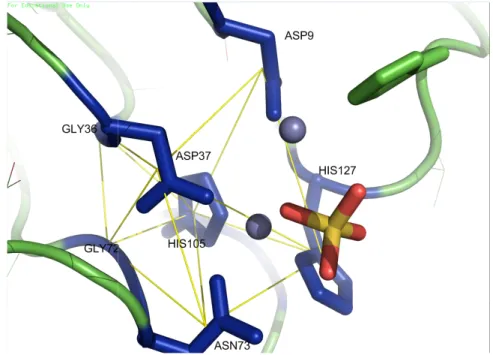
highly suggested that the protein has Metallophos function. DGHN-DHHN’ motif detected by CASIM
presence of metal counter-ions and a phosphate group inside the motifs or in their close proximity. The prediction was supported by the following data: (1) structural and biochemical analysis by the authors revealed that the protein had the Mn
phosphatase activity60, (2) the authors suggested two metal binding sites: HIS11, HIS129 and ASP37 whereas the other one consist
HIS127. We identified 7 conserved key residues; five (Asp9, Asp37, Asn73, His105 and His127) were similar to those suggested by the authors whereas two were neighbors (Gly36, Gly72) to the reported residues. In this case, we di
data because Pfam incorporated protein profile.
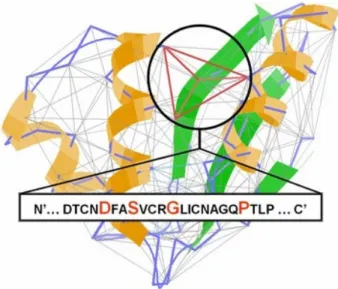
Figure 2.6: Visualization of the Metallophos retrieved by CASIM for protein
39
highly suggested that the protein has Metallophos function. An example of ‘
detected by CASIM is visualized in Figure 2.6. One can see the ions and a phosphate group inside the motifs or in their close The prediction was supported by the following data: (1) structural and
y the authors revealed that the protein had the Mn , (2) the authors suggested two metal binding sites: one include
HIS11, HIS129 and ASP37 whereas the other one consisted of ASP37, ASP73, HIS105 and 7 conserved key residues; five (Asp9, Asp37, Asn73, His105 and His127) were similar to those suggested by the authors whereas two were neighbors (Gly36, Gly72) to the reported residues. In this case, we did not employ Pfam results as a support data because Pfam incorporated protein YfcE into a seed used to generate the Metallophos
Visualization of the Metallophos-specific motif (DDHH-DGGH-DGHN protein YfcE (1su1A (1su1 chain A)).
An example of ‘DDHH-DGGH-. One can see the ions and a phosphate group inside the motifs or in their close The prediction was supported by the following data: (1) structural and y the authors revealed that the protein had the Mn2+ dependent one included ASP9, of ASP37, ASP73, HIS105 and 7 conserved key residues; five (Asp9, Asp37, Asn73, His105 and His127) were similar to those suggested by the authors whereas two were neighbors (Gly36, d not employ Pfam results as a support fcE into a seed used to generate the Metallophos