481
© 2017 by the Serbian Biological Society How to cite this article: Han H, Liu N, Zhang L, Gong M, Cao M, Li B, Kaisa S, Yu X, Tian J. Promotion of cytoplasmic vacuolation-mediated cell death of human prostate cancer PC-3 cells by oxidative stress induced by daucusol, a new guaiane-type sesquiterpenoid from Daucus carota L. Arch Biol Sci. 2017;69(3):481-9.
Promotion of cytoplasmic vacuolation-mediated cell death of human prostate cancer
PC-3 cells by oxidative stress induced by daucusol, a new guaiane-type sesquiterpenoid
from
Daucus carota
L.
Haote Han1, Na Liu2, Lin Zhang1, Minghua Gong2, Ming Cao3, Baoguo Li2, Sulaiman Kaisa4, Xiuying Yu1 and
Jingkui Tian1,*
1The Key Laboratory of Biomedical Engineering, Ministry of Education, Department of Biomedical Engineering, Zhejiang
University, Hangzhou 310027, P.R. China
2 Shandong University of Traditional Chinese Medicine, Jinan 250355, P.R. China
3 Department of Pharmacology, Wenzhou Medical University, Wenzhou 325035, P.R. China
4The Xinjiang Uygur Autonomous Region National Institute of traditional Chinese Medicine, Urumchi 830092, P.R. China
*Corresponding author: [email protected]
Received: September 2, 2016; Revised: October 1, 2016; Accepted: October 28, 2016; Published online: November 9, 2016
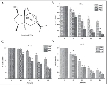
Abstract: We investigated the antitumor activity of daucusol (DS) derived from Daucus carota L. in PC-3, A549 and HeLa cell lines by the MTT assay. Optical microscopy revealed that exposure of PC-3 cells to DS resulted in cytoplasmic vacu-olation. Flow cytometry analysis of the phase of the cell cycle did not reveal a sub-G1 peak, and no caspase-dependent activation was observed after DS treatment. The levels of endoplasmic reticulum (ER) stress biomarkers, LC3B-II and ubiquitinated proteins were increased. It was also observed that oxidative stress played an important role in the activation of the cytoplasmic vacuolation-mediated cell-death pathway. In vivo, DS inhibited tumor growth in nude mice by 39.13% compared to the vehicle. Protein expression in the tumor tissue was consistent with their expression in vitro. Our findings indicate that DS induced cytoplasmic vacuolation-mediated death in PC-3 cells by triggering oxidative stress and suggest that targeting this pathway could serve as a novel therapeutic approach for prostate cancer.
Key words: daucusol; cytoplasmic vacuolation; oxidative stress; endoplasmic reticulum stress; prostate cancer
INTRODUCTION
Prostate cancer is the most common malignant tumor in men, causing the deaths of hundreds of thousands of men each year worldwide [1], making it the sec-ond highest cause of cancer-related deaths after lung cancer. Prostate cancer is heterogeneous, and treat-ment varies depending on the stage of the disease. While guidelines for the treatment of prostate can-cer have been developed and have facilitated treat-ment decisions, its poor response to chemotherapy and frequent recurrence continue to present major obstacles in overcoming the disease. Recent studies showed that chemotherapy induces apoptosis or au-tophagy in prostate cancer cells [2,3]. However, resist-ance to chemotherapy in prostate cresist-ancer can arise via similar antiapoptotic or antiautophagic mechanisms [4,5]. Therefore, targeting a non-apoptotic cell death
pathway may be an effective strategy of avoiding drug resistance in prostate cancer treatment.
In a previous study, we isolated the novel com-pound daucusol (DS) (Fig.1A) [9], a guaiane-type
ses-quiterpenoid from Fructus carotae, the fruit of Daucus
carota L. (Umbelliferae) [10]. In this study, we tested the effects of DS on three human cancer cell lines: prostate cancer PC-3 cells, lung cancer A549 cells and cervical cancer HeLa cells. We found that DS inhibited growth of PC-3 cells via a novel pathway related to cytoplasmic vacuolation-mediated cell death. We next focused our attention on the resistance mechanisms
of prostate cancer to exposure to DS in vitro and in
vivo. Our findings revealed the therapeutic potential
of DS for treating prostate cancer.
MATERIALS AND METHODS
Experimental animals
All animal research was conducted in accordance with the Declaration of Helsinki and with the Guide for the Care and Use of Laboratory Animals as adopted and promulgated by the United National Institutes of Health. All experimental protocols were approved by the Review Committee for the Use of Animal Subjects of Changshu Realistic Technology Co. Ltd (Suzhou, China).
Reagents
Antibodies against the following proteins were used in this study: LC3B, ubiquitin, caspase-3, PARP, β-Actin (Cell Signaling Technology, Beverly, MA); GRP78, GADD153 (Santa Cruz Biotechnology, Santa Cruz, CA); calreticulin (Abcam, Cambridge, MA). The secondary antibodies goat anti-rabbit IgG, goat anti-mouse IgG (Biosharp Biological science and technology, Shanghai, China) and FITC goat anti-rabbit IgG (Huabio, Hangzhou, China) were used. DS was extracted and identified according to meth-ods described in a previous study [9]. The chemicals cycloheximide (CHX, Sigma-Aldrich, St. Louis, MO), 3-(4,5-dimethylthiazol-2-yl)-2,5-diphenyl-2H-tetra-zolium bromide (MTT, Solarbio, Beijing, China), N-acetylcysteine (NAC, Sigma-Aldrich, St. Louis, MO), TritonX-100 (Solarbio, Beijing, China) and dimethyl sulfoxide (DMSO, Gibco, Grand Island, New York, USA) were also used. Cell incubation medium was
purchased from Gino Biomedical Technology Co. Ltd (Hangzhou, China) and fetal bovine serum (FBS) was purchased from Gemini Bio Products (Gemini, CA, USA). The following kits were used in this study: DNA content quantitation assay kit (KeyGEN Nan-jing, China), Reactive Oxygen Species (ROS) determi-nation kit (Jiancheng, Nanjing, China), Giemsa Stain kit (Solarbio, Beijing, China), BCA protein concentra-tion determinaconcentra-tion kit (Beyotime, Shanghai, China) and ECL system (Beyotime, Shanghai, China).
Cell lines and culture conditions
The human cancer cell lines PC-3, A549 and HeLa were obtained from the Chinese Academy of Sciences (Shanghai, China). Cells were cultured in Dulbecco’s modified Eagle’s medium (HeLa) or RPMI-1640 medium (PC-3, A549) supplemented with 10% FBS and 1% penicillin/streptomycin solution (100 IU/ mL penicillin, 100 mg/mL streptomycin) at 37°C in
5% CO2. Experiments were conducted after cells had
been incubated for 24 h. DS was dissolved in DMSO and diluted into culture medium containing 5% FBS, with the final concentration of 0.05% DMSO in each well. The control group received the same amount of DMSO.
Cell viability assay
Cell viability was determined by 3-(4, 5-dimethylth-iazol-2-yl)-2,5-diphenyltetrazolium bromide (MTT) assay in six replicates. Cells were seeded in 96-well
plates at 3000cells/well in a final volume of 100 μL.
After 24-h incubation at 37°C in 5% CO2, cells were
Giemsa staining
PC-3 cells were seeded into 12-well plates at 1×10⁵
cells/well in a final volume of 1 mL. After 24 h, cells were treated with DS (final concentrations 60 and 100 μM) in 2 mL medium or pretreated with CHX (final concentration 5 μg/mL) or NAC (final concentration 5 mM). All treated cells were incubated at 37°C in
5% CO2 for another 48 h. The medium was carefully
removed from each well and cells were washed three times with PBS, fixed for 3 min with methanol (chilled at -20°C), then stained with Giemsa for 30 min. After washing with water, 1 mL PBS was added to each well before optical microscopy.
Flow cytometricy cell cycle analysis
For cell cycle analysis, the DNA content quantitation assay kit was used. Briefly, 48 h after drug treatment,
cells were harvested in ice-cold PBS (3×10⁵ cells in 300
μL) and 700 μL 100% ethanol was added for fixation. After incubation at 4°C overnight, fixed cells were re-suspended in 100 μL RNase A and incubated at 37°C for 30 min, followed by incubation in 400 μL propidium iodide (PI) for 30 min in darkness. The distribution of cells in phases of the cell cycle was determined using a flow cytometer (Cytomics FC 500, Beckman Coulter, USA) and analyzed using Multicycle AV for Windows advanced DNA Cell Cycle Analysis software.
Reactive oxygen species (ROS) assay
Cells were seeded into 6-well plates at 5×10⁵ cells/well
and treated with a range of concentrations of DS for
24 h, or with 20 μM H2O2 for 1 h to serve as a positive
control. The medium was removed and replaced with
1 mL of PBS containing 1 μM CM-H2DCFDA, and
cells were incubated in the CO2 incubator at 37°C for
30 min in the dark. The cells were collected into 15-mL centrifuge tubes. After washing with PBS three times, the ROS level of each concentration was determined using a flow cytometer (Cytomics FC 500, Beckman Coulter, USA) and analyzed using FlowJO 7.6 software.
Immunofluorescence analysis
Cells treated with drugs were washed with cold PBS, fixed with methanol (chilled at -20°C) for 5 min at
room temperature, permeabilized with 0.5% Triton X-100 in PBS for 10 min, and blocked with 1% BSA in PBS for 30 min. After washing with PBS, the cells were incubated overnight with anti-calreticulin an-tibody in 1% BSA at 4°C. The cells were incubated with FITC goat anti-rabbit IgG in 1% BSA for 2 h at room temperature. Nuclei were stained with DAPI in Vectashield mounting medium (Vector Laboratories). Confocal images were obtained using the 63X oil im-mersion lens in a confocal microscope (LSM 780, Carl Zeiss Jena, Germany).
Western blotting
Cells were treated with DS or pretreated with CHX (final concentration 5 μg/mL) or NAC (final tration 5 mM) for 1 h, and then with different concen-trations of DS for 48 h. Cells were collected from 6-cm dishes by scraping and centrifugation. The cells were washed once with ice-cold PBS and subsequently in-cubated in 75 μL lysis buffer for 30 min. Lysates were centrifuged for 15 min at 4°C at 13200 rpm. Protein concentrations in the supernatants were determined using a BCA protein concentration determination kit. Equal amounts of proteins were separated in a 7.5% SDS polyacrylamide gel and then transferred to poly-vinylidene fluoride (PVDF) membranes. Membranes were blocked with 5% non-fat milk in 0.1% Tween-20 in TBS (TBST) for 2 h and incubated in primary anti-body (1:500-1:1000) at 4°C overnight. After washing three times with TBST, the membranes were incu-bated for 2 h at room temperature with the appropri-ate secondary antibody (1:5000). The immunoblots were visualized with an enhanced chemiluminescence (ECL) system.
Xenografts in BALB /C male mice
We established a xenograft tumor model by trans-planting PC-3 cells into nude mice as described pre-viously [11]. BALB/C male mice (5-7 weeks of age, NO.11401300028261) were purchased from HuaFu-Kang Biotechnology Co. Ltd (Beijing, China). Briefly, xenograft tumors were established by subcutaneous
injection of 5×10⁶ PC-3 cells in a total volume of 0.1
mL of serum-free medium. Once tumors were
meas-urable (100-200 mm3), the animals were randomized
ad-ministered the vehicle (10% DMSO, 20%
polyethyl-ene glycol 400, and 5% Tween 80 in sterile H2O), the
chemotherapeutic cyclophosphamide (30 mg/kg, as a positive control), or DS (200 mg/kg in 10% DMSO, 20% polyethylene glycol 400, and 5% Tween 80 in
sterile H2O) daily for 3 weeks. Mouse body weight
was measured daily. Tumor volumes were calculated from caliper measurements using a standard formula:
V= 0.5×(width2×length) [12,13]. The mice were killed
and the tumors were harvested, snap-frozen in liquid nitrogen and stored at -80°C until further use.
Statistical analysis
The data are presented as means±standard deviation (SD). One-way analysis of variance (ANOVA) and least significant difference (LSD) tests were used for all experiments. Differences were considered significant at p<0.05. Statistical analyses were performed using SPSS Statistics 17.0 software.
RESULTS AND DISCUSSION
DS inhibition of cells growth in vitro
The MTT assay was used to evaluate the antitumor activity of DS in the cervical cancer cell line, the PC-3 prostate cancer cell line and the A549 human lung cancer cell line. DS induced dose- and time-depend-ent reductions in cell viability in all three cell lines. These effects were most marked in the HeLa and PC-3
cell lines (Fig. 1B-D). The IC50 values for DS ranged
between 46 μM and 78 μM at 72 h.
DS induction of vacuolization and death signals in PC-3 cells
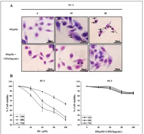
Several forms of non-apoptotic cell death have been described, including oncosis [14], autophagy [15], entosis [16] and necroptosis [17]. Giemsa staining revealed a dramatic change in cell morphology in
response to the DS treatment in PC-3 cells. DS induced a dose-dependent for-mation of cytoplasmic vacuolation. Vacu-oles could be clearly visualized by their lack of cytoplasmic materials (Fig. 2A). The presence of vacuolation suggests that the mechanism by which DS induces cell death in PC-3 cells may be a novel non-apoptotic pathway.
DS-induced cytoplasmic
vacuolization was blocked by CHX
CHX was used to determine its effect on vacuolation in DS-treated cells. Pretreat-ment with CHX diminished the ability of DS to induce vacuolation (Fig. 2A) and cell death (Fig. 2B). This result was consistent with a previous report [8] and suggests that DS-induced cell death is mediated primarily through cytoplas-mic vacuolation. Based on this finding, we hypothesized that ER stress, which is related to the formation of cytoplasmic vacuoles, plays an essential role in DS-induced PC-3 cell death.
Flow cytometry analysis of the cell cycle
To further confirm that DS induced a non-apoptotic form of cell death, we analyzed cell cycle progression by flow cytometry. No sub-G1 peak (Fig. 3), a marker of apoptosis, was observed in the DNA profile. Instead, DS induced S phase retardation, an effect that was decreased by pretreatment with CHX, a general inhibitor of protein synthesis [18]. These results provide evidence that DS-induced non-apoptotic cell death is mediated by cytoplasmic vacuolation. Fig. 3. DS induced S phase retardation but no sub-G1 peak as determined by flow
cytometry. Cells were treated with either DS (0, 80, 100 μM) (left side) or with DS (0, 80, 100 μM) and CHX (5 μg/mL) (right side) for 48 h.
Effect of DS on ER stress, upregulation of LC3B-II and ubiquitinated proteins in the PC-3 cell line
Considering the above findings, we examined the ef-fects of DS on ER stress and autophagy in PC-3 cells by Western blotting. Treatment with DS induced a dose-dependent increase in the expression of the ER biomarkers 78 kDa glucose-regulated protein (GRP78) and GADD153 (Fig. 4) as compared to the control. A previous report demonstrated that knockdown of LC3 conferred significant protection against cytoplasmic vacuolation and cell death, suggesting a novel role of LC3 in a death process other than autophagy [7], and increased protein levels of the autophagy-related protein LC3B-II were also induced by DS as reported herein (Fig. 4). In contrast, treatment with DS did not give rise to detectable levels of caspase-9 activation or PARP-1 cleavage (Fig. 4).
Additional evidence for the occurrence of DS-induced cell death by a cytoplasmic vacuolation-me-diated pathway was provided by the increase in ubiq-uitinated proteins. Mitophagy, the specific autophagic elimination of mitochondria, plays an important role in protein ubiquitination [19]. When mitochondria are damaged, PINK1, a key regulator of mitophagy, is rapidly activated in the mitochondria, resulting in
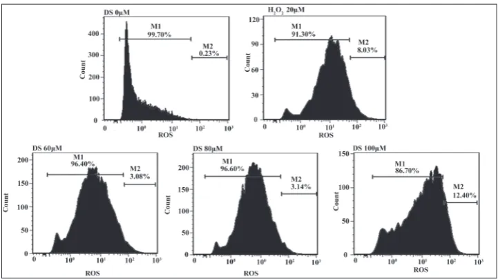
Fig. 5. Induction of ROS production in PC-3 cells by DS. H2O2 (20 μM) was used as a positive control.
the recruitment of the E3 ubiquitin ligase parkin to facilitate mitochondrial protein ubiquitination [20]. These processes can be blocked by CHX, i.e. by inhi-bition of protein synthesis. Pretreatment with CHX abolished the ability of DS to induce ubiquitin accu-mulation (Fig. 4). Therefore, it can be assumed that DS upregulated protein ubiquitination by activating the mitophagy pathway, however, further research remains to be conducted to support this.
DS-induced vacuoles were derived from the ER and triggered by oxidative stress
DS induced the production of ROS, as can be seen in Fig. 5. Immunofluores-cent staining with the ER chaperone calreticulin, which mainly engages in the ER lumen [21], demonstrated that DS-induced vacuoles were derived from the ER (Fig. 6A). N-acetylcysteine (NAC, Fig. 6B), a type of ROS inhibi-tor, blocked the formation of vacuoles, suggesting that oxidative stress likely played an important role in the novel pathway. To prove our assumption, we examined cell morphology and cell vi-ability with Giemsa staining and the MTT assay, respectively, after pre-treatment NAC. NAC abolished the ability of DS to induce cytoplasmic vacuolation (Fig. 7A) and increased the number of living cells (Fig. 7B). Similarly, ER stress (i.e., GRP78 and GADD153) and the increase in LC3B-II and ubiquitinated proteins that were induced by DS were abolished by the treatment with NAC (Fig. 7C). This suggests that the cytoplasmic vacuola-tion-mediated cell death pathway was triggered by oxidative stress.
DS inhibited tumor growth in vivo
The PC-3 xenograft tumor model was conducted to determine the effects of DS treatment on nude mice with PC-3 tumors. Treatment with the chemother-apeutic cyclophosphamide was used as a positive control for successful treat-ment of established tumors. Treattreat-ment with 200 mg/ kg of DS inhibited tumor growth by 39.13% compared with the vehicle (Fig. 8A). Mice treated with DS did not undergo significant weight loss (Fig. 8B). Tumor samples were examined for their expression of pro-teins related to ER stress and autophagy by Western analysis. Consistent with the effects of the DS
treat-ment on protein expression in vitro, in the tumor, DS
Fig. 6. DS-induced vacuoles were derived from the ER and blocked by NAC. A – Immunofluorescent images of prostate cancer PC-3 cells following a 48-h incubation in 0-100 μM DS or in cells pretreated with NAC (5 mM) (B). Confocal images were obtained using a 63X oil immersion lens in a confocal microscope. Green − calre-ticulin (1:500). Blue − DAPI. Arrows – visualized vacuolation.
upregulated the expression of GRP78 and GADD153 proteins, which points to the activation of ER stress. DS also increased the level of protein ubiquitination in the tumor, but had no effect on the levels of PARP-1 and caspase-9 (Fig. 9).
The novel mechanism of cell death mediation by DS in cancer cells is summarized in Fig. 10. DS, a novel guaiane-type sesquiterpenoids natural
com-pound has been shown to be capable of inducing cytoplasmic vacuolation-mediated PC-3 cells death by triggering oxidative stress, which was related to ER stress and upregulation of LC3B-II and ubiquitinated proteins without
caspase-dependent activation in vitro
and in vivo. This pathway could repre-sent a new target in the development of therapeutic approaches for prostate cancer.
This study provides new informa-tion about the mechanisms of non-apoptotic cell death which are not well understood. Structural modification of DS which would enhance its anti-tumor efficacy could promote its future development as a new type of anti-prostate cancer drug.
Acknowledgments: This work was supported by the National
Science Foundation of China [U1303122], the Jiangsu Provincial Science and Technology Project [BE2014654], the Changshu Sci-ence and Technology Project [CS201405] and Jiangsu Provincial Natural Science Foundation of China [BK20161269].
Conflict of interest: The authors report no conflicts of interest.
Fig. 10. Diagram of the cytoplasmic vacuolation-mediated mechanism by which DS induces PC-3 cell death.
Fig. 9. Effect of DS on the expression of proteins in vivo. Tumor proteins were ex-tracted and analyzed by Western blotting.
REFERENCES
1. Jemal A, Bray F, Center MM, Ferlay JJ, Ward E, Forman D. Global cancer statistics. CA. Cancer J Clin. 2011;61(2):69-90. 2. Zhang XS, Zhao C, Tang WZ, Wu XJ, Zhao YQ. Gypensa-pogenin H, a novel dammarane-type triterpene induces cell cycle arrest and apoptosis on prostate cancer cells. Steroids. 2015;104:276-83.
3. Jiang Q, Yeh S, Wang X, Xu DF, Zhang QX, Wen XQ, Xia SJ, Chang CS. Targeting androgen receptor leads to suppres-sion of prostate cancer via induction of autophagy. J Urology. 2012;188(4):1361-8.
4. Mckenzie S, Kyprianou N. Apoptosis evasion: The role of survival pathways in prostate cancer progression and thera-peutic resistance. J Cell Biochem. 2006;97(1):18-32. 5. Farrow JM, Yang JC, Evans CP. Autophagy as a modulator
and target in prostate cancer. Nat Rev Urol. 2014;11(9):508-16. 6. Sperandio S, Belle ID, Bredesen DE. An alternative, non-apoptotic form of programmed cell death. Proc Natl Acad Sci U S A. 2000;97(26):14376-81.
7. Kar R, Singha PK, Venkatachalam MA, Saikumar P. A Novel Role for MAP1 LC3 in Non-Autophagic Cytoplasmic Vacuo-lation Death of Cancer Cells. Oncogene. 2009;28(28):2556-68. 8. Lee WJ, Chien MH, Chow JM, Chang JL, Wen YC, Lin YW, Cheng CW, Lai GM, Hsiao M, Lee LM. Nonautophagic cytoplasmic vacuolation death induction in human PC-3M prostate cancer by curcumin through reactive oxygen spe-cies -mediated endoplasmic reticulum stress. Sci Rep-UK. 2015;5:10420.
9. Fu HW, Zhang L, Yi T, Chen RZ, Wang X, Tian JK. Two new guaiane-type sesquiterpene glycosides from the fruits of Daucus carota L. ChemInform. 2010;41(26):69-71. 10. Chinese Pharmacopoeia Commission. Pharmacopoeia of
the People’s Republic of China. 10th ed. Part 1. Beijing:
Peo-ple’s Medical Publishing House; 2015. p. 245-6.
11. Chou CC, Chuang HC, Salunke SB, Kulp SK, Chen CS. A novel HIF-1α-integrin-linked kinase regulatory loop that facilitates hypoxia-induced HIF-1α expression and epithe-lial-mesenchymal transition in cancer cells. Oncotarget. 2015;6(10):74-80.
12. Chiu HW, Lin JH, Chen YA, Ho SY, Wang YJ. Combination treatment with arsenic trioxide and irradiation enhances cell-killing effects in human fibrosarcoma cells in vitro and in vivo through induction of both autophagy and apoptosis. Autophagy. 2010;6(3):353-65.
13. Li CJ, Chu CY, Huang LH, Wang LH, Wang MH, Sheu LF, Yeh, JL, Hsu HY. Synergistic anticancer activity of triptolide combined with cisplatin enhances apoptosis in gastric cancer in vitro and in vivo. Cancer Lett. 2012;319(2):203-13. 14. Suárez Y, González L, Cuadrado A, Berciano M, Lafarga M,
Muñoz A. Kahalalide F, a new marine-derived compound, induces oncosis in human prostate and breast cancer cells. Mol Cancer Ther. 2003;2(9):863-72.
15. Dipaola RS, Dvorzhinski D, Thalasila A, Garikapaty V, Doram D, May M, Bray K, Mathew R, Beaudoin B, Karp C, Stein M, Foran DJ, White E. Therapeutic starvation and autophagy in prostate cancer: a new paradigm for targeting metabolism in cancer therapy. Prostate. 2008;68(16):1743-52. 16. Overholtzer M, Mailleux AA, Mouneimne G, Normand G,
Schnitt S, King RW, Cibas ES, Brugge JS. A nonapoptotic cell death process, entosis, that occurs by cell-in-cell invasion. Cell. 2007;131(5):966-79.
17. Almagro MCD, Vucic D. Necroptosis: Pathway diversity and characteristics. Semin Cell Dev Biol. 2015;39:56-62. 18. Ennis HL, Lubin M. Cycloheximide: Aspects of
inhibi-tion of protein synthesis in mammalian cells. Science. 1964;146(3650):1474-6.
19. Kanki T, Furukawa K, Yamashita SI. Mitophagy in yeast: Molecular mechanisms and physiological role. Biochim Biophys Acta, Mol Cell Res. 2015;1853:2756-65.
20. Matsuda N, Sato S, Shiba K, Okatsu K, Saisho K, Gautier CA, Sou YS, Saiki S, Kawajiri S, Sato F, Kimura M, Komatsu M, Hattori N, Tanaka K. PINK1 stabilized by drial depolarization recruits Parkin to damaged mitochon-dria and activates latent Parkin for mitophagy. J Cell Biol. 2010;189(2):211-21