ORIGINAL INVESTIGATION
Exercise-induced albuminuria increases
over time in individuals with impaired glucose
metabolism
Rafael Y. Brzezinski
1,2,3,4, Limor Friedensohn
1,2, Itzhak Shapira
1,2, David Zeltser
1,2, Ori Rogowski
1,2,
Shlomo Berliner
1,2, Ayelet Grupper
2,5and Shani Shenhar‑Tsarfaty
1,2*Abstract
Background: Exercise induced albuminuria (EiA) is elevated in patients with metabolic dysfunction and diabetes, and may serve as an early biomarker for endothelial dysfunction and “kidney reserve”. However, the change in EiA lev‑ els over time and its interaction with metabolic dysfunction and glucose metabolism has never been studied. There‑ fore, we sought to determine EiA levels over time in a cohort of individuals attending a routine annual health survey. Methods: We prospectively enrolled 412 patients attending an annual healthy survey at our Medical Center. We col‑ lected urine samples for albumin and creatinine measurements before and immediately after completing an exercise stress test, along with multiple physiologic and metabolic parameters. Participants returned to a second follow up visit after a mean follow up period of 3 years (± 1.7 SD).
Results: Patients with diagnosed diabetes and subjects with HbA1c ≥ 6.5% significantly increased their EiA over time (median [IQR] change between visits = 19.5 [− 10.4–56.1] vs. − 1.1 [− 12.7–4.9] (p = 0.049) for diabetics vs non‑dia‑ betics respectively). Moreover, a diabetes diagnosis was significantly associated with a high increase in EiA over time (top 10th percentile) even after adjusting for age, BMI, eGFR, METs, self‑reported history of heart disease, systolic and diastolic blood pressure; OR = 4.4 (1.01–19.3 95% CI) (p = 0.049). Finally, elevated fasting blood glucose (≥ 100 mg/dl) was the strongest and only significant predictor for a greater increase in EiA over time after adjusting for all five meta‑ bolic syndrome components; blood glucose, waist circumference, blood triglycerides, HDL cholesterol, and BP criteria; OR = 4.0 (1.6–9.8 95% CI) (p < 0.01).
Conclusions: Patients with diabetes and/or elevated fasting blood glucose increase their exercise‑induced urinary albumin excretion over time. The ability of EiA to predict major clinical outcomes in patients with and without diabe‑ tes needs to be determined in future studies.
Keywords: Albuminuria, Blood glucose, Diabetes, Endothelial dysfunction, Exercise
© The Author(s) 2020. This article is licensed under a Creative Commons Attribution 4.0 International License, which permits use, sharing, adaptation, distribution and reproduction in any medium or format, as long as you give appropriate credit to the original author(s) and the source, provide a link to the Creative Commons licence, and indicate if changes were made. The images or other third party material in this article are included in the article’s Creative Commons licence, unless indicated otherwise in a credit line to the material. If material is not included in the article’s Creative Commons licence and your intended use is not permitted by statutory regulation or exceeds the permitted use, you will need to obtain permission directly from the copyright holder. To view a copy of this licence, visit http://creat iveco mmons .org/licen ses/by/4.0/. The Creative Commons Public Domain Dedication waiver (http://creat iveco mmons .org/publi cdoma in/ zero/1.0/) applies to the data made available in this article, unless otherwise stated in a credit line to the data.
Background
Diabetes and impaired glucose metabolism lead to
endothelial dysfunction and kidney injury [1]. Excessive
urinary albumin excretion reflects damage to the kidney’s
vasculature bed and precedes kidney failure [2].
Moreo-ver, even moderately increased levels of urinary albumin excretion (measured by a spot urinary albumin to cre-atine ratio test (UACR)) correlate with elevated risk for cardiovascular disease (CVD) [3–5].
Exercise induced albuminuria (EiA) appears earlier and may be a more sensitive biomarker for kidney endothelial
damage [6]. We and others have shown that elevated EiA
Open Access
*Correspondence: shanis@tlvmc.gov.il
1 Department of Internal Medicine “C”, “D” and “E”, Tel Aviv Sourasky
levels are associated with metabolic syndrome [7], hyper-tension [8], and diabetes [9, 10]. However, the change in an individual’s EiA levels over time and its interaction with metabolic dysfunction has never been studied.
A recent report showed that changes in rest UACR over time modifies the risk of major clinical outcomes
and mortality in diabetic patients [11]. Accordingly, we
hypothesize that measuring changes in EiA over time should enable earlier risk stratification.
Here, we sought to determine the change in EiA levels over time in a cohort of diabetic and apparently healthy individuals, and to reveal the factors responsible for “abnormal” changes.
Methods Study cohort
We prospectively enrolled consecutive patients attend-ing a routine annual health survey at the Tel-Aviv Medi-cal Center Inflammation Survey (TAMCIS), a registered databank of the Israeli Ministry of Justice [12–16]. Sub-jects underwent a physician’s interview and examination, blood and urine tests, and an exercise stress test. The study was approved by the local Ethics committee and informed consent was obtained from all participants.
We collected urine samples for albumin and creati-nine measurements from participants before and after an exercise stress test and analyzed data collected from patients attending at least two consecutive visits at TAM-CIS. Out of 433 subjects with at least two consecutive visits, we excluded 19 subjects due to lack of urinary tests (before or after the exercise test, on one or more visits). Patients with diagnosed kidney failure (eGFR < 30 ml/
min/1.73 m2) and patients with a prior incidence of
stroke were also excluded. The final study cohort com-prised 412 participants. Population characteristics are presented in Table 1.
Study procedures
Blood samples were drawn upon the participants’ arrival to the center after a 12-h overnight fast. An Exercise ECG stress test was preformed according to the Bruce
proto-col [17]. ECG results were manually reviewed on the spot
by a cardiologist. Only after the morning blood test par-ticipants were allowed to drink, and after the exercise test they were invited to eat breakfast.
Urine tests were analyzed by an ADVIA chemistry analyzer (Siemens Healthcare Diagnostics, Tarrytown, NY), which is an improved albumin detection method with an extended analytic range of 3–420 mg/l and coef-ficient of variation of 2% [18]. It is accepted that a test for UACR is the standard method for assessing proteinuria using a spot examination, as advised by National Kidney Foundation Kidney Disease Outcomes Quality Initiative
guidelines [19]. Therefore, we performed calculation of
the UACR with a standard cutoff of 30 mg/g for moder-ately increased albuminuria. eGFR values were calculated
using the CKD-EPI formula [20].
Separation of HbA1c from non-glycated hemoglobin of whole blood samples in EDTA was done by Tosoh’s G7 HPLC (Tosoh Bioscience, Inc. San Francisco, CA). The concentrations of glycated hemoglobin (HbA1c) and the concentration of total hemoglobin were measured, and the ratio reported as % HbA1c. HbA1c levels were cat-egorized into three states, healthy; < 5.7%, pre-diabetic;
5.7–6.4% and diabetic; ≥ 6.5% according to the American
Diabetes Association (ADA) guidelines [21].
EiA measurement
We measured urinary albumin and creatinine before and after the exercise stress test. EiA was defined as the change in albumin excretion following exercise (exercise albumin- rest albumin) divided by the rest urinary cre-atine measurement.
Elevated EiA was defined as > 20 mg/g as this was the lower bound of the top quartile of the cohort. Similar cut-off values have been shown to be associated with metabolic dysfunction and abnormal cardiac findings in our previous reports [8, 22].
Metabolic syndrome definition
Evaluation and diagnosis of metabolic syndrome (and it’s components) was performed based on the joint interim statement of the International Diabetes Federation Task Force on Epidemiology and Prevention; National Heart, Lung, and Blood Institute; American Heart Association; World Heart Federation; International Atherosclerosis Society; and International Association for the Study of
Obesity [23]. Briefly, elevated waist circumference was
defined as ≥ 94 cm (37 in.) in men and ≥ 80 (31.5 in.) in women, as recommended for individuals of European and Middle Eastern descent. Elevated triglycerides were
defined as ≥ 150 mg/dl (1.7 mmol/l) or on drug
treat-ment for elevated triglycerides. Reduced high-density lipoprotein-cholesterol (HDL) was defined as < 40 mg/ dl (1.0 mmol/l) in men and < 50 mg/dl (1.3 mmol/l) in women. Elevated blood pressure was defined
as ≥ 130 mmHg for systolic blood pressure or ≥ 85 mmHg
for diastolic blood pressure or on antihypertensive drug treatment in a patient with a history of hypertension.
Elevated fasting glucose was defined as ≥ 100 mg/dl
(5.55 mmol/l). The diagnosis of metabolic syndrome was based on the existence of at least three abnormal findings out of the five mentioned above.
Table 1 Population characteristics (Total N = 412)
BMI body mass index, HbA1c hemoglobin A1c, BPM beats per minute, BP blood pressure, METs metabolic equivalents, UACR urinary albumin to creatinine ratio, EiA exercise induced albuminuria, eGFR estimated glomerular filtration rate, ACE inhibitors angiotensin converting enzyme inhibitors, ARBs angiotensin receptor blockers Values are presented as mean (SD), or median [interquartile range] for irregular distributed parameters
Elevated EiA: > 20 mg/g (see details in “Methods” section)
Δ refers to the change in a characteristic between visits = measurement on second visit − measurement on first visit Characteristic Normal EiA
on both visits Elevated EiA only on first visit Elevated EiA only on second visit
Elevated EiA on both visits p
N 269 58 37 48
Age, years 48.7 (9.4) 47.3 (10.0) 49.3 (8.4) 47.0 (8.3) 0.48
Men (%) 43 (16) 8 (14) 6 (16) 5 (10) 0.78
BMI, kg/m2 25.7 (3.8) 27.0 (4.6) 26.8 (3.2) 29.5 (5.1) < 0.01
Δ BMI, kg/m2 − 0.4 (3.6) − 0.9 (4.3) − 0.1 (3.4) − 1.0 (2.9) 0.55
Systolic BP, mmHg 125.7 (13.5) 127.5 (13.1) 125.0 (14.0) 128.1 (14.9) 0.55
Δ Systolic BP, mmHg 0.5 (13.4) 0.2 (11.1) − 1.1 (11.3) 3.5 (14.1) 0.39
Diastolic BP, mmHg 78.7 (8.8) 80.9 (9.1) 78.5 (9.7) 82.2 (9.2) 0.04
Δ Diastolic BP, mmHg − 0.5 (10.6) − 0.4 (13.5) − 0.9 (8.5) − 2.2 (10.6) 0.78
Rest heart rate, BPM 68.4 (11.1) 68.6 (12.7) 66.5 (12.7) 68.6 (13.3) 0.81
Δ Rest heart rate, BPM 0.5 (11.6) 1.1 (13.4) − 4.5 (10.8) 1.2 (12.4) 0.10
METs 12.0 (2.2) 12.2 (2.2) 11.8 (2.4) 12.9 (2.2) 0.12
Δ METs 3.2 (52.6) − 0.3 (1.9) 0.5 (2.0) − 0.1 (1.6) 0.94
Fasting blood glucose, mg/dl 86.9 (8.4) 89.4 (15.5) 88.9 (19.2) 92.0 (19.5) 0.04
Δ Fasting blood glucose, mg/dl − 0.6 (9.3) − 3.4 (10.1) 1.0 (21.8) − 1.0 (12.6) 0.26
HbA1c, % 5.4 [5.2, 5.7] 5.5 [5.3, 5.8] 5.5 [5.3, 5.7] 5.6 [5.2, 5.8] 0.35
Δ HbA1c, % 0.1 (0.4) 0.1 (0.3) 0.1 (0.3) 0.1 (0.3) 0.85
Total cholesterol, mg/dl 185.2 (31.6) 187.6 (32.1) 181.3 (38.2) 186.9 (30.7) 0.81
Δ Total cholesterol, mg/dl − 1.1 (27.2) − 5.2 (22.4) − 10.1 (21.4) − 4.5 (28.7) 0.20
High‑density lipoprotein‑cholesterol, mg/ dl
53.1 (13.1) 52.1 (16.3) 52.6 (18.2) 51.8 (16.9) 0.92
Δ High‑density lipoprotein‑cholesterol,
mg/dl −
0.8 (6.9) − 0.7 (10.9) 0.1 (7.2) − 1.0 (9.1) 0.92
Low‑density lipoprotein‑cholesterol, mg/dl 111.6 (25.9) 111.0 (26.4) 107.8 (30.5) 109.1 (24.7) 0.82
Δ Low‑density lipoprotein‑cholesterol,
mg/dl − 0.7 (24.0) − 4.0 (18.6) − 8.9 (16.1) − 3.7 (23.4) 0.18
Triglycerides, mg/dl 109.2 (83.0) 122.6 (64.6) 104.5 (47.6) 130.1 (62.4) 0.21
Δ Triglycerides, mg/dl − 3.8 (61.4) − 2.8 (52.4) − 6.5 (39.6) 1.6 (44.1) 0.92
eGFR, ml/min/1.73 m2 77.4 (12.3) 79.2 (14.0) 79.1 (11.1) 80.0 (11.7) 0.44
Δ eGFR, ml/min/1.73 m2 5.9 (10.4) 4.7 (11.9) 4.9 (7.5) 2.9 (9.8) 0.28
Rest UACR, mg/g 3.2 [1.1, 6.3] 5.8 [1.7, 13.5] 3.7 [1.6, 8.5] 5.7 [2.6, 16.4] < 0.01
Δ Rest UACR, mg/g − 0.2 [− 3.2, 1.5] − 1.8 [− 7.8, 1.1] − 0.4 [− 4.2, 1.9] − 0.3 [− 5.4, 4.4] 0.19
EiA, mg/g 1.1 [− 1.2, 5.1] 33.9 [25.9, 66.6] 2.8 [− 3.5, 8.2] 71.8 [38.3, 154.6] < 0.01
Δ EiA, mg/g − 0.1 [− 4.2, 3.0] − 31.2 [− 62.1, − 21.1] 41.1 [29.0, 80.9] − 17.5 [− 87.6, 30.0] < 0.01
Blood creatinine, mg/dl 1.1 (0.2) 1.1 (0.6) 1.1 (0.1) 1.1 (0.1) 0.46
Δ Blood creatinine, mg/dl − 0.1 (0.1) − 0.1 (0.6) − 0.1 (0.1) − 0.1 (0.1) 0.31
Blood Albumin, g/l 45.2 (2.3) 45.5 (2.8) 45.5 (1.8) 45.8 (2.6) 0.32
Δ Blood Albumin, g/l − 0.9 (2.3) − 1.0 (2.5) − 1.3 (2.2) − 1.0 (2.3) 0.78
Diabetes diagnosis (%) 3 (1) 3 (5) 4 (11) 3 (6) 0.01
Pre‑diabetes diagnosis (%) 39 (15) 15 (26) 6 (16) 12 (26) < 0.01
History of heart disease 12 (5) 4 (7) 4 (11) 5 (10) 0.25
Antihypertensive medications (%) 28 (10) 14 (24) 6 (16) 9 (19) 0.03
ACE inhibitors/ARBs (%) 21 (8) 10 (17) 5 (14) 7 (15) 0.11
pressure of 90 mmHg or higher, or the use of antihyper-tensive medications. Diagnosed diabetes mellitus was defined as a self-reported physician diagnosis of diabetes or current use of diabetic medications.
Statistical analysis
All continuous variables are displayed as means (SD) for normally distributed variables or median [interquartile range (IQR)] for variables with abnormal distribution. Categorical variables are displayed as numbers (%) of patients within each group. The different biomarkers in patients with and without diabetes were compared by a Student’s t-test for normally distributed variables and by
the Mann–Whitney U-test for non-normally distributed
variables. To assess associations among categorical varia-bles, we used a χ2-test. Spearman’s test was used to assess
the correlation between EiA levels on the first and second visit.
We used Kruskal–Wallis’s test with Dunn’s test for multiple comparisons to evaluate the difference in EiA according to categorized HbA1c levels.
To identify possible confounders, we performed a multivariate binary logistic regression to predict a high increase in EiA between visits (top 10th percen-tile; > 30 mg/g). The model was adjusted for the follow-ing covariates measured durfollow-ing the participants` first visit: age, diabetes diagnosis, body mass index (BMI), eGFR, self-reported history of heart disease, systolic and diastolic blood pressure at rest and metabolic equiva-lents (METs). To determine the relative effect of each metabolic syndrome component on the increase in EiA between visits, we used the same model adjusted to all five components (waist criteria, blood pressure criteria, HDL criteria, triglyceride criteria and blood glucose cri-teria). The same model was then used to predict a high increase in rest UACR between visits (top 10th percen-tile, > 6 mg/g). We performed an additional binary logis-tic regression to predict a high increase in EiA adjusted to the following covariates: age, sex and the continuous change between visits in fasting blood glucose, BMI, eGFR, systolic and diastolic blood pressure, and METs.
We used the R statistical package (version 3.3.1, R Foundation for Statistical Computing, Vienna, Austria) along with IBM SPSS Statistics 25.0 statistical pack-age (IBM Corporation, Armonk, New York, USA) and GraphPad Prism version 8.00 (GraphPad Software, La Jolla, CA, USA) for all statistical analysis.
Results
Baseline characteristics are presented in Table 1. Our
study cohort was male dominant (85%) and had relatively low prevalence of pre-existing comorbidities. The mean
follow-up period between the subjects’ first and second visit was 3 years (± 1.7 SD).
Rest UACR levels were 3.7 mg/g [1.3–8.7] during the first visit and 2.4 mg/g [1.2–5.5] for median [IQR] during the follow up visit. The median change in rest UACR was − 0.4 mg/g [− 4.5 to 1.7] [IQR].
Urinary albumin concentrations were elevated after exercise across all consecutive visits. The median change in urinary albumin from before to after exercise was 2.3 mg/l [− 0.1 to 12.1] [IQR] on the first visit and 1.5 mg/l [0–9.4] [IQR] on the second visit. The exercise
induced change in UACR (EiA) was 3.9 mg/g [− 0.2, 21.8]
for median [IQR] on the first visit and 2.4 mg/g [0–13.4] during the follow-up visit.
EiA and metabolic syndrome status
EiA measurements demonstrated a significant
cor-relation between the two visits (Fig. 1a). Elevated EiA
(> 20 mg/g) at only one time point was associated with increased prevalence of metabolic syndrome diagnosis (Fig. 1b). Moreover, subjects with consistent elevated EiA on both visits were more likely to have a metabolic syn-drome diagnosis during their first visit (Fig. 1b). These findings strengthen our past reports linking increased levels of exercise induced albumin excretion to metabolic dysfunction [7, 22]. Notably, patients that had elevated EiA only on their first visit and not during follow-up, demonstrated a greater reduction in fasting blood glu-cose between visits compared with the rest of the cohort; change in blood glucose =− 3 mg/dl [− 8.5 to 2.3] vs − 1 [− 7 to 5] for median [IQR] (p = 0.03) (Table 1).
Diabetes/impaired fasting blood glucose and EiA levels over time
Next, we sought to characterize the change in EiA over time. The majority of the cohort demonstrated a minor change in EiA levels between the two visits; median change in EiA =− 0.9 [− 12.3 to 5.1] [IQR] (Fig. 2a). However, patients with a past diabetes diagnosis and/or
subjects with HbA1c ≥ 6.5% significantly increased their
EiA over time (Fig. 2b, c). Accordingly, diabetic patients had higher EiA levels on their second visit compared with non-diabetic controls; 31.9 [7.4–145.1] vs 2.4 [0–12.4] for median [IQR] (p < 0.01). The change in rest UACR between the two visits was not significantly different in patients with and without diabetes; − 0.7 [− 3.9 to 19.3] vs − 0.4 [− 4.5–1.7] for median [IQR] (p = 0.4).
glucose concentration between visits was the only signifi-cant predictor of a high increase in EiA over time after adjusting for age, sex and the respective changes in BMI, eGFR, systolic and diastolic blood pressure, and METs; OR = 1.05 (95% CI 1.01–1.09) (p = 0.03).
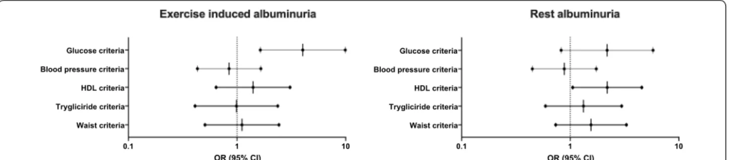
Finally, we wanted to determine which parameters of the metabolic syndrome were associated with the observed increase in EiA levels over time. Elevated
fast-ing blood glucose criteria (≥ 100 mg/dl) was the
strong-est and only significant predictor for an elevated increase in EiA over time after adjusting for all five metabolic syn-drome components; blood glucose, waist circumference, blood triglycerides, HDL cholesterol, and blood pressure criteria (Fig. 3). When comparing EiA to rest albuminuria measurements, elevated blood glucose was not signifi-cantly associated with increased changes in rest UACR,
while decreased HDL cholesterol was the only significant predictor in the rest model (Fig. 3).
Notably, although patients with hypertension had higher EiA levels compared with normotensive subjects
on both visits (median [IQR] = 8 mg/g [0–32] vs 3.5 mg/g
[− 0.2 to 15.8] (p = 0.04) on the first visit and 4.2 [0.3– 19.2] vs 2.2 [− 0.2 to 12.3] (p = 0.08) on the second visit), they did not change their EiA levels over time differently
than normotensive individuals; change in EiA =− 1.9
[− 17.9 to 5.9] vs. − 0.8 [− 10.9 to 5.1] for median [IQR]
(p =0.36) respectively. Hypertensive patients taking
antihypertensive medications (n = 58) had a greater
reduction in EiA between the two visits compared with untreated individuals, albeit not statistically signifi-cant; change in EiA between visits =− 3.1 mg/g [− 24.1 to 11.5] vs. − 1.6 mg/g [− 12.9 to 4.2] for median [IQR] -60 -40 -20 0 20 40 60 80 100 120
-60 -40 -20 0 20 40 60 80 100 120
EiA first visit, mg/g
EiA second visit, mg/g
Correlation of EiA Between visits
Elevated EiA
Elevated EiA
Spearman’s r=0.4 p<0.01
7.4
21.4 21.6
37.5
Normal EiA both visits (n=269)
Elevated EiA only first visit
(n=58)
Elevated EiA only second visit
(n=37)
Elevated EiA both visits
(n=48)
0% 10% 20% 30% 40%
% of Patients with Metabolic Syndrome
Chi-square p< 0.01
a b
Fig. 1 Exercise‑induced albuminuria levels on two consecutive visits. a A scatter plot of exercise‑induced albuminuria (EiA) levels during a
participant’s first (X axis) and second (Y axis) visit. Dotted lines mark the threshold for elevated EiA (> 20 mg/g, bottom limit of top quartile). P value by spearman’s r test. Point color marks are according to the four groups shown in b. b Bar graph depicting the percent of patients diagnosed with metabolic syndrome during their first visit, according to their EiA status (elevated vs normal levels). Colors correspond to the scatter plot in a. p value by Chi square. EiA Exercise‑induced albuminuria
-100 -80 -60 -40 -20 0 20 40 60 80 100 0
25 50 75 100 125
Change in EiA between Visits, mg/g
Frequenc
y
N=412 Median= -0.9 IQR [-12.1, 5.1]
No Yes
-100 -75 -50 -25 0 25 50 75 100
Diabetes Diagnosis
Change in EiA between visits, mg/g
Mann Whitney p=0.049
< 5.7 5.7-6.4 > 6.5 -100
-75 -50 -25 0 25 50 75 100
HbA1c, %
Change in EiA between visits, mg/g
Kruskal Wallis p<0.01
a b c
Fig. 2 Change in exercise‑induced albuminuria levels over time. a A histogram of the change in exercise‑induced albuminuria (EiA) levels among
(p = 0.7). The majority of treated patients used angioten-sin converting enzyme inhibitors (ACEi) and/or
angio-tensin receptor blockers (ARBs) (70%) (Table 1). ACEi
and ARBs have been shown to reduce albuminuria levels
[24] and this might explain the greater reduction in EiA
over time among treated patients.
Discussion
The main finding of this study is that patients with dia-betes and/or elevated fasting blood glucose increase their exercise-induced urinary albumin excretion over time. Metabolic dysfunction was associated with consist-ent elevated levels of exercise induced albuminuria in a cohort of young and apparently healthy individuals with-out overt kidney disease.
Excessive urinary albumin excretion following exercise has been reported in patients with metabolic syndrome [7], including patients with hypertension [8] and type 2 diabetes [9, 10]. We present here, for the first time, a pro-spective study that characterizes EiA variation over time in patients with and without metabolic dysfunction. Our findings show that EiA levels are relatively stable among healthy individuals and do not change significantly over a mean follow-up period of 3 years. However, metabolic dysfunction, and more specifically impaired glucose metabolism seems to drive an increase in urinary albu-min excretion following exercise which continues to ele-vate over time. These differences between patients with and without altered glucose metabolism were not seen using standard rest albuminuria measurements which stayed relatively stable during follow up.
The current study is in line with our past reports on urinary albumin excretion following exercise and its association with multiple risk factors for CVD [8, 22, 25]. Measuring EiA is a potential new sensitive and early bio-marker for the kidney’s “reservoir capacities”, endothelial
dysfunction and other metabolic related end organ dam-age. Results of future reports from this study cohort and others on the ability of EiA to predict major renal and cardiovascular events are highly anticipated. Whether EiA measurements provide additional clinical utility in stratifying risk as opposed to standard rest UACR calcu-lations is still unknown.
A reduction in albuminuria is related to favorable car-diovascular and renal outcomes, especially in patients with diabetes [26–28]. While novel treatment strategies for cardio-renal outcomes such as SGLT-2 inhibitors are effective in lowering rest albuminuria [29, 30], their effect on exercise induced excretion of albumin is not entirely clear. We suggest that monitoring exercise induced levels of albuminuria may serve as a screening tool for pre-dia-betic and diapre-dia-betic individuals who require early initiation of such renal protective therapies [31, 32].
Our study has several limitations. First, the relatively low prevalence of pre-existing co-morbidities in our cohort limit our ability to describe the kinetics of EiA in patients at high risk for CVD or patients with overt kid-ney disease. Future prospective studies in older patients with evident CVD and renal disease are needed to bet-ter define the “normal” range of EiA and its variation over an individual’s life span. Furthermore, the major-ity of our study population improved their metabolic profile during follow up (reduced BMI, total cholesterol and triglycerides) which may imply that these individu-als adopted a healthier life style after their first visit to our annual heath survey. This trend cannot necessar-ily be generalized to other populations. Nonetheless, the group of individuals who regressed their EiA from abnormal values at baseline to normal values on the second visit were the ones who had the greatest reduc-tion in fasting blood glucose levels. This strengthens our main hypothesis that glucose metabolism is the
0.1 1 10
Waist criteria Trygliciride criteria HDL criteria Blood pressure criteria Glucose criteria
OR (95% CI) 0.1 1 10
Waist criteria Trygliciride criteria HDL criteria Blood pressure criteria Glucose criteria
OR (95% CI)
Fig. 3 Multivariate analysis of metabolic syndrome components to predict a high increase in EiA and rest albuminuria over time. We ran a
multivariate binary logistic regression to predict a high increase in EiA (left side) and rest albuminuria (right side) over time (top 10th percentile). The forest plots present the odds ratio (OR) and 95% confidence intervals of each metabolic syndrome component (see definition criteria in “Methods” section). Glucose criteria (elevated fasting blood glucose) was the only significant predictor in the EiA model (OR = 4.0 95% CI = 1.6–9.8, p < 0.01) while presence of the HDL‑cholesterol criteria precited a greater increase in rest measurements (OR = 2.2 95% CI = 1.01–4.6, p = 0.04). EiA
main predictor of EiA in presumably healthy adults. A second limitation is that part of our observed changes in EiA over time could be attributed to the
‘regres-sion to the mean phenomenon’ [33] which needs to be
accounted for whenever assessing changes in a biologi-cal measurement over time. And yet, although patients with diabetes have consistent higher levels of EiA
com-pared with normo-glycemic individuals [22], they still
demonstrated a higher increase in EiA over time. The majority of diabetic patients did not “regress” down to the mean, thus highlighting the presence of a true bio-logical driver for increasing EiA in this sub-population.
It should be noted that our current threshold for an increased change in EiA (top 10th percentile) was sta-tistically driven in order to study the extremities of the cohort and will have to be adjusted in future prospec-tive studies that will measure clinical outcomes.
Finally, the current study cohort was notably male dominant (85% men) and limited our ability to deter-mine statistically significant sex-and-gender related dif-ferences in EiA levels over time. Our past report shows that elevated EiA is associated with abnormal ECG findings in apparently healthy women rather than men
[22]. Thus, future studies should strive towards
imple-menting sex-specific thresholds for EiA levels which may lead to better risk stratification of kidney and CVD.
Conclusion
We conclude that individuals with diabetes and/or ele-vated fasting blood glucose increase their EiA levels over time. EiA should be further studied as an early and sen-sitive biomarker for kidney reserve and endothelial dys-function in patients with metabolic syndrome.
Abbreviations
BMI: Body mass index; CVD: Cardiovascular disease; EiA: Exercise induced albuminuria; HbA1c: Glycated hemoglobin; ECG: Electrocardiogram; UACR : Urinary albumin to creatinine ratio; eGFR: Estimated glomerular filtration rate; HDL: High‑density lipoprotein‑cholesterol; LDL: Low‑density lipoprotein‑cho‑ lesterol; METs: Metabolic equivalents score; SD: Standard deviation; ANOVA: Analysis of variance; CI: Confidence interval; OR: Odds ratio; IQR: Inter quartile range.
Acknowledgements
None.
Authors’ contributions
RYB, SB, AG and SST participated in study conception and design. LF, DZ and IS performed the acquisition of data. SST, RB and AG participated in analysis and interpretation of data. RYB, AG and SST drafted the manuscript and AG, LF, SST, OR and SB helped in critical review of the manuscript. All authors read and approved the final manuscript.
Funding
None.
Availability of data and materials
The data that support the findings of this study are available from the cor‑ responding author upon reasonable request.
Ethics approval and consent to participate
This study was conducted after the acquisition of written informed consent from the participating patients and upon the approval by the ethics commit‑ tee of the Tel Aviv Sourasky Medical Center Institutional Review Board (Helsinki Committee).
Consent for publication
If the manuscript is accepted, we approve it for publication in Cardiovascular Diabetology.
Competing interests
The authors declare that they have no competing interests.
Author details
1 Department of Internal Medicine “C”, “D” and “E”, Tel Aviv Sourasky Medical
Center, 6 Weizmann Street, 64239 Tel Aviv, Israel. 2 Sackler Faculty of Medicine,
Tel Aviv University, Tel Aviv, Israel. 3 Neufled Cardiac Research Institute, Sackler
Faculty of Medicine, Tel Aviv University, Tel Aviv, Israel. 4 Tamman Cardiovascu‑
lar Research Institute, Sheba Medical Center, Tel Hashomer, Israel. 5 Nephrology
Department, Tel‑Aviv Sourasky Medical Center, Tel Aviv, Israel.
Received: 6 April 2020 Accepted: 8 June 2020
References
1. Nakagawa T, Tanabe K, Croker BP, Johnson RJ, Grant MB, Kosugi T, et al. Endothelial dysfunction as a potential contributor in diabetic nephropa‑ thy. Nat Rev Nephrol. 2011;7:36–44.
2. Stevens PE, Levin A, Kidney Disease: Improving Global Outcomes Chronic Kidney Disease Guideline Development Work Group Members. Evalua‑ tion and management of chronic kidney disease: synopsis of the kidney disease: improving global outcomes 2012 clinical practice guideline. Ann Intern Med. 2013;158:825–30.
3. Sung K, Ryu S, Lee J, Lee SH, Cheong E, Hyun Y, et al. Urine albumin/cre‑ atinine ratio below 30 mg/g is a predictor of incident hypertension and cardiovascular mortality. J Am Heart Assoc. 2016;5:e003245.
4. Ärnlöv J, Evans JC, Meigs JB, Wang TJ, Fox CS, Levy D, et al. Low‑grade albuminuria and incidence of cardiovascular disease events in nonhyper‑ tensive and nondiabetic individuals. Circulation. 2005;112:969–75. 5. Kweon S‑S, Shin M‑H, Lee Y‑H, Choi J‑S, Nam H‑S, Park K‑S, et al. Higher
normal ranges of urine albumin‑to‑creatinine ratio are independently associated with carotid intima‑media thickness. Cardiovasc Diabetol. 2012;11:112.
6. Wagner R, Machann J, Lehmann R, Rittig K, Schick F, Lenhart J, et al. Exercise‑induced albuminuria is associated with perivascular renal sinus fat in individuals at increased risk of type 2 diabetes. Diabetologia. 2012;55:2054–8.
7. Greenberg S, Shenhar‑Tsarfaty S, Rogowski O, Shapira I, Zeltser D, Weinstein T, et al. Exercise‑induced albuminuria is related to metabolic syndrome. Am J Physiol Physiol. 2016;310:1192.
8. Grupper A, Ehrenwald M, Schwartz D, Berliner S, Shashar M, Baruch R, et al. Hypertension is associated with increased post‑exercise albumi‑ nuria, which may be attenuated by an active lifestyle. J Clin Hypertens. 2019;21:1171–9.
9. Climie RED, Srikanth V, Keith LJ, Davies JE, Sharman JE. Exercise excess pressure and exercise‑induced albuminuria in patients with type 2 diabe‑ tes mellitus. Am J Physiol Circ Physiol. 2015;308:H1136–42.
10. Tankeu AT, Kaze FF, Noubiap JJ, Chelo D, Dehayem MY, Sobngwi E. Exercise‑induced albuminuria and circadian blood pressure abnormali‑ ties in type 2 diabetes. World J Nephrol. 2017;6:209.
11. Jun M, Ohkuma T, Zoungas S, Colagiuri S, Mancia G, Marre M, et al. Changes in albuminuria and the risk of major clinical outcomes in diabe‑ tes: results from ADVANCE‑ON. Diabetes Care. 2018;41:163–70. 12. Milwidsky A, Steinvil A, Shapira I, Letourneau‑Shesaf S, Limor R,
•fast, convenient online submission
•
thorough peer review by experienced researchers in your field
• rapid publication on acceptance
• support for research data, including large and complex data types
•
gold Open Access which fosters wider collaboration and increased citations maximum visibility for your research: over 100M website views per year
•
At BMC, research is always in progress.
Learn more biomedcentral.com/submissions
Ready to submit your research? Choose BMC and benefit from: is associated with elevated cardiac troponin T concentrations. J Metab
Syndr. 2014;03:1–5.
13. Brzezinski RY, Fisher E, Cohen N, Zwang E, Shefer G, Stern N, et al. Total serum cholinesterase activity predicts hemodynamic changes during exercise and associates with cardiac troponin detection in a sex‑depend‑ ent manner. Mol Med. 2018;24:63.
14. Rogowski O, Shapira I, Shirom A, Melamed S, Toker S, Berliner S. Heart rate and microinflammation in men: a relevant atherothrombotic link. Heart. 2007;93:940–4.
15. Steinvil A, Shirom A, Melamed S, Toker S, Justo D, Saar N, et al. Relation of educational level to inflammation‑sensitive biomarker level. Am J Cardiol. 2008;102:1034–9.
16. Brzezinski RY, Fisher E, Ehrenwald M, Shefer G, Stern N, Shapira I, et al. Elevated high‑sensitive troponin T in negative stress test individuals. Eur J Clin Invest. 2018;48:e12930.
17. Members Task Force, Montalescot G, Sechtem U, Achenbach S, Andreotti F, Arden C, et al. ESC guidelines on the management of stable coronary artery disease: the Task Force on the management of stable coronary artery disease of the European Society of Cardiology. Eur Heart J. 2013;2013(34):2949–3003.
18. Datta P, Dasgupta A. An improved microalbumin method (microALB_2) with extended analytical measurement range evaluated on the ADVIA chemistry systems. J Clin Lab Anal. 2009;23:314–8.
19. Foundation NK. KDOQI clinical practice guideline for diabetes and CKD: 2012 update. Am J Kidney Dis. 2012;60:850–86.
20. Levey AS, Stevens LA, Schmid CH, Zhang YL, Castro AF, Feldman HI, et al. A new equation to estimate glomerular filtration rate. Ann Intern Med. 2009;150:604–12.
21. Association AD. Standards of medical care in diabetes‑2017. Diabetes Care. 2017;2017(40 Suppl 1):S1–20.
22. Brzezinski RY, Etz‑Hadar I, Grupper A, Ehrenwald M, Shapira I, Zeltser D, et al. Sex difference in the risk for exercise‑induced albuminuria correlates with hemoglobin A1C and abnormal exercise ECG test findings. Cardio‑ vasc Diabetol. 2017;16:79.
23. Alberti KGMM, Eckel RH, Grundy SM, Zimmet PZ, Cleeman JI, Donato KA, et al. Harmonizing the metabolic syndrome. Circulation. 2009;120:1640–5. 24. Galle J. Reduction of proteinuria with angiotensin receptor blockers. Nat
Clin Pract Cardiovasc Med. 2008;5:S36–43.
25. Milwidsky A, Fisher E, Brzezinski RY, Ehrenwald M, Shefer G, Stern N, et al. Metabolic syndrome is associated to high‑sensitivity cardiac troponin T elevation. Biomarkers. 2019;24:153–8.
26. Kluger AY, Tecson KM, Lee AY, Lerma EV, Rangaswami J, Lepor NE, et al. Class effects of SGLT2 inhibitors on cardiorenal outcomes. Cardiovasc Diabetol. 2019;18:99.
27. Williams DM, Nawaz A, Evans M. Renal outcomes in type 2 diabetes: a review of cardiovascular and renal outcome trials. Diabetes Ther. 2020;11:369–86.
28. Piperidou A, Sarafidis P, Boutou A, Thomopoulos C, Loutradis C, Alex‑ androu ME, et al. The effect of SGLT‑2 inhibitors on albuminuria and proteinuria in diabetes mellitus: a systematic review and meta‑analysis of randomized controlled trials. J Hypertens. 2019;37:1334–43.
29. Dekkers CCJ, Petrykiv S, Laverman GD, Cherney DZ, Gansevoort RT, Heer‑ spink HJL. Effects of the SGLT‑2 inhibitor dapagliflozin on glomerular and tubular injury markers. Diabetes Obes Metab. 2018;20:1988–93. 30. Kinguchi S, Wakui H, Ito Y, Kondo Y, Azushima K, Osada U, et al. Improved
home BP profile with dapagliflozin is associated with amelioration of albuminuria in Japanese patients with diabetic nephropathy: the Yokohama add‑on inhibitory efficacy of dapagliflozin on albuminuria in Japanese patients with type 2 diabetes study (Y‑AIDA study). Cardiovasc Diabetol. 2019;18:110.
31. Bramlage P, Lanzinger S, Van Mark G, Hess E, Fahrner S, Heyer CHJ, et al. Patient and disease characteristics of type‑2 diabetes patients with or without chronic kidney disease: an analysis of the German DPV and DIVE databases. Cardiovasc Diabetol. 2019;18:33.
32. Leite KM, Long AM, Ostroff ML, Borges L, Braden G. A review of the reno‑ protective effects of novel antidiabetic agents. J Pharm Pract. 2020. https
://doi.org/10.1177/08971 90020 90234 4.
33. Barnett AG, van der Pols JCDA. Regression to the mean: what it is and how to deal with it. Int J Epidemiol. 2004;34:215–20.
Publisher’s Note