ORIGINAL INVESTIGATION
Effect of liraglutide on cardiac function
in patients with type 2 diabetes mellitus:
randomized placebo-controlled trial
Maurice B. Bizino
1*, Ingrid M. Jazet
2, Jos J. M. Westenberg
1, Huub J. van Eyk
2, Elisabeth H. M. Paiman
1,
Jan W. A. Smit
3and Hildebrandus J. Lamb
1Abstract
Background: Liraglutide is an antidiabetic agent with cardioprotective effect. The purpose of this study is to test efficacy of liraglutide to improve diabetic cardiomyopathy in patients with diabetes mellitus type 2 (DM2) without cardiovascular disease.
Methods: Patients with DM2 were randomly assigned to receive liraglutide 1.8 mg/day or placebo in this double-blind trial of 26 weeks. Primary outcome measures were LV diastolic function (early (E) and late (A) transmitral peak flow rate, E/A ratio, early deceleration peak (Edec), early peak mitral annular septal tissue velocity (Ea) and estimated LV filling pressure (E/Ea), and systolic function (stroke volume, ejection fraction, cardiac output, cardiac index and peak ejection rate) assessed with CMR. Intention-to-treat analysis of between-group differences was performed using ANCOVA. Mean estimated treatment differences (95% confidence intervals) are reported.
Results: 23 patients were randomized to liraglutide and 26 to placebo. As compared with placebo, liraglutide significantly reduced E (− 56 mL/s (− 91 to − 21)), E/A ratio (− 0.17 (− 0.27 to − 0.06)), Edec (− 0.9 mL/s2 * 10−3 (
− 1.3 to − 0.2)) and E/Ea (− 1.8 (− 3.0 to − 0.6)), without affecting A (3 mL/s (− 35 to 41)) and Ea (0.4 cm/s (− 0.9 to 1.4)). Liraglutide reduced stroke volume (− 9 mL (− 16 to − 2)) and ejection fraction (− 3% (− 6 to − 0.1)), but did not change cardiac output (− 0.4 L/min (− 0.9 to 0.2)), cardiac index (− 0.1 L/min/m2 (
− 0.4 to 0.1)) and peak ejection rate (− 46 mL/s (− 95 to 3)).
Conclusions: Liraglutide reduced early LV diastolic filling and LV filling pressure, thereby unloading the left ventricle. LV systolic function reduced and remained within normal range. Future studies are needed to investigate if liraglutide-induced left ventricular unloading slows progression of diabetic cardiomyopathy into symptomatic stages.
Trial registration ClinicalTrials.gov: NCT01761318.
Keywords: Diabetes mellitus type 2, Liraglutide, GLP1-receptor agonist, Diastolic heart failure, Cardiac function
© The Author(s) 2019. This article is distributed under the terms of the Creative Commons Attribution 4.0 International License (http://creat iveco mmons .org/licen ses/by/4.0/), which permits unrestricted use, distribution, and reproduction in any medium, provided you give appropriate credit to the original author(s) and the source, provide a link to the Creative Commons license, and indicate if changes were made. The Creative Commons Public Domain Dedication waiver (http://creat iveco mmons .org/ publi cdoma in/zero/1.0/) applies to the data made available in this article, unless otherwise stated.
Introduction
Patients with type 2 diabetes mellitus (DM2) are at increased risk for heart failure, even in the absence of coronary artery disease and hypertension. This so-called diabetic cardiomyopathy is characterized by left ventric-ular (LV) diastolic dysfunction [1] and has an estimated
prevalence of approximately 50% [2]. When heart failure symptoms have developed, most patients with diabetic cardiomyopathy are classified as heart failure with pre-served ejection fraction (HFpEF). HFpEF poses patients with DM2 at a very high morbidity [3] and mortality risk [4]. Therefore, early detection followed by medi-cal therapy to reverse LV diastolic dysfunction seems an attractive goal in diabetes management. Although intense glycaemic control is a primary tool to reduce diabetes complications, tight glucoregulation by itself does not seem to improve LV diastolic function [5]. Nor are there
Open Access
*Correspondence: [email protected]
1 Department of Radiology, Leiden University Medical Center, LUMC
any specific drugs besides diuretics that can be used to treat or prevent HFpEF [6].
The anti-diabetic agent liraglutide is a glucagon-like peptide-1 receptor agonist (GLP-1RA) that improves insulin secretion, suppresses glucagon production and induces weight loss. Although some studies have inves-tigated the effect of GLP-1RA on ischemic heart disease and symptomatic heart failure with reduced ejection fraction (HFrEF) [7], little is known about the effect on LV diastolic function. GLP-1RA induced weight loss by itself might improve LV diastolic function [8]. In addi-tion, a direct cardio-protective effect of GLP-1RA ther-apy has been suggested by preclinical studies and in some but not all human studies [9].
Cardiac magnetic resonance imaging (CMR) has been shown to enable accurate assessment of LV diastolic and systolic function with very high reproducibility [10, 11]. Therefore, the purpose of this randomized placebo con-trolled trial was to evaluate the effect of the GLP1-RA liraglutide on CMR-derived indices of cardiac function in patients with DM2 without prior cardiovascular disease.
Methods
Study design and participants
The MAGNA VICTORIA (MAGNetic resonance Assess-ment of VICTOza efficacy in the Regression of cardio-vascular dysfunction In type 2 diAbetes mellitus) study was an investigator-initiated randomized, double-blind, assessor-blinded, placebo-controlled, single-center clini-cal trial with 26 weeks follow-up. Men and women with DM2 were eligible if aged 18–69 years. Inclusion crite-ria were: BMI 25 kg/m2 or above; glycated haemoglobin (HbA1c) level of 7.0 to 10.0% (53–86 mmol/mol) despite use of maximally tolerable metformin treatment, with or without sulphonyurea derivative (SUD) and/or insulin, with stable dosage in the 3 months before study entry; blood pressure < 150/85 mmHg and stable for at least 1 month. Due to lack of eligible patients use of SUD and insulin was added to inclusion criteria after commence-ment of the trial. Exclusion criteria were: use of other glucose-lowering therapy than mentioned above; history or presence of renal, hepatic or cardiovascular disease; gastric bypass surgery; chronic pancreatitis or previous acute pancreatitis; pregnant or lactating women; and contra-indications for MRI. The trial was approved by the local ethics committee and performed in accordance with the principles of the revised Declaration of Helsinki. Written informed consent was obtained from all par-ticipants before study entry. The trial was conducted at the Leiden University Medical Center (LUMC), Leiden, the Netherlands, and was registered at clinicaltrials.gov (NCT01761318).
Study procedures
Screening visit
Participants underwent pre-screening by telephone to assess eligibility on the basis of drug use, medical his-tory, and anthropometric measures. If potentially eligi-ble, participants were submitted to a screening visit with detailed history taking with special interest to cardiovas-cular symptoms and presence of neuropathy (peripheral sensory neuropathy as detected by monofilament testing and/or erectile dysfunction), nephropathy (micro-albu-minuria) and retinopathy. Height, weight and blood pres-sure were meapres-sured and physical examination, resting electrocardiogram (ECG) and blood examination were performed. The nonattendance of cardiovascular disease was defined as absence of symptoms related to coronary artery disease and heart failure and normal ECG.
Randomization and masking
Patients were randomized to liraglutide (Victoza, Novo Nordisk A/S, Bagsvaerd, Denmark) or placebo (provided by Novo Nordisk A/S, Bagsvaerd, Denmark) once daily subcutaneous injections, added to their pharmacologic treatment at study entry. Participants were randomized with 1:1 stratification for sex and insulin use (block size of 4) to increase likelihood of comparable groups given the relatively low sample size. Randomization was performed by the local research pharmacist (Department of Clinical Pharmacy, LUMC, Leiden, The Netherlands) after inves-tigator had provided information on sex and insulin use (directly after results of the screening visit). All investi-gators, study personnel and participants were blinded to treatment allocation until the study had been completed (including CMR post-processing and analyses).
Study protocol
supine position) were measured at each study visit. Dur-ing the study, glycaemic drugs were titrated accordDur-ing to clinical practice guidelines by means of dose adjustment of insulin and/or SUD. Adjustment of antihypertensive and lipid-lowering drugs were made if necessary. At study entry, week 12 and at end of study, blood examinations were performed after at least six hours of fasting. HbA1c was measured with boronate affinity high-performance liquid chromatography (Primus Ultra, Siemens Health-care Diagnostics, Breda, the Netherlands) throughout the first part of the study. The laboratory chose to change their HbA1c measurement method for logistic reasons while our study was ongoing. The method was changed into ion-exchange high-performance liquid chromatog-raphy (HPLC) (Tosoh G8, Sysmex Nederland B.V., Etten-Leur, the Netherlands). HbA1c values were corrected on the basis of the correlation coefficient that was derived from a validation experiment that used data of 196 sam-ples that were measured on both analysers (data can be provided on request). All other blood samples were cen-trifuged and stored at − 80 °C until analysis. Serum cre-atinine, triglyceride, total cholesterol, HDL-cholesterol, LDL-cholesterol (Friedewald formula) and N-terminal prohormone of brain natriuretic peptide (NTproBNP) concentrations were measured on a Modular P800 ana-lyser (Roche Diagnostics, Mannheim, Germany).
CMR protocol
All participants underwent a cardiac MRI protocol using a clinical 3 Tesla Ingenia whole-body MR system (Philips Medical Systems, Best, the Netherlands) at baseline and follow-up. Subjects were scanned in supine position. The body coil was used for transmission, and reception was achieved with a 16-element anterior, and a 12-element posterior array. The heart was imaged in 2-chamber, 4-chamber and short-axis views with ECG-gated breath-hold balanced steady state free-precession cine imaging. Then, whole-heart 4D velocity encoded flow MRI was performed as described elsewhere [12]. For visualisa-tion of prior myocardial scarring, a free-breathing high spatial resolution delayed enhancement phase-sensitive inversion recovery sequence was acquired after intra-venous administration of gadolinium contrast material (0.3 mL/kg, Dotarem; Guerbet, Bloomington, USA) [13]. All images were blinded for study participant and occa-sion (baseline or follow-up). Image post-processing was performed using validated MASS software (LUMC, Lei-den, the Netherlands). LV diastolic function comprised of early peak mitral annular septal tissue velocity (Ea in cm/s) which was analysed with the use of 4 chamber long-axis view. Early (E) and late (A) peak transmitral flow rate (in mL/s) and E/A ratio were analysed using 4D flow dataset with retrospective valve tracking [12]. E
deceleration peak (Edec) was defined as the maximum downward slope of early peak flow rate. E (in cm/s, with-out background subtraction) divided by Ea is a validated estimate of LV filling pressure [14]. Short axis cine images were used to measure LV systolic function parameters: stroke volume, ejection fraction, cardiac output and car-diac index (carcar-diac output/body surface area). The LV systolic function parameter peak ejection rate was meas-ured with 4D flow MRI. The heart rate during MRI scan was chosen to report because that heart rate most closely reflects cardiac dynamics as assessed with MRI. LV filling volume was analysed with 4D flow. LA volume was calcu-lated using Simpsons rule [10] and then divided by body surface area to obtain LA volume index. LV end-diastolic volume (LVEDV), LV end-systolic volume (LVESV) and LV mass (LVM) were all obtained from short axis cine imaging studies. Parameters LV mass index (LV mass/ body surface area) and LVMI/LVEDVI (LV mass index/ LVEDV index) and LV compliance (LVEDV/E/Ea) were calculated.
Study endpoints
Since integrative assessment of cardiac function encom-passes both LV diastolic and systolic indices, all were marked as primary endpoint. However, for sample size calculation (see below) Edec and LV ejection fraction were used. Predefined secondary endpoint were blood pressure, body weight, HbA1c, LVEDV, LVESV, LVM, LVMI, LVMI/LVEDVI. Other pre-specified endpoints were creatinine and NTproBNP. Endpoints that were not predefined were heart rate, LV filling volume, LA volume, LA volume index and LV compliance. We chose to report these endpoints for interpretation purposes.
Statistical analysis
of change from baseline with randomization arm as the independent variable and the baseline measurement of dependent variable as a covariate. Statistical analyses were performed using SPSS version 23.0 for Windows (IBM Corporation, Chicago, IL). A P value < 0.05 was considered statistically significant.
Role of the funding source
Novo Nordisk (Denmark) funded this investigator-initi-ated study. Novo Nordisk had no role in the design of the study, data collection, data analysis, data interpretation, or writing of the report. All authors had access to all the data and final responsibility for the decision to submit for publication.
Results
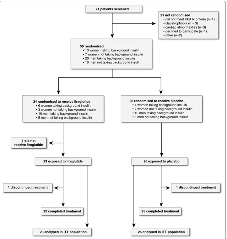
Participants were enrolled between December 2013 and September 2015 with last patient last visit in March 2016. Figure 1 shows the trial profile and baseline character-istics are shown in Table 1. Sex, insulin use, age, blood pressure, use of anti-hypertensive drugs, lipid levels, smoking history and glycaemic control were compa-rable in both groups. Liraglutide patients had slightly higher BMI (+ 1.0 kg/m2). There was an uneven
distribu-tion of nephropathy (9% in liraglutide versus 42% in pla-cebo group). With regard to primary outcome measures there was slightly higher E/A ratio (+ 0.05) and lower E/
Ea (− 0.6) in liraglutide versus placebo at baseline. In the
liraglutide group uptitration was delayed in five patients versus none in placebo group, and study drug dose that patients used was 0.6 mg (n = 2); 1.2 mg (n = 3) and
1.8 mg (n = 18). In placebo group, no patient had delayed
uptitration and all patients used 1.8 mg once daily. The cumulative prescribed study drug dose was 278.4 ± 45 mg
in liraglutide versus 302.4 ± 13.8 mg in placebo with
com-pliance of 98% (± 3) versus 96% (± 4).
Concomitant glucose‑lowering drugs
In liraglutide group use of SUD decreased from 26% at baseline to 18% at 26 weeks, and the use of insulin decreased from 70 ± 46 to 54 ± 43 IU/day (percentage of participants on insulin therapy decreased from 65 to 64%). In placebo group use of SUD increased from 31 to 40%. Number of insulin users increased from 65 to 72% with average daily dose of 69 IU at baseline and 69 IU at 26 weeks.
Anthropometric and laboratory values
Liraglutide group had significantly more weight loss than placebo group (− 4.3 ± 3.8 kg vs 0.1 ± 2.5 kg, p < 0.001). Systolic and diastolic blood pressure changes were not different amongst treatment groups (p = 0.63
and p = 0.23 respectively). In both liraglutide and pla-cebo treated patients an improvement in glycaemic con-trol was noticed. In liraglutide group HbA1c decreased 1.1 ± 1.0% (11.6 ± 11.1 mmol/mol) versus 0.7 ± 0.9% (7.7 ± 9.4 mmol/mol) decline in placebo group, with no significant difference between group changes (esti-mated mean treatment difference: − 2.9 with 95% CI from − 8.1 to 2.3 mmol/mol, p = 0.27). Serum creati-nine slightly increased in both treatment groups but there was no difference between group changes (liraglu-tide: + 4 ± 5 μmol/L; placebo: + 4 ± 5 μmol/L, p = 0.69). NTproBNP levels declined from 45 ± 30 to 37 ± 18 pg/ mL in liraglutide group, and increased in placebo group from 39 ± 29 to 45 ± 29 pg/mL, with estimated mean treatment difference of − 10 pg/mL with 95% CI between − 20 and 1 pg/mL, p = 0.07.
Magnetic resonance imaging and spectroscopy
In one patient in the liraglutide group a small area of delayed contrast enhancement was noted in the infer-oposterior basal segment. On further examination by cardiologist there was no sign of cardiac ischemia during exercise testing. All other patients had no late gadolinium enhancement.
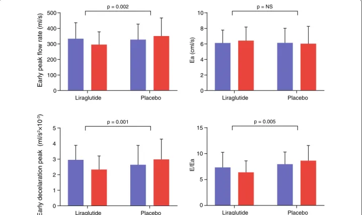
Primary endpoints are shown Table 2 and Fig. 2. LV diastolic function indices that changed significantly between groups were E, E/A ratio, Edec and E/Ea. All these parameters were reduced by liraglutide, as com-pared with placebo. A and Ea were not affected by treat-ment. LV systolic function parameters that changed significantly between groups were stroke volume and ejection fraction. Despite a reduction in these param-eters, cardiac output and cardiac index did not change between groups, due to increased heart rate (Fig. 3).
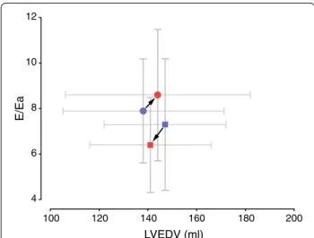
Table 3 displays non-primary outcome measures. In line with reduced stroke volume, the LV filling volume is also reduced in liraglutide as compared to placebo. Furthermore, LVM significantly decreased in liraglutide compared to placebo, but when corrected for reduced body surface area (LVMI) this difference did not persist. LVEDV was significantly reduced by liraglutide as com-pared to placebo treated patients. LV compliance showed a non-significant trend towards increased compliance in liraglutide versus placebo (Fig. 4).
Safety
71 patients screened
23 exposed to liraglutide
22 completed treatment
23 analysed in ITT population 1 did not
receive liraglutide
26 exposed to placebo 50 randomised
• 13 women taking background insulin • 7 women not taking background insulin • 20 men taking background insulin • 10 men not taking background insulin
1 discontinued treatment 1 discontinued treatment
25 completed treatment
26 analysed in ITT population 24 randomised to receive liraglutide
• 6 women taking background insulin • 3 women not taking background insulin • 10 men taking background insulin • 5 men not taking background insulin
26 randomised to receive placebo
• 4 women taking background insulin • 7 women not taking background insulin • 10 men taking background insulin • 5 men not taking background insulin
21 not randomised
• did not meet HbA1c criteria (n=12) • claustrophobia (n = 3)
• cardiac abnormalities (n=3) • declined to participate (n=1) • other (n=2)
origin. There were no cases of acute pancreatitis during the study period.
Discussion
This study shows that in patients with type 2 diabetes mellitus without prior cardiovascular disease, 6-month treatment with liraglutide improved E/Ea, as compared with placebo added to standard care. As such, liraglu-tide beneficially influenced a key pathogenic hallmark of HFpEF: left ventricular filling pressure. Liraglutide did not improve left ventricular myocardial relaxation (Ea). Liraglutide reduced left ventricular systolic func-tion parameters stroke volume and ejecfunc-tion fracfunc-tion, and these remained within normal range.
Interpretation
Diabetes with or without the presence of hypertension is independently associated with abnormal LV dias-tolic filling pattern [1], i.e. diabetic cardiomyopathy. The asymptomatic stage can persist during years or decades, but once symptomatic heart failure has developed, pro-gressive impairment of myocardial relaxation results in compensatory rise in E/Ea to ensure sufficient LV filling during diastole. These final stages of HFpEF are charac-terized by impaired quality of life and life expectancy [6]. The early asymptomatic stage with prevalence up to 50% [2] therefore seems a window of opportunity to reverse or delay progression of diabetic cardiomyopathy. However, there are no pharmacologic agents that have unequivo-cally shown benefit in HFpEF patients [17]. An anti-dia-betic agent that positively affects HFpEF indices would therefore be of great clinical importance. In that regard, the observed reduction in E/Ea, is a promising prospect. Elevated filling pressure has been shown to indepen-dently predict progression of HFpEF in patients with DM2 [18]. Possible underlying cardiac pathologic mecha-nisms include wall stress, diffuse cardiac fibrosis and LV hypertrophy [19]. Liraglutide seems to positively affect these pathologic pathways, as evidenced by reduced E/ Ea, LVM, and a trend towards improved LV compliance and NTproBNP levels, as compared to placebo. As such, it might be postulated that initiation of liraglutide treat-ment in the early asymptomatic stage of diabetic cardio-myopathy, could delay the onset of clinically significant HFpEF. With regard to systolic function, we hypothesize that reduced LV filling volume directly results in reduced stroke volume and ejection fraction. The modest decline of ejection fraction is not considered clinically relevant in this specific study cohort, because it remained within normal range [11]. Furthermore, cardiac output and car-diac index did not change due to rise in heart rate which is a well-documented finding in studies with GLP-1RA therapy [9].
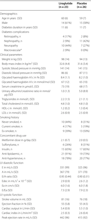
Table 1 Baseline characteristics of trial population
E early transmitral peak flow rate, A late transmitral peak flow rate, Edec peak deceleration of transmitral early peak flow, Ea early peak mitral annular septal tissue velocity
a Macrovascular complications were cerebrovascular or peripheral artery
disease and not cardiovascular
Liraglutide
(n = 23) Placebo (n = 26)
Demographics
Age in years (SD) 60 (6) 59 (7)
Male 14 (61%) 15 (58%)
Diabetes duration in years (SD) 11 (6) 11 (7) Diabetes complications
Retinopathy, n 4 (17%) 2 (8%)
Nephropathy, n 2 (9%) 11 (42%)
Neuropathy 10 (44%) 7 (27%)
Macrovasculara 2 (9%) 0 (0%)
Clinical parameters
Weight in kg (SD) 98 (14) 94 (13)
Body-mass index in kg/m2 (SD) 32.6 (4.4) 31.6 (3.4)
Systolic blood pressure in mmHg (SD) 141 (14) 141 (15) Diastolic blood pressure in mmHg (SD) 86 (6) 87 (11) Glycated haemoglobin A1c in % (SD) 8.4 (1.1) 8.2 (1.0) Glycated haemoglobin A1c in mmol/mol (SD) 67 (12) 65 (10) Serum creatinine in μmol/L (SD) 73 (19) 68 (17) Urinary albumin/creatinine ratio in mmol/
μg (SD)
1.0 (1.3) 5.0 (8.9)
Triglycerides in mmol/L (SD) 2.2 (1.5) 2.1 (1.1) Total cholesterol in mmol/L (SD) 4.8 (1.0) 4.8 (1.0)
HDL-c in mmol/L (SD) 1.2 (0.2) 1.3 (0.4)
LDL-c in mmol/L (SD) 2.6 (0.9) 2.5 (0.9)
Smoking history
Never smoked, n 10 (44%) 8 (31%)
Current smoker, n 4 (17%) 5 (19%)
Ex-smoker, n 9 (39%) 13 (50%)
Concomitant drug use
Metformin dose in g/day (SD) 2.1 (0.7) 2.0 (0.5)
Sulfonylurea, n 6 (26%) 8 (31%)
Insulin, n 15 (65%) 17 (65%)
Anti-lipidaemic, n 21 (91%) 19 (73%)
Anti-hypertensive, n 18 (78%) 20 (77%)
LV diastolic function
E in mL/s (SD) 331 (99) 325 (96)
A in mL/s (SD) 367 (79) 371 (70)
E/A ratio (SD) 0.95 (0.44) 0.90 (0.31)
Edec in mL/s2× 10−3 (SD) 2.9 (0.9) 2.6 (1.2)
Ea in cm/s (SD) 6.0 (1.6) 6.0 (1.8)
E/Ea (SD) 7.3 (2.9) 7.9 (2.3)
LV systolic function
Stroke volume in mL (SD) 81 (16) 76 (18)
Ejection fraction in % (SD) 55 (5.8) 55 (4.5) Cardiac output in L/min (SD) 5.4 (0.9) 5.5 (1.0) Cardiac index in L/min/m2 (SD) 2.5 (0.3) 2.6 (0.4)
Table 2 Primary outcome measures
Within group and between group changes in left ventricular diastolic and systolic function between baseline and 26 weeks (primary outcome)
E early transmitral peak flow rate, A late transmitral peak flow rate, Edec early deceleration peak of transmitral flow rate, Ea early peak mitral annular septal tissue velocity
Mean (SD) change from baseline to 26 weeks Mean (95% CI) changes from baseline
(liraglutide vs placebo) P value
Liraglutide (n = 23) Placebo (n = 26)
LV diastolic function
E in mL/s (SD) − 33 (59) 23 (62) − 56 (− 91 to − 21) 0.002
A in mL/s (SD) 31 (77) 23 (62) 3 (− 35 to 41) 0.88
E/A (SD) − 0.19 (0.31) − 0.00 (0.17) − 0.17 (− 0.27 to − 0.06) 0.003
Edec in mL/s2× 10−3 (SD) − 0.6 (0.6) 0.3 (0.9) − 0.9 (− 1.3 to − 0.4) < 0.001
Ea in cm/s (SD) 0.4 (1.8) − 0.2 (1.7) 0.4 (− 0.6 to 1.4) 0.40
E/Ea (SD) − 0.9 (2.6) 0.6 (1.9) − 1.8 (− 3.0 to − 0.6) 0.005
LV systolic function
Stroke volume in mL (SD) − 4 (13) 5 (12) − 9 (−16 to −2) 0.02
Ejection fraction in (% (SD) − 1 (5) 1 (5) − 3 (−6 to − 0.1) 0.02
Cardiac output in L/min (SD) 0.0 (0.9) 0.3 (1.1) − 0.4 (− 0.9 to 0.2) 0.21
Cardiac index in L/min/m2 (SD) − 0.0 (0.4) 0.1 (0.5) − 0.1 (− 0.4 to 0.1) 0.27
Peak ejection rate in mL/s (SD) − 28 (89) 24 (82) − 46 (−95 to 3) 0.07
p = 0.002 p = NS
p = 0.005 p = 0.001
Liraglutide
Early peak flow rate (ml/s)
Placebo 500
400
300
200
100
0
Liraglutide
Ea (cml/s)
Placebo 10
8
6
4
2
0
Liraglutide
E/Ea
Placebo 15
10
5
0 Liraglutide
Early decelaration peak (ml/s
2×1
0
-3)
Placebo 5
4
3
2
1
0
Possible mechanisms
The design of our study did not facilitate unravelling the mechanism by which liraglutide reduced E/Ea. There
are several potential mechanisms to be addressed. First, liraglutide has been shown to have natriuretic [20] and vasodilatory [21] effect which could have lowered E/ p = 0.02
p = NS p = 0.02
p = 0.04
Liraglutide
Stroke V
olume (ml)
Placebo 150
100
50
0
Liraglutide
Cardiac index (ml/min/
m
2)
Placebo 4
3
2
1
0 Liraglutide
Ejection fraction (%)
Placebo 80
60
40
20
0
Liraglutide
Heart rate (bpm)
Placebo 100
80
60
40
20
0
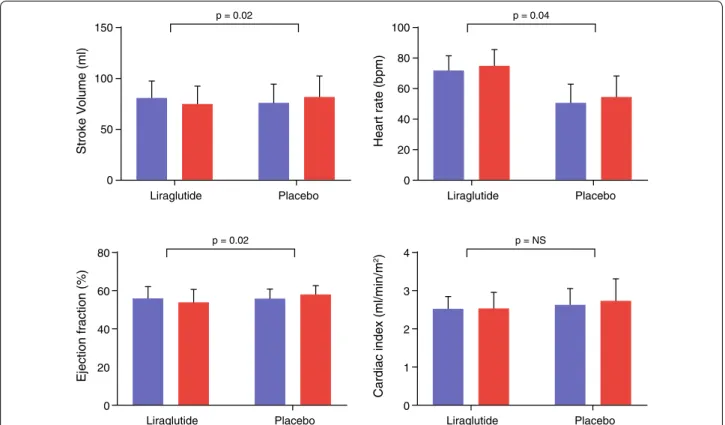
Fig. 3 LV systolic function. Bar graphs of MRI-derived indices of systolic function. Blue bars indicate baseline measurement and red bars follow-up. In the liraglutide group stroke volume decreased, whereas cardiac index remained unchanged because of the increased heart rate. Bpm beats per minute
Table 3 Heart rate and heart dimensions
bpm beats per minute, LA left atrial, LVEDV left ventricular end-diastolic volume, LVESV left ventricular end-systolic volume, LVM left ventricular mass index, LVMI left ventricular mass index, LVEDVI left ventricular end diastolic volume index
Liraglutide (n = 23) Placebo (n = 26) Mean (95% CI)
changes from baseline (liraglutide vs placebo)
p value
Baseline 26 week Mean (SD)
change from baseline
Baseline 26 week Mean (SD)
change from baseline
Heart rate in bpm (SD) 72 (9) 75 (10) 4 (8) 77 (13) 76 (13) − 1 (6) 4.3 (0.2 to 8) 0.04
LV filling volume in
mL (SD) 82 (17) 76 (17) − 5 (15) 74 (17) 82 (21) 7 (10) − 11 (− 18 to − 3) 0.01
LA volume index in mL/
m2 (SD) 36 (8) 35 (7) − 1 (6) 32 (8) 34 (10) 1 (7) − 2 (− 6 to 2) 0.38
LVEDV in mL (SD) 147 (25) 141 (25) − 5 (14) 138 (33) 144 (38) 6 (16) − 11 (− 20 to − 2) 0.02
LVESV in mL (SD) 67 (14) 66 (14) − 0 (9) 62 (17) 63 (20) 1 (9) − 1 (− 7 to 4) 0.69
LVM in g (SD) 107 (18) 105 (18) − 2 (8) 108 (27) 110 (29) 4 (9) − 6 (− 11 to − 1) 0.03 LVMI in g/m2 (SD) 49 (6) 49 (6) − 0 (3) 50 (11) 52 (12) 2 (4) − 1.5 (−3.6 to 0.6) 0.17 LVMI/LVEDVI g/mL/
Ea by reducing cardiac preload. Second, the increase in heart rate could have affected LV diastolic filling pat-tern directly [22]. However, there are two reasons why we do not expect increased heart rate to be the causa-tive effect of diastolic filling pattern changes: 1. the change in heart rate is relatively small in comparison to change in early filling; 2. a study in HFpEF patients using the selective sino-atrial node blocker ivabradine did not change E/Ea [23]. Lastly, a direct effect of GLP-1RA on the heart has been proposed as a mechanism to improve cardiac energy metabolism and thereby cardiac func-tion. Although GLP-1 receptor is expressed in cardio-myocytes, to date it is uncertain what its exact function in humans is [24]. However, if liraglutide had improved myocardial relaxation, an increase in Ea would have been expected. Ea however did not change significantly, which suggests against a direct effect of liraglutide on cardiomy-ocyte relaxation properties. It is unlikely that weight loss explains the observed effect of liraglutide on LV diastolic function, because a previous study from our group [25] has shown that calorie restriction with significant weight loss increased E/A ratio probably as a result of improved LV relaxation and/or filling properties (since LV filling pressure remained unchanged). Another important car-diovascular effect of weight loss in the study by Hammer et al. was a significant decline in heart rate, which is a consistent finding in patients after weight loss. Therefore, the rise in heart rate in the present study is in keeping
with the hypothesis that other mechanisms than weight loss are responsible for the observed changes in LV dias-tolic function.
Comparison with literature
Some studies have investigated the effect of liraglutide on LV diastolic function. Nystrom et al. [26] found no change in echocardiography-derived indices of myo-cardial relaxation, E/Ea or LV ejection fraction in their non-blinded randomized study with 62 DM2 patients with subclinical heart failure receiving either liraglu-tide or glimepiride treatment. A double-blind rand-omized trial in 33 patients with DM2 who underwent a 16 week exercise program with addition of either lira-glutide or placebo, showed significantly lower E/Ea in liraglutide treated patients [27]. Lastly, in two small non-randomized studies in patients with DM2, the effect of liraglutide was evaluated after 6 months using echocar-diography. These studies showed a decrease in E/Ea [28] and improved Ea [28, 29]. The results of our placebo-controlled double-blind randomized study confirm the finding of some preliminary studies to date that 6-month therapy with liraglutide showing lower E/Ea. With regard to LV systolic function, not surprisingly, most studies have been performed in HFrEF patients with or with-out DM2 [30, 31]. In HFrEF GLP-1RA therapy has been shown to have no effect on LV systolic function, although there was a trend towards more frequent hospitalisation for heart failure in the study by Margulies et al. [31]. The small but significant decline in LV ejection fraction in our study is to our knowledge the first study reporting this effect of liraglutide in a DM2 population without prior cardiovascular disease.
Clinical implications
The LEADER trial has shown that liraglutide reduces major adverse cardiovascular event rate (MACE) as compared to placebo in patients with DM2 [32]. The mechanisms responsible for GLP-1RAs beneficial effect on macrovascular diabetes complications remain to be established. Besides improvement of traditional car-diovascular risk factors, GLP-1RA treatment has been shown to reduce atherosclerotic plaque formation in mice by modulating macrophage phenotype [33], and reducing pro-inflammatory cytokines on a systemic level in conjunction with decreased leucocyte adhesion and extravasation into the vascular wall [34]. In addi-tion, a direct effect of GLP-1RA on endothelial cells of injured mouse femoral arteries has been described that pointed towards suppression of restenosis via nitric oxide [35]. Although reduction in cardiovascular event rate is the most important factor for improving prognosis of patients with DM2, it is important to note that heart
12
10
8
6
4
100 120 140 160 180 200
LVEDV (ml)
E/Ea
failure was not amongst the primary endpoints of the LEADER trial and other cardiovascular safety trials. As such, heart failure in patients with DM2 has been pos-tulated to be the forgotten diabetes complication after microvascular and macrovascular complications [6]. This study shows that liraglutide has a significant effect on LV diastolic function. This study shows that short-term use of liraglutide is safe in DM2 patients with LV diastolic dysfunction without heart failure (symptoms). We argue against routine evaluation of cardiac function with imag-ing in these patients, because clinical implications for the individual patient are currently lacking. It is impor-tant to note that HFpEF patients with New York Heart Association class III or IV were excluded in the present study. Since these stages are accompanied by higher E/Ea, effects of GLP-1RA therapy in this group of patients can-not be extrapolated from our study. Since these patients are dependent on increased E/Ea for adequate LV filling, liraglutide might even risk exacerbation of heart failure symptoms and decompensation in this particular sub-group of patients.
Limitations
First, the relatively low sample size was calculated to detect differences in Edec and LV ejection fraction. Other primary outcome measures were not included in sam-ple size calculation. We did indicate the other indices of diastolic and systolic function as primary because they are very strongly causally linked to Edec and ejection fraction. Therefore, we did not correct for multiple test-ing. As a result of low sample size, there was an uneven distribution of BMI (slightly higher in liraglutide) and nephropathy (higher prevalence in placebo). Although BMI [36] and albuminuria [37] are associated with LV diastolic dysfunction, it is unlikely that this affects study outcome because differences are relatively small. More-over, ANOVA analysis tests the differences between groups of within-group changes between baseline and follow-up, with correction for between-group differences at baseline. A second limitation is that we have chosen not to include LV diastolic dysfunction in the inclusion criteria of the study because there are no known cut-off values for LV diastolic dysfunction assessed with CMR. It is very likely that in our study population with mean diabetes duration of 11 years, poor glycaemic control, and high prevalence of hypertension, the vast majority of patients had LV diastolic dysfunction [1, 2, 6]. The third limitation regards the use of CMR. The reason CMR was used is that it is known for its excellent intra-observer reproducibility [11], and CMR is considered the gold standard for LV function and structure. CMR assessment of LV diastolic function has been shown to be a good alternative for echocardiography [10, 14]. It should be
noted though that values for diastolic and systolic func-tion as derived from CMR are not interchangeable with echocardiography [10, 11]. With regard to assessment of Ea, the relatively low temporal resolution of CMR as compared to echocardiography might have resulted in a lower power to detect significant differences. Another possible limitation is the relatively low sample size that does not facilitate reliable subgroup analyses.
Conclusions
In conclusion, this study provides evidence that the GLP-1RA liraglutide influences both left ventricular diastolic and systolic function by unloading the left ventricle in patients with DM2. Because elevated left ventricular fill-ing pressure is a driver for diabetic cardiomyopathy, an interesting hypothesis is that liraglutide could postpone the onset of HFpEF and concomitant morbidity and mor-tality. Liraglutide does not appear to have a direct effect on myocardial relaxation properties. The results of this study emphasize that larger studies specifically focus-ing on cardiac function are warranted in patients with DM2 with and without cardiovascular disease, including HFpEF. These studies will contribute to a more a com-plete understanding of cardiovascular benefit and safety of GLP-1RA therapy.
Abbreviations
A: late transmitral peak flow rate; Bpm: beats per minute; CMR: cardiac mag-netic resonance imaging; DM2: diabetes mellitus type 2; E: early transmitral peak flow rate; Ea: early peak mitral annular septal tissue velocity; Edec: peak deceleration of transmitral early peak flow; GLP-1RA: glucagon-like peptide-1 receptor agonist; HbA1c: glycated haemoglobin; HFpEF: heart failure with preserved ejection fraction; HFrEF: heart failure with reduced ejection fraction; ITT: intention-to-treat; LV: left ventricular; LVEDV: left ventricular end-diastolic volume; LVEDVI: left ventricular end-diastolic volume index; LVESV: left ven-tricular end-systolic volume; LVM: left venven-tricular mass; LVMI: left venven-tricular mass index; NTproBNP: N-terminal prohormone of brain natriuretic peptide; SUD: sulphonylurea derivative.
Authors’ contributions
All authors contributed to study concept and design and analysis and interpretation of data. EP, EK and MB contributed to acquisition of data. IJ, JS and JW supervised the MAGNA VICTORIA study, with HL as study director. EP, HE and MB performed statistical analysis of data. EP, HE and MB drafted the manuscript. All authors contributed to critical revision of the manuscript and approved the final version of the manuscript to be published. MB and HL are the guarantors of this work and, as such, had full access to all the data in the study and takes responsibility for the integrity of the data and the accuracy of the data analysis. All authors read and approved the final manuscript.
Author details
1 Department of Radiology, Leiden University Medical Center, LUMC Postzone
C2S, Albinusdreef 2, 2333 ZA Leiden, The Netherlands. 2 Department of
Medi-cine, Division of Endocrinology, Leiden University Medical Center, Leiden, The Netherlands. 3 Department of Medicine, Radboud University Medical Center,
Nijmegen, The Netherlands.
Acknowledgements
Winde, T.N. Bonten and L. van Duijn) and the physicians and nurses of the HMC Westeinde Hospital, The Hague (P.H.L.M. Geelhoed, A.H. Bootsma and A.V. Kharagjitsingh) for inviting eligible participants. We thank B. Polm and B. Ladan-Eygenraam for technical assistance on data gathering and processing. We thank G. Kracht for assistance on figure.
Competing interests
The authors declare that they have no competing interests.
Availability of data and materials
Please contact author for data requests.
Consent for publication
Not applicable.
Ethics approval and consent to participate
This study was approved by the ethical committee of the Leiden University Medical Center and all study participants provided written informed consent.
Funding
Novo Nordisk (Denmark) funded this investigator-initiated study. Novo Nordisk had no role in the design of the study, data collection, data analysis, data interpretation, or writing of the report. All authors had access to all the data and final responsibility for the decision to submit for publication.
Publisher’s Note
Springer Nature remains neutral with regard to jurisdictional claims in pub-lished maps and institutional affiliations.
Received: 28 January 2019 Accepted: 8 April 2019
References
1. Liu JE, Palmieri V, Roman MJ, et al. The impact of diabetes on left ventricu-lar filling pattern in normotensive and hypertensive adults: the Strong Heart Study. J Am Coll Cardiol. 2001;37:1943–9.
2. Redfield MM, Jacobsen SJ, Burnett JC Jr, Mahoney DW, Bailey KR, Rode-heffer RJ. Burden of systolic and diastolic ventricular dysfunction in the community: appreciating the scope of the heart failure epidemic. JAMA. 2003;289:194–202.
3. Lindman BR, Davila-Roman VG, Mann DL, et al. Cardiovascular phenotype in HFpEF patients with or without diabetes: a RELAX trial ancillary study. J Am Coll Cardiol. 2014;64:541–9.
4. MacDonald MR, Petrie MC, Varyani F, et al. Impact of diabetes on outcomes in patients with low and preserved ejection fraction heart failure: an analysis of the Candesartan in Heart failure: assessment of Reduction in Mortality and morbidity (CHARM) programme. Eur Heart J. 2008;29:1377–85.
5. Jarnert C, Landstedt-Hallin L, Malmberg K, et al. A randomized trial of the impact of strict glycaemic control on myocardial diastolic function and perfusion reserve: a report from the DADD (Diabetes mellitus And Diastolic Dysfunction) study. Eur J Heart Fail. 2009;11:39–47.
6. McMurray JJ, Gerstein HC, Holman RR, Pfeffer MA. Heart failure: a car-diovascular outcome in diabetes that can no longer be ignored. Lancet Diabetes Endocrinol. 2014;2:843–51.
7. Nauck MA, Meier JJ, Cavender MA, Abd El Aziz M, Drucker DJ. Cardio-vascular actions and clinical outcomes with glucagon-like peptide-1 receptor agonists and dipeptidyl peptidase-4 inhibitors. Circulation. 2017;136:849–70.
8. Russo C, Jin Z, Homma S, et al. Effect of obesity and overweight on left ventricular diastolic function: a community-based study in an elderly cohort. J Am Coll Cardiol. 2011;57:1368–74.
9. Ussher JR, Drucker DJ. Cardiovascular actions of incretin-based therapies. Circ Res. 2014;114:1788–803.
10. Westenberg JJ. CMR for Assessment of Diastolic Function. Curr Cardiovasc Imaging Rep. 2011;4:149–58.
11. Petersen SE, Aung N, Sanghvi MM, et al. Reference ranges for cardiac structure and function using cardiovascular magnetic resonance (CMR)
in Caucasians from the UK Biobank population cohort. J Cardiovasc Magn Reson. 2017;19:18.
12. Brandts A, Bertini M, van Dijk EJ, et al. Left ventricular diastolic func-tion assessment from three-dimensional three-direcfunc-tional velocity-encoded MRI with retrospective valve tracking. J Magn Reson Imaging. 2011;33:312–9.
13. Bizino MB, Tao Q, Amersfoort J, et al. High spatial resolution free-breathing 3D late gadolinium enhancement cardiac magnetic resonance imaging in ischaemic and non-ischaemic cardiomyopathy: quantitative assessment of scar mass and image quality. Eur Radiol. 2018;28:4027–35. 14. Paelinck BP, de Roos A, Bax JJ, et al. Feasibility of tissue magnetic
reso-nance imaging: a pilot study in comparison with tissue Doppler imaging and invasive measurement. J Am Coll Cardiol. 2005;45:1109–16. 15. van der Meer RW, Rijzewijk LJ, de Jong HW, et al. Pioglitazone improves
cardiac function and alters myocardial substrate metabolism without affecting cardiac triglyceride accumulation and high-energy phosphate metabolism in patients with well-controlled type 2 diabetes mellitus. Circulation. 2009;119:2069–77.
16. Sokos GG, Nikolaidis LA, Mankad S, Elahi D, Shannon RP. Glucagon-like peptide-1 infusion improves left ventricular ejection fraction and functional status in patients with chronic heart failure. J Cardiac Fail. 2006;12:694–9.
17. Yancy CW, Jessup M, Bozkurt B, et al. 2013 ACCF/AHA guideline for the management of heart failure: a report of the American College of Cardiol-ogy Foundation/American Heart Association Task Force on Practice Guidelines. J Am Coll Cardiol. 2013;62:e147–239.
18. From AM, Scott CG, Chen HH. Changes in diastolic dysfunction in diabe-tes mellitus over time. Am J Cardiol. 2009;103:1463–6.
19. Lekavich CL, Barksdale DJ, Neelon V, Wu JR. Heart failure preserved ejec-tion fracejec-tion (HFpEF): an integrated and strategic review. Heart Fail Rev. 2015;20:643–53.
20. Skov J, Pedersen M, Holst JJ, et al. Short-term effects of liraglutide on kid-ney function and vasoactive hormones in type 2 diabetes: a randomized clinical trial. Diabetes Obes Metab. 2016;18:581–9.
21. Koska J, Sands M, Burciu C, et al. Exenatide protects against glucose- and lipid-induced endothelial dysfunction: evidence for direct vasodilation effect of GLP-1 receptor agonists in humans. Diabetes. 2015;64:2624–35. 22. Johannessen KA, Cerqueira M, Veith RC, Stratton JR. Influence of sympa-thetic stimulation and parasympasympa-thetic withdrawal on Doppler echo-cardiographic left ventricular diastolic filling velocities in young normal subjects. Am J Cardiol. 1991;67:520–6.
23. Pal N, Sivaswamy N, Mahmod M, et al. Effect of selective heart rate slowing in heart failure with preserved ejection fraction. Circulation. 2015;132:1719–25.
24. Baggio LL, Yusta B, Mulvihill EE, et al. GLP-1 receptor expression within the human heart. Endocrinology. 2018;159:1570–84.
25. Hammer S, Snel M, Lamb HJ, et al. Prolonged caloric restriction in obese patients with type 2 diabetes mellitus decreases myocardial triglyc-eride content and improves myocardial function. J Am Coll Cardiol. 2008;52(12):1006–12.
26. Nystrom T, Padro I, Hedberg F, et al. Effects on subclinical heart failure in type 2 diabetic subjects on liraglutide treatment vs. glimepiride both in combination with metformin: a randomized open parallel-group study. Front Endocrinol. 2017;8:325.
27. Jorgensen PG, Jensen MT, Mensberg P, et al. Effect of exercise combined with glucagon-like peptide-1 receptor agonist treatment on cardiac function: a randomized double-blind placebo-controlled clinical trial. Diabetes Obes Metab. 2017;19:1040–4.
28. Saponaro F, Sonaglioni A, Rossi A, et al. Improved diastolic function in type 2 diabetes after a six month liraglutide treatment. Diabetes Res Clin Pract. 2016;118:21–8.
29. Lambadiari V, Pavlidis G, Kousathana F, et al. Effects of 6-month treatment with the glucagon like peptide-1 analogue liraglutide on arterial stiffness, left ventricular myocardial deformation and oxidative stress in subjects with newly diagnosed type 2 diabetes. Cardiovasc Diabetol. 2018;17:8. 30. Jorsal A, Kistorp C, Holmager P, et al. Effect of liraglutide, a
•fast, convenient online submission •
thorough peer review by experienced researchers in your field • rapid publication on acceptance
• support for research data, including large and complex data types •
gold Open Access which fosters wider collaboration and increased citations maximum visibility for your research: over 100M website views per year •
At BMC, research is always in progress.
Learn more biomedcentral.com/submissions
Ready to submit your research? Choose BMC and benefit from: 31. Margulies KB, Hernandez AF, Redfield MM, et al. Effects of liraglutide on
clinical stability among patients with advanced heart failure and reduced ejection fraction: a randomized clinical trial. JAMA. 2016;316:500–8. 32. Marso SP, Daniels GH, Brown-Frandsen K, et al. Liraglutide and
cardiovas-cular outcomes in type 2 diabetes. N Engl J Med. 2016;375:311–22. 33. Bruen R, Curley S, Kajani S, et al. Liraglutide dictates macrophage
phenotype in apolipoprotein E null mice during early atherosclerosis. Cardiovasc Diabetol. 2017;16:143.
34. Rakjpovski G, Rolin B, Nøhr J, et al. The GLP-1 analogs liraglutide and semaglutide reduce atherosclerosis in ApoE−/− and LDLr−/− mice by a mechanism that includes inflammatory pathways. JACC Basic Transl Sci. 2018;3:844–57.
35. Kushima H, Mori Y, Koshibu M, et al. The role of endothelial nitric oxide in the anti-restenotic effects of liraglutide in a mouse model of restenosis. Cardiovasc Diabetol. 2017;16:122.
36. Wang YC, Liang CS, Gopal DM, et al. Preclinical systolic and diastolic dysfunctions in metabolically healthy and unhealthy obese individuals. Circ Heart Fail. 2015;8(5):897–904.