ISSN:1991-8178
Australian Journal of Basic and Applied Sciences
Journal home page: www.ajbasweb.com
Corresponding Author: Raheem Lafta Ali, University Sains Malaysia,Instrumentation & Analytical Science, School of Physics, 11800, Pinang, Malaysia.
Structural Properties of Hydroxyapatite (HA
P) Thin Film on Ti-6Al-4V Alloy Prepared
by Radio Frequency Plasma Sputtering
1.2Raheem Lafta Ali, 1Mohamad Suhaimi and 1Shahrom Mahmud
1University Sains Malaysia, Instrumentation & Analytical Science, School of Physics, 11800, Pinang, Malaysia 2Department of Physics, College of Education, University of Al- Mustansiriya, Baghdad, Iraq
A R T I C L E I N F O A B S T R A C T Article history:
Received 10 January 2016 Accepted 15 February 2016 Available online 28 February 2016
Keywords:
Ti-6Al-4V; hydroxyapatite coating; crystal growth; RF plasma;
Background: Titanium and its alloys have been used in biomedical applications because of their biocompatibility and mechanical properties. However, It has a possible toxic effect resulting from released vanadium and aluminum. The (HAP) coated Ti-6Al-4V alloy prevented releasing of undesirable ions. Objective: The aim of this paper is to investigate the structural and mechanical properties of Ti-6Al-4V alloy by coating with (HAp). This coating was prepared by Radio Frequency Plasma Sputtering (RF) method. Results: The thin film of HAp was annealed at temperature of 800 C° to improve the structural, composition, roughness and hardness were characterized by X-ray diffraction (XRD), Field Emission- Scan Electron Microscope (FE-SEM), atomic force microscope (AFM) and Vickers hardness respectively. The HAp films were amorphous. Conclusion: After annealing at 800 C° the surface amorphous of HAp was converted to crystalline structure. AFM and the Vickers hardness showed different roughness and hardness of Ti-6Al-4V when uncoating, as-deposited HAp thin film and after annealing.
© 2016 AENSI Publisher All rights reserved. To Cite This Article: Raheem Lafta Ali, Mohamad Suhaimi and Shahrom Mahmud., Structural Properties of Hydroxyapatite (HAP) Thin Film on Ti-6Al-4V Alloy Prepared by Radio Frequency Plasma Sputtering. Aust. J. Basic & Appl. Sci., 10(7): 46-50, 2016
INTRODUCTION
Titanium was discovered in 1791 and got its name from Greek mythology; "Titan". It means the enormously strong giant. Titanium is light and transition metal on the periodic table composed of 60% iron and density of 4.5 g/cm. It is a silver-gray material with an atomic number of 22 and an atomic weight of 47.90 (Adnan. 2012). Titanium is allotropic material that exists as a hexagonal close- packed structure (α Ti) up to 82 C° and a body centered cubic structure (bcc) above the temperature, the addition of alloying elements to titanium enables it to have a wide range of properties (Tapash et al. 2011). Commercially, pure (cpTi) titanium and its alloys (such as Ti-6Al-4V) have been utilized for load bearing implants in dentistry, osteosynthesis and orthopedics due to their excellent biocompatibility, lightweight, high-strength, corrosion resistance and relatively low density(Gemelli E et al. 2007). However, these properties are not high enough to promote direct growth of the bone tissue or reduced the healing period and also Ti-6Al-4V alloy has a possible toxic effect resulting from released vanadium and aluminum (C.N.Elias. et al. 2008). Modifications of
metal surfaces have been employed to controlling tissue–titanium interactions; reduce the time for bone fixation and to prevent releasing of undesirable ions from the alloy (Richard C, 2015). These problems can be prevented by using coating layers from bioinert materials as bioceramic HAp. Since these materials are major inorganic component of human bone matrix and has a crystal structure identical to human bone and teeth minerals (Baek-Hee lee et al. 2008). The coated layers do not release ions into surrounding tissue of the body as do metallic biomaterials so that the coating layers on metallic biomaterials to prevent metallic ion release from orthopedic implants made of Ti6Al4V (Davide Zaffe lee et al. 2008 ).
Many techniques, such as sol gel (Sajjad et al. & A. Balakrishnan et al. 2007), Plasma electrolytic oxidation (PEO)[Yeung et al,2013 ], Electrophoretic deposition (EPD) (Corni I et al. 2008 & Lovsky Y et
al. 2010), dip coating process (Seriven, 1988 & M.
(Yunzhi lee et al. 2008). This technique can produce thin, uniform, dense coatings that are homogeneous in structure and composition (Hee et al. 2008).
MATERIALS AND METHODS
Ti-6Al-4V Specimens used in this project were acquired from WG (William Gregor Ltd, London, United Kingdom). Circular (Ti-6Al-4V) Specimens are cut from rod with 30mm diameter and 3 mm thickness. All specimens were abraded successively using SiC grinding paper with different grits started from 80 grit, and continued by 120, 230, 400, 600, 800 , 1000 and 1200 grit to obtain flat and scratch free surface and then polishing with diamond suspension start (1, 3, 6, 9, 15 µ m) for a smooth and mirror polished surface. A grinding and polishing machine model: metaserv 250, buehler was used and then cleaned ultrasonically and washed with acetone and deionized water.
RF sputtering was used to prepare HAp layer on Ti6Al4V alloy by using high purity (99.99%) HAp target, a base pressure was evacuated to (1 10-5 mbar) by a combination of rotary and turbo-molecular pump, then argon was passed till the pressure reached (1 10-3 mbar) , and then the target was cleaned by pre-sputtering (with 30 W RF power) for 15 min. Ar (99.99%) was supplied as reactive gas at a flow ratio of 10 sccm. The distance between the target and the substrate was maintained at 80 mm for all deposition experiments with a RF power 200 W for 1 h duration. During the sputtering process, the temperature rose to approximately 800
because of self-heating.
Various techniques for surface analysis, structure examination and properties evaluation are used to investigate modified surface such as XRD analysis was carried out within the 20–60° range using a (PANalytical X’Pert PRO MRD PW3040, Almelo,Netherlands) with a Cu Kα radiation (λ= 1.541 Å) to identify the phases developed of specimens of Ti6Al4V alloy. FESEM was performed using a (Leo-Supra 50VP, Carl Zeiss, Germany) equipped with an energy dispersive X-ray (EDX) system. . Element distribution in the surface of treated and untreated samples is evaluated using EDX mapping. The surface roughness of the film was measured by using atomic force microscope (AFM) (Dimension Edge, Bruker) in the Tapping operation mode and a NanoDrive dimension-edgetapping, image-processing software. Vickers hardness of the coated composites was evaluated
data of HAp thin films formed on the surface of Ti-6Al-4V alloy respectively. In spite of the existence α
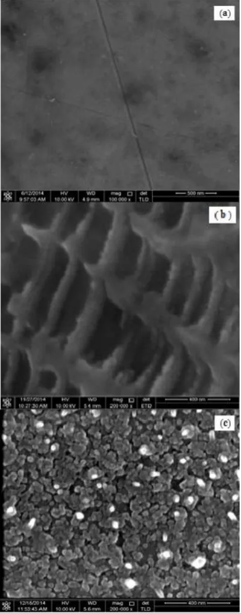
and β phases of Ti-6Al-4V alloy. The HAp thin films were built on whole surface of substrate. The uncoated specimen in figure (1a) seems to have many scratches and smooth surface due to its grinding and polishing. Figure (1b) shows surface morphology of Ti-6Al-4V specimen coated with HAp by using RF plasma sputtering method without annealing. HAp thin film was found to be a porous surface because of amorphous structure of HAp. It was found that annealing treatment at 800 °C for 1hr can cause the amorphous structure of HAp to convert roughness due to create crystalline phase as indicated in Figure (1c).
The EDX spectrums are shown in Figure2 which explain the elemental distribution of the Ti-6Al-4V specimen uncoated, as coated with HAp by RF plasma sputtering and annealing at temperature 800 °C . The EDX for the uncoated specimen shows energy emission (Ti, Al and V) indicated that the substrate is Ti-6Al-4V alloy due to the existence of elements (Ti, Al and V) as indicated in Figure (2a). It can be seen from the patterns of calcium and phosphorus elements as well as the matrix (Ti-6Al-4V). This means the layer formed on the surface is HAp layer which is comprised of calcium and phosphorus elements and diffusion through the substrate as indicated in Figures (2b) and (2c). The composition of calcium and phosphorus elements increases with annealing at temperature 800 °C as indicated in Figure (2c).
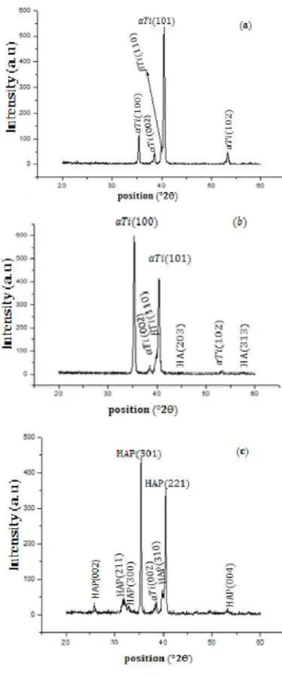
X-ray diffraction pattern of Ti-6Al-4V uncoated, as coated with HAp and annealing at temperature 800
°C are shown in Figure 3. The XRD pattern for the uncoated specimen shows the lines of Ti-6Al-4V are 100, 002, 110, 101 and 102 at 2Ɵ values 35.46,
38.27°, 39.98°, 40.7° and 53.24° respectively which are belong a phase of Ti-6Al-4V as indicated in Figure (3a). As deposit HAp was found to be
amorphous with no crystalline phase; just peaks related to Ti-6Al-4V substrate were observed as indicated in figure (3b). Figure (3c) shows Strongest lines of the XRD profile of annealed the coated specimen of Ti-6Al-4V alloy at 800 °C with the following Miller indices (hkl): (002), (211), (300), (301), (310), (221)and (004) belong to the HAp phase were agreement to the data reported for hexagonal C 5(PO4)3OH (JCDS –ICDD
Fig. 1: Field emission scanning electron microscope images of Ti6Al4V alloy (a) the as-received substrate (b)
HAp coatings deposited on a Ti6Al4Vsubstrate (c) annealing at 800 °C.
AFM micrographs with a scanning area in figure 4 shows the surface topography of the average surface roughness (R ) of the substrate, as deposited and annealing at temperature 800 °C the uncoated specimen in Figure (4a) has many scratches and smooth surface with roughness (R ) is 0.00367 . Figures (4b) and (4c) show the AFM images of the HAp coated Ti-6Al-4V alloy without and with annealing at temperature 800 °C respectively. In annealing at 800 °C convert the surface porosity of HAp with (R ) was 0.104 to crystalline grain with roughness (R ) of 0.203 .
The Vickers hardness value of uncoated specimen was 348.9 HV. The hardness of coating was 423.3 HV and after annealing at 800 °C was
610 HV. It was found the hardness of HAp layer was
greater after annealing at 800 °C
Conclusions:
The present study concludes that coating of Ti-6Al-4V alloy with HAp has amorphous structure and
this structure transforms to crystalline phase after annealing at 800 °C . The surface topography of uncoated Ti-6Al-4V,as deposited HAp film and
Fig. 2: EDX analyses of the samples (a) the as-received substrate (b) HAp coatings deposited on a Ti6Al4Vsubstrate (c) annealing at 800 °C.
Fig. 4:AFM 3D images of Ti-6Al-4V alloy (a) the as-received substrate (b) as-coated (c) annealing at 800 °C.
REFERENCES
Adnan safder, 2012 A study on electron beam melted Ti-6Al-4V.
Balakrishnan, A., B.C. LEE,T.N. Kim and BB. Panigrahi, 2007. Hydroxyapatite coatings on NaOH treated Ti–6Al–4V alloy using sol–gel precursor. Materials Science and Technology, 23(8): 1005.
Agatadude, 2009. Surface properties in titanium with hydroxyapatite coating. Optica Applicata,
XXXIX, 4.
Baek-Hee lee1 and Naoto Koshizaki, 2008. Nanostructured hydroxyapatite/TiO2 Composite
coating applied to commercially pure titanium by a co-sputtering technique. Nanotechnology, 19: 415303 (7pp).
Elias, C.N., J.H.C. Lima, R.Valiev and M.A. Meyers,2008. Biomedical applications of Titanium and its alloys. Biological Materials science.
Corni, I., M.P. Ryan, A.R. Boecacelni, 2008. Electrophoretic deposition: from traditional ceramics to nanotechnology. J Eur ceram Soc., 28: 1353-1367.
Davide Zaffea, Carlo Bertoldib Ugo Consolob , 2004. Accumulation of aluminum in lamellar bone after implantation of titanium plates, Ti–6Al–4V screws, hydroxyapatite granules. Biomaterials, 25: 3837–3844.
Mohseni, E., E.A.R. Zalnezhad, 2014. Comparative investigation on the adhesion of hydroxyapatite coating on Ti–6Al–4V implant: Bushroa, International Journal of Adhesion & Adhesives, 48: 238–257.
Gemelli, E., A. Scariot, N. Heriberto, 2007. Thermal characterization of commercially pure titanium for dental application. Mat.Res., 10-3.
Guocheng Wang and Hala Zreiqat, 2010. Functional Coatings or Films for Hard-Tissue Applications. Materials, 3: 3994-4050.
Hee Ay Ching, Dipanker Choudhury, Md Julker Nine and Noor Azuan Abu Osman, 2014. Review effects of surface coating on reducing friction and wear of orthopaedic. implants. Sci. Technol. Adv. Mater.,15: 014402 (21pp).
Shi, J.Z., C.Z.H.J.Y.U. Chen, S.J. Zhang, 2008. Application of magnetron sputtering for producing bioactive ceramic coatings on implant materials. Bull. Mater. Sci., 31(6): 877–884. © Indian Academy of Sciences.
Lovsky, Y., A. Lewis, C. Sukenik, Y. Grushka, 2010. Atomic-force-controlled capillary electrphoretic nanoprinting of proteins. Anal Bioanal Chem., 396:133-138.
Torrisi, L., R. Setola, 1993. Thermally assisted hydroxyapatite obtained by pulsed-laser deposition on titanium substrates. Thin solid Films, 227: 32-36.
Khalid, M., M. Mujahid, A. Nusair Khan, R.S. Rawat, 2013. Dip Coating of Nano Hydroxyapatite on TitaniumAlloy with Plasma Assisted ɣ- Alumina Buffer Layer: A Novel Coating Approach, J. Mater. Sci. Technol, 29(6): 557-564.
Richard, C., Petersen, 2015. Titanium Implant Osseointegration Problems with Alternate Solutions Using Epoxy/Carbon Fiber – Reinforced Composite. available in PMC January 27.
Seriven, L.E., 1988. Physics and applications of dip coating and spin coating. Better ceramics through chemistry 3rd 717-729.
Sajjad Jafari, Mehdi Mazar Atabaki, Jamaliah Idris, Comparative study on Bioactive coating of Ti-6Al-4V alloy and 316 L stainless steel. Association of Metallurgical Engineers of Serbia, (AMES), UDC: 669.295.5'71'292.018.8 ; 621.794.6.
Xu, S., J.D. Long, K.N. Ostrikov, J.H. Lu and C.H.R.F., 2008. Magnetron Sputtering Deposition of Bioactive Ca–P-Based Coatings on Ti–6Al–4VAlloy. Diong. IEEE TRANSACTIONS ON PLASMA SCIENCE, 30-1.
Tapash, R., Rautray and R. Narayanan, Kyo-han Kim, 2011. Ion implantation of titanium based biomaterials. Materials science, 56: 1137-1177.
Yeung, Wk1, G.C., Reilly, 2013. Matthew. In vitro biological response of plasma electrolytically oxidized and plasma-sprayed hydroxyapatite coating on Ti-6Al-4V alloy. Aug; 101(6): 939- 49. Doi: 10.1002/jbm. b32899.
Yunzhi Yanga, B., Kyo-Han Kimc, Joo B.L. Onga, 2005. A review on calcium phosphate coatingsproduced using a sputtering process—an alternative to plasma spraying. Biomaterials, 26: 327– 337.