Address for correspondence Dilip Kumar SA
Cutis Skin & LASER Clinic, Gourishankar Mandir Raod, Raigarh, Chhattisgarh, India PIN Code 496001
Ph: +91-7024449837
Email: [email protected]
Original Article
Clinico-pathological profile of Systemic Sclerosis in a
tertiary care center
Introduction
Systemic sclerosis (SSc) is a chronic disorder of unknown etiology characterized clinically by thickening and stiffening of skin caused by accumulation of connective tissue and by structural and functional abnormalities of visceral organs including gastrointestinal tract, lung, heart, kidney.1 It is characterized
pathologically by vascular abnormalities,
sclerosis of connective tissue, epidermal atrophy
and autoantibodies. Patients with systemic sclerosis are classified as limited cutaneous systemic sclerosis and diffuse cutaneous systemic sclerosis.2 Based on extent of skin
involvement, limited cutaneous systemic
sclerosis is defined as skin involvement confined to areas distal to elbows and knees and also face and neck whereas diffuse cutaneous systemic sclerosis as that extending proximal to the elbows and knees, face, neck and trunk. Patients with limited cutaneous systemic sclerosis have long duration of Raynaud’s phenomenon, esophageal dysmotility or reflux, gastrointestinal problems, pulmonary fibrosis and pulmonary hypertension late in the course of their disease. They are often positive for anticentromere antibody.2 Patients with diffuse cutaneous
Dilip Kumar SA, Alaka Sahu*
Department of Dermatology, LSLAM Govt. Medical College, Raigarh Chhattisgarh, India * Department of Pathology, VIMSAR, Burla, India
Abstract
Objective To find out the profile of cutaneous and systemic features of systemic sclerosis (SSc) in people of East-Central part of India.Methods This was a cross-sectional descriptive study conducted over eight years in a tertiary care center. All the patients of SSc were evaluated for cutaneous and systemic involvement.
Results 54 cases (48 females and 6 males) of SSc were evaluated. Mean age was 31.81±14.52 years. We found microstomia in 88.8%, sclerodactyly 88.8%, Raynaud’s phenomenon in 81.4%, salt-and-pepper pigmentation in 70.3%, fingertip ulceration in 66.6%, pinched nose in 61.1%, radial furrows around mouth in 33.3%, diffuse alopecia in 18.5%, nail changes in 18.5%, calcinosis cutis in 7.4% and digital gangrene in 5.6% cases. Among the systemic features, we observed arthralgia in 55.5%, dyspnea on exertion in 55.5%, dysphagia in 51.8%, esophageal reflux in 46.2%, cough in 11.1%, and palpitation in 7.4% cases. Antinuclear antibody (ANA) was positive in 85.1%, abnormal pulmonary function test in 59.2%, abnormal X-ray hand in 33.3% and proteinuria in 7.4% cases.
Conclusion Pulmonary, renal and cardiac involvement were less in our study population as compared to other studies.
Key words
systemic sclerosis have short duration of Raynaud’s phenomenon preceding onset of
cutaneous features, fatigue, arthralgia,
gastrointestinal manifestations, pulmonary
fibrosis, cardiac involvement and renal crisis. They often have antibodies to Scl-70.2 SSc
mimics many other skin conditions like
generalized morphea, scleromyxedema,
scleredema of Buschke, primary systemic amyloidosis, chronic graft-versus-host disease, eosinophilic fasciitis etc.3 The criteria for
diagnosis of systemic sclerosis have been established by the subcommittee for scleroderma criteria of American Rheumatism Association
(now called the American College of
Rheumatology).4 The single major criterion is
presence of sclerodermatous involvement
proximal to the digits, affecting limbs, face, neck, or trunk usually in a bilateral and symmetrical pattern.; the minor criteria are sclerodactyly, digital pitted scars or tissue loss of volar pads of fingertips, and bibasilar pulmonary fibrosis.4 The diagnosis is based on
presence of either the major criterion or at least two of minor criteria. These criteria have 97% sensitivity and 98% specificity. The addition of Raynaud’s phenomenon and nail fold capillary microscopy and systemic sclerosis selective antibodies as additional minor criteria improve the sensitivity of these criteria.5 This study
intended to explore the cutaneous and systemic features of patients of SSc in the East-central part of India.
Methods
A cross-sectional, descriptive study was conducted over a period of eight years in a tertiary care center in East-central part of India. All the patients of systemic sclerosis giving written consent for the study and fulfilling the criteria laid down by the subcommittee for scleroderma criteria of the American College of Rheumatology were recruited for the study.4
Patients with overlap syndrome and mixed connective tissue disorders were excluded from the study. Detailed history, clinical examination and necessary investigations were undertaken. Special investigations like high resolution CT scan, skin biopsy, echocardiography etc. were performed in a subset of patients. Data analysis was done using Microsoft Office Excel 2007 and SPSS
Results
A total of 54 cases of SSc were evaluated; of which 36 (66.7%) belonged to diffuse cutaneous and 18 (33.3%) to limited cutaneous variety. Female to male ratio was 8:1 (48 females and 6 males). The mean age of our patients was 31.81±14.52 years with a range of 12 to 65 years. By occupation 38 (70.3%) were housewives, 10 (18.5%) were students, 4 (7.4%) were farmers, 1 (1.8%) was industrial worker and 1 (1.8%) was businessman.
Duration of disease varied from 2 months to 10 years with a mean of 3.2±2.96 years. Raynaud’s phenomenon was present in 44 (81.4%) cases. Duration of Raynaud’s varied from 1 month to 10 years with mean of 4±3.03 years. Raynaud’s phenomenon preceded skin tightening in 49 (90.7%) cases whereas both occurred simultaneously in 5 (9.3%) cases.
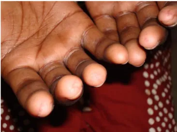
Among the cutaneous features (Table 1), fingertip ulceration was found in 36 (66.7%), sclerodactyly in 48 (88.8%) (Figure 1), digital pitted scars in 30 (55.5%) (Figure 2), digital gangrene in 3 (5.6%), calcinosis cutis in 4 (7.4%), microstomia in 48 (88.8%) (Figure 3), radial furrows around mouth in 18 (33.3%)
(Figure 4), pinched nose in 33 (61.1%), diffuse
Figure 1 Sclerodactyly in a case of diffuse cutaneous systemic sclerosis.
Figure 2 Digital pitted scars in a case of limited cutaneous systemic sclerosis
Figure 3 Microstomia in a case of Systemic Sclerosis
Figure 4 Radial furrows around the mouth in a case of systemic sclerosis
Figure 5 Thinning of epidermis, effacement of rete ridges,
Table1 Cutaneous findings, systemic features and laboratory parameters in Systemic Sclerosis (n=54)
Variables N (%)
Cutaneous
Pigmentary changes 50 (92.6)
Sclerodactyly 48 (88.8)
Microstomia 48 (88.8)
Raynaud’s phenomenon 44 (81.4) Fingertip ulceration 36 (66.6)
Pinched nose 33 (61.1)
Radial furrows around mouth 18 (33.3)
Diffuse alopecia 10 (18.5)
Nail changes 10 (18.5)
Calcinosis cutis 4 (7.4)
Digital gangrene 3 (5.55)
Systemic
Arthralgia 30 (55.5)
Dyspnea on exertion 30 (55.5)
Dysphagia 28 (51.8)
Esophageal reflux 25 (46.2)
Cough 6 (11.1)
Palpitation 4 (7.4)
Laboratory parameters
Anemia 51 (94.4)
Raised ESR 46 (85.1)
ANA positivity 46 (85.1)
Proteinuria 4 (7.4)
Abnormality in X-ray hands 18 (33.3) Abnormality in chest X-ray 10 (18.5) Abnormal pulmonary function tests 32 (59.2)
Abnormality in ECG 3 (5.6)
diffuse hyperpigmentation and 38 (70.3%) had salt-and-pepper pigmentation (mottled pigmentation).
Among the systemic presentations, arthralgia was present in 30 (55.5%) cases, dyspnea on exertion in 30 (55.5%), dysphagia in 28 (51.8%), esophageal reflux in 25 (46.2%), cough in 6 (11.1%) and palpitation in 4 (7.4%) cases.
Hemoglobin level in our patients ranged from 7.6 to 12.6gm/dl with a mean of 10.35±1.52 gm/dl. Anemia was noted in 51 (94.4%) cases. ESR level was raised in 46 (85.1%) patients with a mean ESR of 27.13± 16.31mm first hour (range was 3 to 76). ANA was positive in 46 (85.1%) and proteinuria in 4 (7.4%) patients. Abnormality in X-ray hands was found in 18
(33.3%) cases, of which 8 (14.8%) cases showed acro-osteolysis, 5 (9.3%) cases showed contracture and another 5 (9.3%) cases showed both these features. Abnormality in chest X-ray was found in 10 (18.5%) cases. Pulmonary function test showed restrictive pattern of ventilatory defect in 32 (59.2%) cases. Abnormal ECG was noted in 3 (5.6%) cases. We performed skin biopsy in a subset of patients and observed features like epidermal atrophy, flattening of rete ridges, thickening and hyalinization of collagen bundles, encroachment of fat by collagen bundles and perivascular inflammatory infiltrate (Figure 5).
Discussion
The mean age of presentation in our study was 31.81±14.52 years which is in concordance with Sharma et al.6 (32.75± 11.62years), Ghosh et al.7
(29.6±12.3 years) and Basel et al.8 (36.17±13.9
years). However, the mean age was higher in a study conducted at Sarawak General hospital by
Teh et al.9 We found female preponderance in
our study with female to male ratio of 8:1. Most of the studies in literature also noted female preponderance with female to male ratio varying from 3.9:1 to 8.37:1.6,7,10-12 Occupational history
is important in SSc as scleroderma like lesions occur in workers exposed to chemicals like vinyl chloride, perchlorethylene, trichlorethylene, organic solvents, pesticides, epoxy resins etc.3
None of our patients had exposure to these chemicals. By occupation majority of our patients were housewives followed by students.
Mean duration of disease in our study was 3.2±2.9 years which is shorter than a North Indian study.6 Raynaud’s phenomenon was found in 81.4% patients which is similar to Sharma et al.6, Ghosh et al.7 and Teh et al.9
However, Krishnamurthy et al.10 noted
majority of cases Raynaud’s phenomenon precedes cutaneous changes; this is much shorter in males than in females. In our study, 90.7% cases gave the history of Raynaud’s phenomenon prior to onset of cutaneous changes. Fingertip ulceration was observed in 66.6% patients as compared to 58.6% by Sharma et al.6, and 52% by Teh et al.9 We observed
diffuse hyperpigmentation in 55.5% whereas Sharma et al.6 found this in a higher proportion
of cases (88.1%). Salt-and-pepper pigmentation was found in 70.3% cases as compared to 51.2% cases by Sharma et al.6and 70% by Dia et al.11
Calcinosis cutis most commonly found in fingers and is more common in female than in males. It may lead to ulceration with discharge of chalky materials.3 We noticed calcinosis cutis
in relatively higher proportion of cases compared to Ghosh et al.7 and Al-Adhadh et al.12
Sclerodactyly and digital pitted scars are included as minor criteria by subcommittee for scleroderma criteria.4 Sclerodactyly was also
found in a higher proportion in our study compared to Sharma et al.6 (64.6%) and Teh et
al.9 (65%). We noticed digital pitted scars in
55.5% cases. Microstomia was found in 88.8% as compared to 55.5% in a North Indian study.6
Microstomia may interfere with speaking, eating and maintenance of oral hygiene.3 Radial
furrows around the mouth were found in about one-third of our cases and diffuse alopecia in 21.7% cases.
Esophagus is the most common part of gastrointestinal tract (GIT) to be involved in SSc and gastroesophageal reflux and dysphagia are common gastrointestinal symptoms.3 Dysphagia
was found in 51.8% cases in the present study. Gupta et al.13 noted dysphagia in 82.6% cases of
limited cutaneous and 60.7% cases of diffuse cutaneous SSc. Gastroesophageal reflux was found in 46.2% cases compared to 38.7% observed by an Egyptian study.8 Pulmonary
involvement occurs in about 40% to 90% cases
in SSc.3 Pulmonary fibrosis is usually associated
with diffuse cutaneous SSc and pulmonary hypertension with limited cutaneous SSc.14
Dyspnea on exertion and cough are common
pulmonary symptom in SSc.3 Dyspnea on
exertion was observed in 55.5% cases in our study. This is consistent with the findings by Arakkal et al.15 (64.3%) and North Indian study6
(51.1%). Arakkal et al.15 noted cough in a higher
proportion of patients (32%) compared to our findings. Arthralgia was noted in 55.5% cases which is little higher than the result of North Indian study6 and lower than the finding of
Krishnamurthy et al.10 Malignant hypertension
secondary to renal involvement develops in about 8% patients.16 None of our patients had
malignant hypertension. Cardiac involvement in SSc includes myocardial disease, conduction system abnormalities, arrhythmia and pericardial disease. Resting ECG is abnormal in about 50% cases and includes bifid P waves and T wave changes.17 Only 5.6% of our patients had
abnormal ECG which is very less as compared to observations by Arakkal et al.15
We noted raised ESR in about 85% which is consistent with the result of Sharma et al.6
However, Arakkal et al.15 found raised ESR in a
lower proportion (53.6%) of patients. Mean hemoglobin level in our study was found to be 10.35±1.52 gm/dl and 94.4% of our patients were anemic. Hemoglobin less than 10gm/dl was found in 42.9% cases by Arakkal et al.15
whereas Al-Adhadh et al.12 observed anemia in
only 31% patients. ANA level was raised in 85.1% in our study which is comparable to other studies like Sharma et al.6 (89.1%), Ghosh et al.7
(78.2%) and Pradhan et al.18 (87.01%).
However, Krishnamurthy et al.10 found ANA
other studies like Sharma et al.6, Teh et al.9, Dia
et al.11 and Arakkal et al.15Pulmonary function
tests are a very useful investigation in SSc and it shows restrictive changes with a decreased forced vital capacity and total lung capacity.3
About 59% of our patients showed restrictive changes in PFT. This finding is consistent with Krishnamurthy et al.10 and Dia et al.11 However,
Sharma et al.6 and Arakkal et al.15 noted
restrictive pattern of ventilatory defect in a higher proportion of cases. HRCT shows ground glass appearance in areas of active alveolitis or septal fibrosis and honey combing in areas of interstitial fibrosis. We performed HRCT only in selected cases and found the features of interstitial fibrosis in lungs. Mild
proteinuria is the most common renal
manifestation, often early in the disease. Proteinuria was noted in 7.4% cases which is comparable to results of North Indian6 and
Egyptian studies.8 X-ray hand showed changes
like acro-osteolysis, contracture, calcinosis, erosive arthropathy etc.
Conclusion
Demographic profile and cutaneous features in our patients were almost similar to many other studies, however, pulmonary, renal and cardiac involvement was relatively less in our study population.
References
1. Gilliland BC. Systemic Sclerosis and related disorders. In: Kasper DL, Braunwald E, Fauci AS, Hauser SL, Longo DL, Jameson JL. Harrisons Principles of Internal Medicine, 16th ed. New York: McGraw-Hill; 2005. P. 1979-90.
2. LeRoy EC, Black C, Fleischmajer R, Jablonska S, Krieg T, Medsger TA Jr et al. Scleroderma (systemic sclerosis): classification, subsets and pathogenesis. J Rheumatol. 1988;15:202-5.
3. Goodfield MJD, Jones SK, Veale DJ. The Connective Tissue Diseases. In: Burns T,
Breathnach S, Cox N, Griffiths C eds. Rook’s Textbook of Dermatology, 8th ed. Oxford: Wiley-Blackwell; 2010. 51. P. 64-110.
4. Subcommittee for Scleroderma criteria of the American Rheumatism Association Diagnostic and Therapeutic Criteria Committee. Preliminary criteria for the classification of systemic sclerosis. Arthritis Rheum. 1980;23:581-90.
5. Lonzetti LS, Joyal F, Raynauld JP, Roussin A, Goulet JR, Rich E et al. Updating the American college of Rheumatology preliminary classification criteria for systemic sclerosis: addition of severe nail fold capillaroscopy abnormalities markedly increases the sensitivity for limited scleroderma. Arthritis Rheum. 2001;44:735-6.
6. Sharma VK, Trilokraj T, Khaitan BK, Krishna SM. Profile of systemic sclerosis in a tertiary care center in North India. Indian J Dermatol Venereol Leprol. 2006;72:416-20. 7. Ghosh SK, Bandyopadhyay D, Saha I,
Barua JK. Mucocutaneous and demographic features of systemic sclerosis: A profile of 46 patients from Eastern India. Indian J Dermatol. 2012;57:201-5.
8. Basel ME, Khalil N. Disease characteristics of systemic sclerosis among Egyptian patients. Kasr Al Ainy Med J. 2015;21:41-6. 9. Teh CL, Kuan YC, Wong JS. Systemic
Sclerosis in Sarawak: a profile of patients treated in the Sarawak General Hospital. Rheumatol Int. 2009;29:1243-5.
10. Krishnamurthy V, Porkodi R, Ramakrishnan S, Rajendran CP, Madhavan R, Achuthan K et al. Progressive systemic sclerosis in south India. J Assoc Physicians India.1991;39:254-7.
11. Dia D, Dieng MT, Sy TN, Diallo M, Fall S, Ndongo S et al. Systemic scleroderma: 92 cases in Dakar. Dakar Med. 2003;48 (2):82-6.
12. Al-Adhadh RN, Al-Sayed TA. Clinical features of systemic sclerosis. Saudi Med J. 2001;22:333-6.
13. Gupta R, Bammigatti C, Dinda AK, Marwaha V, Gupta S. Prevalence of renal involvement in Indian patients with systemic sclerosis. Indian J Dermatol Venereol Leprol. 2007;61:91-6.
15. Arakkal G, Chintagunta SR, Chandrika V, Damarla SV, Manchala S, Kumar BU. Cardiopulmonary involvement in systemic sclerosis: A study in a tertiary care center. Indian J Dermatol Venereol Leprol. 2017;83:677-82.
16. Steen VD, Constantino JP, Shapiro AP, Medsger TA Jr. Long term outcomes of renal crisis in systemic sclerosis: relation to
availability of ACE inhibitors. Ann Inter Med. 1990;113:352-7.
17. Champion HC. The heart in scleroderma. Rheum Dis Clin North Am. 2008;34:181-90. 18. Pradhan V, Rajadhyaksha A, Nandkar M,